Replicative Deformed Wing Virus Found in the Head of Adults from Symptomatic Commercial Bumblebee (Bombus terrestris) Colonies
←
→
Page content transcription
If your browser does not render page correctly, please read the page content below
veterinary
              sciences

Article
Replicative Deformed Wing Virus Found in the Head of Adults
from Symptomatic Commercial Bumblebee (Bombus
terrestris) Colonies
Giovanni Cilia              , Laura Zavatta, Rosa Ranalli, Antonio Nanetti *                   and Laura Bortolotti

                                           CREA Research Centre for Agriculture and Environment, Via di Saliceto 80, 40128 Bologna, Italy;
                                           giovanni.cilia@crea.gov.it (G.C.); laura.zavatta@crea.gov.it (L.Z.); rosa.ranalli@crea.gov.it (R.R.);
                                           laura.bortolotti@crea.gov.it (L.B.)
                                           * Correspondence: antonio.nanetti@crea.gov.it

                                           Abstract: The deformed wing virus (DWV) is one of the most common honey bee pathogens. The
                                           virus may also be detected in other insect species, including Bombus terrestris adults from wild and
                                           managed colonies. In this study, individuals of all stages, castes, and sexes were sampled from three
                                           commercial colonies exhibiting the presence of deformed workers and analysed for the presence
                                           of DWV. Adults (deformed individuals, gynes, workers, males) had their head exscinded from the
                                           rest of the body and the two parts were analysed separately by RT-PCR. Juvenile stages (pupae,
                                           larvae, and eggs) were analysed undissected. All individuals tested positive for replicative DWV,
                                           but deformed adults showed a higher number of copies compared to asymptomatic individuals.
                                 Moreover, they showed viral infection in their heads. Sequence analysis indicated that the obtained

                                           DWV amplicons belonged to a strain isolated in the United Kingdom. Further studies are needed to
Citation: Cilia, G.; Zavatta, L.;
                                           characterize the specific DWV target organs in the bumblebees. The result of this study indicates the
Ranalli, R.; Nanetti, A.; Bortolotti, L.
                                           evidence of DWV infection in B. terrestris specimens that could cause wing deformities, suggesting
Replicative Deformed Wing Virus
                                           a relationship between the deformities and the virus localization in the head. Further studies are
Found in the Head of Adults from
                                           needed to define if a specific organ could be a target in symptomatic bumblebees.
Symptomatic Commercial Bumblebee
(Bombus terrestris) Colonies. Vet. Sci.
2021, 8, 117. https://doi.org/
                                           Keywords: bumblebee; commercial Bombus terrestris; spillover; DWV; honey bee pathogens; replica-
10.3390/vetsci8070117                      tive virus; strand-specific RT-PCR

Academic Editor: Yanping
(Judy) Chen
                                           1. Introduction
Received: 20 May 2021                            The deformed wing virus (DWV) belongs to the Picornaviridae family, within the
Accepted: 21 June 2021
                                           Iflavirus genus, with a positive-sense ssRNA [1,2]. As an Apis mellifera pathogen, DWV
Published: 23 June 2021
                                           is deemed to be involved in colony losses and, as such, a significant impact to the ecosys-
                                           tem [3,4]. The virus is spread globally [1,2,5,6], and DWV infections are generally detected
Publisher’s Note: MDPI stays neutral
                                           due to the presence of symptomatic honey bees, characterized by deformed or missing
with regard to jurisdictional claims in
                                           wings and shortened abdomens [1]. Presently, three genetic DWV variants are acknowl-
published maps and institutional affil-
                                           edged and named as type A, B, and C [7,8], with type A being the most prevalent [8].
iations.
                                                 In honey bees, DWV is usually associated with Varroa destructor infestations, even
                                           if, in some cases, the virus was detected in Varroa-free bees [9]. Within the colony, it is
                                           transmitted by the punctures produced in both the juvenile and adult stages [10], but
                                           the infection may also spread horizontally by bee-to-bee contact [11–15] and ingestion of
Copyright: © 2021 by the authors.          contaminated food [16–18]. However, due to the multiple possible transmission routes,
Licensee MDPI, Basel, Switzerland.
                                           DWV may spillover to other sympatric Hymenopterans [19–21], including species used in
This article is an open access article
                                           commercial pollination [22–26] and beetles [27].
distributed under the terms and
                                                 Bumblebees are among the most common and widespread bee pollinators, mainly
conditions of the Creative Commons
                                           in temperate and cold areas. Due to their unique ability to pollinate tomato crops, they
Attribution (CC BY) license (https://
                                           began to be bred and sold in many countries for pollination purposes [28]. Bumblebee
creativecommons.org/licenses/by/
                                           rearing and trading date back to the 1980s, when the first company for the commercial
4.0/).

Vet. Sci. 2021, 8, 117. https://doi.org/10.3390/vetsci8070117                                                       https://www.mdpi.com/journal/vetsci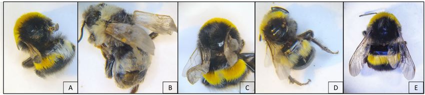
Vet. Sci. 2021, 8, 117                                                                                            2 of 12

                         rearing of B. terrestris was founded in Belgium. Bumblebees were initially used for tomato
                         pollination in greenhouses, but later they extended to other crops both in open fields and
                         greenhouses. Presently, bumblebees are commercially bred in all continents, except Africa,
                         with a global sales volume exceeding 1,000,000 colonies per year. They are sold worldwide,
                         with the main exception of mainland Australia. Due to the emerging risks in the marketing
                         of non-native species and the resulting restrictions, many local species of bumblebee began
                         to be bred for pollination in the countries of origin, but B. terrestris always remains the most
                         used and widespread in Europe, North Africa, and West Asia [29,30].
                              One of the main concerns in the commercialization of bumblebees is the spread of
                         diseases and pathogens [31]. Commercial bumblebee colonies can suffer from various
                         diseases and pests, which can affect their survival. Those that can cause major problems
                         are the protozoan Nosema bombi, the tracheal mite Locustacarus (Bombacarus) buchneri, the
                         Trypanosomatidae Crithidia bombi, and the brood parasites Melittobia acasta and M. chalybii,
                         whose massive presence in breeding facilities requires the stamping out of the infected
                         colony and, in the most severe infestations, of the entire production stock [32–35]. Other
                         less dangerous but still problematic pests are the pyralid moths Vitula edmandsii and Plodia
                         interpunctella whose larvae feed primarily on bumblebee food stores but occasionally also
                         on bumblebee brood [29].
                              Thus far, viral infections have not been considered major threats for wild and com-
                         mercial bumblebees. However, recent findings show the presence of replicative honey
                         bee viruses in wild bees, including bumblebees. The infection with some of these viruses
                         proved to cause negative consequences in B. terrestris, such as reduced fecundity and
                         colony founding associated with Kashmir bee virus (KBV), reduced fecundity due to Israeli
                         acute paralysis virus (IAPV), and mortality due to acute bee paralysis virus (ABPV) [36–39].
                         Conversely, B. terrestris infection with DWV has been found to result in wing deformities
                         and mortality in a limited number of individuals [40,41]. A recent investigation showed
                         that DWV could replicate in Bombus pupae after artificial infection and larval feeding
                         of virus-contaminated food, but none of the infected bumblebees showed signs of wing
                         deformities [23].
                              Since bumblebees are not affected by Varroa infestations, it remains unclear how the
                         infection occurs under natural conditions, although DWV-positive Bombus sp. Were found
                         in areas with a high DWV prevalence in Apoidea species [40,42–44]. Possibly, the virus
                         transmission between sympatric pollinator species is mediated by shared floral resources
                         such as pollen [16,24]. Similarly, in commercial breeding, the most likely infection route
                         is by feeding the bumblebees with pollen from honey bee colonies. In effect, batches of
                         honey bee pollen used to feed bumblebees revealed the presence of several pathogens
                         (Crithidia spp., Ascosphaera bombi, A. apis, Nosema ceranae, Nosema thomsoni, Microsporidium
                         sp. Oise), including viruses (sacbrood virus—SBV, DWV, IAPV, and chronic bee paralysis
                         virus—CBPV) [45]. This study aimed to investigate the DWV distribution in the body of
                         symptomatic and asymptomatic Bombus terrestris specimens reared in a commercial colony
                         after the first detection in a worker with relevant wing deformities.

                         2. Materials and Methods
                         2.1. Sample Collection
                              On 15 January 2021, three commercial bumblebee colonies (B. terrestris) arrived from a
                         North European breeding company to our laboratory (Bologna, Italy) for an experimental
                         test requiring colonies in an advanced stage of development. The colonies contained
                         about a hundred workers and pupal stages of gynes and males. Moreover, they appeared
                         healthy and in good condition, although the founder queen was found dead in all of
                         them. The colonies were kept in a climate room for a total of three weeks, under constant
                         environmental conditions of temperature (25 ± 1 ◦ C) and relative humidity (40 ± 10%).
                         Darkness (0:24 L:B) was maintained throughout the rearing period, and all the laboratory
                         manipulations were conducted with the aid of red lights [46]. Flight activity was not
                         allowed. The colonies had arrived from the breeding company with food supplies that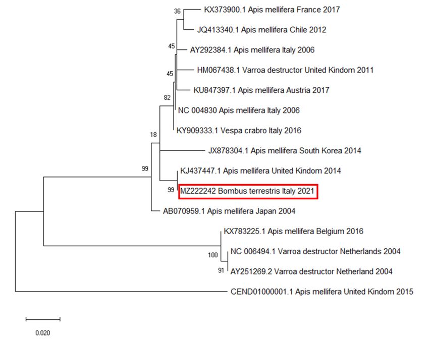
Vet. Sci. 2021, 8, 117 3 of 12
were integrated onsite with frozen pollen from local honey bee colonies. On 17 January
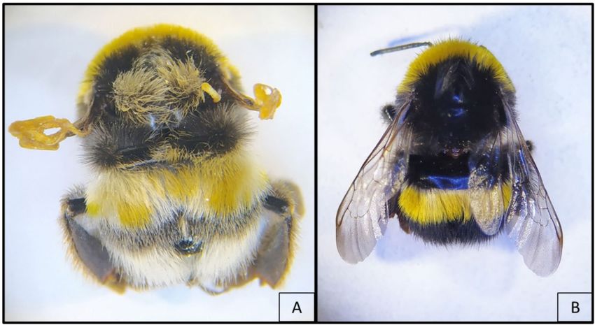
2021, a routine check allowed for the detection in one of the colonies of one worker with
crippled wings (Figure 1).
Figure 1. Bombus terrestris workers with crippled (A) and normal (B) wings. Individual A shows yellow and white patches
of anomalous pigmentation.
As this observation was compatible with a DWV infection, a sampling plan created
to collect and analyse all kinds of individuals present in the colonies. Accordingly, the
following asymptomatic individuals were sampled from each colony: five gynes, five
workers, five males, five pupae, five larvae, and three pools of eggs (n = 5). All the adults
that were found with crippled wings were also individually collected.
2.2. Extraction of Total RNA
Before dissection and extraction of viral RNA, all samples were washed, through full
immersion in 95% ethanol for 10 s to remove any external viral contamination that may
have been present. Each adult was dissected under sterile conditions with a scalpel to
exscind the head from the rest of the body (thorax and abdomen), which were collected
in separate 2-ml microtubes. To avoid possible cross contamination, new scalpels were
used for each individual. The juvenile stages (pupae, larvae, and the pool of eggs) were
introduced undissected into separate 2-ml microtubes. Beads and 80 µL of Lysis Buffer
provided by GeneJET RNA Purification Kit (ThermoFisher Scientific, Waltham, MA, USA)
were added to each sample, which was crushed with a TissueLyser II (Qiagen, Milan, Italy)
for 3 min at 25 Hz, as previously described [47,48].
Total RNA was extracted from each sample with GeneJET RNA Purification Kit
(ThermoFisher Scientific, Waltham, MA, USA) following the manufacturer’s instruction.
All samples were eluted in 100l RNase-free water. The RNA extracts were stored at −80 ◦ C
until use. High pure sterile DNA- and RNA-free water was used as a negative control in
all analytical steps.
2.3. qRT-PCR Assays to Detect and Quantify the Deforming Wing Virus (DWV)
The extracted RNAs were analysed by qRT-PCR to detect and quantify the presence
of DWV in bumblebees. Primers amplified a 132-bp fragment within the highly conserved
region coding for the RNA-dependent RNA polymerase (Rd-Rp) commonly expressed in all
virus variants. The sequences were: DWV Fw 50 - TTTGACATTGAGCTACAAGACTCG-30
(nt. 8685–8708), DWV Rev 50 - ACAATCCGTGAATATAGTGTGAGG-30 (nt. 8816–8793) [16].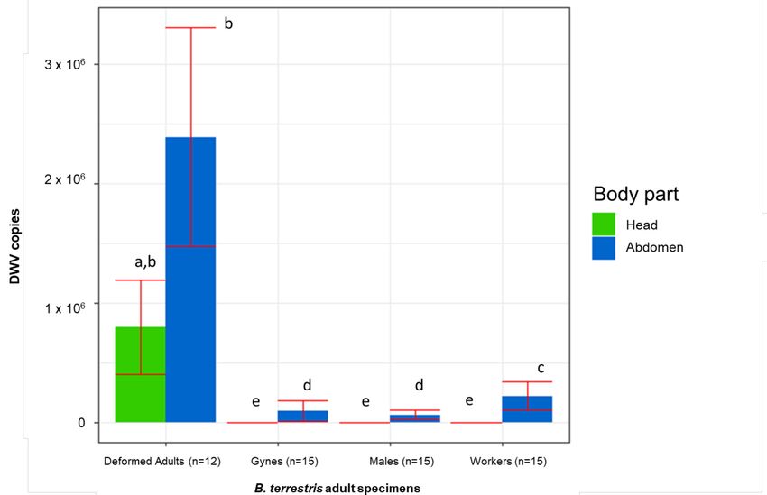
Vet. Sci. 2021, 8, 117                                                                                        4 of 12

                         The viral genomes were amplified using Power SYBR® Green RNA-to-Ct™ 1-Step Kit
                         (ThermoFisher Scientific, Waltham, MA, USA), following the manufacturer’s instruction.
                         The qPCR assay was performed on Applied Biosystems® 7500 fast and 7500 Real-Time
                         PCR (ThermoFisher Scientific, Waltham, MA, USA). For the target gene, a total reaction
                         volume of 20 µL was used following the protocols previously described [16,19].
                              The successful amplification of reference gene β-Actin, with the previously described
                         primers [49], was used to confirm the sample integrity from the RNA extraction to the
                         qPCR analysis and to normalize the results to reach an absolute quantification.
                              Virus loads were quantified with absolute quantification of number of DWV copies in
                         each ng of RNA (copies/ng RNA). The amplified fragment was gel purified using GeneJET
                         Gel Extraction and DNA Cleanup Micro Kit (ThermoFischer Scientific, Waltham, MA, USA)
                         and cloned using CloneJET PCR Cloning Kit with DH10B Competent Cells (ThermoFischer
                         Scientific), following the manufacturer’s instructions, and sequenced (BMR Genomics,
                         Padova, Italy). Following plasmid DNA removal by RNase-free DNase treatment (RNase
                         Free DNase Set, Qiagen, Hilden, Germany), the transcript was purified and concentrated
                         with the RNeasy Minelute Cleanup Kit (Qiagen, Hilden, Germany) and quantified with
                         the Quant-iT™ RiboGreen™ RNA Assay Kit (ThermoFisher, Waltham, MA, USA). A
                         standard curve was created with six tenfold dilutions of cloned RNA fragment (2 × 10⁵ to
                         2 copies/ng) [16].

                         2.4. Strand-Specific RT-PCR
                              The DWV replication was evaluated through a strand specific RT-PCR using specific
                         primers Fw 8450: 50 - TGGCATGCCTTGTTCACCGT-30 (nt. 8450–8469) or Rev 8953: 50 -
                         CGTGCAGCTCGATAGGATGCCA-30 (nt. 8953–8932), which amplify a 504-bp fragment
                         of the Rd-Rp, as previously described [16]. All amplicons were visualized on a 1.5%
                         agarose gel.
                              The DWV sequence described in this study was submitted to the GenBank database
                         under the accession number MZ222242.

                         2.5. Phylogenetic Analysis
                              The strand specific RT-PCR-obtained amplicons were sequenced (BMR Genomics,
                         Padua, Italy) and analysed using BLASTn to standard databases with default parameters
                         for megablast [50]. The sequences with a high Max Score and a Query ≥70% in the BLAST
                         analysis were selected to build the phylogenetic tree. The phylogenetic analysis was
                         performed by the maximum likelihood method based on the Tamura–Nei model with a
                         bootstrap test using MEGA software [51].

                         2.6. Statistical Analysis
                               As the batch of colonies was provided by the same producer and all of them were pre-
                         vented from flying, the ‘colony’ as an explanatory factor for the DWV titre was considered
                         unimportant. This consideration elicited the decision to pool the data together by the kind
                         of individual, as if they all belonged to the same colony.
                               The results were analysed with a parametric approach. Before the statistical anal-
                         ysis, significant violations to the assumptions of parametric tests were removed by log
                         transformation of data. However, text and illustrations report untransformed data.
                               The number of DWV copies detected in the dissected body parts from the same adult
                         was summed up to calculate the total individual DWV titre. This was considered as the
                         dependent variable in a one-way analysis of variance (ONE-WAY ANOVA) with the ‘kind
                         of individual’ (deformed adults, gynes, workers, males, pupae, larvae, and eggs) as the
                         categorical factor.
                               A factorial ANOVA was conducted on the amount of DWV copies detected in the
                         adults against the categorical independent variables ‘kind of individual’ (deformed adults,
                         gynes, workers, males) and ‘body part’ (head, rest of the body). The interaction between
                         the two factors mentioned above was also calculated.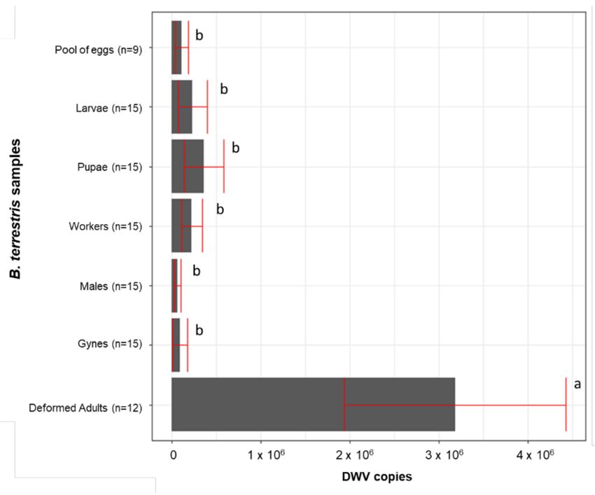
Vet. Sci. 2021, 8, 117                                                                                                      5 of 12

                                      When the F-test resulted in a significant effect, a pairwise Newman-Keuls post hoc
                                 test was conducted to spot significant differences between the groups.
                                      For all tests, the protection level from statistical Type I error was set at p ≤ α = 0.05.

                                 3. Results
                                      Wing deformity (Figure 2) was detected in adults of all colonies. In detail, the deformi-
                                 ties were found, respectively, in one and six workers of two colonies and in four workers
                                 and one newly hatched male (Figure 2B) of the third one.

      Figure 2. Evidence of several degrees of wing deformities in symptomatic DWV infected Bombus terrestris. Wings of
      adult bumblebees from the inspected colonies: bilateral (A,C) and unilateral (D) deformity in workers; bilateral defor-
      mity in newly hatched male (B); asymptomatic worker (E). Deformed individuals show yellow and white patches of
      anomalous pigmentation.

                                       All samples scored positive for DWV, with a number of genomic copies ranging
                                 from 2.01 × 10² to 5.10 × 10⁶ . The viral titre varied significantly in the different kinds of
                                 individuals that were sampled (F(6, 69) = 6.130, p = 0.000); a Newman-Keuls test showed
                                 that the amount of DWV copies was significantly higher in the deformed adults compared
                                 to the asymptomatic adults and the eggs, larvae, and pupae). Among the nondeformed
                                 individuals, no significant differences were detected despite belonging to different stages,
                                 sexes, and castes (Figure 3).

                                 Figure 3. DWV titre of deformed and asymptomatic adults and juvenile individuals. Averages +/−
                                 standard error are shown. The same letter indicates a nonsignificant difference.

                                      In the adults, the DWV titre was significantly influenced by the kind of individual (F(3,
                                 66) = 127.687, p = 0.000), the body part (F(1, 66) = 337.772, p = 0.000), and their interaction
Vet. Sci. 2021, 8, 117 6 of 12
(F(3, 66) = 36.342, p = 0.000). A Newman-Keuls test did not show a significantly different
DWV abundance in the head and the rest of the body of deformed individuals. However,
all the considered types of asymptomatic adults had significantly fewer copies in their
heads. Furthermore, in both the heads and the rest of the body of deformed individuals,
the number of DWV copies was significantly higher compared to the asymptomatic adults
(Figure 4).
Figure 4. DWV copies detected in both head and rest of the body of deformed and asymptomatic B. terrestris adults.
Averages (columns) and standard errors (vertical bars) are shown. The same letters highlight nonsignificant differences
(p ≤ 0.05).
The strand-specific PCR demonstrated the active DWV replication in all tested samples.
The BLAST analysis performed on the obtained amplicons confirmed the specificity of the
sequences, with high similarity (99% of percent identity, 0.0 of E-value, 100% of Query
Cover) to specific DWV genomes deposited in GenBank. The same sequence was recorded
in all positive samples. The phylogenetic analysis and pairwise distance analysis indicated
the highest homology to DWV strains isolated from A. mellifera in the United Kingdom
(Figure 5).Vet. Sci. 2021, 8, 117 7 of 12
Figure 5. Molecular phylogenetic analysis for RNA-dependent RNA polymerase of deformed wing virus (DWV) by
maximum likelihood method. The evolutionary history was inferred using the maximum likelihood method based on the
Tamura–Nei model. The branch lengths of the tree measured the number of substitutions per site. The analysis involved
28 nucleotide sequences. There were 255 positions in the final dataset. Accession number, host, state, and year of available
GenBank DWV sequences are shown. DWV sequence accession numbers are reported and associated with year and site of
origin. The DWV sequence obtained from the tested B. terrestris samples is highlighted by a red box.
4. Discussion
Several studies highlight that bees other than A. mellifera can be infected by the
DWV [22,24–26,41,52]. Additionally, the virus has been detected in the hornets Vespa crabro
and Vespa velutina [19,20], wasp Vespula vulgaris [53], invasive ant Linepithema humile [21],
and beetle Aethina tumida [27,54,55]. This shows that spillover may occur between both close
superfamilies (Apoidea, Vespidae) and insect species belonging to relatively distant groups.
In Bombus spp., DWV has been found in several instances worldwide and con-
sidered a significant factor of decline in both wild and managed bumblebee popula-
tions [56,57]. DWV infections have been reported in B. terrestris [25,40,52,58–60], B. pas-
cuorum [25,41,52,58,60], B. impatiens [24,25,61–63], B. atratus [64,65], B. vagans [24,62], B.
huntii [63], B. ruderatus [59], B. ternarius [24], B. lapidarius, B. lucorum, and B. monticola [40].
Despite the fact that DWV infections are a frequent occurrence in B. terrestris [22,25],
symptomatic adults with crippled and deformed wings have been seldom reported. To
the best of our knowledge, only B. terrestris queens and B. pascuorum workers have been
found showing symptoms linked to DWV infection, reporting replicative viral RNA in the
thorax and abdomen [41]. Moreover, in asymptomatic B. hunti, DWV was found in the
brain and antennae of wild males and reared males and workers, even if it was not found
to be replicative [63].Vet. Sci. 2021, 8, 117 8 of 12
In A. mellifera, DWV is chiefly transmitted by the Varroa mites [1,6,15,17,22]. Missing
a known vector, in bumblebees, the infections are more likely to propagate by a feeding
route through the consumption of contaminated food and trophallaxis.
Indeed, commercial bumblebee producers may use honey bee collected pollen as a
protein source to feed the queens and support the colony development [24,66,67]. In this
respect, fresh frozen pollen has a shown higher effectiveness compared to dry pollen, which
makes the first generally preferred to feed both queens and developing colonies [68]. How-
ever, the pollen collected by the honey bees may contain pathogens, including viruses [45],
which may have likely played a role in developing the infections detected in this study. Al-
though the investigated colonies were fed fresh frozen pollen in our laboratory, they already
had adults with crippled wings when they arrived, indicating that possible pollen-mediated
DWV infections must have occurred at the breeding site. Furthermore, flowers may become
contaminated with DWV when visited by infected honey bees, thus representing sites for
interspecific transmission to other pollinators, wild bumblebees included [16,17,52,69].
Additionally, newly emerged honey bee workers may be used in commercial breeding
to stimulate bumblebee queens to initiate their nesting activity after hibernation [41], but
this method is rarely used as it is less effective in inducing oviposition than the use of the
bumblebee pupa [70]. Finally, the vertical transmission is also not to be excluded because a
DWV-positive queen could deposit infected eggs, from which infected adults can be born,
as suggested by the deformed and positive newly emerged male collected.
The total viral load found in each investigated B. terrestris individual is comparable
to values measured in asymptomatic honey bees (from 102 to 106 copies) and lower than
the viral titre of symptomatic honey bees (> 107 copies) [16]. In all investigated samples,
DWV was found in replicative form, demonstrating its adaptability and capability to
replicate in Bombus cells [22,41]. Nonsignificant differences in DWV titre were observed
in adults considering the head and the rest of the body separately. As for DWV-positive
honey bees [41], the symptomatic bumblebees investigated here were characterized by
virus presence in the head. Additionally, our results highlighted a relationship between
deformities in adults and the presence of DWV in the head. Previously, the B. terrestris
and B. pascuorum specimens with crippled wings only resulted positive for the presence
of DWV in the thorax and abdomen, but their head scored negative [41]. Additionally, in
asymptomatic artificially infected B. huntii specimens, the DWV was found in the antennae
and brain [63]. This study could not clarify whether a relationship existed between DWV
infection and abnormal pigmentations in symptomatic individuals.
Although other causes of wing malformation cannot be excluded, they should be
seen as most unlikely. Upon arrival at the laboratory, the three colonies showed healthy,
well-developed, and without signs of mishandling or malnutrition. The fact that in all the
colonies the founder queen had died is not uncommon at this stage of colony development,
although conventionally it makes the colonies unsuitable for commercialization. In many
insect species, the development of deformed adults can also be caused by thermal shock,
but in bumblebee colonies, this usually occurs at the early stages of development, when
there are still only few workers tending the brood [71]. However, during the three weeks
of observation by our laboratory, the colonies were kept at a constant temperature and
humidity and carefully manipulated.
Finally, the phylogenetic analysis highlighted that the obtained DWV amplicons
showed high similarity (99% identity) with a DWV sequence isolated from the honey bees
in the United Kingdom in 2014 [9], suggesting that DWV could be associated with the
commercial origin of colonies.
5. Conclusions
The results of this study, other than indicating evidence of DWV infection in B. terrestris
specimens causing wing deformities similar to the clinical lesion in honey bees, suggest a
relationship between the wing deformities and the virus localization in the head. Further
studies are needed to define if a specific organ could be a target in symptomatic bumblebees.Vet. Sci. 2021, 8, 117 9 of 12
Furthermore, the high similarity of sequenced amplicons with DWV isolated from honey
bees in the United Kingdom indicates the North European origin of the virus and suggests
that transmission could occur by contaminated pollen administered to bumblebees. The
finding of adults with deformed wings and the premature death of the queen raises the
question of to what extent the presence of the virus can be associated with a reduction of
vitality at the individual level.
Several studies demonstrate that commercially produced bumblebee colonies can
carry multiple infectious parasites, posing a significant risk to other native and managed
pollinators through pathogen spillover [72]. Currently, commercial colonies must be accom-
panied by a parasite-free certification, and rearing facilities may be subject to inspection
by the national veterinary services. If the colonies are intended for exportation, the certifi-
cates may also cover honey bee parasites and pests (e.g., Varroa destructor, Tropilaelaps spp.,
Aethina tumida, and American foulbrood) but not honey bee viruses.
The geographical origin of the DWV strains found in our colonies and the fact that
most commercial B. terrestris breeding is located in Northern Europe raise concerns about
the possible future increase of virus spread by long-range export of infected colonies. This
advocates for intensified controls in bumblebee rearing operations and the application of
preventive measures against virus spread, such as pollen sterilization by gamma radiation,
which has already been proven effective against IAPV present in honey bee-collected
pollen [73].
Author Contributions: Conceptualization, G.C. and L.B.; methodology, G.C., L.Z., R.R., and L.B.;
investigation, G.C., L.Z., R.R., and L.B.; data curation, G.C., L.Z., R.R., A.N., and L.B.; writing—
original draft preparation, G.C. and L.B.; writing—review and editing, G.C., L.Z., R.R., A.N., and
L.B.; supervision, L.B. All authors have read and agreed to the published version of the manuscript.
Funding: This research received no external funding.
Institutional Review Board Statement: Not applicable.
Informed Consent Statement: Not applicable.
Acknowledgments: The authors are grateful to Marta Barberis of the Department of Biological,
Geological, and Environmental Sciences, University of Bologna, for her valuable technical support.
Conflicts of Interest: The authors declare no conflict of interest.
References
1. De Miranda, J.R.; Genersch, E. Deformed wing virus. J. Invertebr. Pathol. 2010, 103, S48–S61. [CrossRef]
2. Genersch, E.; Aubert, M. Emerging and re-emerging viruses of the honey bee (Apis mellifera L.). Vet. Res. 2010, 41, 54. [CrossRef]
3. McMenamin, A.J.; Genersch, E. Honey bee colony losses and associated viruses. Curr. Opin. Insect Sci. 2015, 8, 121–129. [CrossRef]
4. Steinhauer, N.; Kulhanek, K.; Antúnez, K.; Human, H.; Chantawannakul, P.; Chauzat, M.P.; van Engelsdorp, D. Drivers of colony
losses. Curr. Opin. Insect Sci. 2018, 26, 142–148. [CrossRef] [PubMed]
5. Buendía, M.; Martín-Hernández, R.; Ornosa, C.; Barrios, L.; Bartolomé, C.; Higes, M. Epidemiological study of honeybee
pathogens in Europe: The results of Castilla-La Mancha (Spain). Span. J. Agric. Res. 2018, 16, e0502. [CrossRef]
6. Martin, S.J.; Highfield, A.C.; Brettell, L.; Villalobos, E.M.; Budge, G.E.; Powell, M.; Nikaido, S.; Schroeder, D.C. Global honey bee
viral landscape altered by a parasitic mite. Science 2012, 336, 1304–1306. [CrossRef] [PubMed]
7. Mordecai, G.J.; Wilfert, L.; Martin, S.J.; Jones, I.M.; Schroeder, D.C. Diversity in a honey bee pathogen: First report of a third
master variant of the Deformed Wing Virus quasispecies. ISME J. 2016, 10, 1264–1273. [CrossRef]
8. McMahon, D.P.; Natsopoulou, M.E.; Doublet, V.; Fürst, M.; Weging, S.; Brown, M.J.F.; Gogol-Döring, A.; Paxton, R.J. Elevated
virulence of an emerging viral genotype as a driver of honeybee loss. Proc. Biol. Sci. 2016, 283, 20160811. [CrossRef] [PubMed]
9. Ryabov, E.V.; Wood, G.R.; Fannon, J.M.; Moore, J.D.; Bull, J.C.; Chandler, D.; Mead, A.; Burroughs, N.; Evans, D.J. A virulent strain
of deformed Wing Virus (DWV) of honeybees (Apis mellifera) prevails after varroa destructor-mediated, or in vitro, transmission.
PLoS Pathog. 2014, 10, e1004230. [CrossRef]
10. Yue, C.; Schroder, M.; Gisder, S.; Genersch, E. Vertical-transmission routes for deformed wing virus of honeybees (Apis mellifera).
J. Gen. Virol. 2007, 88, 2329–2336. [CrossRef]
11. Ball, B.V.; Allen, M.F. The prevalence of pathogens in honey bee (Apis mellifera) colonies infested with the parasitic mite Varroa
jacobsoni. Ann. Appl. Biol. 1988, 113, 237–244. [CrossRef]
12. Nordström, S. Distribution of deformed wing virus within honey bee (Apis mellifera) brood cells infested with the ectoparasitic
mite Varroa destructor. Exp. Appl. Acarol. 2003, 29, 293–302. [CrossRef] [PubMed]Vet. Sci. 2021, 8, 117 10 of 12
13. Shen, M.; Cui, L.; Ostiguy, N.; Cox-Foster, D. Intricate transmission routes and interactions between picorna-like viruses (Kashmir
bee virus and sacbrood virus) with the honeybee host and the parasitic varroa mite. J. Gen. Virol. 2005, 86, 2281–2289. [CrossRef]
[PubMed]
14. Lanzi, G.; de Miranda, J.R.; Boniotti, M.B.; Cameron, C.E.; Lavazza, A.; Capucci, L.; Camazine, S.M.; Rossi, C. Molecular and
biological characterization of deformed wing virus of honeybees (Apis mellifera L.). J. Virol. 2006, 80, 4998–5009. [CrossRef]
15. Gisder, S.; Aumeier, P.; Genersch, E. Deformed wing virus: Replication and viral load in mites (Varroa destructor). J. Gen. Virol.
2009, 90, 463–467. [CrossRef] [PubMed]
16. Mazzei, M.; Carrozza, M.L.; Luisi, E.; Forzan, M.; Giusti, M.; Sagona, S.; Tolari, F.; Felicioli, A. Infectivity of DWV associated to
flower pollen: Experimental evidence of a horizontal transmission route. PLoS ONE 2014, 9, e113448. [CrossRef]
17. Mockel, N.; Gisder, S.; Genersch, E. Horizontal transmission of deformed wing virus: Pathological consequences in adult bees
(Apis mellifera) depend on the transmission route. J. Gen. Virol. 2011, 92, 370–377. [CrossRef]
18. Chen, Y.; Evans, J.; Feldlaufer, M. Horizontal and vertical transmission of viruses in the honey bee, Apis mellifera. J. Invertebr.
Pathol. 2006, 92, 152–159. [CrossRef]
19. Mazzei, M.; Forzan, M.; Cilia, G.; Sagona, S.; Bortolotti, L.; Felicioli, A. First detection of replicative deformed wing virus (DWV)
in Vespa velutina nigrithorax. Bull. Insectol. 2018, 71, 211–216.
20. Forzan, M.; Sagona, S.; Mazzei, M.; Felicioli, A. Detection of deformed wing virus in Vespa crabro. Bull. Insectol. 2017, 70, 261–265.
21. Sébastien, A.; Lester, P.J.; Hall, R.J.; Wang, J.; Moore, N.E.; Gruber, M.A.M. Invasive ants carry novel viruses in their new range
and form reservoirs for a honeybee pathogen. Biol. Lett. 2015, 11, 20150610. [CrossRef] [PubMed]
22. Gisder, S.; Genersch, E. Viruses of commercialized insect pollinators. J. Invertebr. Pathol. 2017, 147, 51–59. [CrossRef] [PubMed]
23. Gusachenko, O.N.; Woodford, L.; Balbirnie-Cumming, K.; Ryabov, E.V.; Evans, D.J. Evidence for and against deformed wing
virus spillover from honey bees to bumble bees: A reverse genetic analysis. Sci. Rep. 2020, 10, 16847. [CrossRef] [PubMed]
24. Singh, R.; Levitt, A.L.; Rajotte, E.G.; Holmes, E.C.; Ostiguy, N.; van Engelsdorp, D.; Lipkin, W.I.; dePamphilis, C.W.; Toth, A.L.;
Cox-Foster, D.L. RNA viruses in hymenopteran pollinators: Evidence of Inter-Taxa Virus transmission via pollen and potential
impact on non-apis hymenopteran species. PLoS ONE 2010, 5, e14357. [CrossRef]
25. Tehel, A.; Brown, M.J.; Paxton, R.J. Impact of managed honey bee viruses on wild bees. Curr. Opin. Virol. 2016, 19, 16–22.
[CrossRef] [PubMed]
26. Ravoet, J.; de Smet, L.; Meeus, I.; Smagghe, G.; Wenseleers, T.; de Graaf, D.C. Widespread occurrence of honey bee pathogens in
solitary bees. J. Invertebr. Pathol. 2014, 122, 55–58. [CrossRef]
27. Eyer, M.; Chen, Y.P.; Schäfer, M.O.; Pettis, J.; Neumann, P. Small hive beetle, Aethina tumida, as a potential biological vector of
honeybee viruses. Apidologie 2009, 40, 419–428. [CrossRef]
28. Potts, S.G.; Imperatriz-Fonseca, V.; Ngo, H.T.; Aizen, M.A.; Biesmeijer, J.C.; Breeze, T.D.; Dicks, L.V.; Garibaldi, L.A.; Hill, R.;
Settele, J.; et al. Safeguarding pollinators and their values to human well-being. Nature 2016, 540, 220–229. [CrossRef] [PubMed]
29. Velthuis, H.H.W.; van Doorn, A. A century of advances in bumblebee domestication and the economic and environmental aspects
of its commercialization for pollination. Apidologie 2006, 37, 421–451. [CrossRef]
30. Lecocq, T.; Rasmont, P.; Harpke, A.; Schweiger, O. Improving international trade regulation by considering intraspecific variation
for invasion risk assessment of commercially traded species: The Bombus terrestris case. Conserv. Lett. 2016, 9, 281–289. [CrossRef]
31. Colla, S.R.; Otterstatter, M.C.; Gegear, R.J.; Thomson, J.D. Plight of the bumble bee: Pathogen spillover from commercial to wild
populations. Biol. Conserv. 2006, 129, 461–467. [CrossRef]
32. Imhoof, B.; Schmid-Hempel, P. Colony success of the bumble bee, Bombus terrestris, in relation to infections by two protozoan
parasites, Crithidia bombi and Nosema bombi. Insectes Soc. 1999, 46, 233–238. [CrossRef]
33. Folly, A.J.; Koch, H.; Stevenson, P.C.; Brown, M.J.F. Larvae act as a transient transmission hub for the prevalent bumblebee
parasite Crithidia bombi. J. Invertebr. Pathol. 2017, 148, 81–85. [CrossRef] [PubMed]
34. Shykoff, J.A.; Schmid-Hempel, P. Incidence and effects of four parasites in natural populations of bumble bees in Switzerland.
Apidologie 1991, 22, 117–125. [CrossRef]
35. Otterstatter, M.C.; Whidden, T.L. Patterns of parasitism by tracheal mites (Locustacarus buchneri) in natural bumble bee
populations. Apidologie 2004, 35, 351–357. [CrossRef]
36. McMenamin, A.J.; Flenniken, M.L. Recently identified bee viruses and their impact on bee pollinators. Curr. Opin. Insect Sci. 2018,
26, 120–129. [CrossRef]
37. Meeus, I.; de Miranda, J.R.; de Graaf, D.C.; Wäckers, F.; Smagghe, G. Effect of oral infection with Kashmir bee virus and Israeli
acute paralysis virus on bumblebee (Bombus terrestris) reproductive success. J. Invertebr. Pathol. 2014, 121, 64–69. [CrossRef]
[PubMed]
38. Pascall, D.J.; Tinsley, M.C.; Clark, B.L.; Obbard, D.J.; Wilfert, L. Virus prevalence and genetic diversity across a wild bumblebee
community. Front. Microbiol. 2021, 12. [CrossRef]
39. Wang, H.; Meeus, I.; Piot, N.; Smagghe, G. Systemic Israeli acute paralysis virus (IAPV) infection in bumblebees (Bombus
terrestris) through feeding and injection. J. Invertebr. Pathol. 2018, 151, 158–164. [CrossRef]
40. Fürst, M.A.; McMahon, D.P.; Osborne, J.L.; Paxton, R.J.; Brown, M.J.F.F. Disease associations between honeybees and bumblebees
as a threat to wild pollinators. Nature 2014, 506, 364–366. [CrossRef]
41. Genersch, E.; Yue, C.; Fries, I.; de Miranda, J.R. Detection of Deformed wing virus, a honey bee viral pathogen, in bumble bees
(Bombus terrestris and Bombus pascuorum) with wing deformities. J. Invertebr. Pathol. 2006, 91, 61–63. [CrossRef]Vet. Sci. 2021, 8, 117 11 of 12
42. Radzevičiūtė, R.; Theodorou, P.; Husemann, M.; Japoshvili, G.; Kirkitadze, G.; Zhusupbaeva, A.; Paxton, R.J. Replication of honey
bee-associated RNA viruses across multiple bee species in apple orchards of Georgia, Germany and Kyrgyzstan. J. Invertebr.
Pathol. 2017, 146, 14–23. [CrossRef] [PubMed]
43. Alger, S.A.; Burnham, P.A.; Boncristiani, H.F.; Brody, A.K. RNA virus spillover from managed honeybees (Apis mellifera) to wild
bumblebees (Bombus spp.). PLoS ONE 2019, 14, e0217822. [CrossRef] [PubMed]
44. Manley, R.; Temperton, B.; Doyle, T.; Gates, D.; Hedges, S.; Boots, M.; Wilfert, L. Knock-on community impacts of a novel vector:
Spillover of emerging DWV-B from Varroa-infested honeybees to wild bumblebees. Ecol. Lett. 2019, 22, 1306–1315. [CrossRef]
45. de Sousa Pereira, K.; Meeus, I.; Smagghe, G. Honey bee-collected pollen is a potential source of Ascosphaera apis infection in
managed bumble bees. Sci. Rep. 2019, 9, 1–9. [CrossRef]
46. Bogo, G.; de Manincor, N.; Fisogni, A.; Galloni, M.; Bortolotti, L. Effects of queen mating status, pre-diapause weight and pupae’s
sex on colony initiation in small-scale rearing of Bombus terrestris. Apidologie 2017, 48, 845–854. [CrossRef]
47. Cilia, G.; Cabbri, R.; Maiorana, G.; Cardaio, I.; Dall’Olio, R.; Nanetti, A. A novel TaqMan ® assay for Nosema ceranae quantification
in honey bee, based on the protein coding gene Hsp70. Eur. J. Protistol. 2018, 63, 44–50. [CrossRef]
48. Cilia, G.; Sagona, S.; Giusti, M.; dos Santos, P.E.J.; Nanetti, A.; Felicioli, A. Nosema ceranae infection in honeybee samples from
Tuscanian Archipelago (Central Italy) investigated by two qPCR methods. Saudi J. Biol. Sci. 2019, 26, 1553–1556. [CrossRef]
49. Chen, Y.P.; Higgins, J.A.; Feldlaufer, M.F. Quantitative real-time reverse transcription-PCR analysis of deformed wing virus
infection in the honeybee (Apis mellifera L.). Appl. Environ. Microbiol. 2005, 71, 436–441. [CrossRef]
50. Altschul, S.F.; Gish, W.; Miller, W.; Myers, E.W.; Lipman, D.J. Basic local alignment search tool. J. Mol. Biol. 1990, 215, 403–410.
[CrossRef]
51. Kumar, S.; Stecher, G.; Li, M.; Knyaz, C.; Tamura, K. MEGA X: Molecular evolutionary genetics analysis across computing
platforms. Mol. Biol. Evol. 2018, 35, 1547–1549. [CrossRef]
52. Dalmon, A.; Diévart, V.; Thomasson, M.; Fouque, R.; Vaissière, B.E.; Guilbaud, L.; le Conte, Y.; Henry, M. Possible spillover of
pathogens between bee communities foraging on the same floral resource. Insects 2021, 12, 122. [CrossRef]
53. Brenton-Rule, E.C.; Dobelmann, J.; Baty, J.W.; Brown, R.L.; Dvorak, L.; Grangier, J.; Masciocchi, M.; McGrannachan, C.; Shortall,
C.R.; Schmack, J.; et al. The origins of global invasions of the German wasp (Vespula germanica) and its infection with four honey
bee viruses. Biol. Invasions 2018, 20, 3445–3460. [CrossRef]
54. Huwiler, M.; Papach, A.; Cristina, E.; Yañez, O.; Williams, G.R.; Neumann, P. Deformed wings of small hive beetle independent of
virus infections and mites. J. Invertebr. Pathol. 2020, 172, 107365. [CrossRef] [PubMed]
55. Nanetti, A.; Ellis, J.D.; Cardaio, I.; Cilia, G. Detection of Lotmaria passim, Crithidia mellificae and replicative forms of deformed
Wing Virus and Kashmir Bee Virus in the Small Hive Beetle (Aethina tumida). Pathogens 2021, 10, 372. [CrossRef] [PubMed]
56. McMahon, D.P.; Fürst, M.A.; Caspar, J.; Theodorou, P.; Brown, M.J.F.; Paxton, R.J. A sting in the spit: Widespread cross-infection
of multiple RNA viruses across wild and managed bees. J. Anim. Ecol. 2015, 84, 615–624. [CrossRef]
57. Meeus, I.; Brown, M.J.F.; de Graaf, D.C.; Smagghe, G. Effects of invasive parasites on bumble bee declines. Conserv. Biol. 2011, 25,
662–671. [CrossRef]
58. Jabal-Uriel, C.; Martín-Hernández, R.; Ornosa, C.; Higes, M.; Berriatua, E.; de la Rua, P. First data on the prevalence and
distribution of pathogens in bumblebees (Bombus terrestris and Bombus pascuorum) from Spain. Span. J. Agric. Res. 2017,
15, e05SC01. [CrossRef]
59. Arismendi, N.; Riveros, G.; Zapata, N.; Smagghe, G.; González, C.; Vargas, M. Occurrence of bee viruses and pathogens associated
with emerging infectious diseases in native and non-native bumble bees in southern Chile. Biol. Invasions 2021, 1–15. [CrossRef]
60. Evison, S.E.F.; Roberts, K.E.; Laurenson, L.; Pietravalle, S.; Hui, J.; Biesmeijer, J.C.; Smith, J.E.; Budge, G.; Hughes, W.O.H.
Pervasiveness of parasites in pollinators. PLoS ONE 2012, 7, e30641. [CrossRef] [PubMed]
61. Sachman-Ruiz, B.; Narváez-Padilla, V.; Reynaud, E. Commercial Bombus impatiens as reservoirs of emerging infectious diseases
in central Mexico. Biol. Invasions 2015, 17, 2043–2053. [CrossRef]
62. Levitt, A.L.; Singh, R.; Cox-Foster, D.L.; Rajotte, E.; Hoover, K.; Ostiguy, N.; Holmes, E.C. Cross-species transmission of honey bee
viruses in associated arthropods. Virus Res. 2013, 176, 232–240. [CrossRef]
63. Li, J.; Peng, W.; Wu, J.; Strange, J.P.; Boncristiani, H.; Chen, Y. Cross-species infection of deformed wing virus poses a new threat
to pollinator conservation. J. Econ. Entomol. 2011, 104, 732–739. [CrossRef] [PubMed]
64. Gamboa, V.; Ravoet, J.; Brunain, M.; Smagghe, G.; Meeus, I.; Figueroa, J.; Riaño, D.; de Graaf, D.C. Bee pathogens found in
Bombus atratus from Colombia: A case study. J. Invertebr. Pathol. 2015, 129, 36–39. [CrossRef]
65. Reynaldi, F.; Sguazza, G.; Albicoro, F.; Pecoraro, M.; Galosi, C. First molecular detection of co-infection of honey bee viruses in
asymptomatic Bombus atratus in South America. Braz. J. Biol. 2013, 73, 797–800. [CrossRef]
66. Graystock, P.; Goulson, D.; Hughes, W.O.H. The relationship between managed bees and the prevalence of parasites in bumblebees.
PeerJ 2014, 2, e522. [CrossRef] [PubMed]
67. Graystock, P.; Blane, E.J.; McFrederick, Q.S.; Goulson, D.; Hughes, W.O.H. Do managed bees drive parasite spread and emergence
in wild bees? Int. J. Parasitol. Parasites Wildl. 2016, 5, 64–75. [CrossRef] [PubMed]
68. Ribeiro, M.F.; Duchateau, M.J.; Velthuis, H.H.W. Comparison of the effects of two kinds of commercially available pollen on
colony development and queen production in the bumble bee Bombus terrestris L (Hymenoptera, Apidae). Apidologie 1996, 27,
133–144. [CrossRef]Vet. Sci. 2021, 8, 117 12 of 12
69. Yañez, O.; Piot, N.; Dalmon, A.; de Miranda, J.R.; Chantawannakul, P.; Panziera, D.; Amiri, E.; Smagghe, G.; Schroeder, D.;
Chejanovsky, N. Bee viruses: Routes of infection in hymenoptera. Front. Microbiol. 2020, 11, 943. [CrossRef]
70. Gurel, F.; Gosterit, A. Effects of different stimulation methods on colony initiation and development of Bombus terrestris L.
(Hymenoptera: Apidae) queens. Appl. Entomol. Zool. 2008, 43, 113–117. [CrossRef]
71. Dalmon, A.; Peruzzi, M.; le Conte, Y.; Alaux, C.; Pioz, M. Temperature-driven changes in viral loads in the honey bee Apis
mellifera. J. Invertebr. Pathol. 2019, 160, 87–94. [CrossRef] [PubMed]
72. Graystock, P.; Yates, K.; Evison, S.E.F.; Darvill, B.; Goulson, D.; Hughes, W.O.H. The Trojan hives: Pollinator pathogens, imported
and distributed in bumblebee colonies. J. Appl. Ecol. 2013, 50, 1207–1215. [CrossRef]
73. Meeus, I.; Mosallanejad, H.; Niu, J.; de Graaf, D.C.; Wäckers, F.; Smagghe, G. Gamma irradiation of pollen and eradication of
Israeli acute paralysis virus. J. Invertebr. Pathol. 2014, 121, 74–77. [CrossRef] [PubMed]You can also read