Implants - OEMUS MEDIA AG
←
→
Page content transcription
If your browser does not render page correctly, please read the page content below
1/20
issn 1868-3207 • Vol. 21 • Issue 1/2020

implants
international magazine of oral            implantology

research
The dos and don’ts in the handling of PRF

case report
Peri-implant bone regeneration
through laser decontamination

industry
The next generation biomaterial
for soft-tissue augmentation
IT'S SIMPLE TO BE A

WINNER                                                                                                        GLOBAL
                                                                                                          CONFERENCE
                                                                                                            May 14 -17, 2020
                                                                                                                 Marrakech
                                                                                                                   Morocco

                                                                                                               1

                                                                                          1

       1                  1

                                                                                                      1

           PROVEN SUCCESS MEETS ENHANCED
                    STABILITY. MAKE IT SIMPLE                                                 ®

   The biological stability and predictable esthetics of the SEVEN, combined with the
   extensive research and development process have given the SEVEN a potential                    ®
   advantage in soft tissue preservation and growth as well as an array of restorative
   benefits. Learn more about the SEVEN implant system and MIS at: www.mis-implants.com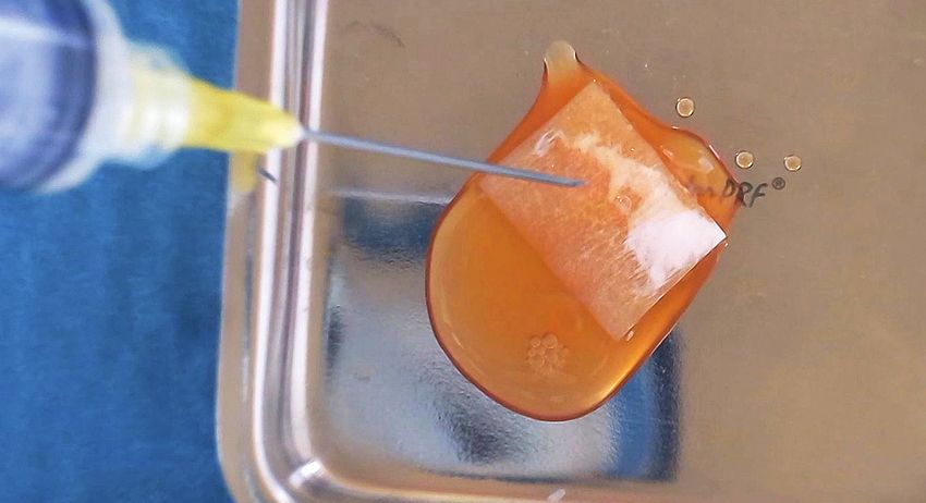
editorial   |

   Dr Rolf Vollmer

   First Vice President and Treasurer of DGZI

Join DGZI on
the track for success
With a 50-year history, the German Association of Dental       In 2019 the American Board of Oral Implantology (ABOI)
Implantology (DGZI) represents a practice-oriented and         in the US decided to make the ABOI/ID Diplomate ex-
evidence-based approach to implantology in Germany.            amination available for experienced dental practitioners
DGZI always keeps pushing boundaries in this particu-          internationally. The ABOI has an independent examina-
larly innovative field of dentistry. We place an overarching   tion committee chartered by the American Academy of
focus on actively supporting our practising colleagues         Implant Dentistry (AAID), the official US partner of
and dental technicians by offering a wide range of spe-        DGZI. Now, especially graduates of the DGZI implanto-
cial training courses. I can proudly say that DGZI is one      logy curriculum, as well as holders of both Expert in
of the best internationally networked German expert            Dental Implantology and DGZI Specialist in Oral Implan-
societies for dental implantology, boasting more than          tology certification can take this examination to become
registered 4,000 members in Germany alone and more             an ABOI/ID Diplomate in addition to the already gained
than 13,000 cooperating members in 66 countries ab-            credentials. DGZI can offer individual training and/or
road. In addition, the annual meeting of DGZI is consid-       preparatory seminars for those wishing to pursue this
ered a definite highlight and it is fair to say that it has    prestigious certification on request.
become an integral part of the annual schedules of
numerous clinicians from all around the globe.                 Last but not least we should not forget our endeavours in
                                                               publishing: The German language Implantologie Journal
With more than 300 holders of the DGZI specialist in oral      and the English language implants—international maga-
implantology certification, our expert society can recom-      zine of oral implantology. The former is DGZI’s personal
mend a vast number of highly qualified clinicians who          member journal and delivered to our German DGZI mem-
continue to break the mould every single day in their den-     bers, oral surgeons, prosthodontists and dental techni-
tal practices. Thanks to our close ties to universities and    cians on a monthly basis. The latter, with a circulation of
research facilities we can implement the latest findings       10,000 copies and four issues every year, is popular in
effortlessly into their daily practice. Fully recognised by    over 90 countries around the world.
the “Konsensuskonferenz Implantologie” (a joint initiative
of German professional associations involved in dental         I wish you a great and interesting time reading this first
implantology), the one-year DGZI implantology curricu-         2020 issue of implants—international magazine of oral
lum is the base for the practitioner. It features an inno-     implantology.
vative e-learning concept, which consists of regularly
updated compulsory and optional modules, and is now            Yours,
offered in English too. The DGZI implantology curriculum
is a state-of-the-art beacon of education for young as-
piring dentists looking to delve deeply into the specialist
field that is oral implantology.                               Dr Rolf Vollmer

                                                                                                                        1 2020   03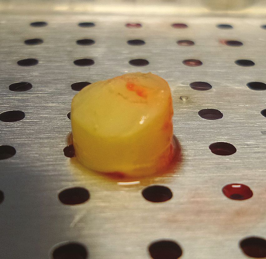
| content

                                                                             editorial
                                                                             Join DGZI on the track for success                                03
                                                                             Dr Rolf Vollmer

                                                                             research
                                                                             The dos and don’ts in the handling of PRF                         06
                                                                             Prof. Shahram Ghanaati, Dr Sarah Al-Maawi, Dr Eva Dohle &
                                                                             Dr Torsten S. Conrad

                                                                             Success rate of dental implants in heavy smokers                  12
                                                                             Drs Branislav Fatori & Inge Schmitz
                                                                   page 06

                                                                             case report
                                                                             Implant-retained overdenture on a very thin bone ridge            16
                                                                             Drs Nicola Alberto Valente, Murali Srinivasan & Nicole Kalberer

                                                                             Peri-implant bone regeneration through laser decontamination      22
                                                                             Prof. Wilfried Engelke, Dr Christian Engelke,
                                                                   page 16   Dr Victor Beltrán & Dr Marcio Lazzarini

                                                                             Sixteen years of follow-up after insertion of a Z1 implant        28
                                                                             Dr Virgilio Masini

                                                                             industry
                                                                             The next generation biomaterial for soft-tissue augmentation      32

                                                                             practice management
                                                                             Feedback done right                                               34
                                                                             Andrea Stix, MSc, MBA

                                                                   page 32   interview
                                                                             Reshaping implantology                                            36
                                                                             The link between oral disease and oxidative stress                38
Cover image courtesy of
TBR Dental Group
www.tbr.dental
                                                                             news
                                                                             manufacturer news                                                 40
                                                          1/20
 issn 1868-3207 • Vol. 21 • Issue 1/2020

 implants
 international magazine of oral            implantology
                                                                             news                                                              44

                                                                             about the publisher
                                                                             imprint                                                           50
 research
 The dos and don’ts in the handling of PRF

 case report
 Peri-implant bone regeneration
 through laser decontamination

 industry
 The next generation biomaterial
 for soft-tissue augmentation

04                                                        1 2020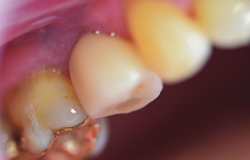
TRULY CONICAL PRECISION

                               CONELOG®
                               PROGRESSIVE-LINE
                               conical performance 1,2
                               at bone level

                               Precise conical connection
                                  long conus for reduced micromovements
                                  superior positional stability in comparison
                                  to other conical systems 1,2
                                  easy positioning with excellent tactile feedback
                                  integrated platform switching supporting the
                                  preservation of crestal bone

                  [1] Semper-Hogg, W, Kraft, S, Stiller, S et al. Analytical and experimental position stability
                  of the abutment in different dental implant systems with a conical implant–abutment
                  connection Clin Oral Invest (2013) 17: 1017
                  [2] Semper Hogg W, Zulauf K, Mehrhof J, Nelson K. The influence of torque
                  tightening on the position stability of the abutment in conical implant-abutment
                  connections. Int J Prosthodont 2015;28:538-41
X.J7513_01-2020

                  www.biohorizonscamlog.com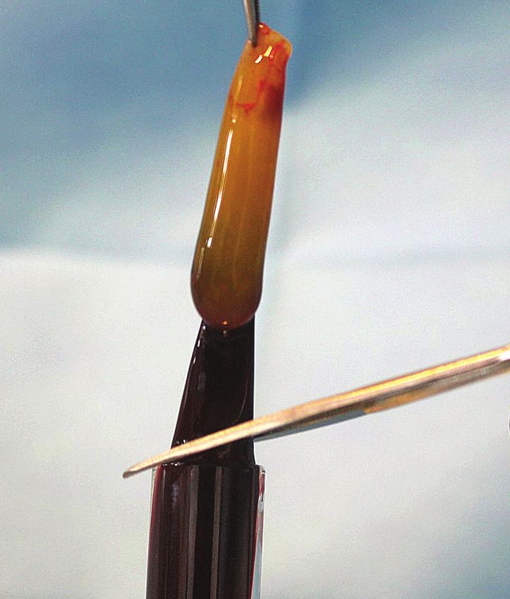
| research
The dos and don’ts
in the handling of PRF
Prof. Shahram Ghanaati, Dr Sarah Al-Maawi, Dr Eva Dohle & Dr Torsten S. Conrad, Germany
ative cells.6, 7 The composition and bioactivity of PRF
30° depends primarily on the centrifugal force that is used
during centrifugation.3
Several recent studies have demonstrated the influence
Vein of the centrifugal force on the composition and bioactivity
of the obtained PRF.8–12 It has been shown that the ap-
Fig. 1: Visualisation of the puncture direction during blood collection. plication of a low centrifugal force for accumulation leads
to a significantly higher number of platelets and leuko-
cytes in PRF compared to a medium or high centrifugal
Autologous blood concentrates, and platelet-rich force.3, 10 Growth factors are released in a similar way.
fibrin (PRF) in particular, are increasingly used today to PRF matrices that are prepared with a low centrifugal
support wound healing and regenerative processes.1 force release significantly higher concentrations of differ-
PRF is made from the patient’s own peripheral blood ent growth factors (such as VEGF, PDGF, EGF, TGF-b1)
without the addition of anticoagulants. A solid or liquid compared to PRF matrices that are prepared using a
PRF matrix can be obtained through a single centrifu- higher centrifugal force.8–12 As a result, the so-called
gation process, depending on the collection tube that Low-Speed Centrifugation Concept (LSCC) was intro-
is used.2 Through this centrifugation process, the blood duced, which aims at standardising the production of
components are separated according to the centrifugal blood concentrates and enabling reproducible treatment
force used. The red blood cells move towards the bot- protocols or clinical results.3 This article will particularly
tom of the tube.3 The platelets and leucocytes are con- focus on the technical aspects of the clinical applica-
centrated in the upper layer, the remaining fibrin ma- tion and handling of PRF. The tubes used for the pro-
trix. Thus, this autologous blood concentrate, which duction of PRF have been specifically developed for this
also contains further plasma proteins, is capable of particular purpose. Depending on the clinical indication,
actively releasing different growth factors such as vas- two different variants of PRF matrices exist. PRF tubes
cular endothelial growth factors (VEGF), epidermal with a glass surface promote coagulation. During cen-
growth factors (EGF), or platelet-derived growth factors trifugation, a solid PRF matrix is formed. In contrast, the
(PDGF) over a relatively long period of time (up to fif- coagulation process can be slowed down by means of
teen days).4, 5 These growth factors play a key role in the plastic-coated tubes. Accordingly, coagulation is slowed
support of wound healing and regenerative processes, down during centrifugation. At room temperature, a
since they contribute to the formation of new vessels, PRF matrix remains liquid for about thirty minutes after
epithelialisation and the stimulation of further regener- centrifugation until it eventually coagulates.
Protocol RPM (x 100) Duration (min) Centrifugal force (x G)
High concentration RCF 2,400 8 710
Medium concentration RCF 1,200 8 177
Low concentration RCF 600 8 44
RCF, relative centrifugal force; RPM, revolutions of the centrifuge per minute.
Table 1: Visualisation of the different LSCC protocols (Low-Speed Centrifugation Concept for a centrifuge with a radius of 110 mm).
06 1 2020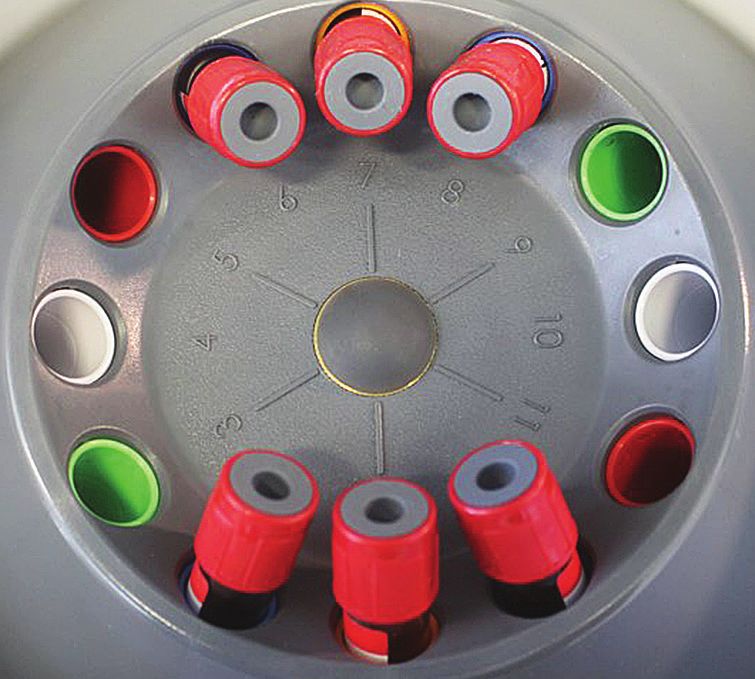
research   |

Blood collection                                                                    flattened once the vein has been hit. The vacuum system
                                                                                    of the PRF tube then fills the tube with venous blood un-
For the production of PRF the patient’s own venous blood                            til an amount of 10 ml is reached and the blood supply
is required, which is taken from the peripheral veins after                         can be stopped automatically. After loosening the tour-
the patient has been fully briefed on the procedure. This                           niquet tube, the butterfly cannula can be removed. Sub-
blood collection is a routine method and is particularly                            sequently, sufficient pressure should be exerted on the
used in diagnostics. The blood collection should be car-                            puncture site with a sterile swab in order to avoid second-
ried out according to the guidelines of the World Health                            ary bleeding underneath the skin.
Organization (WHO).13 In order to find a suitable puncture
site, the anatomical position of the peripheral veins should                        Centrifugation
first be palpated. For this purpose, the vena mediana
cubiti, which is located in the antecubital fossa (inner                            In order to avoid early physiological coagulation of the
bend of the elbow), is ideally suited. Gloves must be worn                          blood, the PRF tubes must be centrifuged quickly after
and the tourniquet must be placed approximately 5 cm                                blood collection in a dedicated centrifuge that stands
above the puncture site of the vein, which must be dis-                             on a table nearby in the same treatment room. Through
infected with a skin antiseptic according to the manufac-                           centrifugation, a separation process is triggered, which
turer’s instructions. A butterfly needle is then inserted into the                  sediments cells and/or biomolecules from a suspen-
vein at an angle of 30° to the skin surface (Fig. 1). To avoid                      sion (i.e. blood), depending on the relative centrifu-
completely piercing through the vein, the angle should be                           gal force and the size, shape and density of the vari-

 2

Fig. 2: Balance pattern of the centrifuge when loading two, three, four and six tubes. Centrifugation of five, seven, nine or eleven tubes is not possible. For this
purpose, an additional tube filled with water must be used.

                                                                                                                                                                 1 2020    07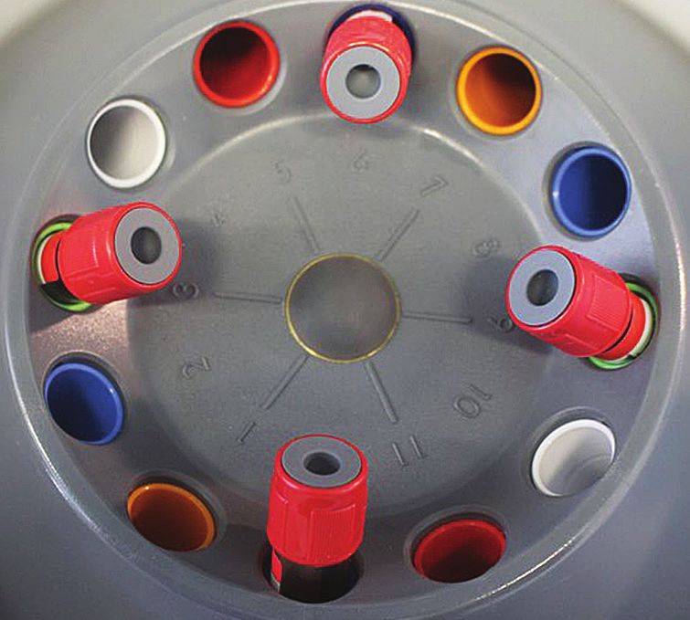
| research

3                                4                                                           5

             Fig. 3: Separating the red phase from the solid PRF phase. Fig. 4: A PRF plug. Fig. 5: A thinly pressed PRF matrix.

             ous suspension components. The relative centrifugal                              is given in Table 1 and should be set on the centrifuge
             force (RCF) represents the centrifugal force as a multi-                         according to the clinical indication (r = 110 mm). In gen-
             ple of the Earth’s gravity and is often expressed as the                         eral, the centrifuge should be placed on a stable and even
             value G. Moreover, it is a decisive factor for the result-                       fundament. When loading the centrifuge with the blood-
             ing concentration of the sedimented cells and biomol-                            filled tubes, it is imperative to ensure that any imbalance
             ecules in PRF. The value G indicates exactly the force                           is eliminated. This means that the tubes must be placed
             required for an optimal centrifugation of a correspond-                          inside of the rotor in such a way that the weight of the tube
             ing suspension (in this case blood) to obtain the desired                        placed exactly opposite the other is identical (Fig. 2). If the
             sediment (in this case PRF) as final product, and allows                         number of tubes is uneven, a tube filled with the equiva-
             the calculation of the necessary speed of the centrifuge                         lent volume (such as sodium chloride, for example) must
             rotor for a corresponding tube and centrifuge.                                   be added to compensate for the weight.

             For centrifuges where only the rotational speed (revolu-                         Processing of PRF
             tions of the centrifuge per minute, RPM) can be set, the
             RCF or the necessary G-value must first be calculated                            Immediately after centrifugation, the tubes are carefully
             by means of a fixed formula. The relation between ro-                            removed from the centrifuge and transferred to an ap-
             tational speed (RPM) and RCF depends on the size of                              propriate tube holder. Owing to the applied RCF and de-
             the rotor (r = radius of rotation = distance between the                         pending on the size, shape and density of the blood com-
             axis of rotation and the bottom of the tube). Here, the fol-                     ponents, only two phases can now be visually identified:
             lowing formula is used for conversion:14 RCF = 1.12 x ra-                        a red phase at the bottom of the tube, which contains
             dius x (RPM/1,000)2. The relative centrifugal force re-                          mainly erythrocytes and a PRF phase on top of it, fill-
             quired for PRF production using the established LSCC                             ing up the upper part of the tube. In the case of solid
                                                                                              PRF, which is obtained by centrifuging the blood in the
                                                                                              red PRF tubes, these two phases coagulate very quickly.
                                                                                              In order to separate the solid PRF matrix from the red
                                                                                              lower phase, it is recommended to first separate the two
                                                                                              phases roughly by cutting them with scissors. In concrete
                                                                                              terms, this means carefully lifting up the upper phase of
                                                                                              solid PRF with sterile tweezers (the lower red phase is
                                                                                              lifted as well) and then roughly separate the two phases
                                                                                              in the upper part of the red phase (Fig. 3). The PRF phase
                                                                                              (with remains of the red phase) is then transferred into a
                                                                                              dedicated PRF box provided for this particular purpose.
                                                                                              This PRF box, which has been specially developed for
                                                                                              various PRF indications, consists of a stainless-steel
                                                                                              container with a self-weighted lid and a large and small
                                                                                              stamp. In this box, the remaining parts of the red phase
                                                                                              can now be removed from the PRF phase by carefully
6
                                                                                              wiping it off with a blunt object (such as a closed pair of
             Fig. 6: Separating liquid PRF from the red phase by means of a syringe.          scissors). Thereafter, solid PRF matrices can be further

08                  1 2020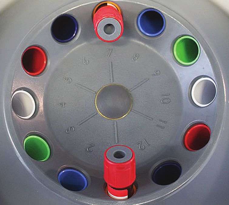
Conical? Parallel?

copaSKY !
The unique hybrid connection
for sophisticated restorations!

                                                                                                                                                                                              Photo: © Getty Images

                   Leading in Immediate Restoration – powered by physiological Prosthetic!

S I N C E       1 9 7 4

bredent UK Ltd · Unit 26, Tapton Park Innovation Centre · Brimington Road · Chesterfield S41 0TZ · T: +44 1246 559599 · F: +44 1246 557777 · www.bredent.co.uk · @: enquiries@bredent.co.uk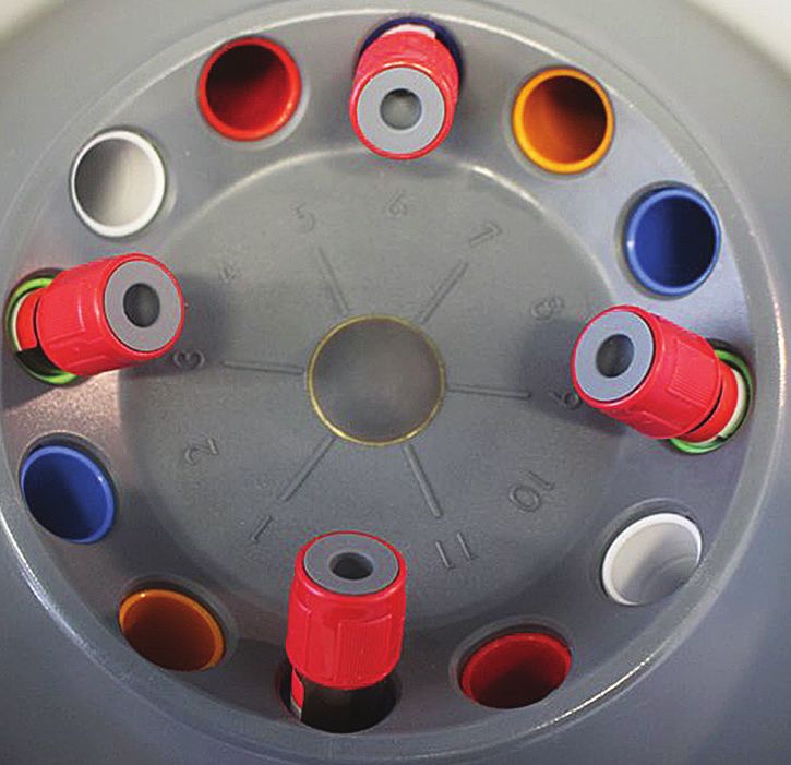
| research
after 10 to 15 days, facilitating the formation of cavities in
the augmentation sites as a result.
In order to biologise biomaterials of different classes
and origin, such as bone grafting materials and colla-
gen-based membranes (allogeneic, xenogeneic, synthetic),
they can be combined with solid or liquid PRF. Here,
biomaterials that do not trigger a foreign body reaction
should ideally be used. With the help of PRF it is possible
to biologise or functionalise these biomaterials, since they
themselves do not have any bioactive components, but
serve only as a mere scaffold. The functionalisation of
biomaterials with endogenous cells, plasma and growth
7
factors that are found in great concentration in PRF
Fig. 7: Biologising a collagen-based matrix with liquid PRF. should lead to an improved integration of the biomaterial
in certain defects and, as a consequence, to an increased
regenerative capacity. When combining PRF with bone
processed either into a PRF plug (Fig. 4) or into a thinly grafting material and collagen-based membranes, PRF
pressed PRF matrix (Fig. 5), depending on the clinical in- in liquid form should be used. In order to biologise and
dication. For PRF application in the extraction sockets of stabilise these bone grafting materials or membranes,
wisdom teeth, the fibrin clot is placed in the containers in liquid PRF collected in the syringe is drizzled onto them,
the box, which are specifically provided for this very in- which also simplifies clinical handling (Fig. 7).
dication, and carefully compressed with the small stamp
until the appropriate size of the PRF plug is reached. A comprehensive list of references can be obtained from
the authors.
In order to process PRF into a thinly pressed matrix, the
fibrin clot is carefully placed on the grid of the PRF box
and processed into a pressed PRF matrix using the self- Dr Sarah Al-Maawi Dr Eva Dohle Dr Torsten S. Conrad
Author details Author details Author details
weighted lid of the PRF box. This pressed PRF matrix can
then, for example, be placed as a wound cover on the
surgical sites where autologous palatal soft-tissue trans-
plants have been removed to support the wound heal-
ing process. In addition, it can be used during implant
surgeries as a support of the mucosal flap. As opposed
to the earlier-described solid PRF, which is produced
through centrifugation in glass tubes, the two phases of about the author
the blood remain liquid at first for a few minutes if cen-
trifugation is done with plastic tubes. The production of Frankfurt am Main-based Prof. Shah-
liquid PRF opens up the possibility of further PRF appli- ram Ghanaati is a specialist in maxillo-
cations in the field of regenerative medicine, such as the facial surgery and oncology. In 2013, he
combination of PRF with bone grafting materials and was appointed Director of the University
collagen membranes. The upper, still liquid PRF phase Cancer Center of the Frankfurt Univer-
is carefully removed from the remaining lower red phase sity Hospital. He is the Senior Physician
using a syringe. A 5 ml syringe with a long and wide and Deputy Director of the Department
21-gauge cannula is best suited for this purpose (Fig. 6). of Oral and Maxillofacial Plastic Surgery
It is recommended that the tube is slightly tilted so that of the Frankfurt University Hospital. In
the boundary between the two phases can be seen more addition, he is the Director of the research laboratory FORM-Lab
easily. In this way as much liquid PRF as possible can (Frankfurt Orofacial Regenerative Medicine).
be extracted from the tube, without extracting too much
of the red phase. With a liquid matrix, the wetting of the contact
biomaterial surface can be carried out in a reproducible
fashion. In contrast, based on the preliminary results of Prof. Shahram Ghanaati Author details
the ongoing clinical studies, it is no longer recommended Universitätsklinikum Frankfurt
to cut up a solid PRF matrix and mix the obtained pieces Theodor-Stern-Kai 7
with bone grafting material in order to create so-called 60590 Frankfurt am Main, Germany
sticky bone. Current research findings indicate that this Phone: +49 69 6301-3744
can lead to a loss of volume, since the fibrin is resorbed shahram.ghanaati@kgu.de
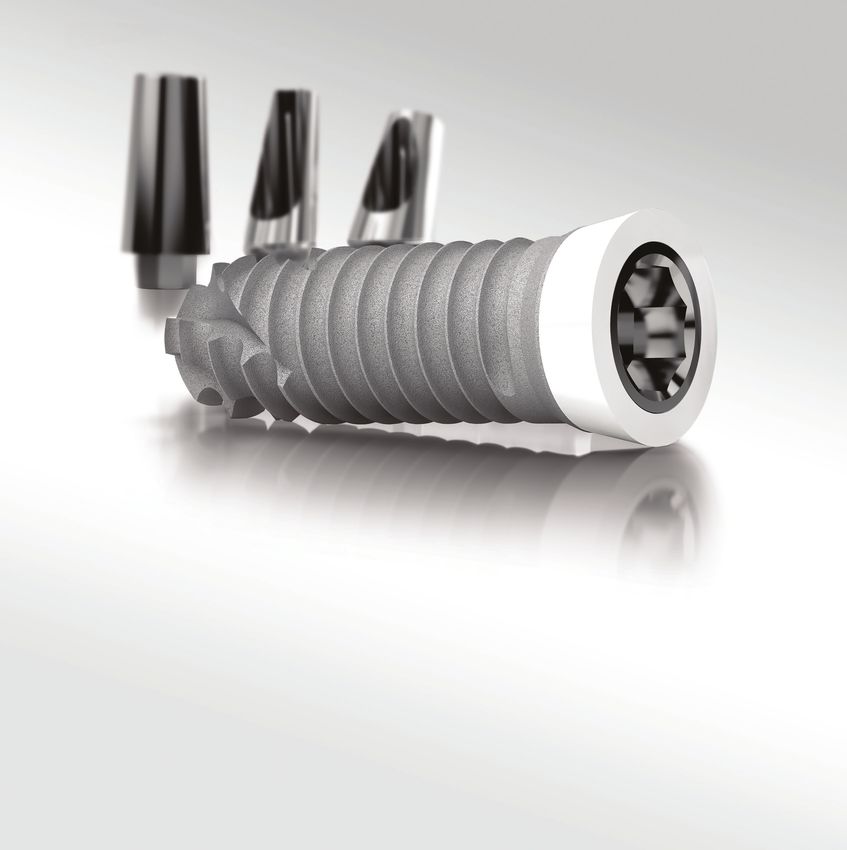
10 1 2020The unique Tissue Level
Implant with Zirconia Collar
Aesthetic
gingival area
Zirconia collar
Gingival integration
&
Osseointegration
Bone area
Pure Titaninum body
Z1 Implant
®
*LYLQJ\RXFRQȴGHQFH
in implantology.
Proven Technology Proven economics for a
SURȴWDEOHLPSODQWSUDFWLFH
✓ 98.6%* success rate ✓ Reduced chairtime
✓ Pure Titanium and Y-TZP Zirconia ✓ Practice development
data of the Smiletranquility® Program based on 15.534 patients with Z1 implants from 01/2014 to 01/2016.
*Z1 implants are medical devices of class IIb manufactured by SUDIMPLANT SAS. Information collected from the
✓ Suitable for all prosthetic solutions ✓ Patient satisfaction
Proven clinical outcomes Proven surgical protocols
for patient safety IRUDVLPSOLIHGZRUNȵRZ
✓ Anti-bacterial shield ✓ Only 1 surgery
✓ Ideal in fresh extraction sockets ✓ No healing abutment
✓ Immediate aesthetic result ✓ Visibility of the connection
Find us online
www.tbr.dental Unique, like your smile| research
Success rate of dental implants
in heavy smokers
A longitudinal study
Drs Branislav Fatori & Inge Schmitz, Germany
Objective smokers, a specific protocol was established in our den-
tal surgery. In the following, the results of two patients are
The failure rate of dental implants is reported to be higher reported in detail.
in smokers than in non-smokers. The aim of the study
described in this article was to compare the success Introduction
rate of 721 dental implants inserted in 181 smokers with
given reports in the literature. In our study, implants from In general, smoking is reported to increase implant failure
one factory were used and the implants were inserted and favour peri-implantitis. One possible mechanism that
by one surgeon to exclude individual factors. In order to might lead to increased failure rates is a lowering of the
increase the success rate for dental implants inserted in blood flow and direct adverse effects on the osteoblasts.
If smokers are treated with implants, good bone quality
is required. In our study, bone augmentation procedures
Diameter Length No. Successful Failed were necessary in 62 of the cases.
(mm) (mm)
With our study, we set out to investigate whether there
3.70 8.00 4 4 0 is a significantly enhanced risk of implant failure due to
3.75 10.00 182 167 15 the increased number of cigarettes smoked per day.
3.75 12.00 200 182 18 Though smoking is a risk factor for implant failure, it is
3.75 14.00 1 1 0 not considered an absolute contra-indication. When im-
3.30 12.00 38 35 3 plant treatment is planned, the patient’s smoking history
4.10 8.00 2 2 0 should first be obtained, including the duration, the in-
4.10 10.00 101 88 13 tensity (past and present), the present status of smoking,
the number of cigarettes smoked each day and whether
4.10 12.00 193 177 16
there is any notable passive smoking. Here, the surgeon
Total 721 656 65 has to rely completely on the correctness of the infor-
mation provided by the patient. To achieve a satisfactory
Table 1a result regarding implant survival, a number of different
1 2 3
Case 1—Fig. 1: The 54-year-old female patient had an extreme periodontal defect at tooth #44 with bone loss at the apical side. Fig. 2: Radiographic
close-up. Fig. 3: Final result.
12 1 2020AD
the implantation in order to promote
Jaw Regions No. osseointegration.
Upper 17–14 48
13–11 149 Materials and methods
21–23 151
24–27 64 We assessed 181 patients (97 women
and 121 men with an average age of
Lower 47–44 150 49.2 years) with 721 implants. In detail,
43–41 9
384 bone-level implants, 289 bone-level
31–33 9
34–37 141 tapered implants and 48 tissue-level im-
plants were inserted (Table 2). The im- Shortest Implants –
Table 1b plants were classified according to their Longest History.
location in the upper and lower jaws. As
for the smoking history of the patients,
factors have to be taken into consider- the number of cigarettes smoked per
ation, such as bone type and quality, day ranged from 20 to 60 cigarettes. Think Short!
bone density, placement and location
of the inserted implants (Tables 1a & b), Surgical technique
the patient’s personal situation, health For more than 30 years Bicon® short implants
risks and unrelated diseases, such as Implant placement was performed un- are unchanged in clinical use.
diabetes.1 der local anaesthesia (40 mg of Dexa-
ratiopharm, intramuscular; ratiopharm)
On smoking after premedication with antibiotics. The According to the 11th European Consensus
osteotomy was extended gradually ac- Conference (EuCC) 2016 in Cologne, provided
Smoking tobacco reduces leucocyte cording to the intended implant diam- the specific treatment parameters are ob-
activity. It has an influence on blood eter. After carrying out the incision, the
served, the use of short, angulated or diam-
vessels and reduces the body’s heal- oral cavity was cleaned and necrotic or
ing capacity and osseointegration of inflammatory tissue was removed. The eter-reduced implants in sites with reduced
dental implants. Smoking has a direct osteotomy sites were prepared with bone volume can be a reliable treatment
influence on osteoblastic function.2 a sequential order of drills, as recom- option, given the risks associated with the use
The exact mechanism by which smok- mended by the manufacturer. Implants
of standard-dimension implants in combina-
ing compromises wound healing is were inserted in the prepared osteotomy
still unknown. Smoking enhances the sites at an insertion torque of 45 Ncm. tion with augmentation procedures.
risk for ingress of bacteria which may
cause peri-implantitis. It is hypothe- Postoperative treatment
sised that nicotine and chemicals con-
For more Information:
tained in tobacco smoke induce a state Postoperative periapical radiographs
of oxidative stress in the tissue (gingiva were taken, which confirmed the accu- Bicon Europe Ltd.
and alveolar bone) around implants.3, 4 racy of the implant placement. Postop- Dietrichshoehe 2
Abstention from smoking should be erative medication included antibiotics. 55491 Buechenbeuren
extended to at least eight weeks after Digital radiographic images were taken
Germany
Phone +49 (0)6543 818200
germany@bicon.com
www.bicon.de.com
4 5
Case 2—Fig. 4: Defect situation after explantation and guided bone regeneration in a 67-year-old male patient who
smoked 42 cigarettes per day and suffered bone loss 27 years after implantation. Fig. 5: Implant with a new crown.
Popular sizes: 3x6, 3x8, 3.5x8, 4x5, 4x6, 4x8, 4x11, 4.5x6, 4.5x8,
5x5, 5x6, 5x8, 6x5, 6x6, 6x8 mm| research
Type Diameter (mm) Length (mm) No.
at the time of surgery, after 24 hours and one month later
Bone-level 3.30 8 0 in order to evaluate the success of the implant treatment.
3.30 10 0 Inflammatory processes were found in 24.1 % of the
3.30 12 47 patients. If necessary, augmentation was done by means
3.75 8 9 of NanoBone (Artoss), Geistlich Bio-Oss bone substi-
3.75 10 43 tute and Geistlich Bio-Gide membranes (both Geistlich
3.75 12 62 Biomaterials).
4.10 8 18
4.10 10 94 Indication for implants
4.10 12 92
4.10 14 7 The indications for inserting implants in our study were
as follows:
4.80 8 4
–– treatment of the edentulous jaw;
4.80 10 4
–– single-tooth replacement;
4.80 12 4
–– treatment of larger interdental gaps; and
–– free-end situation.
Bone-level 3.30 8 0
tapered 3.30 10 0
Results
3.30 12 29
3.75 8 4 Of the 721 implants inserted, 65 implants failed. Conclu-
3.75 10 48 sively, the success rate was at 90.98 %, which is lower
3.75 12 97 compared with our previous study on non-smokers, in
4.10 8 19 which the success rate was 98.70 %.2 In the group of
4.10 10 39 failed implants, most of them (75.4 %) were lost two to
4.10 12 50 four weeks after implant placement owing to a lack of
4.10 14 3 osseointegration. Peri-implantitis occurred in 20 % of
4.80 8 0 the failed implants. This could be traced back to poor
4.80 10 0 oral health and plaque formation. In 4.6 % of the cases,
4.80 12 0
Tissue-level 3.30 8 0
3.30 10 0
3.30 12 6
3.75 8 0
3.75 10 0
3.75 12 0
4.10 8 8
4.10 10 9
4.10 12 22
4.10 14 0
4.80 8 0 6
4.80 10 0
4.80 12 0
Tissue-level 3.30 8 0
ROX-CERA 3.30 10 0
3.30 12 0
4.10 8 0
4.10 10 1
4.10 12 0
Tissue-level WP 4.80 8 0
4.80 10 0
4.80 12 2 7
Table 2 Case 2—Fig. 6: Cemented crown in situ. Fig. 7: Final result.
14 1 2020research |
peri-implantitis occurred between one and three years Conflict of interest: Dr Inge Schmitz declares that she has
after implant placement owing to mechanical issues after no conflict of interest.
bone loss. There was no correlation to be found between
implant length and diameter and the implant failure rate, Acknowledgement: Dr Branislav Fatori would like to ex-
and neither did we find a correlation between the number press his gratitude to DENTAL RATIO, and to Ulf Hen-
of cigarettes smoked and the implant failure rate. schen in particular, for the technical support and the
donation of implants. In addition, he would like to thank
Discussion Dr Walter Gerike from Artoss in Rostock, Germany, for
his long-standing support.
As established earlier, the failure rate of dental implants
in smokers is higher than in non-smokers, which is due All images: © Branislav Fatori
to lack of early osseointegration and the occurrence of
peri-implantitis. Peri-implantitis was obvious in 62 cases
included in our study. Failed osseointegration was the
Literature
main reason for implant failure. However, in conclusion, it
must be stated that the results we obtained were excellent.
Dental implant therapy is a treatment of choice for treat-
ing patients with missing teeth. However, certain condi-
tions, such as smoking, hypertension and diabetes, have
a negative influence on the success of dental implants. about the authors
Nicotine is found to cause osteoclastic changes. Based
on the cases described here and the results in other pa- Dr Branislav Fatori has more than
tients, it can be concluded that today good results can 41 years of experience in implantol-
be obtained in heavy smokers. It is difficult to evaluate ogy and has placed more than 8,000
the role of a single risk factor such as smoking regard- implants. He was trained at prominent
ing positive treatment outcomes of dental implants, since clinics in Germany, the US, Sweden,
many patients have additional co-risk factors, including Serbia and Switzerland. In addition,
diabetes, advanced age or low bone density. In addi- he has worked as a long-term training
tion, there is great variance in smoking behaviour regard- consultant for professional societies and
ing the actual number of cigarettes smoked per day and implant manufacturers.
the years for which a patient has been smoking. Further-
more, the location of implants, placed in either the maxilla Dr Inge Schmitz has worked at the
or the mandible, may have an influence on osseointegra- Institute of Pathology of the Ruhr-Uni-
tion success. Marginal bone loss around implants placed versity Bochum in Germany since 1990.
in smokers is more pronounced in the maxilla. Implant Her main interests are implant den-
failure may vary with implant location in connection to tistry, stents, electron microscopy and
the quality and quantity of the alveolar bone in which the osteology. She studied biology at the
implant is placed. The alveolar bone varies in terms of Ruhr-University Bochum and completed
mineral density, microarchitecture and trabecular bone her PhD in anatomy at the University of
thickness. Essen in Germany in 1989.
In the relevant literature, it is reported that smoking of contact
Author details
more than 30 cigarettes per day and for a duration of
longer than ten years promotes implant failure. There is Dr Branislav Fatori
little data available, however, on passive smoking and Rolandstraße 11
ex-smokers in this regard. 45128 Essen, Germany
Phone: +49 201 82188890
Summary and outlook info@fatori.de
The risk of implant failure increases with increasing num- Dr Inge Schmitz
ber of cigarettes smoked per day. We found a correla- Georgius Agricola Stiftung Ruhr Author details
tion between heavy smoking and implant loss. Smoking Institut für Pathologie
influences the survival rate of dental implants. Thus, pa- Ruhr-Universität Bochum
tients should be educated thoroughly and be advised to Bürkle-de-la-Camp-Platz 1
discontinue the habit before implant placement can be 44789 Bochum, Germany
carried out. inge.schmitz@rub.de
1 2020 151a 1b 2a
2b 3a 3b
4a 4b 4c
Fig. 1a: Occlusal view before the treatment showing a very thin ridge. Fig. 1b: Frontal view of the ill-fitting mandibular denture. Fig. 2a: Frontal view of the
initial clinical situation. Fig. 2b: Preoperative radiograph. Fig. 3a: A flap was raised to obtain a clear view of the underlying bone. Fig. 3b: Preparation of the
four implant sites. Fig. 4a: Placement of the four implants. Fig. 4b: The flap was closed with a 4/0 polyamide continuous suture. Fig. 4c: Radiograph taken
immediately after surgery.
Implant-retained overdenture
on a very thin bone ridge
Drs Nicola Alberto Valente, Murali Srinivasan & Nicole Kalberer, Switzerland
Initial situation in vestibule depth, which would be unfavourable for the
rendition of the prosthesis (Figs. 1 & 2). After evaluating
A healthy 60-year-old female patient with no medical his- the patient’s motivation, the decision was made to use
tory presented at our clinic with a non-fitting full mandib- the new Straumann® Mini Implants (2.4 mm diameter)
ular denture. Her chief complaints at this point included with the integrated Optiloc® retention system for a new
the lack of retention of her mandibular denture and poor denture supported by four implants. The implants were
aesthetics, coupled with difficulty in chewing and embar- planned for placement in the regions 34, 32, 42 and 44.
rassment at social events. The treatment plan comprised Due to the very limited width of the ridge, open flap sur-
the rehabilitation of jaw function and aesthetics with a gery was planned in order to place the implants safely
new set of dentures, including a conventional maxillary under direct vision.
complete denture (CD) and a mandibular implant-sup-
ported overdenture (IOD) retained by four implants. For Surgical procedure
standard implants, the ridge would have had to be re-
duced by a vertical osteotomy in order to gain thickness After a careful crestal incision, keeping the edge of the
and to reach the wider portion of basal bone. However, blade always in contact with the thin bone ridge, a cen-
this would cause both a loss of height and a reduction tral release incision was performed. The flap was raised
16 1 2020case report |
to obtain a clear view of the underlying bone (Fig. 3a). Prosthetic procedure
In the area of the left incisor, the ridge appeared to be
too thin for implant placement, probably owing to a pre- After a healing period of six weeks, the patient was re-
vious cystic lesion. The implant that had initially been ferred to the Division of Gerodontology and Removable
planned in region 32 was therefore moved to region 33. Prosthodontics at the University Clinic of Dental Medi-
For the implants in regions 42 and 34, the site was pre- cine in Geneva in Switzerland for the final rehabilitation
pared sequentially with the needle drill (1.6 mm diame- of her completely edentulous maxilla and mandible, with
ter) and the pilot drill (2.2 mm diameter), while only the the Straumann® Mini Implants placed in the latter. During
same needle drill was used for the implants in regions the first consultation, preliminary impressions were taken
44 and 33. During the preparation of the implant sites, using an irreversible hydrocolloid impression material.
parallelism was verified at all times through the parallel Simultaneously, the patient’s conventional mandibular
posts (Fig. 3b). Finally, the four implants were placed in CD was relined using a functional impression tissue con-
the respective sites, initially using the vial caps and then ditioning material for better interim retention. In the max-
inserted and stabilised with the Optiloc® ratchet adapter illa, a conventional impression was taken using a custom-
and the ratchet itself (Fig. 4a). The flap was closed with ised impression tray, enabling a mucodynamic border
a 4/0 polyamide continuous suture (Figs. 4b & c). Owing moulding followed by a mucostatic final impression
to the thin bone crest, immediate loading was avoided using zinc oxide eugenol impression material. In the man-
by grinding resin from the existing prosthesis in order to dible, the Optiloc® impression/fixation matrices were
prevent contact with the transgingival part of the implants placed on the Optiloc® before a mucodynamic impres-
during the healing phase. sion was taken with an elastomeric polyvinyl siloxane
5a 5b
7b
6a 6b
7a 7c 7d
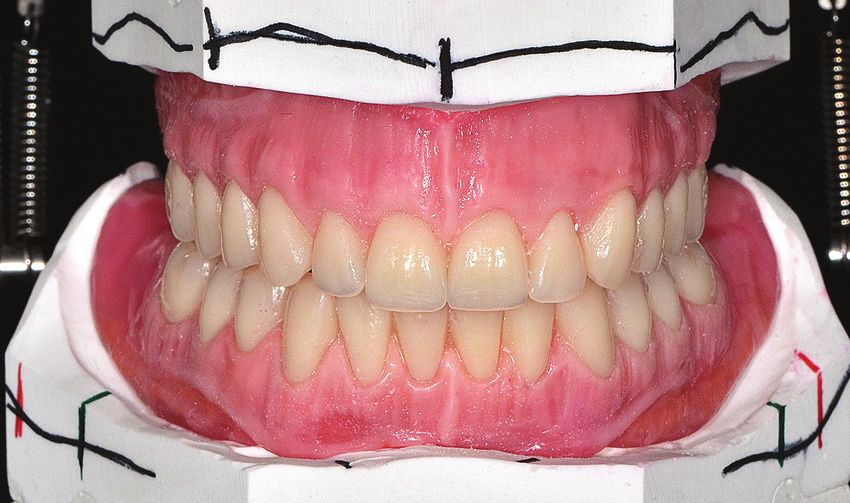
Figs. 5 & 6: A mucodynamic impression was taken. Fig. 7a: The master models were prepared using the Optiloc® analogues and standard techniques.
Figs. 7b–d: Aesthetic teeth exposure was ensured (b), the occlusal planes were checked (c), and the vertical dimension of occlusion was defined (d).
1 2020 17| case report
8a 8b
9a 9b
Fig. 8a: Final bite registration was performed. Fig. 8b: Photographs of the patient’s natural dentition helped in preparing the final teeth set-up. Figs. 9a & b: The
final set-up was checked during try-in.
(PVS) impression material (Figs. 5 & 6). The preparation was able to suggest modifications and give her consent
of the master models and corresponding wax rims and before the final prostheses were prepared. To prevent
all subsequent laboratory works were carried out in the fractures and ensure the longevity of the mandibular IOD,
Swiss-based dental laboratory Zahnmanufaktur Zimmer- a polyether ether ketone (PEEK) reinforcement was in-
mann und Mäder in Bern using the Optiloc® analogues corporated in the final prosthesis (Fig. 10). The new
and standard techniques (Fig. 7a). The next clinical steps conventional maxillary CD and mandibular IOD on the
included verification of the upper and lower lip support Optiloc® retention system was then finalised in the dental
(ensuring aesthetic teeth exposure), checking the occlu- laboratory, placing the Optiloc® housings and process-
sal planes, defining the vertical dimension of occlusion, ing inserts on all Optiloc® model analogues and following
and final bite registration (Figs. 7b–d). the usual manufacturing procedures. The dental labo-
ratory delivered the completed maxillary CD and man-
Communication with the dental laboratory using photo dibular IOD (Figs. 11a & b). During the final consultation,
graphs of the patient’s natural dentition was a key fac- the appropriate retention inserts (low force) Optiloc®
tor for successfully preparing the final teeth set-up were selected and inserted into the housings using the
(Figs. 8a & b). During try-in, the final set-up was checked Optiloc® retention insert placement tool (Figs. 12 & 13).
for lip support, occlusal planes, teeth exposure and The completed conventional maxillary CD and mandibu
occlusal contacts (Figs. 9a & b). Moreover, the patient lar IOD were then inserted into the patient’s mouth, and
10 11a 11b
Fig. 10: A PEEK reinforcement was incorporated in the final prosthesis. Figs. 11a & b: The dental laboratory delivered the completed maxillary CD and man-
dibular IOD.
18 1 2020Making the
difference together
Comprehensive Professional and Digital planning Regenerative Implant systems Restorative
solutions business development
Follow Dentsply Sirona Implants
dentsplysirona.com/implants| case report
Figs. 12a & b: Retention inserts (low force) Optiloc® were selected and
inserted into the housings using the Optiloc® retention insert position-
ing tool. Figs. 13a & b: Occlusal and frontal view at the final consultation.
Figs. 14a & b: Frontal view of the inserted completed conventional maxillary
CD and mandibular IOD.
final post-insertion and denture hygiene instructions were
given to the patient (Figs. 14a & b).
12a
Conclusion
The case was successfully handled. The patient was
highly satisfied and reported increased functional com-
fort and social confidence. The use of four 2.4 mm diam-
eter Straumann® Mini Implants to support a mandibular
overdenture has proved to be a reliable technique, which
guaranteed satisfactory results both for the operator and
the patient in a case where traditional techniques with
larger diameter implants were not possible.
12b
Editorial note: The surgical procedures were performed
by Dr Nicola Alberto Valente and prosthetic procedures
by Dr Nicole Kalberer supervised by Dr Murali Srinivasan.
13a
about the author
Dr Nicola Alberto Valente graduated
in dentistry from the University Cattol-
ica del Sacro Cuore of Rome, Italy. He
completed his Master of Science in Oral
Sciences and his specialty program
13b
in periodontics at the State University
of New York at Buffalo, NY, USA. He
is a Diplomate of the American Board
of Periodontology and has had an ITI
Scholarship from the University of Geneva, Switzerland. He has
worked as Chef de Clinique in the Unit of Oral Surgery, Service
of Maxillofacial Surgery of the University Hospitals of Geneva,
University of Geneva until 2019. He will start his new duties as
Clinical Assistant Professor at the State University of New York
14a at Buffalo in 2020.
contact
Dr Nicola Alberto Valente Author details
University at Buffalo
School of Dental Medicine
250 Squire Hall, 3435 Main Street
Buffalo, NY 14214, USA
14b
www.dental.buffalo.edu
20 1 2020PRECISE 3D IMAGING
Show what you’re made of—with precision from PreXion.
In 2019 the Japanese technology group
PreXion successfully introduced their new
CBCT system PreXion3D EXPLORER. Espe-
cially developed for European and US mar-
kets, the high-performance system allows
for an extraordinary combination of the most
precise imaging, great picture detail, low
radiation exposure, safe diagnostics and
digital planning, covering the entire range
of indications in modern dentistry.
Show what you’re made of—
with precision from PreXion.
e a free O W.
a ng n N
Arr s e s sio prexion-eu.de)
ng 78558 | info@
traini 40
9 614
2
e: + 4
( Ph o n
PreXion (Europe) GmbH
Stahlstraße 42–44 · 65428 Rüsselsheim · Germany
Phone: +49 6142 4078558 · info@prexion-eu.de · www.prexion.eu| case report
Peri-implant bone regeneration
through laser decontamination
Endoscopic paracrestal tunnel technique
Prof. Wilfried Engelke, Dr Christian Engelke, Germany;
Dr Victor Beltrán, Chile & Dr Marcio Lazzarini, Germany
Introduction increasing probing depth, pain and radiographic bone
resorption. Implant loosening requires a high degree of
The recently published S3 guidelines of the German As- bone resorption in the case of peri-implantitis. Microbio-
sociation of Oral Implantology (DGI) and the German So- logical tests are rather unspecific regarding peri-implant
ciety of Dentistry and Oral Medicine (DGZMK) state that mucositis and peri-implantitis.
peri-implant infections can be categorised into peri-
implant mucositis and peri-implantitis.1 In peri-implant The goal of non-surgical peri-implantitis therapy is to elim-
mucositis, only the supracrestal soft-tissue interface is inate the clinical signs of the infection. In addition to a par-
involved; in peri-implantitis, the bony implant site is also tial or complete reduction in bleeding on probing (BOP),
involved.2 Smoking is the main risk factor for peri-implant an effective therapy should lead to a reduction in the
mucositis, but it is likely that there are further contributing depth of periodontal pockets.6 To date, deep peri-implant
factors, such as cement residue, diabetes mellitus and pockets have not been clearly defined, but in most cases,
sex.2 The development of peri-implantitis is particularly a probing depth of less than 6 mm is considered a treat-
favoured by a history of periodontal disease, smoking ment success.7 There are various treatment protocols
and interleukin-1 polymorphism.4, 5 The main diagnostic used for non-surgical therapy: procedures for biofilm re-
criterion for distinguishing peri-implantitis from peri-im- moval, antiseptic therapy and adjuvant antibiotic therapy.
plant mucositis is the lack of reversibility of the condition. Surgical peri-implantitis treatment includes surface decon-
Peri-implantitis can be characterised by putrid secretion, tamination, adjuvant resectional therapy and, if necessary,
Illu
s tr
a ti o
nI
mp
lan
t: ©
Jut
/ Sh
u t te
r s to
ck.
com
Fig. 1a: Open surgical peri-implantitis therapy with basal stemmed flap: application and operating direction of the laser for sulcular decontamination
(yellow), implant surface decontamination (blue) and bone decontamination (white).
22 1 2020case report |
adjuvant augmentative therapy. Surface decontamination reas outside the physiological barrier of current augmen-
a
by means of a modified ultrasonic system (hydroxyapa- tation procedures. Augmentation (xenogeneic bone sub-
tite suspension) led to a comparable reduction in mucosal stitute material of bovine origin and a barrier membrane)
bleeding and probing depth after six months to mechan- was carried out only in the area of intraosseous defects,
ical debridement using carbon fibre or titanium curettes.8 whereby the adjacent implant surfaces were preserved in
After an observation period of 12 months, BOP values in- their original structure, and these surfaces were decon-
creased again, especially in initially deep pockets.9 In con- taminated before augmentation. Over an observation pe-
ventional flap surgery for surface decontamination, the use riod of four years, combination therapy after open wound
of special decontamination methods (e.g. 980 nm diode healing led to a clinically relevant reduction in BOP and ST
laser, carbon dioxide laser, chlorhexidine digluconate and values. A difference between the two investigated decon-
cetylpyridinium chloride) did not lead to significantly better tamination methods was not observed.13
clinical or radiographic results than in the respective con-
trol groups, in which air polishing, chlorhexidine solutions In summary, it is not possible at this point to clearly de-
and placebo solutions were used.10, 11 termine which protocol should be preferred, based on
current literature. In the case of surgical therapy, gran-
The clinical effectiveness of an adjuvant augmentative ulation tissue should first be entirely removed. The de-
measure for flap surgery alone (titanium curettes and sur- contamination of exposed implant surfaces should be of
face conditioning with 24 % ethylenediaminetetraacetic central importance. Mechanical procedures (for reduc-
acid and covered wound healing for six months) was in- ing biofilm) and chemical procedures (for reducing and
vestigated in a prospective clinical study using a porous inactivating biofilm) are often combined. At this point in
titanium granulate for treating intraosseous defect compo- time, the additional benefit of peri- and/or postoperative
nents.12 After the primarily covered wound healing, a very antibiotic therapy cannot be assessed. Analogous to the
high exposure rate was observed in both groups (control guideline for perioperative antibiotic prophylaxis, a sup-
group: 12/16; test group: 13/16). After 12 months, both portive once-off administration can be done as part of
procedures showed a comparable reduction in prob- surgical peri-implantitis therapy. After decontamination,
ing depth and only minor improvements in peri-implant augmentative measures can lead to a radiographically
bleeding values. However, in the test group, a significantly detectable filling of intraosseous defect components. It
higher decrease in radiographic translucency in the in- should be noted that all surgical therapy approaches
traosseous defect area, as well as an increase in implant involve a high risk of postoperative mucosal recession.
stability, was observed.12 For advanced, complex defect Soft-tissue augmentation can be performed to stabilise
configurations, surgical augmentative and resectional the peri-implant mucosa.14
procedures were combined as part of an implantoplasty
procedure. An implantoplasty was aimed at smoothing In addition to these general explanations based on the
the macro- and microstructure of the implant body in guidelines, a number of techniques have been described
Illu
s tr
a ti o
nI
mp
lan
t: ©
Jut
/ Sh
u t te
r s to
ck.
com
Fig. 1b: Surgical peri-implantitis therapy with closed endoscopic paracrestal tunnel technique: application and operating direction of the laser for sulcular
decontamination (yellow), implant surface decontamination (blue) and bone decontamination (white).
1 2020 23| case report
Fig. 2: Endoscopic equipment to guide laser fibres for peri-implant bone decontamination.
that could support modern peri-implantitis treatment Kirchner and Engelke emphasised the satisfaction of the
based on a minimally invasive therapy concept, given that patients owing to the minimally invasive nature of the pro-
their concepts can be combined in order to safely de- cedure.19 However, there has not been a good solution,
contaminate the implant surface. Kim et al. made a small thus, far to the problem of accessing contaminated and
labial incision with subperiosteal tunnelling for horizon- infected implants, since most endoscopes do not feature
tal ridge augmentation.15 They used bone grafts, which working shafts particularly designed for this kind of appli-
were placed in the soft-tissue pocket created by tunnel- cation. This paper presents a concept that allows for tar-
ling and subsequently fixed by conventional means so geted and visually controlled implant decontamination,
that they could successfully integrate implants into the removal of granulation tissue and simultaneous augmen-
alveolar ridge in the context of a two-stage procedure.15 tation without the need for open-flap reflection.
Montevecchi et al. reported cases of peri-implantitis in
which fibres of dental floss attached themselves to the Case report
implant superstructure and, as a result, gave rise to
peri-implantitis.16 They were able to remove these fibres A 48-year-old female patient presented with an in alio loco
using a periodontal endoscopic technique and, in doing placed exposed titanium screw-retained implant. Upon
so, promote healing. The healing was confirmed over a examination, a triangular bony defect situation was noted,
six-year period. An endoscopically supported therapy in extending into the middle third of the implant. In addition,
implant dentistry was described by our working group there was secretion of pus. Upon pressure, the patient
for implant cavities and for sinus floor augmentation in experienced a feeling of tension and local pain. Explan-
a closed procedure.17, 18 In this context, a tunnel tech- tation of the implant and bone regeneration measures for
nique was carried out laterally for the augmentation of the the purpose of a new restoration were discussed. Various
sinus floor, in which the entire basal maxillary sinus mu- possible treatment protocols were explained to the pa-
cosa was detached and tunnelled through without hav- tient, and minimally invasive microsurgical treatment us-
ing to cut a bony window, which made the procedure ing the tunnel technique was proposed. The patient was
less invasive. thoroughly informed about possible risks and the overall
problematic prognosis. In the tunnel technique, the im-
In 2003, Sennhenn-Kirchner and Engelke reported on a plant surface is reached through an entrance fashioned
procedure in which peri-implantitis can be successfully away from the implant, without interrupting the continu-
treated by endoscopic tunnelling and the use of a di- ity of the peri-implant tissue cuff. In order to gain an opti-
ode laser.19 The laser is used for decontaminating the ex- mal view in the tunnelled area throughout the procedure,
posed implant surfaces, followed by augmentation of the support immersion endoscopy is used (Fig. 1b).
peri-implant bone defects.19 The authors found that ra-
diographic defect filling and a reduction in probing depths The operation was performed via a mesial tunnel en-
can be achieved, with no postoperative infections and no trance outside the surgical field and under local anaes-
augmentation losses observed in five patients with eight thesia. After access away from the implant through a ver-
implants.19 Prior to the operation which their research is tical mucosal periosteal incision, subperiosteal tunnelling
based on, the probing depths were deeper than 6 mm was performed up to the affected implant. The surface of
and, afterwards, between 3 and 4 mm.19 Sennhenn- the implant was visualised by advancing the endoscope
24 1 2020SWISS PREMIUM ORAL CARE
| case report
while perfusing the tunnel with a sterile sodium chloride tion. Support immersion endoscopy allows a minimally
solution. The gingival cuff could be mobilised towards the invasive approach away from the implant. The different
occlusal plane via a high vestibular periosteal slit. Gran- types of support and irrigation shafts allow preparation
ulation tissue was removed and the implant surface de- under immersion. Blood and secretion are immediately
contaminated under direct endoscopic vision without ir- removed by the irrigation flow and do not interfere with
rigation. Decontamination was done with a GaAlAs laser the preparation of the operation site. After exposure of
set at 1 W and at a wavelength of 809 nm (Fig. 3). The the infected part of the implant surface inside the tunnel,
exposure time was 20 seconds. Four repetitions in con- laser decontamination should be done in an aerobic en-
tact mode were enough to produce sterile conditions.20 vironment, reducing heat generation and, thus, allowing
After filling the defect with tricalcium phosphate ceramic for targeted decontamination. Using intermittent irriga-
and locally obtained autogenous bone particles, the tion, the operating field can be freed from detritus and
minimally invasive access was closed with two button secretion at any time. Finally, surface decontamination is
sutures. The postoperative medication consisted of an done in the open operation area. The size of the tunnel
analgesic (paracetamol, 500 mg, if necessary) and a sin- entrance and its localisation can be reduced to such an
gle dose of antibiotic (clindamycin, 600 mg). The post- extent that large-area detachment of the flap and basal
operative course was inconspicuous, and the augmenta- flap extension by periosteal slitting can be avoided with-
tion height showed that the defect had been completely out compromising visualisation of the contaminated im-
regenerated. In the re-entry to expose the implant after plant surface.
four months, a complete bony covering of the implant
could be observed vestibularly (Fig. 4). The prosthetic Bleeding in the tunnel can be stopped by means of vaso-
restoration was performed by the family dentist. constrictors or direct laser coagulation so that an opti-
cally perfect assessment of the critical parts of the bone
Discussion pockets is possible using support immersion endoscopy.
Removal of granulation tissue with a laser has the ad-
The concept of microsurgical peri-implant bone regener- vantage that a low-bleeding preparation technique facili-
ation using the tunnel technique complies with the DGI/ tates the precision of the subsequent steps significantly.
DGZMK guidelines and has two significant advantages: This advantage of the endoscopic technique can also
firstly, the cervical gingival cuff around the implant is pre- be used for tunnel procedures in primary bone augmen-
served, and secondly, augmentation material can be se- tation, allowing reliable intraoperative quality control of
curely positioned in a zone of optimal perfusion through the microsurgical measures even without flap reflection.
the local periosteum. This significantly reduces the risk If dealing with fixed implants, it is not advantageous to
of postoperative recession and promotes bone regenera- remove the superstructure before the operation, since
the operating direction is apical. Removal should only be
carried out in pathological situations, for example inac-
curacies in fit. In the case of extensive interdental or oral
defects, multiple tunnelling sessions might be necessary.
Their indication should be clarified beforehand by means
of 3D imaging. In the case that is described in this arti-
cle, 3D diagnosis was not desired by the patient. Based
on the extensive experience of the authors with the de-
scribed procedure, it can be stated that the tunnelling
of apicoapproximal peri-implantitis is advantageous for
the majority of referred peri-implantitis cases and that
a b the frequency of dehiscence may be significantly re-
duced by modifying the approach.
The recommended treatment sequence for the peri-
implantitis therapy described in this article is as follows:
– Granulation tissue is first removed completely.
– The implant surfaces exposed in the tunnel are safely
decontaminated.
– After decontamination, suitable augmentative proce-
dures are performed for radiographically detectable
c d
filling of intraosseous defects. The choice of suitable
Fig. 3: Intra-op situation: mucosal incision away from implant (a), vestibular procedures depends on the clinician’s experience. The
mucosa (b), laser fibre in the fundus of the bone pocket (immersion) (c), use of bone block grafts can also be considered if the
decontamination of the bone pocket (without immersion) (d). tunnel entrance is wide enough.
26 1 2020You can also read