RNAseq shows an all pervasive day night rhythm in the transcriptome of the pacemaker of the heart - Nature
←
→
Page content transcription
If your browser does not render page correctly, please read the page content below
www.nature.com/scientificreports
OPEN RNAseq shows an all‑pervasive
day‑night rhythm
in the transcriptome
of the pacemaker of the heart
Yanwen Wang1, Cali Anderson1, Halina Dobrzynski1, George Hart1, Alicia D’Souza1 &
Mark R. Boyett2*
Physiological systems vary in a day-night manner anticipating increased demand at a particular
time. Heart is no exception. Cardiac output is primarily determined by heart rate and unsurprisingly
this varies in a day-night manner and is higher during the day in the human (anticipating increased
day-time demand). Although this is attributed to a day-night rhythm in post-translational ion
channel regulation in the heart’s pacemaker, the sinus node, by the autonomic nervous system, we
investigated whether there is a day-night rhythm in transcription. RNAseq revealed that ~ 44% of
the sinus node transcriptome (7134 of 16,387 transcripts) has a significant day-night rhythm. The
data revealed the oscillating components of an intrinsic circadian clock. Presumably this clock (or
perhaps the master circadian clock in the suprachiasmatic nucleus) is responsible for the rhythm
observed in the transcriptional machinery, which in turn is responsible for the rhythm observed in
the transcriptome. For example, there is a rhythm in transcripts responsible for the two principal
pacemaker mechanisms (membrane and Ca2+ clocks), transcripts responsible for receptors and
signalling pathways known to control pacemaking, transcripts from genes identified by GWAS as
determinants of resting heart rate, and transcripts from genes responsible for familial and acquired
sick sinus syndrome.
Resting heart rate is associated with cardiovascular health: an elevated resting heart rate is an independent risk
factor for cardiovascular mortality and morbidity even in healthy individuals1–3, whereas a slow heart rate can com-
promise cardiac output and even lead to heart f ailure4–7. Heart rate is affected by many factors such as p regnancy8,
development9 and a geing10, physical activity, long-term physical t raining11 and d
isease12. Another factor is the time
of day and night—the resting heart rate oscillates from day to night and is lower at night in the case of diurnal
species such as the h uman13. Clinically, this is important because bradyarrhythmias (slow heart rhythms because
of sinus bradycardia or atrioventricular block) occur at night in humans (diurnal) and during the day in rats
(nocturnal)13–15. This is particularly evident in veteran athletes, who have nocturnal pauses between heart beats;
the longest documented nocturnal pause is 15 s 14. Based principally on heart rate variability e.g.16, but also in part
on autonomic blockade17, the day-night rhythm in heart rate is attributed to high vagal tone during sleep and
changes in ionic conductances (as a result of a post-translational regulation of the corresponding ion channels) in
the pacemaker of the heart, the sinus node. However, it is possible that the day-night rhythm in heart rate is the
result of transcriptional changes in the sinus node. Most day-night rhythms are the result of a circadian clock and,
whereas there is a master circadian clock in the suprachiasmatic nucleus, there are peripheral clocks in peripheral
tissues. The heart is known to have its own circadian clock, and 6–13% of the transcriptome of the mouse heart
(likely to be exclusively or mainly the ventricles) has been reported to vary in a day-night manner, presumably
under the control of the local clock in the heart18–21 or the master circadian clock in the suprachiasmatic nucleus.
In ventricular muscle, a day-night rhythm in 10 ion channel transcripts has been reported13,22. As well as ion
channel expression, a day-night rhythm has been reported in metabolism in the heart e.g.23.
The aim of the present study was to measure the transcriptome of the sinus node using RNAseq and determine
whether there is a functional circadian clock and a day-night rhythm in pacemaker genes in the sinus node.
Because RNAseq yields the whole transcriptome of a tissue, a secondary aim was to determine if other systems
1
Division of Cardiovascular Sciences, University of Manchester, Manchester, UK. 2Department of Biomedical
Sciences, University of Copenhagen, Blegdamsvej 3, 2200 Copenhagen, Denmark. *email: mark.richard.boyett@
gmail.com
Scientific Reports | (2021) 11:3565 | https://doi.org/10.1038/s41598-021-82202-7 1
Vol.:(0123456789)
www.nature.com/scientificreports/
in the sinus node cell, especially those which may impact pacemaking, show a day-night rhythm. The data show
an all-pervasive day-night rhythm in the transcriptome of the pacemaker of the heart, the sinus node—there is
a day-night rhythm in pacemaker genes and in many other systems as well.
Results
In total, 55,450 transcripts were identified in the mouse sinus node. Expression of the transcripts was measured
at six time points (at 4 h intervals) over 24 h. At each time point, expression was measured in three mice. The
average expression of each transcript over 24 h was calculated and it varied from 910,910 to 0 normalised reads.
The average expression of 16,387 transcripts with an expression greater than 10 normalised reads is plotted in
Fig. S1A in the Data Supplement. It was assumed that transcripts poorly expressed do not play a functional role
in the sinus node cell, and transcripts with an average expression of less than 10 normalised reads were arbitrarily
excluded from further analysis. JTK Cycle software was developed by Hughes et al.24 to test whether a variable
shows a statistically significant day-night rhythm. The 16,387 transcripts plotted in Fig. S1A were analysed using
JTK Cycle and Fig. S1B shows that of these 7134 transcripts (~ 44%) showed a significant day-night rhythm
(permutation-based P value < 0.05). Figure S1C shows the amplitude of the day-night variation, i.e. deviation
from the mean; the day-night variation from day to night (2 × amplitude) could be substantial. Figure S1 dem-
onstrates that the sinus node transcriptome has a day-night rhythm.
Functioning circadian clock in the sinus node. Figure 1 shows a schematic diagram of the circadian
clock. There are two feedback loops. First, CLOCK and BMAL1 proteins dimerise and activate transcrip-
tion of period (Per1, Per2, and Per3) and cryptochrome (Cry1 and Cry2) genes. PER and CRY proteins then
dimerise and repress their own transcription by inhibiting the activity of the CLOCK:BMAL1 dimer. Secondly,
CLOCK:BMAL1 dimer activates the transcription of Nr1d1 (and Rora). NR1D1 then represses transcription of
Bmal1 (whilst RORA activates it). All components of the circadian clock are present in the sinus node, includ-
ing paralogs of Clock and Bmal1 (Npas2 and Bmal2), and all show significant day-night rhythms except Bmal2
(poorly expressed) and Per1 (Fig. 1). Table 1 shows that of 19 circadian clock transcripts present, ~ 79% showed a
significant day-night rhythm. It is concluded that there is a functioning circadian clock in the sinus node and this
could be responsible for the day-night rhythm in other transcripts, selected examples of which are shown below.
Circadian clocks in peripheral tissues are known to be entrained to the master circadian clock in the supra-
chiasmatic nucleus via neurohumoral regulation. Potential entrainment signals have been suggested to be plasma
glucocorticoids25 and thyroid h ormone26 as well as the autonomic nervous system25. It is therefore interesting
that transcripts for glucocorticoid and thyroid hormone receptors (Fig. 1) as well as receptors for autonomic
transmitters (see below) all showed significant day-night rhythms.
Day‑night rhythm in pacemaker genes. Pacemaking in the sinus node is widely regarded as the result
of two mechanisms, the membrane and Ca2+ clocks27,28. The main contributor to the membrane clock are the
HCN (funny) channels. All four Hcn isoform transcripts were present and Hcn4 > > Hcn1≈Hcn2 > > Hcn3 as
expected (Fig. 2). Hcn1 and Hcn2 showed significant day-night rhythms, whereas Hcn3 did not and Hcn4 showed
a trend towards a rhythm (P = 0.072) (Fig. 2). Using quantitative PCR, we have previously shown a qualitatively
similar, but significant, day-night rhythm in Hcn4 in the mouse sinus n ode13. TRPM7 (a divalent-permeant
channel-kinase)29, AMP kinase30 and phosphoinositide 3-kinase31 have been shown to regulate funny channels
and/or current, and their transcripts all showed significant day-night rhythms (Fig. 2). CLCN2 is an ion channel
responsible for a Cl- current that functions in a similar way to the funny current and has been shown to contrib-
ute to pacemaking32, and Clcn2 too showed a significant rhythm (Fig. 2). Figure 3 shows a schematic diagram
a2+-handling and, therefore, the main components of the C
of intracellular C a2+ clock: surface membrane Ca2+
2+
channels and their accessory subunits, the sarcoplasmic reticulum (SR) Ca release channel (RYR2), FKBP12.6,
the SR Ca2+ pump (SERCA2), phospholamban/sarcolipin, calsequestrin 2, and the N a+-Ca2+ exchanger (NCX1).
For completeness, the diagram also shows SR Cl channels (which balance charge associated with Ca2+ move-
-
ments in and out of the SR)33 and surface membrane C a2+ pumps (PMCAs) (Fig. 3). Not all, but many, of the cor-
responding transcripts showed a significant day-night rhythm (Fig. 3). Two of the most important players in the
Ca2+ clock are RYR2 and NCX1 and transcripts for both showed prominent and significant day-night rhythms
(Fig. 3). Table 1 shows that of 21 transcripts involved in the C a2+ clock, ~ 62% showed a significant day-night
rhythm. It is concluded that there is a day-night rhythm in transcript expression for many of the key players
involved in pacemaker activity in the sinus node.
Other ion channels, gap junction channels and the Na+‑K+ pump. The HGNC website provides
a list of 328 ion channel subunits, of which 199 were present in the RNAseq dataset. Of these ~ 32% showed
a significant day-night rhythm—listed in Table S1. Examples are shown in Fig. S2. One example, the TASK-1
channel, is highly abundant and its transcript showed a significant day-night rhythm and yet the role of this
channel in the sinus node is not known. In the case of surface membrane ion channels, the day-night rhythm
could potentially impact pacemaking. Transcripts for twelve gap junction subunits were identified; Gja5 (Cx40)
showed a significant day-night rhythm and Gja1 (Cx43; P = 0.092) and Cx45 (Gjc1; P = 0.072) showed a trend
towards one (Fig. S3). In addition, TMEM65, which interacts with and functionally regulates C x4334, showed
a significant day-night rhythm (Fig. S3). Gap junctions, by controlling the interaction of the pacemaking sinus
node with the hyperpolarized non-pacemaking neighbouring atrial muscle, affect pacemaking35 and, therefore,
a day-night rhythm in connexin protein could potentially impact pacemaking. Ionic currents are dependent on
ionic gradients across the cell membrane set up by the Na+-K+ pump. There are three Na+-K+ pump α-subunits
and transcripts for all three tended to or showed a significant day-night rhythm (Fig. S4). The Na+-K+ pump is
Scientific Reports | (2021) 11:3565 | https://doi.org/10.1038/s41598-021-82202-7 2
Vol:.(1234567890)
www.nature.com/scientificreports/
Figure 1. Circadian clock. Abundance (normalised counts) of circadian clock transcripts and some potentially
related transcripts is shown over 24 h. The inset shows a schematic diagram of the circadian clock. In this and
similar figures: gene name and common name (in parenthesis) given; mean ± SEM transcript abundance shown
(n = 3) at six ZT time points over 24 h (24 h data repeat of 0 h data); the permutation-based P value (corrected
for multiple testing) from JTK Cycle for significance of a day-night rhythm is given; significant transcripts
(permutation-based P value < 0.05) are shown in red and have been fitted with a sine wave by a least squares
fitting method and the R 2 value given; transcripts showing a trend towards significance (permutation-based P
value > 0.05 and < 0.1) are shown in black and have again been fitted with a sine wave by a least squares fitting
method and the R2 value given; non-significant transcripts (permutation-based P value > 0.1) are shown in black
and have not been fitted with a sine wave.
Scientific Reports | (2021) 11:3565 | https://doi.org/10.1038/s41598-021-82202-7 3
Vol.:(0123456789)
www.nature.com/scientificreports/
Total number of transcripts Number showing significant day- Percentage showing significant
Group of transcripts Source of transcripts with > 10 normalised reads night rhythm day-night rhythm
Histone acetyltransferases HGNC 15 12 80.0%
Circadian clock Custom selection 19 15 78.9%
Citric acid cycle RGD 30 20 66.7%
Eukaryotic initiation factors Custom selection 55 35 63.6%
CaMKII pathway Kreusser and Backs (2014) 19 12 63.2%
Ca2+clock pacemaker mechanism Custom selection 21 13 61.9%
Glycolysis pathway RGD 54 32 59.3%
Autonomic receptors and their
Custom selection 31 18 58.1%
pathways
Mitochondrial transporters (Slc25
– 40 23 57.5%
transcripts)
Histone deacetylases Wikipedia 17 9 52.9%
RNA degradation pathway Houseley and Tollervey79 38 20 52.6
Transcription factors Custom selection 703 347 49.4%
Fatty acid β-oxidation MGI 61 30 49.2%
Ubiquitin and proteasome Custom selection 46 22 47.8%
G-protein α β and γ subunits HGNC 25 12 48.0%
All transcripts – 16,387 7134 43.5%
Electron transport chain MGI 74 32 43.2%
RNA polymerases Custom selection 26 11 42.3%
Solute carriers (Slc transcripts) – 278 114 41.0%
Ca2+ ion transport MGI 348 128 36.8%
G-protein coupled receptors (Gpr
– 52 17 32.7%
genes)
Extracellular matrix MGI 378 122 32.3%
Ion channel subunits HGNC 199 64 32.2%
Gap junction subunits Custom selection 13 2 15.4%
microRNAs – – 67 –
Table 1. Summary of the percentage of transcripts showing a significant day-night rhythm in different groups
and pathways. HGNC, HUGO Gene Nomenclature Committee; MGI, MGI Gene Ontology Browser; RGD, Rat
Genome Database.
regulated by phospholemman36 and its transcript too showed a significant day-night rhythm (Fig. S4). Is Na+-K+
pump expression (and therefore activity) greater during the awake period when the heart rate is higher in order
to counter the greater movement of ions through ion channels across the cell membrane?
Autonomic receptors and downstream pathways. Heart rate is regulated by the autonomic nerv-
ous system. The day-night rhythm in heart rate is widely attributed to a day-night rhythm in the autonomic
innervation of the heart: high vagal tone during sleep resulting in rapid changes in ionic conductances in the
sinus node and thus heart rate. However, curiously, transcripts for some of the autonomic receptors and their
immediate downstream mediators showed a day-night rhythm. Binding of catecholamine to the α1 adrenergic
receptor activates phospholipase C via a G protein ( Gq) and this ultimately results in the activation of the I P3
receptor and protein kinase C (Fig. S5). Of the three α1 adrenergic receptor transcripts expressed, the α1B adren-
ergic receptor transcript was the most abundant and this showed a significant day-night rhythm (Fig. S5). Many
of the downstream transcripts including transcripts for the α subunit of Gq, phospholipase C, two I P3 receptor
isoforms and protein kinase C, also showed a significant day-night rhythm (Fig. S5). Binding of catecholamine
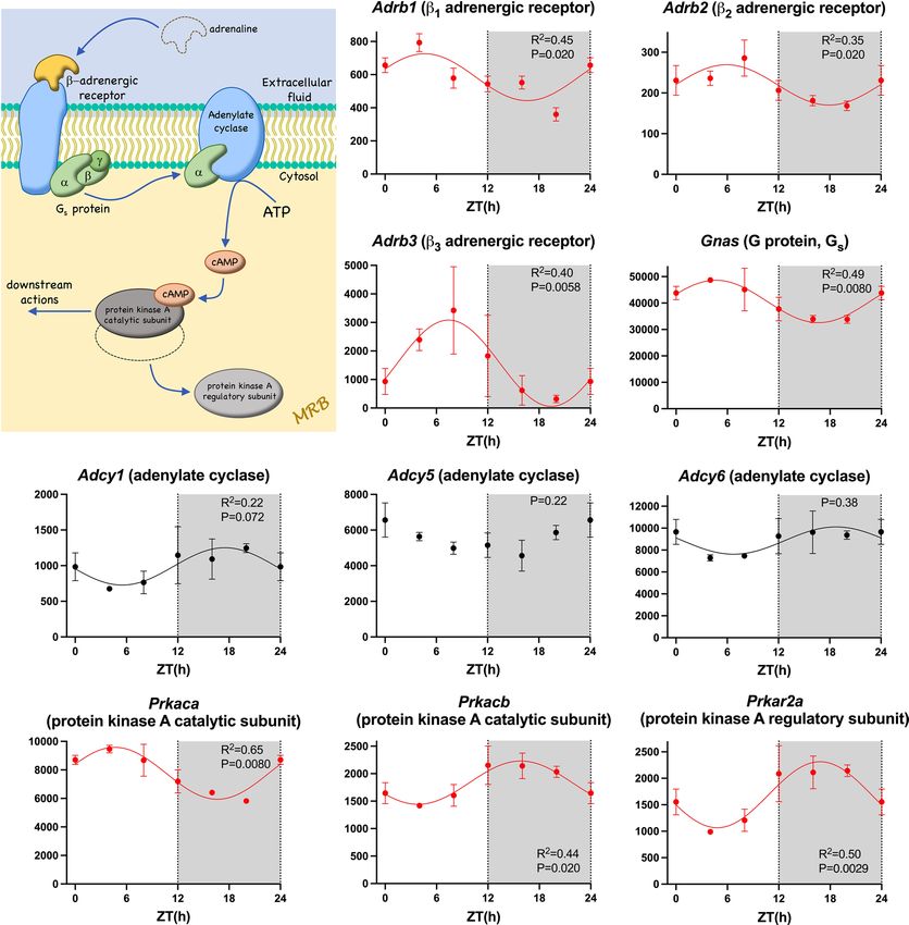
to the β adrenergic receptor activates protein kinase A via a G protein (Gs) and adenylate cyclase. β1, β2 and β3
adrenergic receptor transcripts were present; surprisingly, whereas the β1 adrenergic receptor is considered the
most important in the heart, the β3 adrenergic receptor transcript was the most abundant. Transcripts for β1, β2
and β3 adrenergic receptors, the α subunit of Gs, and the catalytic subunit of protein kinase A showed a signifi-
cant day-night rhythm (Fig. 4). Binding of ACh to the M2 receptor activates the ACh-activated K + channel via a
G protein (Gi), and transcripts for the receptor, the α subunit of G
i, and one of the two subunits making up the
channel (Kir3.1) showed a significant day-night rhythm (Fig. S6). The same pathway is activated by the adeno-
sine A1 receptor37 and this too showed a significant day-night rhythm (Fig. S6).
Signalling pathways. There are many signalling pathways in the heart and just a few examples were investi-
gated. Ca2+/calmodulin-dependent protein kinase II (CaMKII) plays an important role in the heart and the sinus
node. Whereas acute activation of CaMKII results in phosphorylation of downstream targets by the kinase38,
chronic activation results in transcriptional remodelling39. For example, constitutive activation of CaMKII (by
Scientific Reports | (2021) 11:3565 | https://doi.org/10.1038/s41598-021-82202-7 4
Vol:.(1234567890)
www.nature.com/scientificreports/

                                  Figure 2.  Membrane clock pacemaker mechanism. Abundance (normalised counts) of pacemaker ion channel
                                  transcripts and some potentially related transcripts is shown over 24 h. See Fig. 1 legend for further details.

Scientific Reports |   (2021) 11:3565 |               https://doi.org/10.1038/s41598-021-82202-7                                                5

                                                                                                                                           Vol.:(0123456789)
www.nature.com/scientificreports/
Figure 3. Ca2+ clock pacemaker mechanism. Abundance (normalised counts) of Ca2+ clock transcripts is
shown over 24 h. The inset shows a schematic diagram of the Ca2+ clock. See Fig. 1 legend for further details.
oxidation) in heart failure is reported to be responsible for sinus node disease common in heart failure40. Fig-
ure S7 shows a schematic diagram of the pathway (based on Kreusser and Backs39) by which CaMKII regulates
gene transcription. Transcripts for CaMKII (Camk2d) and many of its downstream mediators showed a signifi-
cant rhythm (Fig. S7). Mitogen-activated protein kinases (MAP kinases) are involved in cardiac development,
physiological adaptation and pathological manifestation41. They act on ion channels42. They form a three-tiered
kinase cascade in which a MAP kinase kinase kinase activates a MAP kinase kinase, which in turn activates a
MAP kinase41. Transcripts for some kinases in all three tiers of the cascade showed significant rhythms (Figs. S8-
S10). Nitric oxide (NO) is an important regulator of the cardiovascular system and in part NO is derived from
NO synthases (NOSs)43. Of the three NOS transcripts present, Nos3 (inducible NOS, iNOS) showed a significant
day-night rhythm (data not shown). Any signalling pathway impacting surface membrane ion channels or intra-
cellular Ca2+ handling has the potential to affect pacemaking.
Myofilaments. Some of the transcripts for the contractile apparatus of the sinus node myocyte showed a
significant day-night rhythm, including transcripts for myosin light chain 4 and titin, which are linked to sick
sinus syndrome44,45. In addition, transcripts for actin, tropomyosin 1 and troponin I showed significant day-
night rhythms (Fig. S11).
Metabolism. It is well known that metabolism shows a day-night rhythm, including in the heart e.g.23. The
RNAseq data suggests that this is also true of the sinus node. ~ 70–90% of cardiac ATP is produced by the oxida-
tion of fatty acids, which are transported into the mitochondria as acyl-CoA46. Carnitine palmitoyltransferases
I and II and translocase are involved in the transport and transcripts for carnitine palmitoyltransferase II and
translocase showed a significant day-night rhythm (Fig. S12). The long-chain acyl-CoA enters the fatty acid
β-oxidation pathway. ~ 49% of the transcripts for the fatty acid β-oxidation pathway showed a significant day-
night rhythm (Table 1)—examples are shown in Fig. S13. The acetyl-CoA then enters the citric acid cycle to
Scientific Reports | (2021) 11:3565 | https://doi.org/10.1038/s41598-021-82202-7 6
Vol:.(1234567890)www.nature.com/scientificreports/
Figure 4. β-adrenergic receptor pathway. Abundance (normalised counts) of β-adrenergic receptor pathway
transcripts is shown over 24 h. The inset shows a schematic diagram of the β-adrenergic receptor pathway.
Transcripts for two other protein kinase A subunits, Prkar1b and Prkar2b, also showed a significant circadian
rhythm (data not shown). See Fig. 1 legend for further details.
generate ATP. Glycolysis is the metabolic pathway that converts glucose into pyruvate; the pyruvate is then con-
verted to acetyl-CoA, which again then enters the citric acid cycle to generate ATP. ~ 59% of the transcripts for
the glycolysis pathway showed a significant day-night rhythm (Table 1)—examples are shown in Fig. S14. ~ 67%
of the transcripts for the citric acid cycle itself showed a significant day-night rhythm (Table 1)—examples are
shown in Fig. 5. The NADH generated by the citric acid cycle is fed into the oxidative phosphorylation path-
way—involving the electron transport chain—to form ATP. ~ 43% of the electron transport chain transcripts
showed a significant day-night rhythm (Table 1)—examples are shown in Fig. S15. The day-night rhythm in
metabolism has the potential to impact pacemaking, because AMP kinase (Fig. 2) both conserves cellular energy
homeostasis and also affects the heart rate via the pacemaker funny current carried by HCN channels30. For
example, ATPIF1 is an inhibitor of mitochondrial ATPase, the engine of oxidative phosphorylation, and conse-
quently a regulator of energy m etabolism47. ATPIF1 has a link to AMP k inase48 and Atpif1 showed a trend of a
day-night rhythm (P = 0.072; Fig. S15).
Scientific Reports | (2021) 11:3565 | https://doi.org/10.1038/s41598-021-82202-7 7
Vol.:(0123456789)www.nature.com/scientificreports/
Figure 5. Citric acid cycle. Abundance (normalised counts) of citric acid cycle transcripts is shown over 24 h.
The inset shows a schematic diagram of the citric acid cycle. See Fig. 1 legend for further details.
Extracellular matrix. Of 378 transcripts involved in the extracellular matrix, ~ 32% showed a significant
day-night rhythm (Table 1)—examples are shown in Fig. 6. The extracellular matrix, as well as being essential for
the structural integrity of the heart, is a load that the contractile apparatus must deform in the cardiac cycle—it is
therefore an energetic cost. Could the extracellular matrix change from day to night as the heart rate and cardiac
output change to obtain the necessary structural integrity at the least energetic cost? Proliferation of the extracel-
lular matrix has long been linked to sinus node dysfunction.
Immune system. The immune system is known to show a circadian rhythm49. The class I major histo-
compatibility complex (HMC) of a tissue presents self-antigens to cytotoxic T-cells of the immune system and
ultimately prevents the animal’s immune system targeting its own cells, whereas the class II MHC presents path-
ogen-derived proteins ultimately resulting in the elimination of infected cells by the immune system. The class
II MHC is linked to heart failure50,51. In the sinus node, two transcripts involved with the class I MHC (H2-k1
and H2-q1052), a transcript for a class I MHC-like molecule (Cd1d153), two transcripts potentially involved with
the class I MHC (Rpp21 and Trim39) and one transcript (H2-dmb252) involved with the class II MHC showed a
significant day-night rhythm (Fig. S16). Respiratory (or oxidative) burst is the rapid release of reactive oxygen
species (ROS), superoxide anion, and hydrogen peroxide. Macrophages and neutrophils are especially impli-
Scientific Reports | (2021) 11:3565 | https://doi.org/10.1038/s41598-021-82202-7 8
Vol:.(1234567890)www.nature.com/scientificreports/
Figure 6. Extracellular matrix. Abundance (normalised counts) of example extracellular matrix transcripts is
shown over 24 h. See Fig. 1 legend for further details.
cated in the respiratory burst. They are phagocytic, and the respiratory burst is vital for the subsequent degrada-
tion of internalised bacteria or other pathogens. This is an important aspect of immunological defence. There
was a significant day-night rhythm in Cybb (NOX2) encoding the major component of NADPH oxidase, which
plays a key role in the respiratory burst (Fig. S16). Interleukin 33 is an alarmin (type of cytokine) and is released
under conditions of stress to induce protective m easures54. For example, it has been shown to antagonise cardiac
hypertrophy and remodelling in mice subject to transverse aortic constriction54. The transcript for interleukin
33 (Il33) showed a significant day-night rhythm (Fig. S16) raising the possibility that the heart’s resistance to
stress may also show a day-night rhythm. Heart disease is frequently associated with sinus node d ysfunction55
50
and the immune system has been linked to the adverse remodelling of the diseased h eart . Other transcripts
linked to the immune system also showed a significant day-night rhythm and are illustrated in Fig. S17 or listed
in Table S2.
Scientific Reports | (2021) 11:3565 | https://doi.org/10.1038/s41598-021-82202-7 9
Vol.:(0123456789)www.nature.com/scientificreports/
GWAS‑identified genes associated with resting heart rate. Various genome-wide association stud-
ies (GWAS) have been carried out to identify genetic variants (and therefore genes) affecting the resting heart
rate. The majority of these genes have not previously been identified as involved in pacemaking. Of the genes
identified by these GWAS studies, transcripts for 46 showed a significant day-night rhythm (P < 0.05) and, there-
fore, could potentially contribute to a day-night rhythm in pacemaking—the transcripts are listed in Table S2
together with transcripts which showed a trend of a day-night rhythm (0.1 > P > 0.05). In most cases, the nature
of the relationship between the gene and pacemaking is unknown. In a few cases, there is a plausible link to
pacemaking (cAMP-dependent protein kinase type II-alpha regulatory subunit, Prkar2a—Fig. 4; muscarinic
M2 receptor, Chrm2—Fig. S6; acetylcholine esterase, Ache—Fig. S6; titin, Ttn—Fig. S1144; Hcn4—Fig. 2; Cx43,
Gja1—Fig. S3; desmoplakin, Dsp—Fig. S356). Some genes are related to ion channels, ion transport, receptors
or a signalling pathway (Alg10, Slc12a9, Calcrl, Gng11, Map3k10) and, therefore, a relationship with pacemak-
ing is not implausible. Some genes are involved in transcription, translation or degradation of mRNA/protein
and could potentially be involved with pacemaker genes (Mkln1, Klhl42, Canx, Ppargc1a, Tbx20, Rnf220, Ppil1,
Ddx17, Srrt, Srebf1, Ufsp1, Cby1, Cdc23). Intriguingly, one of the genes, Gtpbp1, promotes degradation of tar-
get mRNA species and plays a role in the regulation of circadian mRNA s tability57. In some cases, a link with
pacemaking can be speculated on: Met is a transcript for a receptor tyrosine kinase, which is known to target
phosphoinositide 3-kinase58, which in turn is known to target H CN431. Ephb4 is a transcript for another receptor
tyrosine kinase, and could it act in a similar way? Inositol hexakisphosphate kinase 1 (Ip6k1) has a link to AMP
kinase59, which is known to regulate HCN430.
Transcription, translation, and mRNA and protein degradation. Figure 7D shows the lag time of
the 7134 transcripts showing a significant day-night rhythm. The lag time corresponds to the maximum expres-
sion during the 24 h period. There are two lag time peaks, one in the day (~ ZT 4–6) and one at night (~ ZT 18),
the most prominent being the one at night (Fig. 7D). This suggests that there are two peaks in the process of
transcription. Transcription is controlled by histone acetyltransferases (HATs), which make chromatin acces-
sible for transcription, and transcription factors, which drive transcription. Fifteen HAT transcripts were identi-
fied and 80% showed a significant day-night rhythm (Table 1). Recently, Zhou et al.60 have published an atlas
of 941 mouse transcription factors. Of these, transcripts for 703 were identified in the sinus node and ~ 49%
showed a significant day-night rhythm (Table 1)—examples are shown in Fig. 8. The chosen examples include
transcripts for transcription factors known to be involved in determining the sinus node pacemaking phenotype
(Tbx1861), sinus node development (Mef2c62), cardiac development (Homez, Sp3, Gata163–65), cardiac develop-
ment and adult function (Tbx2066), cardiac development and possibly the circadian clock (Nrd1d267,68) and car-
diac disease (Atf6, Meox169,70). Sp1 is a regulator of many cardiac genes including Serca271 and genes involved in
metabolism72. Smad signalling has been linked to cardiac remodelling following myocardial i nfarction73. Sox10-
positive cardiomyocytes of neural crest origin contribute to myocardial regeneration in the Zebrafish74. Mef2
directly targets HCN475. Shox2 is essential for the differentiation of cardiac pacemaker c ells76. The oscillating
HAT and transcription factor transcripts peaked at approximately the same time during the day and night (~ ZT
4 and 18; Fig. 7A,B) as all transcripts (Fig. 7D). Could these be responsible for the two peaks in transcription
(Fig. 7D)? Histone deacetylases (HDACs) close chromatin to transcription—of 17 HDAC transcripts identi-
fied, ~ 53% showed a significant day-night rhythm (Table 1). Again HDAC transcripts showed the same two
peaks (Fig. 7C). This is not unexpected—if transcription occurs primarily in two relatively brief spurts, the
HATs and HDACs are expected to be present at roughly the same times. Intuitively, HDACs are expected to peak
later than HATS to provide a time window when chromatin is accessible for transcription, but perhaps such a
time window will only be evident with more frequent sampling than once every 4 h. Translation is governed by
eukaryotic initiation factors (which guide transcripts to the ribosomes) and RNA polymerases. Of 55 transcripts
for eukaryotic initiation factors identified, ~ 66% showed a significant day-night rhythm (Table 1). Of 26 RNA
polymerase transcripts identified, ~ 42% showed a significant day-night rhythm (Table 1). It was only transcripts
for RNA polymerase 2 (responsible for transcribing precursors of mRNA and most snRNA and microRNA) and
3 (responsible for transcribing housekeeping genes: 5S ribosomal RNA, tRNA and other small RNAs) which
showed day-night rhythms—RNA polymerase 1 (responsible for transcribing ribosomal RNA) did not. Once
again, transcripts for eukaryotic initiation factors and RNA polymerases showed the same two peaks (Fig. 7E,F).
microRNAs are short non-coding RNAs—they downregulate the expression level of transcripts (and thereby
downregulate translation) or they directly downregulate translation of transcripts. In a separate study, we meas-
ured the expression of microRNAs at four time points at 6 h intervals over 24 h using q PCR77. 74 microRNAs
showed a significant day-night rhythm and unlike transcription factors and all transcripts showed a single peak
at ~ ZT 10 (Fig. 7G). microRNAs are, therefore, in antiphase to transcription factors etc. This is not unexpected,
because whereas transcription factors upregulate transcription/translation, microRNAs downregulate them.
Two peaks in transcripts during the day and at night have been seen before in mouse heart (presumably
dominated by ventricular muscle) by Zhang et al.20 However, there are important differences between the two
studies: in ventricle, 1335 oscillating transcripts were identified, which Zhang et al.20 reports is consistent with
the 3–10% of the transcriptome oscillating in different tissues. In contrast, in the present study of the sinus node
7134 oscillating transcripts (~ 44% of the transcriptome) were identified. This is a much higher percentage than
in ventricle and other tissues and reasons for this are speculated on in the Discussion. In addition, the segrega-
tion into two peaks is much more marked in sinus node (Fig. 7D) than in ventricle20. In ventricle, Zhang et al.20
argued that the transcripts in the two peaks were different in nature and the biphasic distribution of transcripts
was under the control of the transcription factor, Klf15. However, no evidence of this was found in the sinus node:
the types of transcripts said to be restricted to one peak in ventricle were not restricted to the same peak in the
sinus node, and analysis of all significantly rhythmic transcripts in the two peaks in the sinus node by Ingenuity
Scientific Reports | (2021) 11:3565 | https://doi.org/10.1038/s41598-021-82202-7 10
Vol:.(1234567890)www.nature.com/scientificreports/
Figure 7. Transcription, translation, and mRNA transcript and protein breakdown. (A–I), histogram of
lag times (times of peak transcript abundance) of different groups of transcripts involved in transcription,
translation, and mRNA transcript and protein breakdown. (J), Venn diagram of pathways involving the
transcripts peaking during the day and night; data were analysed through the use of IPA (QIAGEN Inc., https://
www.qiagenbioinformatics.com/products/ingenuitypathway-analysis)103. Inset, schematic diagram of the cycle
of transcription, translation, and RNA and protein degradation.
Pathway Analysis (IPA) software revealed no pattern and the enriched pathways in the two peaks were mostly
similar (494 of 628 pathways enriched in the peaks were common to both; Fig. 7J). Furthermore, Klf15, did not
show a significant day-night rhythm in the sinus node (P = 0.22; data not shown). There is further discussion
about the phasing of transcripts in the Supplementary Information.
The potential targets of the 74 oscillating microRNAs were investigated using IPA, which utilises TargetScan
predictions, experimentally validated TarBase and miRecords targets and manual curations from the literature.
Experimentally observed targets as well as high and moderate confidence predictions were considered, although
it is acknowledged that there is some likelihood of false positive predictions resulting from these analyses. Oscil-
lating microRNAs were screened against the rhythmic transcripts using the microRNA target filter function in
IPA, with a mouse species filter applied. Of the 7134 oscillating transcripts, 4982 (70%) were predicted to be
targeted by 57 of the oscillating microRNAs.
It is assumed that the day-night rhythms in transcripts are the result of the oscillating transcription factors
and microRNAs. In total, 16,387 transcripts were detected and 703 transcription factors were detected. In a
previous study we have detected 715 microRNAs in the mouse sinus node78. Therefore, the ratio of transcription
factors:transcripts is ~ 1:23 and the ratio of microRNAs:transcripts is 1:23. There are 7134 oscillating transcripts,
Scientific Reports | (2021) 11:3565 | https://doi.org/10.1038/s41598-021-82202-7 11
Vol.:(0123456789)www.nature.com/scientificreports/
Atf6 Gata4
6000
5000
4000
4000
3000
2000 2000
R2=0.66 1000 R2=0.59
P=0.00017 P=0.026
0 0
0 6 12 18 24 0 6 12 18 24
ZT(h) ZT(h)
Homez Mef2a Mef2c
2000
4000
200
1500
150 3000
2000 1000
100
1000 500
50 R2=0.58 R2=0.59 R2=0.61
P=3.71E-05 P=0.0014 P=0.0029
0 0 0
0 6 12 18 24 0 6 12 18 24 0 6 12 18 24
ZT(h) ZT(h) ZT(h)
Meox1 Shox2 Smad9
2500 2500 350
2000 2000 300
250
1500 1500
200
1000 1000 150
100
500 R2=0.64 500 R2=0.38
P=0.14 50 P=0.034
P=2.13E-05
0 0 0
0 6 12 18 24 0 6 12 18 24 0 6 12 18 24
ZT(h) ZT(h) ZT(h)
Sox10 Sp1 Sp3
800
1000
1500
600 800
1000 600
400
400
200 500
R2=0.38 R2=0.63 200 R2=0.75
P=0.011 P=0.0020 P=0.00010
0 0 0
0 6 12 18 24 0 6 12 18 24 0 6 12 18 24
ZT(h) ZT(h) ZT(h)
Tbx18 Tbx20
1000
5000
800
4000
600
3000
400 2000
200 R2=0.39 1000 R2=0.48
P=0.057 P=0.0029
0 0
0 6 12 18 24 0 6 12 18 24
ZT(h) ZT(h)
Figure 8. Transcription Factors. Abundance (normalised counts) of example transcription factor transcripts is
shown over 24 h. See Fig. 1 legend for further details.
Scientific Reports | (2021) 11:3565 | https://doi.org/10.1038/s41598-021-82202-7 12
Vol:.(1234567890)www.nature.com/scientificreports/
347 oscillating transcription factors and a minimum of 74 oscillating microRNAs—therefore the ratio of oscil-
lating transcription factors:oscillating transcripts is ~ 1:21 and the ratio of oscillating microRNAs:oscillating
transcripts is ~ 1:96.
At steady-state, RNA and protein degradation has to match transcription and translation. The many path-
ways of RNA degradation were reviewed by Houseley and Tollervey79; 38 components of these pathways were
identified and ~ 53% showed a significant day-night rhythm (Table 1). Proteins are tagged for degradation by
ubiquitination catalysed by ubiquitin ligases. Once ubiquinated, the protein is degraded by the proteasome. 47
transcripts for this pathway were identified and ~ 49% showed a significant day-night rhythm (Table 1). Once
again these showed the same two peaks (Fig. 7H,I).
Discussion
The sinus node has two clocks, the membrane and C a2+ clocks, operating on a time scale of seconds, and which
are responsible for the rhythmic beating of the heart. For the first time, this study has shown that the sinus node
has another clock, the circadian clock, operating on a time scale of days and which could be responsible for, or
at least involved in, a rhythmic change in the transcriptome of the sinus node. ~ 44% of the transcriptome of the
sinus node is changing in a day-night manner and the day-night rhythm is all-pervasive affecting all systems
looked at including the membrane and Ca2+ clocks, neurohumoral receptors, important signalling pathways,
metabolism and extracellular matrix. The interested reader is likely to find other systems affected—a list of all
transcripts together with the permutation-based P value from JTK Cycle for a day-night rhythm is available as
part of the Supplementary Data (AllTranscripts.xlsx).
Day‑night rhythm in heart rate. Based on heart rate variability and autonomic blockade, the day-night
rhythm in heart rate is currently attributed to changes in the autonomic innervation of the heart and in particu-
lar to high vagal tone at night in the case of the human13. According to this hypothesis, ACh released from vagal
nerve endings binds to muscarinic M2 receptors and activates the ACh-activated K + channel, and this causes
the slowing of heart rate at night. However, we have argued that heart rate variability cannot be used to measure
autonomic innervation of the heart80, and data from autonomic blockade has been reported to both block and
have no discernible effect on the day-night rhythm in heart rate13. However, although there is doubt concerning
the evidence for a day-night rhythm in autonomic innervation of the heart, it is clear that there is a day-night
rhythm in the plasma level of catecholamine (presumably coming from the adrenal medulla under the action of
the sympathetic nervous system), which is higher during the day in the human81. Therefore, it is possible that
a day-night rhythm in the autonomic nervous system is responsible for the day-night rhythm in heart rate via
rapid regulation of ionic conductances. This study does not resolve this controversy, but it does show that at the
transcript level the two major pacemaking mechanisms of the sinus node, the membrane and Ca2+ clocks, show a
profound day-night rhythm (Figs. 2,3). Therefore, it is possible that there is a day-night rhythm in pacemaking as
a result of changes in gene transcription in the sinus node. In another study, we have shown that there is indeed
an intrinsic day-night rhythm in both funny current density and pacemaking82. However, although an intrinsic
day-night rhythm in pacemaking could be responsible for the day-night rhythm in heart rate, it may only be
responsible for a day-night rhythm in ‘pacemaker reserve’ so that the sinus node is prepared to deliver higher
heart rates during the awake period when called upon to do so by the autonomic nervous system. The final
answer to the controversy of whether the day-night rhythm in heart rate is the result of the post-translational
regulation of ion channels by the autonomic nervous system or transcriptional changes is unlikely to be simple.
This study has shown that there is a day-night rhythm in expression of autonomic receptors (adrenergic and
muscarinic receptors; Figs. 4, S5 and S6) and, therefore, there may be a day-night rhythm in the responsiveness
to the autonomic receptor stimulation. Another possibility is that the autonomic nervous system is involved, but
in a different way to that originally conceived: Tong et al.83,84 have shown that autonomic blockade abolishes the
day-night rhythm in the expression of various K+ channels and connexin subunits in the ventricles (see below
for further comment).
Day‑night rhythm in the transcriptome. Based on the use of Affymetrix GeneChip oligonucleotide
arrays, Storch et al.21 estimated that about 10% of the mouse liver transcriptome shows a significant day-night
rhythm; they detected 4,805 transcripts in the liver of which 575 transcripts oscillated with a day-night rhythm,
but they attributed 16% of these to noise rather than a genuine day-night rhythm. Using Affymetrix GeneChip
oligonucleotide arrays, Martino et al.18 detected 12,488 transcripts in mouse heart (likely to be exclusively or
mainly the ventricles based on tissue mass), of which 1,634 (~ 13%) showed a significant day-night rhythm
(based on use of COSOPT) during a normal 12 h light:12 h dark lighting regime. In a more recent study, using
RNAseq, Zhang et al.20 identified 1,335 transcripts (based on use of JTK Cycle) showing a significant day-night
rhythm in mouse heart (presumably ventricle) during a normal 12 h light:12 h dark lighting regime. In a variety
of studies on mouse heart (presumably ventricle), 6–13% of the transcriptome has been reported to vary in
a day-night m anner18–21. Using both RNAseq and Affymetric MoGene oligonucleotide arrays, Zhang et al.19
looked at the day-night rhythm in the transcriptome in 12 mouse organs; they reported that the transcripts
oscillating varied from 3% in the hypothalamus, 6% in the heart and 16% in the liver. In the present study,
16,387 transcripts were selected, of which 7134 (~ 44%) showed a significant day-night rhythm and the fraction
of oscillating genes is clearly much higher than in other studies. The data from the present study are robust and it
is concluded that the difference is a tissue difference. The reason why the fraction of transcripts under day-night
control is large in the sinus node can only be speculated on. One possibility is the importance of heart rate for
an organ that is continuously beating every ~ 1 s throughout life; cardiac output is primarily determined by vari-
ation in heart rate rather than by variation in stroke volume. The pacemaker activity of the sinus node has to be
Scientific Reports | (2021) 11:3565 | https://doi.org/10.1038/s41598-021-82202-7 13
Vol.:(0123456789)www.nature.com/scientificreports/
tuned for higher heart rates during the day in the human and this perhaps not only involves a day-night rhythm
in the membrane and C a2+ clock pacemaker mechanisms, but also in closely associated systems involving recep-
tors and signalling for example. Presumably because the heart is continuously active, O2 utilisation per 100 g of
tissue is highest for the heart. Because work carried out by the heart is primarily determined by the heart rate, O
2
utilisation will be primarily determined by the heart rate. Perhaps for this reason, pacemaking and metabolism
have to be controlled together including in a day-night manner and there have to be links between the two; AMP
kinase could be one of these links30,85. For a similar reason, perhaps pacemaking and the extracellular matrix
have to be controlled together to tune the extracellular matrix for the higher heart rate and pressures during the
day in the human.
Day‑night rhythm in transcription, translation, and mRNA and protein degradation. Tran-
scripts changing in a day-night manner peaked either during the day (~ ZT 4–6) or during the night (ZT 18)
(Fig. 7D). This pattern has been seen before: in mouse liver and heart (presumably ventricle) there were peaks at
ZT 6–14 and ZT 2 021, and in other studies of mouse heart (presumably ventricle) there were peaks at ZT 1 and
ZT 1918 or ~ ZT 1 and ZT 1 720. However, in all of these cases the day-time peak was larger than the night-time
peak, whereas the opposite was true in the case of the sinus node (Fig. 7D). The present study has shown the
likely immediate cause of this pattern. Transcripts for the transcription apparatus (HATs, transcription factors,
HDACs) peaked at ~ ZT 4–6 and then at ZT 18 and this ultimately was likely to be responsible for the peak in
transcripts at these two time points (Fig. 7). Transcripts for the translation apparatus (eukaryotic initiation fac-
tors, RNA polymerases) also peaked at the same time points and therefore generation of protein is expected to
peak at the same time points (Fig. 7). Finally, transcripts for the apparatus for the breakdown of both transcripts
and proteins (RNA degradation pathway, ubiquitine and proteasome) also peaked at the same two times (Fig. 7).
However, what this study does not address is how these day-night rhythms impact on the level of proteins. This
will depend on the life time of the protein, which can vary from minutes to y ears86. If the lifetime of the protein
is short, there will be a day-night rhythm in the protein, but if it is longer than 24 h this will not be the case.
Potential systemic regulators. The master circadian clock in the suprachiasmatic nucleus is entrained to
light via the eyes and neuronal circuitry, and peripheral circadian clocks like that in the sinus node are entrained
by the master clock and other physiological stimuli. The peripheral clocks are entrained by neurohumoral fac-
tors and systemic regulators, including the autonomic nervous s ystem87, corticosteroids87,88 and possibly thyroid
hormone89. Presumably the same is true of the sinus node. It is also possible that these same neurohumoral
factors and systemic regulators may directly affect pacemaking. It is interesting that transcripts for receptors for
catecholamines, ACh, corticosteroids and thyroid hormone all showed day-night rhythms (Figs. 1, 4, S5 and S6).
Spoor and J ackson90 reported that the heart rate response of isolated atria of the rat (nocturnal like the mouse)
to ACh is greater during the day than at night, suggesting that the day-night rhythm in the muscarinic pathway
(Fig. S6) has a functional corollary, but perhaps lagging behind mRNA by ~ 12 h. Also Peliciari-Garcia et al.89
reported greater triiodothyronine sensitivity (induction of transcript levels) at the end of the night. Therefore, it
is possible that the effects of the autonomic nervous system, corticosteroids and thyroid hormone on the sinus
node will not just depend on the known day-night rhythms of the autonomic nervous system, corticosteroids
and thyroid hormone—they may also depend on the responsiveness of the sinus node to the factors.
Genes underlying familial and acquired sick sinus syndrome. Familial sick sinus syndrome has been
linked to mutations in Hcn491, and acquired sick sinus syndrome in ageing92, heart f ailure93, atrial fibrillation94,
diabetes95, pulmonary h ypertension96 and even athletes11 has been linked to a downregulation of Hcn4. If there
is a day-night rhythm in Hcn4 (P = 0.072), this may impact sick sinus syndrome. For example, athletes have a
sinus bradycardia as a result of a downregulation of Hcn411,78 and the bradycardia is most marked at night and
athletes can have long nocturnal pauses between heart beats at night14. Familial sick sinus syndrome has also
been linked to mutations in KvLQT1 (Kcnq1), Kir2.1 (Kcnj2), and calsequestrin 2 (Casq2)27, all of which show
a significant day-night rhythm (Kcnq1, P = 0.015; Kcnj2, P = 0.00017; Ryr2, P = 0.044; Casq2, P = 0.0080). Once
again, the day-night rhythm in the ion channels may impact the phenotype caused by the mutation. Recently,
Mesirca et al.97 have suggested block of the ACh-activated K+ channel to treat sick sinus syndrome. Figure S6
shows that the Kir3.1 subunit of the channel shows a significant day-night rhythm, and this may impact the effect
of channel block.
Conclusion
In conclusion, there is an all-pervasive day-night rhythm in the transcriptome of the pacemaker of the heart,
the sinus node. Whether this is responsible for the day-night rhythm in the heart rate or whether it prepares the
pacemaker for the demands placed on it during the awake period remains to be determined.
Methods
Care and use of laboratory animals conformed to the UK Animals (Scientific Procedures) Act 1986 and Directive
2010/63/EU of the European Parliament. Ethical approval for all experimental procedures was granted by the
University of Manchester Animal Welfare and Ethical Review body. 12–14 week old adult male C57bl/6j mice
were maintained in a 12 h light:12 h dark cycle. The work flow is shown below:
Tissue harvesting at ZT 0, 4, 8, 12, 16 and 20 h → library preparation → RNAseq → Analysis
Scientific Reports | (2021) 11:3565 | https://doi.org/10.1038/s41598-021-82202-7 14
Vol:.(1234567890)www.nature.com/scientificreports/
Biopsies were collected from the sinus node as we have done previously in many other studies (e.g. Linscheid
et al.98). We have previously reported the anatomy of the mouse sinus n ode99 and this dictated the position of
the biospies. We collected biopsies from the smooth intercaval region between the superior and inferior vena
cavae centred on the bifurcation of the sinus node artery. The high expression of sinus node markers (e.g. Hcn4)
and absence of atrial markers (e.g. atrial natriuretic peptide) have confirmed the nature of the biopsies. Biopsies
were collected at six time points at four hourly intervals over the 24 h period: at zeitgeber time (ZT) 0, 4, 8, 12,
16 and 20 h. At each time point, biopsies were collected from three mice. RNA was isolated from the sinus node
as described previously78. Quantity and integrity of the RNA samples were measured using a 2200 TapeStation
(Agilent Technologies) to ensure their suitability. Subsequently, TruSeq Stranded mRNA assays (Illumina) were
used in order to produce libraries of more stable, single-stranded cDNA as follows. Total RNA was purified to
polyadenylated mRNA via magnetic separation technology, which works through hybridisation of covalent
interactions of oligo d(T)25 to poly (A) regions present in most eukaryotic mRNA. The mRNA sequences were
fragmented into parts via divalent cations at higher temperature, and random primers were used to reverse tran-
scribe the mRNA fragments into single-stranded cDNA. DNA polymerase and RNase H mediated the synthesis
of the second cDNA strand produced from RNA oligonucleotides, originating from the 5´ end of the mRNA.
The final cDNA library was generated by an addition of a single ‘A’ base, binding of adapters to the fragments
and purification and enrichment via a PCR reaction. The cDNA libraries were incorporated into a multiplex
system using the adapters; they were then pooled and clustered using a cBlot instrument (Illumina). Optical
flow-cells containing the mRNA samples were then paired-end sequenced and mRNA was quantified through
repeating 76 cycles twice, using a HiSeq4000 instrument (Illumina). Unmapped paired-end sequences from an
Illumina HiSeq4000 sequencer were tested by FastQC (http://www.bioinformatics.babraham.ac.uk/projects/
fastqc/). Sequence adapters were removed, and reads were quality trimmed using Trimmomatic_0.36100. The
reads were mapped against the reference mouse genome (mm10/GRCm38) and counts per gene were calculated
using annotation from GENCODE M21 (http://www.gencodegenes.org/) using STAR_2.5.3101. Normalisation
was carried out using DESeq2_1.18.1102. DESeq2 performs an internal normalisation in which a geometric mean
is calculated for each gene across all samples. The counts for a gene in each sample is then divided by this mean.
The median of these ratios in a sample is the size factor for that sample. This procedure corrects for library size
and RNA composition bias, which can arise for example when only a small number of genes are very highly
expressed in one experimental condition but not in the other. In figures, the mean ± SEM transcript expression
(normalised counts) from the three mice at each time point is plotted. To guide the eye, the data for ZT 0 are
also shown for ZT 24 and the data may have been fitted with a sine wave (based on all time points including
ZT 24); if fitted, the R2 value for the fitted curve is shown in figures. JTK Cycle24 was used to test whether a
transcript showed a significant day-night rhythm (based on the data at ZT 0, 4, 8, 12, 16 and 20 (but not ZT 24);
permutation-based P values are shown in figures. In figures, transcripts showing a significant day-night rhythm
(permutation-based P value < 0.05) are shown in red and are fitted with a sine wave, transcripts showing a trend
of a day-night rhythm (permutation-based P value > 0.05 but < 0.1) are shown in black and are fitted with a sine
wave, and transcripts not showing a significant day-night rhythm (permutation-based P value > 0.1) are shown
in black and are not fitted with a sine wave. IPA was used to identify potential targets of microRNAs.
Received: 5 June 2020; Accepted: 1 January 2021
References
1. Fox, K. et al. Resting heart rate in cardiovascular disease. J. Am. Coll. Cardiol. 50, 823–830 (2007).
2. Cooney, M. T. et al. Elevated resting heart rate is an independent risk factor for cardiovascular disease in healthy men and
women. Am. Heart J. 159, 612-619.e613 (2010).
3. Zhang, D., Shen, X. & Qi, X. Resting heart rate and all-cause and cardiovascular mortality in the general population: A meta-
analysis. Can. Med. Assoc. J. 188, E53–E63 (2016).
4. Fabritz, L. et al. Gene dose-dependent atrial arrhythmias, heart block, and brady-cardiomyopathy in mice overexpressing A3
adenosine receptors. Cardiovasc. Res. 62, 500–508 (2004).
5. Caliskan, K., Balk, A. H., Jordaens, L. & Szili-Torok, T. Bradycardiomyopathy: The case for a causative relationship between
severe sinus bradycardia and heart failure. J. Cardiovasc. Electrophysiol. 21, 822–824 (2010).
6. Milano, A. et al. HCN4 mutations in multiple families with bradycardia and left ventricular noncompaction cardiomyopathy.
J. Am. Coll. Cardiol. 64, 745–756 (2014).
7. Tsuji, Y. et al. Ionic mechanisms of acquired QT prolongation and torsades de pointes in rabbits with chronic complete atrio-
ventricular block. Circulation 106, 2012–2018 (2002).
8. El Khoury, N. et al. Upregulation of the hyperpolarization-activated current increases pacemaker activity of the sinoatrial node
and heart rate during pregnancy in mice. Circulation 127, 2009–2020 (2013).
9. Abd Allah, E. S. H. et al. Changes in the expression of ion channels, connexins and Ca2+-handling proteins in the sinoatrial node
during postnatal development. Exp. Physiol. 96, 426–438 (2011).
10. Yanni, J. et al. Ageing-dependent remodelling of ion channel and Ca2+ clock genes underlying sinoatrial node pacemaking. Exp.
Physiol. 96, 1163–1178 (2011).
11. D’Souza, A. et al. Exercise training reduces resting heart rate via downregulation of the funny channel HCN4. Nat. Commun.
5, 3775 (2014).
12. Yanni, J. et al. Silencing miR-370-3p rescues funny current and sinus node function in heart failure. Sci. Rep. 10, 11279 (2020).
13. Black, N. et al. Circadian rhythm of cardiac electrophysiology, arrhythmogenesis, and the underlying mechanisms. Heart Rhythm
16, 298–307 (2019).
14. Northcote, R. J., Canning, G. P. & Ballantyne, D. Electrocardiographic findings in male veteran endurance athletes. Br. Heart J.
61, 155–160 (1989).
15. Otsuka, K. et al. Experimental study on the relationship between cardiac arrhythmias and sleep states by ambulatory ECG-EEC
monitoring. Clin. Cardiol. 9, 305–313 (1986).
Scientific Reports | (2021) 11:3565 | https://doi.org/10.1038/s41598-021-82202-7 15
Vol.:(0123456789)www.nature.com/scientificreports/
16. Vandewalle, G. et al. Robust circadian rhythm in heart rate and its variability: Influence of exogenous melatonin and photoperiod.
J. Sleep Res. 16, 148–155 (2007).
17. West, A. C. et al. Misalignment with the external light environment drives metabolic and cardiac dysfunction. Nat. Commun.
8, 417 (2017).
18. Martino, T. et al. Day/night rhythms in gene expression of the normal murine heart. J. Mol. Med. 82, 256–264 (2004).
19. Zhang, R., Lahens, N. F., Ballance, H. I., Hughes, M. E. & Hogenesch, J. B. A circadian gene expression atlas in mammals:
Implications for biology and medicine. Proc. Natl. Acad. Sci. U.S.A. 111, 16219–16224 (2014).
20. Zhang, L. et al. KLF15 establishes the landscape of diurnal expression in the heart. Cell Reports 13, 2368–2375 (2015).
21. Storch, K. F. et al. Extensive and divergent circadian gene expression in liver and heart. Nature 417, 78–83 (2002).
22. Durgan, D. J. & Young, M. E. The cardiomyocyte circadian clock: Emerging roles in health and disease. Circ. Res. 106, 647–658
(2010).
23. Young, M. E. Circadian control of cardiac metabolism: Physiologic roles and pathologic implications. Methodist DeBakey Car-
diovasc. J. 13, 15–19 (2017).
24. Hughes, M. E., Hogenesch, J. B. & Kornacker, K. JTK_CYCLE: An efficient nonparametric algorithm for detecting rhythmic
components in genome-scale data sets. J. Biol. Rhythms 25, 372–380 (2010).
25. Mohawk, J. A., Green, C. B. & Takahashi, J. S. Central and peripheral circadian clocks in mammals. Annu. Rev. Neurosci. 35,
445–462 (2012).
26. Hardman, J. A., Haslam, I. S., Farjo, N., Farjo, B. & Paus, R. Thyroxine differentially modulates the peripheral clock: Lessons
from the human hair follicle. PLoS ONE 10, e0121878 (2015).
27. Dobrzynski, H. et al. Structure, function and clinical relevance of the cardiac conduction system, including the atrioventricular
ring and outflow tract tissues. Pharmacol. Ther. 139, 260–288 (2013).
28. Lakatta, E. G. & DiFrancesco, D. What keeps us ticking: A funny current, a calcium clock, or both?. J. Mol. Cell. Cardiol. 47,
157–170 (2009).
29. Sah, R. et al. Ion channel-kinase TRPM7 is required for maintaining cardiac automaticity. Proc. Natl. Acad. Sci. U.S.A. 110,
E3037-3046 (2013).
30. Yavari, A. et al. Mammalian gamma2 AMPK regulates intrinsic heart rate. Nat. Commun. 8, 1258 (2017).
31. Lin, R. Z. et al. Regulation of heart rate and the pacemaker current by phosphoinositide 3-kinase signaling. J. Gen. Physiol. 151,
1051–1058 (2019).
32. Huang, Z. M. et al. Functional role of CLC-2 chloride inward rectifier channels in cardiac sinoatrial nodal pacemaker cells. J.
Mol. Cell. Cardiol. 47, 121–132 (2009).
33. Takeshima, H., Venturi, E. & Sitsapesan, R. New and notable ion-channels in the sarcoplasmic/endoplasmic reticulum: Do they
support the process of intracellular Ca2+ release? J. Physiol. 593, 3241–3251 (2015).
34. Sharma, P. et al. Evolutionarily conserved intercalated disc protein Tmem65 regulates cardiac conduction and connexin 43
function. Nat. Commun. 6, 8391 (2015).
35. Inada, S. et al. Importance of gradients in membrane properties and electrical coupling in sinoatrial node pacing. PLoS ONE 9,
e94565 (2014).
36. Pavlovic, D., Fuller, W. & Shattock, M. J. Novel regulation of cardiac Na pump via phospholemman. J. Mol. Cell. Cardiol. 61,
83–93 (2013).
37. Mustafa, S. J., Morrison, R. R., Teng, B. & Pelleg, A. Adenosine receptors and the heart: Role in regulation of coronary blood
flow and cardiac electrophysiology. Handb. Exp. Pharmacol. 193, 161–188 (2009).
38. Wu, Y. & Anderson, M. CaMKII in sinoatrial node physiology and dysfunction. Front. Pharmacol. 5, 48 (2014).
39. Kreusser, M. M. & Backs, J. Integrated mechanisms of CaMKII-dependent ventricular remodeling. Front. Pharmacol. 5, 36
(2014).
40. Swaminathan, P. D. et al. Oxidized CaMKII causes cardiac sinus node dysfunction in mice. J. Clin. Investig. 121, 3277–3288
(2011).
41. Rose, B. A., Force, T. & Wang, Y. Mitogen-activated protein kinase signaling in the heart: Angels versus demons in a heart-
breaking tale. Physiol. Rev. 90, 1507–1546 (2010).
42. Chowdhury, S. K. et al. Stress-activated kinase mitogen-activated kinase kinase-7 governs epigenetics of cardiac repolarization
for arrhythmia prevention. Circulation 135, 683–699 (2017).
43. Liu, V. W. T. & Huang, P. L. Cardiovascular roles of nitric oxide: A review of insights from nitric oxide synthase gene disrupted
mice. Cardiovasc. Res. 77, 19–29 (2008).
44. Zhu, Y.-B., Luo, J.-W., Jiang, F. & Liu, G. Genetic analysis of sick sinus syndrome in a family harboring compound CACNA1C
and TTN mutations. Mol. Med. Rep. 17, 7073–7080 (2018).
45. Peng, W. H., Li, M. X., Li, H. L., Tang, K., Zhuang, J. H., Zhang, J. G., Xiao, J. J., Jiang, H., Li, D. L., Yu, Y. C., Sham, P. C., Nattel,
S. & Xu, Y. W. Dysfunction of myosin light-chain 4 (MYL4) leads to heritable atrial cardiomyopathy with electrical, contractile,
and structural components: Evidence from genetically-engineered rats. J. Am. Heart Assoc. 6, 11 (2017).
46. Doenst, T., Nguyen, T. D. & Abel, E. D. Cardiac metabolism in heart failure: Implications beyond ATP production. Circ. Res.
113, 709–724 (2013).
47. García-Bermúdez, J. & Cuezva, J. M. The ATPase inhibitory factor 1 (IF1): A master regulator of energy metabolism and of cell
survival. Biochim. Biophys. Acta Bioenerg. 1857, 1167–1182 (2016).
48. Yang, K. et al. Knockout of the ATPase inhibitory factor 1 protects the heart from pressure overload-induced cardiac hypertrophy.
Sci. Rep. 7, 10501–10501 (2017).
49. Labrecque, N. & Cermakian, N. Circadian clocks in the immune system. J. Biol. Rhythms 30, 277–290 (2015).
50. Blanton, R. M., Carrillo-Salinas, F. J. & Alcaide, P. T-cell recruitment to the heart: Friendly guests or unwelcome visitors? Am.
J. Physiol. Heart Circulatory Physiol. 317, H124–H140 (2019).
51. Strassheim, D., Dempsey, E. C., Gerasimovskaya, E., Stenmark, K. & Karoor, V. Role of inflammatory cell subtypes in heart
failure. J. Immunol. Res. 2019, 2164017 (2019).
52. Shiina, T., Blancher, A., Inoko, H. & Kulski, J. K. Comparative genomics of the human, macaque and mouse major histocompat-
ibility complex. Immunology 150, 127–138 (2017).
53. Sundararaj, S. et al. Differing roles of CD1d2 and CD1d1 proteins in type I natural killer T cell development and function. Proc.
Natl. Acad. Sci. 115, E1204 (2018).
54. Ghali, R. et al. IL-33 (Interleukin 33)/sST2 axis in hypertension and heart failure. Hypertension 72, 818–828 (2018).
55. Bloch Thomsen, P. E. et al. Long-term recording of cardiac arrhythmias with an implantable cardiac monitor in patients with
reduced ejection fraction after acute myocardial infarction: The Cardiac Arrhythmias and Risk Stratification After Acute Myo-
cardial Infarction (CARISMA) study. Circulation 122, 1258–1264 (2010).
56. Mezzano, V. et al. Desmosomal junctions are necessary for adult sinus node function. Cardiovasc. Res. 111, 274–286 (2016).
57. Kim, S. H. et al. Rhythmic control of mRNA stability modulates circadian amplitude of mouse Period3 mRNA. J. Neurochem.
132, 642–656 (2015).
58. Hervieu, A. & Kermorgant, S. The role of PI3K in met driven cancer: A recap. Front. Mol. Biosci. 5, 86–86 (2018).
59. Zhu, Q. et al. Adipocyte-specific deletion of Ip6k1 reduces diet-induced obesity by enhancing AMPK-mediated thermogenesis.
J. Clin. Investig. 126, 4273–4288 (2016).
Scientific Reports | (2021) 11:3565 | https://doi.org/10.1038/s41598-021-82202-7 16
Vol:.(1234567890)You can also read