Roles of Neuropeptides, VIP and AVP, in the Mammalian Central Circadian Clock
←
→
Page content transcription
If your browser does not render page correctly, please read the page content below
MINI REVIEW
                                                                                                                                             published: 15 April 2021
                                                                                                                                     doi: 10.3389/fnins.2021.650154

                                            Roles of Neuropeptides, VIP and AVP,
                                            in the Mammalian Central Circadian
                                            Clock
                                            Daisuke Ono 1,2* , Ken-ichi Honma 3 and Sato Honma 3*
                                            1
                                             Department of Neuroscience II, Research Institute of Environmental Medicine, Nagoya University, Nagoya, Japan,
                                            2
                                             Department of Neural Regulation, Nagoya University Graduate School of Medicine, Nagoya, Japan, 3 Research
                                            and Education Center for Brain Science, Hokkaido University Graduate School of Medicine, Sapporo, Japan

                                            In mammals, the central circadian clock is located in the suprachiasmatic nucleus
                                            (SCN) of the hypothalamus. Individual SCN cells exhibit intrinsic oscillations, and their
                                            circadian period and robustness are different cell by cell in the absence of cellular
                                            coupling, indicating that cellular coupling is important for coherent circadian rhythms
                                            in the SCN. Several neuropeptides such as arginine vasopressin (AVP) and vasoactive
                                            intestinal polypeptide (VIP) are expressed in the SCN, where these neuropeptides
                                            function as synchronizers and are important for entrainment to environmental light
                                            and for determining the circadian period. These neuropeptides are also related to
                            Edited by:      developmental changes of the circadian system of the SCN. Transcription factors are
                       Masayuki Ikeda,
          University of Toyama, Japan
                                            required for the formation of neuropeptide-related neuronal networks. Although VIP
                         Reviewed by:
                                            is critical for synchrony of circadian rhythms in the neonatal SCN, it is not required
               Elizabeth S. Maywood,        for synchrony in the embryonic SCN. During postnatal development, the clock genes
MRC Laboratory of Molecular Biology         cryptochrome (Cry)1 and Cry2 are involved in the maturation of cellular networks, and
              (LMB), United Kingdom
                   William David Todd,      AVP is involved in SCN networks. This mini-review focuses on the functional roles of
University of Wyoming, United States        neuropeptides in the SCN based on recent findings in the literature.
                *Correspondence:
                                            Keywords: circadian rhythm, suprachiasmatic nucleus, AVP, VIP, neuronal coupling, synchronization, entrainment
                       Daisuke Ono
        dai-ono@riem.nagoya-u.ac.jp
                       Sato Honma
       sathonma@med.hokudai.ac.jp           INTRODUCTION
                   Specialty section:       Creatures on earth anticipate cyclic changes in the surrounding environment, such as day–night,
         This article was submitted to      seasonal, and annual changes, to adapt their own physiological functions so that they can minimize
       Sleep and Circadian Rhythms,         risks to survival. Among them, rhythmic changes adapting to environmental changes caused by the
               a section of the journal     rotation of the earth are driven by intrinsic oscillatory mechanisms called the circadian clock. In
            Frontiers in Neuroscience
                                            mammals, the circadian system comprises a hierarchical structure involving the master clock in
         Received: 06 January 2021          the brain and a number of peripheral clocks situated throughout the body. Almost all cells in
          Accepted: 10 March 2021
                                            our body possess a circadian clock in which the molecular machinery, namely, transcription–
           Published: 15 April 2021
                                            translation feedback loops involving several clock genes and their protein products, generates
                             Citation:      circadian rhythmicity (Reppert and Weaver, 2002). Circadian rhythms of cells and tissues are
     Ono D, Honma K and Honma S
                                            coordinated by the master circadian clock located in the suprachiasmatic nucleus (SCN) of the
  (2021) Roles of Neuropeptides, VIP
  and AVP, in the Mammalian Central
                                            hypothalamus (Weaver, 1998). The SCN is the only clock that can entrain to the environmental
                     Circadian Clock.       light–dark (LD) time cue from the retina via the retinohypothalamic tract (RHT) (LeGates et al.,
          Front. Neurosci. 15:650154.       2014). Ultimately, the circadian clock in the SCN regulates rhythms within a variety of physiological
     doi: 10.3389/fnins.2021.650154         output functions such as sleep/wakefulness, body temperature, and endocrine functions.

Frontiers in Neuroscience | www.frontiersin.org                                   1                                           April 2021 | Volume 15 | Article 650154
Ono et al.                                                                                                                 Neuropeptides in the SCN

   The SCN contains approximately 20,000 neurons that show                   robust circadian rhythms with regard to clock gene expression
circadian rhythms in clock gene expression, cytosolic Ca2+ , and             and intracellular Ca2+ levels. Since TTX treatment suppresses
spontaneous firing, and the rhythms are also maintained in                   action potential, neuronal firing may itself affect the circadian
culture conditions, in the slice and in dispersed cells (Welsh et al.,       molecular rhythms in solitary SCN neurons.
1995; Ikeda et al., 2003; Yamaguchi et al., 2003). The distribution
of the circadian period from dispersed SCN cells is larger than
that from slice cultures (Herzog et al., 2004; Honma et al.,                 ROLES OF VASOACTIVE INTESTINAL
2004). Furthermore, the robustness of circadian rhythmicity in               POLYPEPTIDE NEURONS IN THE
dispersed cells is weaker than that in slices (Liu et al., 2007;             SUPRACHIASMATIC NUCLEUS
Ono et al., 2013). These results suggest that cellular networks are
crucial for coherent circadian rhythms in the SCN.                           VIP is known as a synchronizer of neuronal networks in the
                                                                             SCN, and approximately 10% of the SCN consists of VIP-positive
                                                                             neurons (Abrahamson and Moore, 2001). Mice lacking VIP or
HETEROGENEOUS CELL TYPES IN THE                                              VIP receptor 2 (VPAC2) show desynchronized circadian rhythms
SUPRACHIASMATIC NUCLEUS                                                      in SCN neurons in culture as well as attenuated behavioral
                                                                             rhythms (Harmar et al., 2002; Colwell et al., 2003; Aton et al.,
The SCN is, neuroanatomically and cytochemically, divided into               2005; Maywood et al., 2006). Similarly, the application of an
two subregions—dorsal and ventral subregions (Figure 1). The                 excessive amount of VIP onto cultured SCN slices also attenuates
ventral SCN receives major afferent pathways, including those                the synchrony of cellular circadian rhythms (An et al., 2013).
from the retina, midbrain raphe nucleus, and intergeniculate                 The rhythmic release of VIP in cultured SCN slices has been
leaflet (Hattar et al., 2006; McNeill et al., 2011). It contains             previously reported (Shinohara et al., 1995; Shinohara et al.,
vasoactive intestinal peptide (VIP), gastrin-releasing peptide               1999). These results suggest that rhythmic release of VIP is
(GRP), neurotensin (NT), and calretinin (CALR)-expressing                    important for synchrony of circadian rhythms in SCN neurons.
neurons (Abrahamson and Moore, 2001). Arginine vasopressin                   Chemogenetic activation of VIP neurons also changes the
(AVP), met-enkephalin (mENK), and angiotensin II (AII)-                      spatiotemporal patterns of PER2:LUC rhythms, with lengthening
expressing neurons are located in the dorsal SCN.                            of the circadian period and attenuated amplitude of circadian
   Taking advantage of bioluminescence reporters, circadian                  rhythms (Brancaccio et al., 2013). Maywood et al. (2011)
rhythms can be recorded in individual SCN cells. In dispersed                demonstrated that paracrine signaling in the SCN is critical for
cell culture, individual SCN cells show autonomous circadian                 the synchronization of circadian rhythms. The authors cultured
rhythms, but not all cells maintain rhythmicity. Webb et al.                 VIP or VPAC2 knockout (KO) SCN containing a PER2:LUC
(2009) measured PER2:LUC bioluminescence from dispersed                      reporter with a wild-type (WT) graft SCN, and they placed
SCN cells and performed post hoc immunostaining to identify                  a semipermeable membrane that only passed small molecules,
which neurons (i.e., those expressing AVP or VIP) are responsible            such as neuropeptides, between two SCN slices. Importantly, the
for rhythm generation. They found that the circadian period and              circadian rhythms of VIP KO SCN were restored by coculture
percentage of rhythmic cells did not differ between AVP and VIP              of WT SCN, indicating that small diffusible molecules such
neurons and concluded that intrinsic circadian oscillation was               as neuropeptides from the WT SCN are critical for rhythm
not restricted to a single class of neuropeptidergic neurons in              synchronization. However, it took longer to restore the circadian
the SCN. They also treated SCN slices with tetrodotoxin (TTX)                rhythms of VPAC2 KO SCN as compared to VIP KO SCN.
for 6 days twice at 6-day intervals and examined PER2:LUC                    These results indicate that VIP and other molecules, such as
rhythmicity. Since TTX suppresses neural communication,                      AVP and GRP, are crucial for the synchronization of circadian
neurons exhibiting circadian rhythms via neural inputs no                    rhythms in the SCN.
longer exhibit circadian rhythms following TTX treatment, while                 VIP and gamma aminobutyric acid (GABA) signaling work
neurons with a cell-autonomous circadian clock continue to show              synergistically to sustain circadian phase differences in the
circadian PER2:rhythms even under TTX treatment. If circadian                SCN. For example, a long-day photoperiod shortens the activity
peacemaking neurons are unique to certain cell types, repeated               time and changes cellular coupling in the SCN (Inagaki
TTX treatments would reveal circadian rhythms in the same cells.             et al., 2007). Interestingly, under a long-day photoperiod, the
However, the results showed that the cells exhibiting circadian              circadian phase of PER2:LUC rhythms between the dorsal
rhythms under TTX treatment were not the same between the                    and ventral SCN is decoupled at the beginning of culture,
two trials. Furthermore, the location of neurons that maintained             which gradually resynchronizes under culture conditions (Evans
circadian rhythms in both TTX treatments was not regionally                  et al., 2013; Myung et al., 2015). This resynchronization
specific; rather, these neurons were scattered throughout the                is blocked by a VIP or GABAA receptor antagonist. VIP
SCN. These results suggest that neurons throughout the SCN                   receptor antagonists attenuate both the advance and delay
are intrinsic but unstable circadian oscillators that are capable            portions of the coupling response curve (i.e., the resetting
of showing robust oscillation by neural networks. A recent study             responses of SCN core neurons as compared to the initial
by Hirata et al. (2019) demonstrated that by culturing dispersed             phase relationship with SCN shell neurons). However, in
SCN cells on microfabricated islands, a substantial number of                this study, the GABAA receptor antagonist mainly attenuated
solitary SCN neurons with no contact with other cells exhibited              the advance portion of the curve. Furthermore, the effects

Frontiers in Neuroscience | www.frontiersin.org                          2                                     April 2021 | Volume 15 | Article 650154
Ono et al. Neuropeptides in the SCN FIGURE 1 | Neuropeptides expressed in the suprachiasmatic nucleus (SCN) and intracellular signaling. (A) A variety of neuropeptides expressed in the SCN (top). Cells with an autonomous circadian oscillator are schematically shown in the SCN in coronal plane. Circles of different sizes indicate the percentage of neurons expressed in the SCN. Arginine vasopressin (AVP), enkephalin (ENK), and angiotensin II (AII) are expressed mainly in the dorsal area of the SCN, whereas vasoactive intestinal polypeptide (VIP), gastrin-releasing peptide (GRP), neurotensin (NT), and calretinin (CALR) are mainly expressed in the ventral SCN. Neuromedin-S (NMS) is broadly expressed in the SCN, and gamma aminobutyric acid (GABA) in almost all SCN neurons. Schematic view of the intracellular pathways mediating neuropeptide signals in the SCN (bottom). Neuropeptides bind G-protein-coupled receptors (Gq, Gs) and modulate second messenger signaling such as cAMP or Ca2+ . These signals facilitate the phosphorylation of CREB proteins and change the transcription of Per genes. Some signaling from transcription–translation feedback loop (TTFL) could modulate cAMP or Ca2+ rhythms. (B) Schematic drawing demonstrating the distribution of eight types of peptidergic neurons within the unilateral coronal SCN slice. Directions are shown in the lower right SCN. of GABA receptor antagonists are not evident for circadian VIP neurons exhibit circadian rhythms during spontaneous rhythms in the cultured WT SCN (Aton et al., 2006; firing in cultured SCN slices (Hermanstyne et al., 2016). Taking Ono et al., 2019), though GABA dramatically restores the advantage of the in vivo fiber photometry method, Jones synchrony of circadian rhythms in the VIP KO cultured SCN et al. (2018) demonstrated that VIP neurons exhibit circadian (Freeman et al., 2013). rhythms in spontaneous calcium activity in vivo under LD and Application of VIP into the culture medium induces a constant dark (DD) conditions, but not under constant light phase-dependent phase delay shift in the circadian PER2:LUC (LL) conditions. In addition, calcium activity was evoked by rhythm of the SCN slice (An et al., 2011). The VIP-mediated light pulses with daily peaks occurring at approximately CT 12, phase shift could be due to cAMP signaling because VIP which is consistent with light-induced circadian phase shifts. application increases intracellular cAMP and activity of the Chemogenetic suppression of neuronal activity of VIP neurons cAMP response element (CRE) using cAMP FRET sensor and attenuates light-induced phase shifts. These results indicate CRE-luc reporter, respectively (An et al., 2011; Hamnett et al., that VIP neurons are important for light-mediated resetting of 2019). In addition, extracellular signal-regulated kinases (ERKs) circadian rhythms. 1/2 and dual specificity phosphatase (DUSP) 4 signaling may VIP neurons may play an important role in the circadian also be involved in the VIP-induced responses of circadian output rhythms. Optogenetic activation of VIP neurons in the rhythms in the SCN, which is independent of the cAMP response SCN suppresses paraventricular nucleus (PVN) neurons via element-binding protein (CREB) pathway (Hamnett et al., 2019). GABA release, and chemogenetic suppression of these neurons Similarly, optogenetic activation of VIP neurons causes a phase in turn increases corticosterone concentration (Paul et al., delay shift in circadian PER2:LUC rhythms in the SCN slice as 2020). This regulation could be mediated by corticotropin- well as behavioral rhythms (Mazuski et al., 2018; Patton et al., releasing factor (CRF) neurons in the PVN because activation 2020). Intriguingly, Mazuski et al. (2018) found that VIP neurons of SCN neurons suppresses CRF neuronal activity (Ono showed tonic or irregular firing patterns and optogenetically et al., 2020). The SCN to PVN neuronal pathways also mimicked irregular firing induced large phase shifts in both regulate sleep and wakefulness (Ono et al., 2020). VIP neurons PER2:LUC rhythms in the SCN and behavioral rhythms. This regulate nighttime sleep called “siesta” in mice, which is result shows the physiological significance of irregular firing in due to a specific group of VIP neurons that is active SCN VIP neurons for the entrainment of circadian rhythms. during nighttime in the SCN (Collins et al., 2020). Recently, Frontiers in Neuroscience | www.frontiersin.org 3 April 2021 | Volume 15 | Article 650154
Ono et al.                                                                                                             Neuropeptides in the SCN

single-cell RNA sequencing analysis revealed two types of VIP           with lengthening of the free-running period than WT mice
neurons in the SCN: pacemaker and non-pacemaker cells.                  (Mieda et al., 2015), but VIP neuron-specific Bmal1 KO did not
Such techniques help categorize cell types in the SCN that              affect circadian behavioral rhythms (Lee et al., 2015). PER2:LUC
regulate a variety of physiological functions (Todd et al., 2020;       rhythms in the dorsal SCN of Avp-Bmal1−/− mice exhibited a
Wen et al., 2020).                                                      lengthened circadian period and attenuated amplitude (Mieda
                                                                        et al., 2015) and failed to resynchronize circadian rhythms in
                                                                        the SCN after the washing out of TTX (Shan et al., 2020). Lee
ROLES OF ARGININE VASOPRESSIN                                           et al. (2015) demonstrated similar results in neuromedin-S (NMS)
NEURONS IN THE SUPRACHIASMATIC                                          Cre mice. Overexpression of PER2 or deletion of Bmal1 in NMS
NUCLEUS                                                                 neurons in the SCN abolished circadian wheel-running activity
                                                                        rhythms under constant darkness (DD). NMS KO, per se, did not
Approximately 20% of SCN consists of AVP-positive neurons,              change circadian behavioral rhythms, indicating that NMS itself
which is the second largest peptide population in the SCN               has no role in the SCN circadian clock. Approximately 40% of
(Abrahamson and Moore, 2001). The Avp promoter contains                 the SCN consists of NMS-positive neurons, and VIP and AVP
an E-box element, which is the target of CLOCK/BMAL1                    are coexpressed in NMS neurons. These results suggest that AVP
transcription factors, and Avp mRNA levels exhibit a robust             neurons may modulate the coupling of the SCN network for
daily rhythm with daytime peak (Uhl and Reppert, 1986; Jin              morning and evening behavioral rhythms.
et al., 1999). Avp-Eluc reporter mice also show circadian Avp               AVP neurons are also important for anticipatory thirst prior
expression rhythms in SCN slice (Yoshikawa et al., 2015). Since         to sleep (Gizowski et al., 2016). Optogenetic activation of AVP
the Avp promoter contains an E-box, Avp mRNA levels in                  neurons in the SCN increased water intake around ZT23 via AVP
the SCN are low throughout the day in Clock mutant mice                 release in the organum vasculosum laminae terminalis (OVLT),
(Jin et al., 1999). CRY1 and CRY2 are negative elements of              an important brain area for water intake. In contrast, optogenetic
E-box-related transcription. Thus, Avp expression is expected to        suppression of neuronal activity in AVP neurons decreased water
increase without CRYs. However, Avp expression was suppressed           intake. These results suggest that the anticipatory thirst prior
and arrhythmic in the SCN of Cry1 and Cry2 double-deficient             to sleep is driven by excitatory peptidergic neurotransmission
(Cry1/Cry2 KO) mice, suggesting that suppression of E-box-              mediated by AVP release in the SCN.
related transcription is not only due to Cry genes.
    Although VIP is a strong synchronizer in the SCN, the AVP
functions as a weak synchronizer. For example, the circadian            NEUROPEPTIDE-RELATED NEURONAL
rhythms of VPAC2 KO SCN are restored by the coculture of                NETWORKS IN THE SUPRACHIASMATIC
WT SCN, but this restoration is inhibited by the application            NUCLEUS DURING DEVELOPMENT
of AVP receptor antagonists (Maywood et al., 2011). Edwards
et al. (2016) demonstrated that the arrhythmicity of Cry1/Cry2          During development, dramatic changes in genetic, cellular, and
KO SCN was restored by AAV-mediated transduction of Cry1.               circuit levels were observed in the SCN. Neurogenesis mainly
Interestingly, treatment of Cry1/Cry2 KO SCN with AVP receptor          occurs in the SCN around embryonic days 11–15 in mice
antagonists only induced low-amplitude circadian rhythms when           (Shimada and Nakamura, 1973; Okamura et al., 1983; Kabrita
applied simultaneously with Cry1 gene transduction, but it did          and Davis, 2008; Shimogori et al., 2010), and synaptogenesis
not attenuate restored rhythms when treated after circadian             occurs rapidly around postnatal days 4–10 (Moore and Bernstein,
rhythms were restored by Cry1 transduction, suggesting that AVP         1989). Transcription factors such as Six3, Six6, Lhx1, and Fzd5 are
signaling is required for the induction, but not maintenance,           expressed during the embryonic period in the SCN (Shimogori
of CRY-dependent circadian rhythms in Cry1/Cry2 KO SCN                  et al., 2010; VanDunk et al., 2011). AVP and VIP mRNA are
(Edwards et al., 2016). Loss of the AVP receptors V1a and V1b           observed from around embryonic days 17–18 (Romero and
in the SCN can modify neuronal networks in the SCN. The clock           Silver, 1990; Isobe and Muramatsu, 1995; Ban et al., 1997;
gene, Per1, expression rhythm in the SCN is known to show               VanDunk et al., 2011). Interestingly, Six6-deficient mice show
a phase gradient with phase-leading at the dorsomedial SCN              weak wheel-running rhythms and no clear AVP and VIP
and -lagging at the ventrolateral area (Yamaguchi et al., 2003).        expression in the SCN (Clark et al., 2013). Six6-Cre-dependent
This spatiotemporal pattern was perturbed by the application            Lhx1 deficiency (Six3-Cre;Lhx1loxp/loxp ) attenuates neuropeptide
of cycloheximide (CHX), a translation inhibitor, and restored           expression in the SCN, including Avp, Vip, Grp, Prok2, Enk, and
after washout in WT mice, but not in V1a and V1b KO                     Nms (Bedont et al., 2014). In a study of Six3-Cre;Lhx1loxp/loxp
mice (Yamaguchi et al., 2013). These mice show immediate re-            mice, synchrony of circadian PER2:LUC rhythms in the SCN
entrainment to a new LD environment after an abrupt shift in            was suppressed and the circadian rhythmicity of wheel-running
LD cycles, suggesting that V1a and V1b confer SCN resistance to         rhythms was attenuated; these results are phenotypically similar
external perturbation.                                                  to Vip or VPAC2-deficient mice (Harmar et al., 2002; Colwell
    The functional roles of the circadian clock of AVP neurons          et al., 2003). Rorα-Cre;Lhx1loxP/loxP mice also exhibited similar
in the SCN were evaluated by genetically manipulating AVP               circadian phenotypes (Hatori et al., 2014). These results suggest
neurons using the Cre-loxP system. AVP neuron-specific                  that transcription factors are required for the maturation of
Bmal1 KO (Avp-Bmal1−/− ) mice showed longer activity time               neuronal networks in the SCN.

Frontiers in Neuroscience | www.frontiersin.org                     4                                      April 2021 | Volume 15 | Article 650154
Ono et al. Neuropeptides in the SCN
Circadian rhythms in the SCN have been observed before VIP neurons are suggested to be important for the maturation
birth. For example, day/night differences in 2-deoxyglucose of neuronal networks in the SCN during development. Ablation
uptake were observed at embryonic day 19 in vivo (Reppert and of VIP neurons in the adult SCN using AAV-mediated expression
Schwartz, 1983). Per1 expression or PER2:LUC rhythms in the of caspase3 induced shortening of circadian behavioral rhythms
SCN are detectable around embryonic days 15–18 (Shimomura (Mazuski et al., 2020), but only 15% of mice became behaviorally
et al., 2001; Ansari et al., 2009; Wreschnig et al., 2014; Landgraf arrhythmic. In contrast, 50% of the mice were arrhythmic when
et al., 2015; Carmona-Alcocer et al., 2018). Notably, in the VIP or VPAC2 was deleted globally. In addition, ablation of VIP
fetal SCN, synchronization of circadian PER2:LUC rhythms was neurons in the neonatal SCN significantly dampens the circadian
not affected by the sodium channel inhibitor, TTX, VPAC2 PER2:LUC rhythm (Mazuski et al., 2020). These results suggest
receptor antagonist, GABAA receptor antagonist, or gap junction that VIP neurons are important for synchrony in the SCN during
inhibitor, suggesting that VIP, GABA, gap junction, and neuronal early development but not in adulthood.
firing are not required for synchronization of circadian rhythms On the other hand, Todd et al. (2020) reported that
in the fetal SCN (Carmona-Alcocer et al., 2018). even in adult mice, ablation of VIP neurons in the SCN by
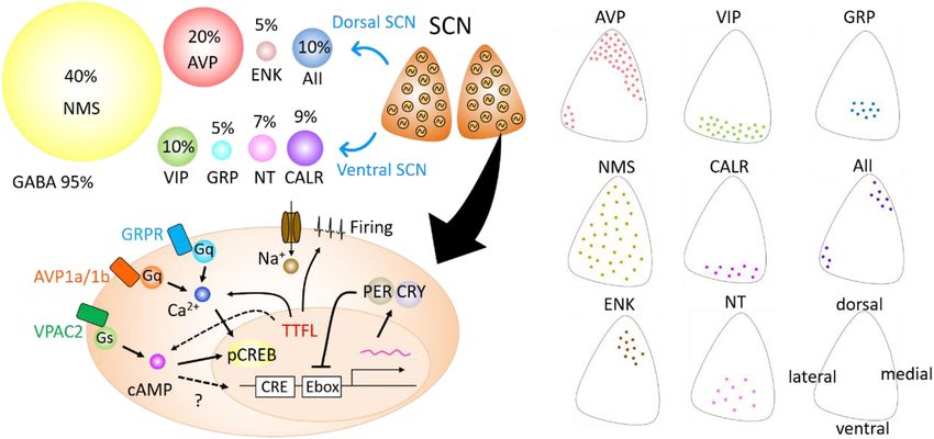
FIGURE 2 | Developmental changes in cellular coupling mechanisms in the suprachiasmatic nucleus (SCN). (A) Tissue-level PER2:LUC bioluminescence from the
cultured SCN in neonate or adult mice (left). Wild-type (WT) SCNs show circadian rhythms in both neonatal and adult SCNs. Cryptochrome (Cry)1- / - /Cry2- / - SCNs
show robust circadian rhythms in the neonate SCN, but not in the adult SCN. Whereas Cry1- / - /Cry2- / - /vasoactive intestinal polypeptide (Vip)2r- / - SCN is arrhythmic
both in the neonate and adult, which is due to desynchronization of cellular circadian rhythms. Arginine vasopressin (Avp)-ELuc bioluminescence from the cultured
SCN in neonate or adult mice (right). Circadian Avp expression rhythms are observed in the WT SCN, but they are arrhythmic and expression is attenuated in
Cry1- / - /Cry2- / - SCN both at neonatal and adult periods. (B) Schematic view of a model of cellular coupling in the SCN during postnatal development. CRY1 and
CRY2 are involved in the developmental shift from CRY-independent (via VIP) to CRY-dependent (via AVP) networks in the SCN. Green and pink circles indicate VIP
and AVP neurons, respectively. Green and pink arrows indicate signaling via VIP and AVP, respectively. This shift is observed around postnatal days 14–21. Modified
from Ono et al., 2016.
Frontiers in Neuroscience | www.frontiersin.org 5 April 2021 | Volume 15 | Article 650154Ono et al. Neuropeptides in the SCN
AAV-mediated diphtheria toxin A subunit (DTA) expression Stephan and Zucker, 1972), SCN circadian rhythms have been
profoundly disrupted circadian locomotor activity recorded by recorded in slice culture as well as in situ. Since culturing
a telemetry transmitter and running wheel. The difference in neonatal SCN is easier than culturing adult SCN, researchers
behavioral phenotypes in the two studies might be related to have used neonatal SCN to explain phenotypes of behavior
the number of VIP neurons spared from deletion and recording of mice lacking specific genes. However, recent studies have
methods. The role of VIP neurons in circadian behavioral demonstrated that neuronal networks in the SCN are modulated
rhythms is still controversial and remains to be studied. during development. CRY1 and CRY2 are involved in the
The importance of neuropeptides and clock gene, Cry, maturation of neuronal networks in the SCN during postnatal
for coherent circadian rhythms in the SCN during postnatal development. Although the functions of neuropeptides in the
development has also been reported. Cry1 and Cry2 are thought SCN cellular network have been investigated, their roles as output
to be essential genes for circadian rhythms (Okamura et al., signals regulating peripheral circadian oscillators have not been
1999; van der Horst et al., 1999; Albus et al., 2002; Yamaguchi clearly identified. Recent advances in various methodologies
et al., 2003). However, circadian Per1-luc, PER2:LUC, and allow us to manipulate cell type- and developmental stage-
spontaneous firing rhythms have been observed in single SCN specific gene expression, cellular signaling, and neuronal activity,
cells in Cry1/Cry2-deficient mice (Maywood et al., 2011; Ono which can help identify mechanisms for the developmental
et al., 2013; Figure 2). Importantly, the cellular circadian aspects of circadian rhythms in the SCN.
rhythms of Cry1/Cry2-deficient SCN were synchronized during
the neonatal period but became desynchronized around postnatal
day 21, where circadian behavioral rhythms could be measured. AUTHOR CONTRIBUTIONS
Furthermore, synchronized circadian rhythms in neonatal
Cry1/Cry2-deficient SCN were desynchronized without VIP DO and SH wrote the manuscript with support of KH. All authors
signaling. Avp expression was also attenuated in Cry1/Cry2- contributed to the article and approved the submitted version.
deficient SCN in both neonatal and adult SCN (Ono et al.,
2016; Figure 2B). These results indicate that VIP and
AVP are responsible for CRY-independent and -dependent FUNDING
cellular coupling, respectively, and that these neuropeptides
are differentially involved in the maturation of circadian This work was supported in part by the Uehara Memorial
networks in the SCN. Foundation, Kowa Life Science Foundation, Takeda Science
Foundation, Kato Memorial Bioscience Foundation, DAIKO
FOUNDATION, SECOM Science and Technology Foundation,
DISCUSSION Research Foundation for Opto-Science and Technology,
The Nakatani Foundation for Advancement of Measuring
Since the first report demonstrating the importance of the SCN Technologies in Biomedical Engineering, and the JSPS
for circadian rhythms in behavior (Moore and Eichler, 1972; KAKENHI (18H02477, 15H04679, and 15K12763).
REFERENCES Aton, S. J., Huettner, J. E., Straume, M., and Herzog, E. D. (2006). GABA and G(i/o)
differentially control circadian rhythms and synchrony in clock neurons. Proc.
Abrahamson, E. E., and Moore, R. Y. (2001). Suprachiasmatic nucleus in the Natl. Acad. Sci. USA 103, 19188–19193. doi: 10.1073/pnas.0607466103
mouse: retinal innervation, intrinsic organization and efferent projections. Ban, Y., Shigeyoshi, Y., and Okamura, H. (1997). Development of vasoactive
Brain Res. 916, 172–191. doi: 10.1016/s0006-8993(01)02890-6 intestinal peptide mRNA rhythm in the rat suprachiasmatic nucleus. J. Neurosci.
Albus, H., Bonnefont, X., Chaves, I., Yasui, A., Doczy, J., van der Horst, G. T. J., et al. 17, 3920–3931. doi: 10.1523/jneurosci.17-10-03920.1997
(2002). Cryptochrome-deficient mice lack circadian electrical activity in the Bedont, J. L., LeGates, T. A., Slat, E. A., Byerly, M. S., Wang, H., Hu, J. F.,
suprachiasmatic nuclei. Curr. Biol. 12, 1130–1133. doi: 10.1016/s0960-9822(02) et al. (2014). Lhx1 controls terminal differentiation and circadian function of
00923-5 the suprachiasmatic nucleus. Cell Rep. 7, 609–622. doi: 10.1016/j.celrep.2014.
An, S. W., Irwin, R. P., Allen, C. N., Tsai, C., and Herzog, E. D. (2011). 03.060
Vasoactive intestinal polypeptide requires parallel changes in adenylate Brancaccio, M., Maywood, E. S., Chesham, J. E., Loudon, A. S. I., and Hastings,
cyclase and phospholipase C to entrain circadian rhythms to a M. H. (2013). A Gq-Ca2+ Axis controls circuit-level encoding of circadian time
predictable phase. J. Neurophysiol. 105, 2289–2296. doi: 10.1152/jn.00966. in the suprachiasmatic nucleus. Neuron 78, 714–728. doi: 10.1016/j.neuron.
2010 2013.03.011
An, S., Harang, R., Meeker, K., Granados-Fuentes, D., Tsai, C. A., Mazuski, C., et al. Carmona-Alcocer, V., Abel, J. H., Sun, T. C., Petzold, L. R., Doyle, F. J., Simms,
(2013). A neuropeptide speeds circadian entrainment by reducing intercellular C. L., et al. (2018). Ontogeny of circadian rhythms and synchrony in the
synchrony. Proc. Natl. Acad. Sci. USA 110, E4355–E4361. suprachiasmatic nucleus. J. Neurosci. 38, 1326–1334. doi: 10.1523/jneurosci.
Ansari, N., Agathagelidis, M., Lee, C., Korf, H. W., and von Gall, C. (2009). 2006-17.2017
Differential maturation of circadian rhythms in clock gene proteins in the Clark, D. D., Gorman, M. R., Hatori, M., Meadows, J. D., Panda, S., and
suprachiasmatic nucleus and the pars tuberalis during mouse ontogeny. Eur. Mellon, P. L. (2013). Aberrant development of the suprachiasmatic nucleus
J. Neurosci. 29, 477–489. doi: 10.1111/j.1460-9568.2008.06605.x and circadian rhythms in mice lacking the homeodomain protein Six6. J. Biol.
Aton, S. J., Colwell, C. S., Harmar, A. J., Waschek, J., and Herzog, E. D. Rhythm 28, 15–25. doi: 10.1177/0748730412468084
(2005). Vasoactive intestinal polypeptide mediates circadian rhythmicity and Collins, B., Pierre-Ferrer, S., Muheim, C., Lukacsovich, D., Cai, Y. C., Spinnler, A.,
synchrony in mammalian clock neurons. Nat. Neurosci. 8, 476–483. doi: 10. et al. (2020). Circadian VIPergic neurons of the suprachiasmatic nuclei sculpt
1038/nn1419 the sleep-wake cycle. Neuron 108, 486–499.e5.
Frontiers in Neuroscience | www.frontiersin.org 6 April 2021 | Volume 15 | Article 650154Ono et al. Neuropeptides in the SCN
Colwell, C. S., Michel, S., Itri, J., Rodriguez, W., Tam, J., Lelievre, V., et al. (2003). Landgraf, D., Achten, C., Dallmann, F., and Oster, H. (2015). Embryonic
Disrupted circadian rhythms in VIP- and PHI-deficient mice. Am. J. Physiol. development and maternal regulation of murine circadian clock function.
Reg. I 285, R939–R949. Chronobiol. Int. 32, 416–427. doi: 10.3109/07420528.2014.986576
Edwards, M. D., Brancaccio, M., Chesham, J. E., Maywood, E. S., and Hastings, Lee, I. T., Chang, A. S., Manandhar, M., Shan, Y. L., Fan, J. M., Izumo, M., et al.
M. H. (2016). Rhythmic expression of cryptochrome induces the circadian clock (2015). Neuromedin S-Producing neurons act as essential pacemakers in the
of arrhythmic suprachiasmatic nuclei through arginine vasopressin signaling. suprachiasmatic nucleus to couple clock neurons and dictate circadian rhythms.
Proc. Natl. Acad. Sci. U S A, 113, 2732–2737. Neuron 85, 1086–1102. doi: 10.1016/j.neuron.2015.02.006
Evans, J. A., Leise, T. L., Castanon-Cervantes, O., and Davidson, A. J. (2013). LeGates, T. A., Fernandez, D. C., and Hattar, S. (2014). Light as a central modulator
Dynamic interactions mediated by nonredundant signaling mechanisms couple of circadian rhythms, sleep and affect. Nat. Rev. Neurosci. 15, 443–454. doi:
circadian clock neurons. Neuron 80, 973–983. doi: 10.1016/j.neuron.2013. 10.1038/nrn3743
08.022 Liu, A. C., Welsh, D. K., Ko, C. H., Tran, H. G., Zhang, E. E., Priest, A. A., et al.
Freeman, G. M. Jr., Krock, R. M., Aton, S. J., Thaben, P., and Herzog, E. D. (2013). (2007). Intercellular coupling confers robustness against mutations in the SCN
GABA networks destabilize genetic oscillations in the circadian pacemaker. circadian clock network. Cell 129, 605–616. doi: 10.1016/j.cell.2007.02.047
Neuron 78, 799–806. doi: 10.1016/j.neuron.2013.04.003 Maywood, E. S., Chesham, J. E., Meng, Q. J., Nolan, P. M., Loudon, A. S. I.,
Gizowski, C., Zaelzer, C., and Bourque, C. W. (2016). Clock-driven vasopressin and Hastings, M. H. (2011). Tuning the period of the mammalian circadian
neurotransmission mediates anticipatory thirst prior to sleep. Nature 537, clock: additive and independent effects of CK1 epsilon(Tau) and Fbxl3(Afh)
685–688. doi: 10.1038/nature19756 mutations on mouse circadian behavior and molecular pacemaking. J. Neurosci.
Hamnett, R., Crosby, P., Chesham, J. E., and Hastings, M. H. (2019). Vasoactive 31, 1539–1544.
intestinal peptide controls the suprachiasmatic circadian clock network via Maywood, E. S., Reddy, A. B., Wong, G. K. Y., O’Neill, J. S., O’Brien,
ERK1/2 and DUSP4 signalling. Nat. Commun. 10:542. J. A., McMahon, D. G., et al. (2006). Synchronization and maintenance
Harmar, A. J., Marston, H. M., Shen, S. B., Spratt, C., West, K. M., Sheward, W. J., of timekeeping in suprachiasmatic circadian clock cells by neuropeptidergic
et al. (2002). The VPAC(2) receptor is essential for circadian function in the signaling. Curr. Biol. 16, 599–605. doi: 10.1016/j.cub.2006.02.023
mouse suprachiasmatic nuclei. Cell 109, 497–508. doi: 10.1016/s0092-8674(02) Mazuski, C., Abel, J. H., Chen, S. P., Hermanstyne, T. O., Jones, J. R., Simon, T.,
00736-5 et al. (2018). Entrainment of circadian rhythms depends on firing rates and
Hatori, M., Gill, S., Mure, L. S., Goulding, M., O’Leary, D. D. M., and Panda, S. neuropeptide release of VIP SCN neurons. Neuron 99, 555–563.e5.
(2014). Lhx1 maintains synchrony among circadian oscillator neurons of the Mazuski, C., Chen, S. P., and Herzog, E. D. (2020). Different roles for VIP neurons
SCN. Elife 3:e03357. in the neonatal and adult suprachiasmatic nucleus. J. Biol. Rhythm 35, 465–475.
Hattar, S., Kumar, M., Park, A., Tong, P., Tung, J., Yau, K.W., and Berson, D.M. doi: 10.1177/0748730420932073
(2006). Central projections of melanopsin-expressing retinal ganglion cells in McNeill, D. S., Sheely, C. J., Ecker, J. L., Badea, T. C., Morhardt, D., Guido, W.,
the mouse. J. Comp. Neurol. 497, 326–349. doi: 10.1002/cne.20970 and Hattar, S. (2011). Development of melanopsin-based irradiance detecting
Hermanstyne, T. O., Simms, C. L., Carrasquillo, Y., Herzog, E. D., and Nerbonne, circuitry. Neural Dev. 6:8. doi: 10.1186/1749-8104-6-8
J. M. (2016). Distinct firing properties of vasoactive intestinal peptide- Mieda, M., Ono, D., Hasegawa, E., Okamoto, H., Honma, K., Honma, S., et al.
expressing neurons in the suprachiasmatic nucleus. J. Biol. Rhythm 31, 57–67. (2015). Cellular clocks in AVP neurons of the SCN are critical for interneuronal
doi: 10.1177/0748730415619745 coupling regulating circadian behavior rhythm. Neuron 85, 1103–1116. doi:
Herzog, E. D., Aton, S. J., Numano, R., Sakaki, Y., and Tei, H. (2004). Temporal 10.1016/j.neuron.2015.02.005
precision in the mammalian circadian system: a reliable clock from less reliable Moore, R. Y., and Bernstein, M. E. (1989). Synaptogenesis in the rat
neurons. J. Biol. Rhythm 19, 35–46. doi: 10.1177/0748730403260776 suprachiasmatic nucleus demonstrated by electron-microscopy and Synapsin-
Hirata, Y., Enoki, R., Kuribayashi-Shigetomi, K., Oda, Y., Honma, S., and Honma, 1 immunoreactivity. J. Neurosci. 9, 2151–2162. doi: 10.1523/jneurosci.09-06-
K. (2019). Circadian rhythms in Per1, PER2 and Ca2+ of a solitary SCN neuron 02151.1989
cultured on a microisland. Sci. Rep. 9:18271. Moore, R. Y., and Eichler, V. B. (1972). Loss of a circadian adrenal corticosterone
Honma, S., Nakamura, W., Shirakawa, T., and Honma, K. (2004). Diversity in rhythm following suprachiasmatic lesions in rat. Brain Res. 42, 201–206. doi:
the circadian periods of single neurons of the rat suprachiasmatic nucleus 10.1016/0006-8993(72)90054-6
depends on nuclear structure and intrinsic period. Neurosci. Lett. 358, 173–176. Myung, J., Hong, S., DeWoskin, D., De Schutter, E., Forger, D. B., and Takumi, T.
doi: 10.1016/j.neulet.2004.01.022 (2015). GABA-mediated repulsive coupling between circadian clock neurons in
Ikeda, M., Sugiyama, T., Wallace, C. S., Gompf, H. S., Yoshioka, T., Miyawaki, the SCN encodes seasonal time. Proc. Natl. Acad. Sci. USA 112, E3920–E3929.
A., et al. (2003). Circadian dynamics of cytosolic and nuclear Ca2+ in single Okamura, H., Fukui, K., Koyama, E., Tsutou, H. L. O., Tsutou, T., Terubayashi, H.,
suprachiasmatic nucleus neurons. Neuron 38, 253–263. doi: 10.1016/s0896- et al. (1983). Time of vasopressin neuron origin in the mouse hypothalamus
6273(03)00164-8 - examination by combined technique of immunocytochemistry and [H-
Inagaki, N., Honma, S., Ono, D., Tanahashi, Y., and Honma, K. (2007). 3]thymidine autoradiography. Dev. Brain Res. 9, 223–226.
Separate oscillating cell groups in mouse suprachiasmatic nucleus couple Okamura, H., Miyake, S., Sumi, Y., Yamaguchi, S., Yasui, A., Muijtjens, M., et al.
photoperiodically to the onset and end of daily activity. Proc. Natl. Acad. Sci. (1999). Photic induction of mPer1 and mPer2 in Cry-deficient mice lacking a
USA 104, 7664–7669. doi: 10.1073/pnas.0607713104 biological clock. Science 286, 2531–2534. doi: 10.1126/science.286.5449.2531
Isobe, Y., and Muramatsu, K. (1995). Day-night differences in the contents Ono, D., Honma, K., Yanagawa, Y., Yamanaka, A., and Honma, S. (2019). GABA
of vasoactive-intestinal-peptide, gastrin-releasing peptide and arg-vasopressin in the suprachiasmatic nucleus refines circadian output rhythms in mice.
in the suprachiasmatic nucleus of rat pups during postnatal-development. Commun. Biol. 2:232.
Neurosci. Lett. 188, 45–48. doi: 10.1016/0304-3940(95)11391-9 Ono, D., Honma, S., and Honma, K. (2013). Cryptochromes are critical for the
Jin, X. W., Shearman, L. P., Weaver, D. R., Zylka, M. J., De Vries, G. J., and Reppert, development of coherent circadian rhythms in the mouse suprachiasmatic
S. M. (1999). A molecular mechanism regulating rhythmic output from the nucleus. Nat. Commun. 4:1666.
suprachiasmatic circadian clock. Cell 96, 57–68. doi: 10.1016/s0092-8674(00) Ono, D., Honma, S., and Honma, K. (2016). Differential roles of AVP and VIP
80959-9 signaling in the postnatal changes of neural networks for coherent circadian
Jones, J. R., Simon, T., Lones, L., and Herzog, E. D. (2018). SCN VIP rhythms in the SCN. Sci. Adv. 2:e1600960. doi: 10.1126/sciadv.1600960
neurons are essential for normal light-mediated resetting of the circadian Ono, D., Mukai, Y., Hung, C. J., Chowdhury, S., Sugiyama, T., and Yamanaka,
system. J. Neurosci. 38, 7986–7995. doi: 10.1523/jneurosci.1322-18. A. (2020). The mammalian circadian pacemaker regulates wakefulness via
2018 CRF neurons in the paraventricular nucleus of the hypothalamus. Sci. Adv.
Kabrita, C. S., and Davis, F. C. (2008). Development of the mouse suprachiasmatic 6:eabd0384. doi: 10.1126/sciadv.abd0384
nucleus: determination of time of cell origin and spatial arrangements within Patton, A. P., Edwards, M. D., Smyllie, N. J., Hamnett, R., Chesham, J. E.,
the nucleus. Brain Res. 1195, 20–27. doi: 10.1016/j.brainres.2007.12.020 Brancaccio, M., et al. (2020). The VIP-VPAC2 neuropeptidergic axis is a
Frontiers in Neuroscience | www.frontiersin.org 7 April 2021 | Volume 15 | Article 650154Ono et al. Neuropeptides in the SCN cellular pacemaking hub of the suprachiasmatic nucleus circadian circuit. Nat. van der Horst, G. T. J., Muijtjens, M., Kobayashi, K., Takano, R., Kanno, S., Takao, Commun. 11:3394. M., et al. (1999). Mammalian Cry1 and Cry2 are essential for maintenance of Paul, S., Hanna, L., Harding, C., Hayter, E. A., Walmsley, L., Bechtold, D. A., et al. circadian rhythms. Nature 398, 627–630. doi: 10.1038/19323 (2020). Output from VIP cells of the mammalian central clock regulates daily VanDunk, C., Hunter, L. A., and Gray, P. A. (2011). Development, maturation, physiological rhythms. Nat. Commun. 11:1453. and necessity of transcription factors in the mouse suprachiasmatic nucleus. Reppert, S. M., and Schwartz, W. J. (1983). Maternal coordination of the J. Neurosci. 31, 6457–6467. doi: 10.1523/jneurosci.5385-10.2011 fetal biological clock inutero. Science 220, 969–971. doi: 10.1126/science. Weaver, D. R. (1998). The suprachiasmatic nucleus: a 25-year retrospective. J. Biol. 6844923 Rhythm 13, 100–112. doi: 10.1177/074873098128999952 Reppert, S. M., and Weaver, D. R. (2002). Coordination of circadian timing in Webb, A. B., Angelo, N., Huettner, J. E., and Herzog, E. D. (2009). Intrinsic, mammals. Nature 418, 935–941. doi: 10.1038/nature00965 nondeterministic circadian rhythm generation in identified mammalian Romero, M. T., and Silver, R. (1990). Time course of peptidergic expression in fetal neurons. Proc. Natl. Acad. Sci. USA 106, 16493–16498. doi: 10.1073/pnas. suprachiasmatic nucleus transplanted into adult hamster. Dev. Brain Res. 57, 0902768106 1–6. doi: 10.1016/0165-3806(90)90177-z Welsh, D. K., Logothetis, D. E., Meister, M., and Reppert, S. M. (1995). Individual Shan, Y., Abel, J. H., Li, Y., Izumo, M., Cox, K. H., Jeong, B., et al. (2020). Dual- Neurons dissociated from rat suprachiasmatic nucleus express independently color single-cell imaging of the suprachiasmatic nucleus reveals a circadian role phased circadian firing rhythms. Neuron 14, 697–706. doi: 10.1016/0896- in network synchrony. Neuron 108, 164–179 e167. 6273(95)90214-7 Shimada, M., and Nakamura, T. (1973). Time of neuron origin in mouse Wen, S., Ma, D., Zhao, M., Xie, L., Wu, Q., Gou, L., et al. (2020). Spatiotemporal hypothalamic nuclei. Exp. Neurol. 41, 163–173. doi: 10.1016/0014-4886(73) single-cell analysis of gene expression in the mouse suprachiasmatic nucleus. 90187-8 Nat. Neurosci. 23, 456–467. doi: 10.1038/s41593-020-0586-x Shimogori, T., Lee, D. A., Miranda-Angulo, A., Yang, Y. Q., Wang, H., Jiang, Wreschnig, D., Dolatshad, H., and Davis, F. C. (2014). Embryonic development of L. Z., et al. (2010). A genomic atlas of mouse hypothalamic development. Nat. circadian oscillations in the mouse hypothalamus. J. Biol. Rhythm 29, 299–310. Neurosci. 13, U767–U153. doi: 10.1177/0748730414545086 Shimomura, H., Moriya, T., Sudo, M., Wakamatsu, H., Akiyama, M., Miyake, Yamaguchi, S., Isejima, H., Matsuo, T., Okura, R., Yagita, K., Kobayashi, M., Y., et al. (2001). Differential daily expression of Per1 and Per2 mRNA in the et al. (2003). Synchronization of cellular clocks in the suprachiasmatic nucleus. suprachiasmatic nucleus of fetal and early postnatal mice. Eur. J. Neurosci. 13, Science 302, 1408–1412. doi: 10.1126/science.1089287 687–693. doi: 10.1046/j.0953-816x.2000.01438.x Yamaguchi, Y., Suzuki, T., Mizoro, Y., Kori, H., Okada, K., Chen, Y. L., et al. (2013). Shinohara, K., Honma, S., Katsuno, Y., Abe, H., and Honma, K. (1995). 2 Distinct Mice genetically deficient in vasopressin V1a and V1b receptors are resistant to Oscillators in the rat suprachiasmatic nucleus in-Vitro. Proc. Natl. Acad. Sci. jet lag. Science 342, 85–90. doi: 10.1126/science.1238599 USA 92, 7396–7400. doi: 10.1073/pnas.92.16.7396 Yoshikawa, T., Nakajima, Y., Yamada, Y., Enoki, R., Watanabe, K., Yamazaki, M., Shinohara, K., Tominaga, K., and Inouye, S. I. T. (1999). Phase dependent et al. (2015). Spatiotemporal profiles of arginine vasopressin transcription in response of vasoactive intestinal polypeptide to light and darkness in the cultured suprachiasmatic nucleus. Eur. J. Neurosci. 42, 2678–2689. doi: 10.1111/ suprachiasmatic nucleus. Neurosci. Res. 33, 105–110. doi: 10.1016/s0168- ejn.13061 0102(98)00122-9 Stephan, F. K., and Zucker, I. (1972). Circadian-rhythms in drinking behavior and Conflict of Interest: The authors declare that the research was conducted in the locomotor activity of rats are eliminated by hypothalamic-lesions. Proc. Natl. absence of any commercial or financial relationships that could be construed as a Acad. Sci. USA 69, 1583–1586. doi: 10.1073/pnas.69.6.1583 potential conflict of interest. Todd, W. D., Venner, A., Anaclet, C., Broadhurst, R. Y., De Luca, R., Bandaru, S. S., et al. (2020). Suprachiasmatic VIP neurons are required for normal Copyright © 2021 Ono, Honma and Honma. This is an open-access article circadian rhythmicity and comprised of molecularly distinct subpopulations. distributed under the terms of the Creative Commons Attribution License (CC BY). Nat. Commun. 11:4410. The use, distribution or reproduction in other forums is permitted, provided the Uhl, G. R., and Reppert, S. M. (1986). Suprachiasmatic nucleus vasopressin original author(s) and the copyright owner(s) are credited and that the original messenger-rna - circadian variation in normal and brattleboro rats. Science 232, publication in this journal is cited, in accordance with accepted academic practice. No 390–393. doi: 10.1126/science.3961487 use, distribution or reproduction is permitted which does not comply with these terms. Frontiers in Neuroscience | www.frontiersin.org 8 April 2021 | Volume 15 | Article 650154
You can also read