Simultaneous perception of prosthetic and natural vision in AMD patients
←
→
Page content transcription
If your browser does not render page correctly, please read the page content below
ARTICLE
                  https://doi.org/10.1038/s41467-022-28125-x                 OPEN

                  Simultaneous perception of prosthetic and natural
                  vision in AMD patients
                  D. Palanker         1 ✉,   Y. Le Mer2, S. Mohand-Said3 & J. A. Sahel                      2,3,4,5

                  Loss of photoreceptors in atrophic age-related macular degeneration (AMD) results in severe
                  visual impairment. Since the low-resolution peripheral vision is retained in such conditions,
1234567890():,;

                  restoration of central vision should not jeopardize the surrounding healthy retina and allow
                  for simultaneous use of the natural and prosthetic sight. This interim report, prespecified in
                  the study protocol, presents the first clinical results with a photovoltaic substitute of the
                  photoreceptors providing simultaneous use of the central prosthetic and peripheral natural
                  vision in atrophic AMD. In this open-label single group feasibility trial (NCT03333954,
                  recruitment completed), five patients with geographic atrophy have been implanted with a
                  wireless 2 x 2 mm-wide 30 µm-thick device, having 378 pixels of 100 µm in size. All 5
                  patients achieved the primary outcome of the study by demonstrating the prosthetic visual
                  perception in the former scotoma. The four patients with a subretinal placement of the chip
                  demonstrated the secondary outcome: Landolt acuity of 1.17 ± 0.13 pixels, corresponding to
                  the Snellen range of 20/460–20/565. With electronic magnification of up to a factor of 8,
                  patients demonstrated prosthetic acuity in the range of 20/63–20/98. Under room lighting
                  conditions, patients could simultaneously use prosthetic central vision and their remaining
                  peripheral vision in the implanted eye and in the fellow eye.

                  1 Department of Ophthalmology and Hansen Experimental Physics Laboratory, Stanford University, Stanford, CA, USA. 2 Department of Ophthalmology,

                  Fondation Ophtalmologique A. de Rothschild, Paris, France. 3 Clinical Investigation Center INSERM-DGOS 1423, Quinze-Vingts National Eye Hospital,
                  Paris, France. 4 Department of Ophthalmology, University of Pittsburgh School of Medicine, Pittsburgh, PA, USA. 5 Sorbonne Université, INSERM, CNRS,
                  Institut de la Vision, Paris, France. ✉email: palanker@stanford.edu

                  NATURE COMMUNICATIONS | (2022)13:513 | https://doi.org/10.1038/s41467-022-28125-x | www.nature.com/naturecommunications                                1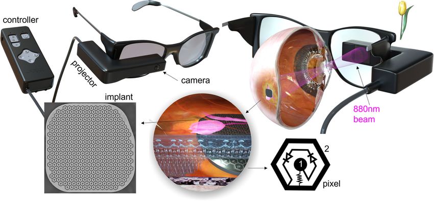
ARTICLE NATURE COMMUNICATIONS | https://doi.org/10.1038/s41467-022-28125-x
A
ge-related macular degeneration (AMD) is a leading cause Images captured by the camera are processed and projected onto
of irreversible vision loss1, with its prevalence dramati- the retina from video glasses using intensified light (Fig. 1). To
cally increasing in the aging population: from 1.5% in the avoid photophobic and phototoxic effects of bright illumination,
US residents above 40 years to more than 15% in the subjects we use near-infrared (NIR, 880 nm) wavelength9. Photovoltaic
older than 802. Currently, there is no efficient therapy for pre- pixels in the implant directly convert the projected pulsed light
venting the AMD progression, except for suppression of the into local electric current flowing through the retina between the
neovascularization3, although research in this field continues4. active and return electrodes6,10.
The atrophic form of AMD (also known as geographic atrophy,
GA) results in a gradual loss of photoreceptors in the central
macula, which is responsible for high-resolution vision, and Results
severely impairs reading and face recognition. Low-resolution Five patients with GA were implanted in Paris during 2017–2018
peripheral vision is retained in this condition, enabling orienta- (NCT03333954). In four of them, the implant was placed in the
tion and the use of eccentric fixation for visual discrimination at subretinal space, but in one it ended up inside the choroid due to
reduced acuity. Therefore, the goal of any treatment strategy patient’s accidental movement during surgery. In one of the four
should be to restore functional central vision without jeopardizing patients, the implant accidentally shifted by about 2 mm from the
the surrounding retina and allowing for their simultaneous use. central position after the fluid-air exchange since the patient did
While photoreceptors gradually disappear in GA, the inner not keep the head in a prone position post implantation. Due to
retinal cells survive to a large extent5. To restore sight in the wireless nature of the implant, surgical procedure was relatively
scotoma, we replace the lost photoreceptors with photovoltaic short—about 2 h11. As shown in Table 1, residual natural acuity
pixels in the subretinal implant, which convert light into electric in the operated eye did not decrease in any of the subjects.
current to selectively stimulate the secondary neurons in the Interestingly, in some patients, acuity improved compared to
retina6. These electronic substitutes of photoreceptors replace the baseline, which could be attributed to either a neurotrophic
two main functions of the natural photoreceptors: (a) the light-to- benefit of subretinal surgery12 or of electrical stimulation13 or just
current conversion, corresponding to the function of the outer improvement with eccentric fixation after training.
segment, and (b) transfer of the visual information to secondary The primary endpoint of the study—prosthetic light perception
neurons by their polarization in extracellular electric field, sub- measured in the visual field test (Octopus 900; Haag-Streit, Koniz,
stituting the function of the synapse. Switzerland), demonstrated that visual perception was elicited by
To avoid irreversible electrochemical reactions at the the PRIMA implant in all subjects, as reported earlier for the time
electrode–electrolyte interface, stimulation current is pulsed and period of 6–12 months11. During the 18–24 months follow-up
charge-balanced. On the other hand, to provide steady visual period, sensitivity improved in all subjects, as shown in Table 1,
percepts under pulsatile illumination, repetition rate should except for patient #3, who passed away due to unrelated cause
exceed the frequency of flicker fusion. In preclinical studies, we before the second phase of the trial. In the first phase of the
demonstrated that selective stimulation of bipolar cells without trial11, prosthetic vision was assessed independently from the
direct activation of the downstream neurons results in preserva- remaining natural vision. For this purpose, opaque virtual reality
tion of multiple features of the natural retinal signal processing, glasses (VR, PRIMA-1) have been used. The projected images
including flicker fusion, adaptation to static images6, ON and covered a horizontal field of 5.1 mm (17.5° on the retina), with
OFF responses with antagonistic center-surround7, and non- approximate resolution of 10.5 μm. Maximum peak retinal irra-
linear summation of subunits in the retinal ganglion cells’ (RGC) diance was 3 mW/mm2, well within the thermal safety limits for
receptive fields6. We have also shown that grating visual acuity chronic use of near-infrared light14. Brightness of the percept was
(VA) matches the pixel pitch with 75 and 55 μm pixels6,8. controlled by pulse duration, between 0.7 and 9.8 ms, in 0.7 ms
The first generation of the human-grade photovoltaic sub- increments.
retinal prosthesis PRIMA (Pixium Vision SA., Paris, France) is The four patients with subretinal implant placement demon-
2 mm in width (~7° of the visual angle in a human eye), 30 μm in strated monochromatic (white-yellowish “sun-color”) form per-
thickness, containing 378 hexagonal pixels of 100 μm in width. ception, with flicker fusion above 30 Hz. In three patients with
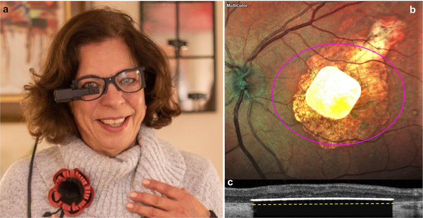
Fig. 1 Diagram of the PRIMA system. Top row: Artistic rendering of the augmented reality glasses with a projector and a camera. The 880 nm beam
projects the video stream onto the retina. Bottom row: PRIMA implant with a hexagonal array of 100 μm pixels. Implant is placed under the degenerate
retina without damaging the peripheral healthy retina. Pixels are composed of two photodiodes connected in series between the active (1) and a
circumferential return (2) electrode.
2 NATURE COMMUNICATIONS | (2022)13:513 | https://doi.org/10.1038/s41467-022-28125-x | www.nature.com/naturecommunications
NATURE COMMUNICATIONS | https://doi.org/10.1038/s41467-022-28125-x                                                                                              ARTICLE

 Table 1 Residual natural vision, anatomical and functional outcomes with the implant.

 Test/Patient                                                 1                          2                     3                    4                       5
 Patients’ data
 Age at baseline                                              83                         66                    82                   69                      74
 Years of VA < 20/400 in implanted eye                        6                          2                     3                    5                       10
 Residual peripheral vision
 Pre-op natural letter acuity in the study eye                20/400                     20/800                20/1000              20/500                  20/500
 Postop natural letter acuity in the study eye                20/320; 20/160             20/800; 20/200        20/800*              20/400; 20/500          20/400; 20/550
 at 12 months and at 24 months
 Pre-op letter acuity in the fellow eye                       20/100                     20/50                 20/125               20/400                  20/100
 Postop letter acuity in the fellow eye                       20/160; 20/160             20/50; 20/50          20/200*              20/400; 20/640          20/125; 20/125
 at 12 months and at 24 months
 Implant location
 Implant location in the macula                               Intra-choroidal            Central subretinal    Central subretinal   Off-center subretinal   Central subretinal
 Stimulation threshold and sensitivity
 Perceptual threshold with PRIMA-1                            2.1                        0.8                   0.7                  1.0                     0.8
 glasses, ms
 Perceptual threshold with PRIMA-2                            1.28 ± 0.84                0.75 ± 0.19           *                    0.82 ± 0.29             0.70 ± 0.00
 glasses, ms
 Central perceptual threshold in OCTOPUS, dB                  0.9; 2.5                   1.3; 1.9              3.1*                 0.4; 2.9                1.3; 10.1
 at 6–12 and at 18–24 months
 Prosthetic visual acuity
 PRIMA-1 (VR), 12 months; no magnification                     Light perception           20/550;               20/500;              20/800;                 20/460;
 Min. Landolt C gap, pix                                                                 logMAR 1.44           logMAR 1.40          logMAR 1.60             logMAR 1.37
                                                                                         1.3 pix               1.2 pix              1.9 pix                 1.1 pix
 PRIMA-2 (AR), 18–24 months, no                               Light perception           20/564;               *                    20/438;                 **
 magnification                                                                            logMAR 1.45                                logMAR 1.34
 Min. Landolt C gap, pix                                                                 1.34 pix                                   1.04 pix
 PRIMA-2 (AR), Landolt VA with preferred                      Light perception           20/98;                *                    20/71; logMAR 0.55      20/63;
 magnification, 18–24 months                                                              logMAR 0.69                                                        logMAR 0.50
 Natural Landolt VA in the study eye,                         20/182;                    20/246;               *                    20/428;                 20/332;
 18–24 months                                                 logMAR 0.96                logMAR 1.09                                logMAR 1.33             logMAR 1.22
 LogMAR gain due to PRIMA 18–24 months                        0                          0.4                   *                    0.78                    0.72
 Background lighting
 Background light threshold [cd/m2]                           NA, no shape               >256                  *                    >256                    64
                                                              perception
 Attenuation for bright room lighting                         NA                         Clear                 *                    Clear                   65%
 Simultaneous perception of prosthetic and natural vision
 Bar orientation, % correct                        NA,                                   100                   *                    100                     96
 Monocular                                         No shape                              100                                        100                     92
 Binocular                                         perception

 *Patient 3 passed away before the second phase of the trial.
 **Patient 5 was not available for this measurement because of the COVID restrictions.

central location of the subretinal implant, acuity closely matched                                  thresholds measured during the first 6 months—in the first phase
the pixel size: 20/460, 20/500, and 20/550 (1.1, 1.2, and 1.3 pixels).                              of the trial11.
Patient with the off-center implant demonstrated lower acuity:                                         Prosthetic visual acuity with PRIMA-2 glasses was measured
20/800 (1.9 pixels)11. Patient #1 with the intra-choroidal implant                                  using Landolt C optotypes. To mimic the crowding effect of the
had blurry prosthetic vision, with no discernable acuity.                                           letter charts, the Landolt rings were surrounded by a square frame
   In the second phase of the study, starting at 18–24 months                                       (Fig. 3a). At each trial, subjects reported the font orientation (up,
post-op, we introduced augmented reality glasses (AR, PRIMA-                                        down, left, or right), and its size was then adjusted, depending on
2), which allow unobstructed natural vision by the fellow eye and                                   the response. The visual acuity was determined using the Freiburg
by the peripheral field of the operated eye, simultaneously with                                     Visual Acuity Test (FrACT) software15,16. For a stable perception
prosthetic central vision in the treated eye (Fig. 2a). The images,                                 under pulsatile illumination, 30 Hz repetition rate was applied. In
projected through the glasses adapted to patient’s refraction,                                      the first set of the tests, computer-generated Landolt optotypes
covered a horizontal field of 5.3 mm (18.5°) on the retina, with a                                   were projected into the eye directly from the AR glasses without
resolution of 6.7 μm, as illustrated in Fig. 2b. This design provided                               using a camera. As shown in Table 1, patients #2 and #5
improved beam homogeneity and easier alignment, compared to                                         demonstrated prosthetic acuity at the level similar to that observed
VR glasses (PRIMA-1). The maximum retinal irradiance was                                            with VR glasses in the first phase of the trial (20/500, 20/460), but
increased to 3.5 mW/mm², with the same range of pulse dura-                                         patient #4 improved compared to the earlier result—from 20/800
tions as with the VR glasses. This system allows the use of elec-                                   to 20/438. This is potentially due to easier alignment of the display
tronic magnification (×1, ×2, ×4, and ×8) between the camera and                                     to the off-center location of the implant with improved glasses. The
the image projection onto the implant. As shown in Table 1,                                         average acuity in the four patients with the subretinal implant
perceptual thresholds 18–24 months after the implantation,                                          placement was 1.17 ± 0.13 pixels at the latest measurement, cor-
measured with PRIMA-2 glasses, were slightly lower than the                                         responding to logMAR 1.39, or 20/500 on a Snellen scale.

NATURE COMMUNICATIONS | (2022)13:513 | https://doi.org/10.1038/s41467-022-28125-x | www.nature.com/naturecommunications                                                      3
ARTICLE NATURE COMMUNICATIONS | https://doi.org/10.1038/s41467-022-28125-x Fig. 2 PRIMA system in practice. a PRIMA-2 glasses on a person. b Fundus photo of a patient with the PRIMA implant inside the geographic atrophy area. Magenta oval illustrates the size of the beam (5.3 × 4.3 mm) projected onto the retina. c OCT image demonstrates the implant in subretinal space 6 months post-op. Yellow dash line depicts the approximate position of the back side of the implant resting on the Bruch’s membrane. Fig. 3 Assessment of prosthetic vision. a Landolt C in the frame mimicking the crowding effect. b Testing setup with a patient sitting 40 cm from the screen. Horizontal green bar shown on a large display (1) can be seen with the remaining natural vision, while the diagonal bar (2) is presented only on the NIR display inside the glasses. In the second set of the acuity tests, Landolt C optotypes were varied. In this experiment, subjects with both eyes open were displayed at 40 cm distance from the subject, so patients used placed 40 cm in front of a wide LCD screen, where a homo- the camera and were allowed to apply their preferred electronic geneous white illumination at 16 levels (ranging from 1.4 to magnification (1, 2, 4, or 8). To ensure that prosthetic acuity is 256 cd/m2) was presented. Prosthetic patterns were presented at measured rather than the residual natural vision, in these tests the maximum brightness: 3.5 mW/mm2 of NIR irradiance with fellow eye was covered. In addition, contrast of the electronic 9.8 ms pulse duration. As shown in Table 1, subjects 2 and 4 did image was inverted from the original black optotypes on a white not have a problem seeing the Landolt C in front of the screen background to white optotypes on a black background (white even with the highest background luminance (256 cd/m2). Subject patterns stimulate the retina), and patients were asked about the 5 had difficulties with luminance above 64 cd/m2, and therefore color of the percept. With magnification, all three participants of was provided later with a shaded lens (65% attenuation of white the second trial could recognize optotypes equating to 20/98, 20/ light) to allow using the device in a bright office environment. 71, and 20/63 acuity, respectively. As shown in Table 1, these It is important to note that patients could simultaneously use values significantly exceeded their residual natural acuity in the prosthetic and residual natural vision from both, the study eye, treated eye, and for patients 4 and 5, even in the (better) and the fellow eye. For example, in a setup shown in Fig. 3b, fellow eye. green bars of various orientations were presented on a large Video S1 illustrates a patient’s letter recognition using pros- screen for natural vision and another set of bars was simulta- thetic vision, with ×4 magnification and a contrast reversal. neously presented just on the NIR display inside the glasses. The Video S2 illustrates a word reading test with ×4 magnification and patients were asked about both orientations and colors, as illu- a control experiment (Video S3), where patient is attempting to strated in the Video S4 for binocular vision and in the Video S5 read the same word without the PRIMA glasses. It is important to for monocular vision. In both cases, the bars were perceived emphasize that these videos are for illustration purposes only. simultaneously at correct orientations, as summarized in the Quantitative measurements of the prosthetic acuity were con- Table 1. ducted with Landolt C optotypes. To evaluate the effect of background light on prosthetic vision when the transparent AR glasses are used, Landolt C optotypes Discussion have been presented on the glasses display directly, without using This study was limited primarily to the clinical evaluation of the camera, while intensity of the background visible light was prosthetic vision. Future testing will be expanded to include a 4 NATURE COMMUNICATIONS | (2022)13:513 | https://doi.org/10.1038/s41467-022-28125-x | www.nature.com/naturecommunications
NATURE COMMUNICATIONS | https://doi.org/10.1038/s41467-022-28125-x                                                                                             ARTICLE

home use, which will increase the active time and also help assess                        was considered the daily result. The final result was defined as the median of the
the functional benefits of the device, considering the residual                            three daily measurements. To mimic the crowding effect of the letters in vision
                                                                                          charts, Landolt C was presented with a frame around it (Fig. 3a).
vision available in the fellow eye. Future studies will also assess a
practical value of the electronic magnification for the central
                                                                                          Background illumination. To provide a uniform and controllable background
vision in the PRIMA system.                                                               illumination, a 71 cm-wide Samsung U28E590D LCD screen has been used.
   In summary, this trial confirmed the safety and stability of the                        Subjects were placed 40 cm in front of a screen, where a homogeneous white
PRIMA implant over 18–24 months follow-up in five patients                                 illumination at 16 levels was presented: 256, 181, 128, 90.5, 64, 45.3, 32, 22.6, 16,
with geographic atrophy. Prosthetic central vision in the former                          11.3, 8, 5.7, 4, 2.8, 2, 1.4 cd/m², while room lights were turned off.
                                                                                              The best-PEST (Parameter Estimation by Sequential Testing) method20 was
scotoma represents monochromatic form perception matching                                 used to determine the maximum luminance which still allowed identifying the
the presented patterns and, most importantly, is perceived in                             prosthetic patterns (Landolt C optotypes with four different orientations) with
conjunction with the residual peripheral vision, thus enabling                            accuracy exceeding 62.5% using the following parameters: 20 iterations, 0.25 false
natural orientation and central discrimination. Spatial resolution                        positive rate, 0 false negative rate, and sigmoid slope of 0.5. The last value of the
was, on average, 1.2 pixels of the implant, corresponding to acuity                       best-PEST was used as the resulting threshold luminance. Data for the three
                                                                                          patients with a subretinal implant are presented, but not for subject #1 since this
of about 20/500, and using electronic magnification, all patients                          subject with intra-choroidal chip placement could not recognize Landolt C.
with subretinal implant demonstrated acuity exceeding 20/100.                             Prosthetic patterns were presented at maximum brightness: 3.5 mW/mm2 of NIR
Recent advancements in the photovoltaic pixel design, which                               irradiance with 9.8 ms pulse duration and 30 Hz repetition rate. The patterns were
enable five times the smaller pixels17,18 may improve acuity of the                        presented for up to 30 s, with 10 s break between the stimuli.
future PRIMA devices up to 20/100, and with electronic zoom—
potentially up to 20/20. Further improvements with video glasses                          Simultaneous perception of prosthetic and natural vision. A green bar was
                                                                                          displayed on an LCD screen placed at 40 cm in front of the subject in one of the
may widen the visual field, while the advanced image processing                            four possible orientations (vertical, horizontal, 45° diagonal from the upper left, 45°
and stimulation protocol may help enhance the dynamic range                               diagonal from the lower left). Simultaneously another bar is also projected in
and contrast sensitivity, promising even more functional                                  Artificial Pattern Mode (APM) on the PRIMA Glasses in one of the four orien-
restoration of sight for numerous patients suffering from atrophic                        tations (Fig. 3b). The width of the bars corresponds to 0.4 mm on the retina (four
                                                                                          implant pixels). The bar orientation on LCD screen is random, but 50% of the
macular degeneration.                                                                     times orientation of the bar displayed on the glasses matches the bar orientation on
                                                                                          LCD. The subject is expected to see the bar displayed on the LCD screen with the
                                                                                          natural peripheral vision and report it as green, and the bar projected by the
Methods                                                                                   PRIMA Glasses with prosthetic vision, perceived as white. At each repetition, the
Patients. The aim of this study (NCT03333954) was to test functionality of the            subject is asked about orientation of each bar individually.
PRIMA system in 5 patients with atrophic AMD. The study adhered to the                        A total of 48 bar pairs were presented (24 presentations with the fellow eye open
Declaration of Helsinki and received the ethics committee approval from the               and 24 presentation with the fellow eye closed), each for a duration of up to 20 s.
Comité de Protection des Personnes Ile de France II and approval by the Agence            The subjects were not allowed to move their head—only the eyes.
Nationale de Sécurité du Médicament et des Produits de Santé in France (ASNM).
Study participants were above 60 years of age and had advanced atrophic AMD
with an atrophic zone of at least three optic disc diameters and best corrected visual    Reporting summary. Further information on research design is available in the Nature
acuity of ≤20/400 in the worse-seeing study eye; no foveal light perception               Research Reporting Summary linked to this article.
(absolute scotoma) but visual perception in the periphery, with preferred retinal
locus determined by micro-perimetry; absence of photoreceptors and presence of
the inner retina in the atrophic area as confirmed by optical coherence tomography
                                                                                          Data availability
                                                                                          All the data supporting the findings of this study are available within the paper and its
(OCT); absence of choroidal neovascularization verified by retinal angiography.
Patient #2 had glaucoma, while all other ocular and general pathologies that could        supplementary information files. The visual acuity data analysis software can be
contribute to the low visual acuity were excluded. All patients, except for #1,           downloaded from the publicly available database21. Protocol for the studies of
underwent visual rehabilitation for improving eccentric fixation before they were          simultaneous perception of prosthetic and natural vision can be provided upon a
recruited for the trial. Patients provided written informed consent to participate in     reasonable request. Pixium Vision is responsible for approval of the clinical study
the study and to publish clinical information and videos in anonymized form.              protocol and for reporting the results to the regulatory authorities in Europe and in US.
Person shown in Fig. 2a is a healthy volunteer who provided a written informed            As such, it was informed about the study on a continuous basis and performed its own
consent to publish her photo in a scientific journal. Patients recruitment was             data analysis. The paper was written by its authors based on their data analysis, and they
completed in 2018.                                                                        are responsible for its content.
    Implantation took place at the Foundation A Rothschild Hospital (Paris,
France). The patients’ rehabilitation and visual function assessment were carried
                                                                                          Received: 12 January 2021; Accepted: 11 January 2022;
out at the Clinical Investigation Center of Quinze-Vingts National Eye Hospital
(Paris, France). In addition to the primary endpoint of the study, the prosthetic
light perception measured in the visual field test, a secondary endpoint was added
in 2019: the visual acuity measured by Landolt C optotypes, as well as exploratory
studies of the visual function, including the simultaneous use of the natural and
prosthetic vision. These modifications were approved by ASNM and by the same
ethics committee as the original protocol. Results in this paper represent an interim     References
report prespecified in the study protocol.                                                 1.   Wong, W. L. et al. Global prevalence of age-related macular degeneration and
    Lab tests were conducted, on average, once a week before COVID, but during                 disease burden projection for 2020 and 2040: a systematic review and meta-
the pandemic the tests frequency significantly decreased due to restrictions on                 analysis. Lancet Glob. Health 2, e106–e116 (2014).
patients’ travel. As these limitations have been relaxed, the frequency of the tests is   2.   Friedman, D. S., Tomany, S. C., McCarty, C., & De Jong, P. Prevalence of age-
being increased again.                                                                         related macular degeneration in the United States. Arch Ophthalmol. 122,
                                                                                               564–572 (2004).
                                                                                          3.   Wong, I. Y., Koo, S. C. & Chan, C. W. Prevention of age-related macular
Assessment of prosthetic vision. Visual acuity was assessed using a computer-                  degeneration. Int. Ophthalmol. 31, 73–82 (2011).
generated Landolt C in four different orientations (gap at the top, bottom, right or      4.   Ammar, M. J., Hsu, J., Chiang, A., Ho, A. C. & Regillo, C. D. Age-related
left), so that a random response corresponds to 25% accuracy. The threshold
                                                                                               macular degeneration therapy: a review. Curr. Opin. Ophthalmol. 31, 215–221
optotype size was defined as the proper symbol recognition with at least 62.5%
                                                                                               (2020).
accuracy. To minimize the number of presentations, the study was conducted using
                                                                                          5.   Kim, S. Y. et al. Morphometric analysis of the macula in eyes with disciform
the method of the Freiburg Acuity and Contrast Test (FrACT)16, which was shown
                                                                                               age-related macular degeneration. Retina 22, 471–477 (2002).
to yield equivalent VA when measured with Landolt C optotypes to that obtained
                                                                                          6.   Lorach, H. et al. Photovoltaic restoration of sight with high visual acuity. Nat.
with ETDRS charts19. In this protocol, a single Landolt C is presented in a fixed
central position on a display, and the best Parameter Estimation by Sequential                 Med. 21, 476–482 (2015).
Testing (best PEST) procedure20 was used to estimate the VA. Since it is an               7.   Ho, E. et al. Spatiotemporal characteristics of retinal response to network-
adaptive test, the number of times that each optotype is presented varies depending            mediated photovoltaic stimulation. J. Neurophysiol. 119, 389–400 (2018).
on the patient responses. The test was performed three times on three different           8.   Ho, E. et al. Characteristics of prosthetic vision in rats with subretinal flat and
days. At each day, 24 trials were performed twice, and the mean of these two runs              pillar electrode arrays. J. Neural Eng. 16, 066027 (2019).

NATURE COMMUNICATIONS | (2022)13:513 | https://doi.org/10.1038/s41467-022-28125-x | www.nature.com/naturecommunications                                                           5
ARTICLE NATURE COMMUNICATIONS | https://doi.org/10.1038/s41467-022-28125-x
9. Goetz, G. A., Mandel, Y., Manivanh, R., Palanker, D. V. & Cizmar, T. Institutes of Health (R01-EY027786). J.A.S. is supported in part by the NIH CORE grant
Holographic display system for restoration of sight to the blind. J. Neural Eng. P30 EY08098, and by unrestricted grant from Research to Prevent Blindness, New York.
10, 056021 (2013).
10. Mathieson, K. et al. Photovoltaic retinal prosthesis with high pixel density.
Nat. Photonics 6, 391–397 (2012).
Author contributions
Y.L.M., S.M.S., J.A.S., and D.P. conceptualized the study, Y.L.M. screened the patients,
11. Palanker, D., Le Mer, Y., Mohand-Said, S., Muqit, M. & Sahel, J. A.
performed implantations and post-op imaging. S.M.S. guided the rehabilitation and
Photovoltaic restoration of central vision in atrophic age-related macular
assessment of prosthetic vision. D.P., Y.L.M., J.A.S., and S.M.S. analyzed the data and
degeneration. Ophthalmology 127, 1097–1104 (2020).
wrote the manuscript.
12. Pardue, M. T. et al. Neuroprotective effect of subretinal implants in the RCS
rat. Invest. Ophthalmol. Vis. Sci. 46, 674–682 (2005).
13. Castaldi, E. et al. Visual BOLD response in late blind subjects with argus II Competing interests
retinal prosthesis. PLoS Biol. 14, e1002569 (2016). D.P.: Consultant, and Patent Royalties with Pixium Vision, Y.L.M.: Consultant with
14. Lorach, H. et al. Retinal safety of near infrared radiation in photovoltaic Pixium Vision, J.A.S.: Equity owner at Pixium Vision, S.M.S.: Consultant with Pixium
restoration of sight. Biomed. Opt. Express 7, 13–21 (2016). Vision.
15. Bach, M. The Freiburg visual acuity test-variability unchanged by post-hoc re-
analysis. Graef Arch. Clin. Exp. 245, 965–971 (2007).
16. Bach, M. The Freiburg Visual Acuity test—automatic measurement of visual Additional information
acuity. Optom. Vis. Sci. 73, 49–53 (1996). Supplementary information The online version contains supplementary material
17. Huang, T. W. et al. Vertical-junction photodiodes for smaller pixels in retinal available at https://doi.org/10.1038/s41467-022-28125-x.
prostheses. J. Neural Eng. 18, 036015 (2021).
18. Wang, B.-Y. et al. Electronic “photoreceptors” enable prosthetic vision with Correspondence and requests for materials should be addressed to D. Palanker.
acuity matching the natural resolution in rats. bioRxiv. https://doi.org/
10.1101/2021.07.12.452093 (2021). Peer review information Nature Communications thanks James Weiland, Maureen
19. Schulze-Bonsel, K., Feltgen, N., Burau, H., Hansen, L. & Bach, M. Visual Maguire and the other anonymous reviewer(s) for their contribution to the peer review
acuities “hand motion” and “counting fingers” can be quantified with the this work. Peer reviewer reports are available.
freiburg visual acuity test. Invest. Ophthalmol. Vis. Sci. 47, 1236–1240 (2006).
20. Lieberman, H. R. & Pentland, A. P. Microcomputer-based estimation of Reprints and permission information is available at http://www.nature.com/reprints
psychophysical thresholds—the best pest. Behav. Res. Meth. Instr. 14, 21–25
Publisher’s note Springer Nature remains neutral with regard to jurisdictional claims in
(1982).
published maps and institutional affiliations.
21. Bach, M. Freiburg Vision Test (‘FrACT’). Free, multi-platform Freiburg Visual
Acuity Test + Contrast Test + Vernier Test + Grating Test. Available from:
https://michaelbach.de/fract/ (2021).
Open Access This article is licensed under a Creative Commons
Attribution 4.0 International License, which permits use, sharing,
adaptation, distribution and reproduction in any medium or format, as long as you give
Acknowledgements appropriate credit to the original author(s) and the source, provide a link to the Creative
The authors thank the patients who participated in the study; the Pixium Vision team Commons license, and indicate if changes were made. The images or other third party
who designed, fabricated, and tested the PRIMA system; the Scientific and Medical material in this article are included in the article’s Creative Commons license, unless
Advisory Board of Pixium Vision for its guidance on the clinical trial design; and all the indicated otherwise in a credit line to the material. If material is not included in the
scientific, research and development, medical, and clinical research staff who continue article’s Creative Commons license and your intended use is not permitted by statutory
the patient care, rehabilitation, and evaluation. Studies were supported by: Pixium Vision regulation or exceeds the permitted use, you will need to obtain permission directly from
SA; the Sight Again project (via Structural R&D Projects for Competitiveness and the copyright holder. To view a copy of this license, visit http://creativecommons.org/
Investment for the Future funding managed by BpiFrance) and the Clinical Investigation licenses/by/4.0/.
Center at the Quinze-Vingts National Hospital, which is supported in part by the
Inserm-DGOS, France and by LabEx LIFESENSES (ANR-10-LABX-65) and IHU
FOReSIGHT (ANR-18-IAHU-01) grants. D.P. is supported in part by the National © The Author(s) 2022
6 NATURE COMMUNICATIONS | (2022)13:513 | https://doi.org/10.1038/s41467-022-28125-x | www.nature.com/naturecommunicationsYou can also read