Single molecule force spectroscopy on ligand-DNA complexes: from molecular binding mechanisms to biosensor applications
←
→
Page content transcription
If your browser does not render page correctly, please read the page content below
Journal of Biotechnology 112 (2004) 5–12

                                                              Mini-review
   Single molecule force spectroscopy on ligand–DNA complexes:
   from molecular binding mechanisms to biosensor applications
   Robert Ros a,∗ , Rainer Eckel a , Frank Bartels a , Andy Sischka a , Birgit Baumgarth b ,
                Sven David Wilking c , Alfred Pühler b , Norbert Sewald c ,
                            Anke Becker b , Dario Anselmetti a
             aExperimental Biophysics, Faculty of Physics, Bielefeld University, Universitätsstrasse 25, 33615 Bielefeld, Germany
                    b Genetics, Faculty of Biology, Bielefeld University, Universitätsstrasse 25, 33615 Bielefeld, Germany
     c   Organic and Bioorganic Chemistry, Faculty of Chemistry, Bielefeld University, Universitätsstrasse 25, 33615 Bielefeld, Germany

                          Received 15 December 2003; received in revised form 8 April 2004; accepted 16 April 2004

Abstract

  Recent developments in single molecule force spectroscopy (SMFS) allow direct observation and measurements of forces that
hold protein–DNA complexes together. Furthermore, the mechanics of double-stranded (ds) DNA molecules in the presence of
small binding ligands can be detected. The results elucidate molecular binding mechanisms and open the way for ultra sensitive
and powerful biosensor applications.
© 2004 Elsevier B.V. All rights reserved.

Keywords: Single molecule force spectroscopy; Atomic force microscopy; Optical tweezers; Ligand–DNA interaction

1. Introduction                                                            commonly used as anti-cancer drugs and antibiotics.
                                                                           Recent developments of ultrasensitive force transduc-
   The interaction of ligands with double-stranded                         ers (Bustamante et al., 2000a) allow the investigation
(ds) DNA is fundamental for many intracellular pro-                        and manipulation of these interactions at the single
cesses and therefore of biotechnological relevance.                        molecule level. The most prominent techniques are
For example proteins that bind to specific DNA target                      based on atomic force microscopy (AFM) (Binnig
sequences control processes of transcription, transla-                     et al., 1986) and optical tweezers (OT) (Ashkin et al.,
tion and regulation of genes. Small binding ligands                        1986).
with reduced or non sequence specificity are often                            In contrast to the well-established ensemble mea-
able to interfere with those processes and thus are                        surements, experiments dealing with single molecules
                                                                           have the potential to detect fast intermediate transition
   ∗ Corresponding author. Tel.: +49-521-1065388;                          states, details of the energy landscape, and struc-
fax: +49-521-1062959.                                                      tural changes, and identifying “individual” behavior.
   E-mail address: robert.ros@physik.uni-bielefeld.de (R. Ros).            Force is a direct observable to describe the interac-

0168-1656/$ – see front matter © 2004 Elsevier B.V. All rights reserved.
doi:10.1016/j.jbiotec.2004.04.029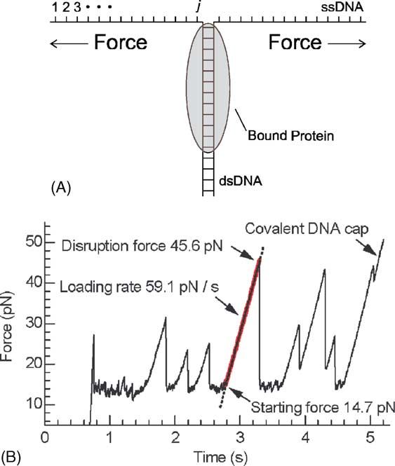
6 R. Ros et al. / Journal of Biotechnology 112 (2004) 5–12
tion between different molecules or compartments cantilever is mostly detected by the deflection of a
in a macromolecular assembly. In single molecule laser beam (Meyer and Amer, 1988). Spring constants
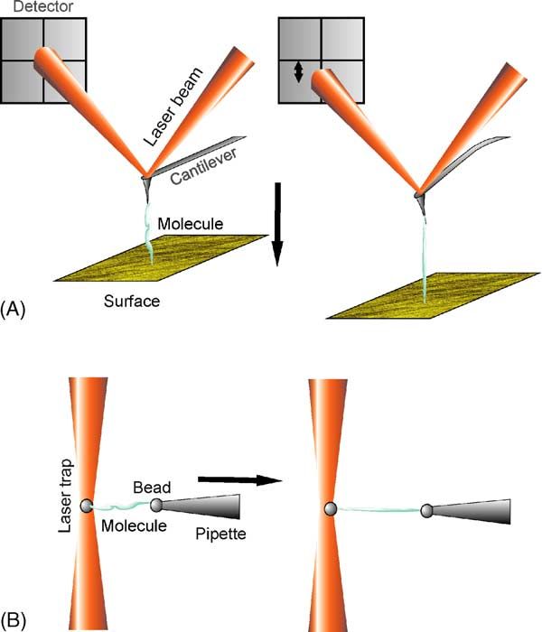
force spectroscopy (SMFS) experiments the force of the cantilever in the order of 10–100 nN/nm and de-
response is detected while a complex formed by two flection sensitivities in the sub nanometer range allow
molecules is separated or a single chain-like molecule measurements of forces from about 10 pN to 10 N.
is stretched. At the end of the cantilever, a tip with a radius of
In this paper, we review SMFS experiments of the about 10 nm is integrated, which results in a unique
binding of sequence specific proteins to dsDNA and spatial resolution in the nanometer range. In OT ex-
of small ligands which bind in a mainly sequence un- periments a micrometer sized bead with a different
specific manner to dsDNA. refractive index is trapped in an electric field gradi-
ent of a laser focus by optical forces (Gordon, 1973;
Ashkin, 1997). Displacements of the bead from the
2. Single molecule force spectroscopy center of the laser focus can be converted into forces.
Optical tweezers possess a superb force resolution in
In force spectroscopy experiments, the complex or
the sub piconewton range. However, because of the
molecule of interest is stretched with a sub nanome-
low spring constants of the optical trap (R. Ros et al. / Journal of Biotechnology 112 (2004) 5–12 7
iments on complexes allow direct insights in the
energy landscape by means of mapping activation
energy barriers (Merkel et al., 1999). In addition,
the biological function of the observed molecule
can be addressed by the extraction of the thermal
off-rates (Schwesinger et al., 2000). Intramolecular
forces, on the other hand, have a direct influence on
the mechanics of a molecule. Pathbreaking elasticity
studies on single DNA molecules were shown with
OT (Bustamante, 1994) and AFM (Rief et al., 1999).
The latter describing the sequence dependence of
the mechanics and extracts the base pair unzipping
force for G–C and A–T. Conformational changes in
polysaccharides were observed and corroborated by
molecular dynamic calculations (Rief et al., 1997b).
The fundamental field of protein folding was ad-
dressed by unfolding individual domains of the giant
muscle protein titin (Rief et al., 1997a) and the detec-
tion of intermediates (Marszalek et al., 1999). Here
the combination with molecular dynamic simulations
results in an atomistic view of structural chances in
the unfolding process. Fig. 2. Specific enzyme–DNA interaction analyzed by unzipping
DNA. (A) Schematic setup and (B) typical force vs. time graph
acquired at a constant loading rate. (Reprinted figure with per-
mission from S.J. Koch, M.D. Wang, Phys. Rev. Lett. 91 (2003).
3. Sequence specific binding of proteins to DNA Copyright (2004) by American Physical Society.)
The specific binding of proteins to a DNA target se-
quence is of fundamental interest for many biological 3.2. Direct force spectroscopy on protein–DNA
questions. Recently, this interaction was investigated complexes
by two different SMFS techniques.
Direct force measurements by mechanically un-
3.1. Unzipping force analysis of binding protein–DNA complexes were reported by
protein association Bartels et al. for a transcriptional regulator protein
that binds to three promotor regions in a gene cluster
Koch et al. (2002) investigated the binding of the (Bartels et al., 2003) (see Fig. 3). Three DNA frag-
restriction enzymes BsoBI, XhoI, and EcoRI with a ments comprising the target sequences were selected
new technique based on unzipping the two strands for the force spectroscopy measurements and attached
of dsDNA with OT (Fig. 2). The tension force and covalently via a long flexible polymer linker to the
extension of the DNA were recorded continuously. A AFM tip while the protein was anchored covalently on
bound protein is indicated by an increase in the force. the surface. DFS experiments with loading rates from
This method allows the containment of the binding 70 pN/s to 79 nN/s allow discrimination of the com-
site on the DNA to about 25 bp, whereas repetition of plex lifetimes and the detection of inner barriers in the
the unzipping experiments lead to predictions about energy landscape. From the lower loading rate regime,
equilibrium constants. Life times from 92 s for BsoBI a thermal off-rate of koff = (1.2 ± 1.0) ×10−3 s−1
to 6000 s for XhoI and information about the energy corresponding to a life time of about 830 s was de-
landscape were extracted by DFS measurements with rived for all promotor regions. In the higher loading
loading rates from 12 to 1200 pN/s (Koch and Wang, rate regime distinct differences, that were attributed
2003). to the molecular binding mechanism, were observed.8 R. Ros et al. / Journal of Biotechnology 112 (2004) 5–12
Fig. 3. Specific regulatory protein–DNA interaction analyzed with AFM based SMFS (A) schematic setup (B) typical force distance curve
(only the retracting part) with a rupture event. The rupture forces of a given series are combined to form a histogram with a gaussian
like distribution (inset). (C–E) Loading rate dependent force measurements of three different DNA fragments with a regulatory protein.
(F) Comparison of the fits from (C) to (E). Different slopes correspond to inner and outer barriers. (Reprinted from publication (Bartels,
2003), with permission from Elsevier.)
4. Interaction of small ligands with DNA tural transition from B-DNA to a stretched form, the
so-called S-DNA. In this cooperative transition the
The mechanical properties of single dsDNA were molecule can be elongated by a factor of 0.6–0.8 by
investigated in details in the pioneering works of an only weak increase in the restoring force (Smith
Bustamante (1994) with OT and Rief et al. (1999) et al., 1996; Cluzel et al., 1996).
with AFM. Briefly (see Fig. 4b), the DNA molecule The force value of this transition depends on the
in solution behaves like an entropic coil. Pulling on dsDNA sequence (Rief et al., 1999) and experimental
the two ends induces a restoring force due to de- conditions (Wenner et al., 2002). Higher forces induce
creasing degrees of freedom. This effect dominates the melting of the two complementary DNA strands
the stretching behavior up to about 50–60 pN and can followed by single strand stretching and the detach-
be described in good agreement with the worm-like ment of the molecule from the force transducer or
chain (WLC) elasticity model (Bustamante et al., translation stage. Fig. 4 shows the mechanics of a sin-
2000b). The extension at this point can be interpreted gle poly(dG-dC) dsDNA molecule explored with AFM
as the contour length of the dsDNA. At higher forces force spectroscopy (Eckel et al., 2003). This charac-
a distinct plateau is visible, corresponding to a struc- teristic fingerprint of free dsDNA changes drasticallyR. Ros et al. / Journal of Biotechnology 112 (2004) 5–12 9 Fig. 4. Identification of binding mechanisms in single molecule-DNA complexes. (A) Experimental setup. (B) Force-extension trace of free poly(dG-dC) dsDNA. DNA complexed with (C) the minor groove binder distamycin A, (D) major groove binding ␣-helical peptide, (E) with the intercalant ethidium bromide and (F) with the bis-intercalant YOYO-1. (Graphs adapted from Eckel et al., 2003.) in the presence of small DNA binding ligands (Figs. 4 et al. (2002a,b) investigated the crosslinking agent cis- and 5). First experiments with the intercalant ethid- platin, the intercalants proflavine, ethidium bromide, ium bromide in the low force regime were published psoralen, and the minor groove binder netropsin, bere- by Cluzel et al. (Cluzel et al., 1996) and Bennink nil and Hoechst 33258. Eckel et al. (2003) expanded et al. with OT (Bennink et al., 1999). The force re- the studies to major groove binding helical peptides sponse at higher forces was studied by Anselmetti and the bis-intercalant YOYO-1 and compared the re- et al. (2000) and Krautbauer et al. (2000) with AFM sults with the intercalants ethidium bromide, YO-1, force spectroscopy. In systematic studies Krautbauer daunomycin, and the minor groove binder distamycin
10 R. Ros et al. / Journal of Biotechnology 112 (2004) 5–12
plateau without any tilt at a minimal lower force com-
pared with free DNA (Eckel et al., 2003). In contrast,
optical tweezers data for this minor groove binder with
-DNA (51% GC) indicates an increasing force value
of the transition and a slight tilt that indicates a repres-
sion of the cooperative effects (Sischka et al., 2004).
Similar results were found for netropsin and Hoechst
33258 (Krautbauer et al., 2002a). For the major groove
binder, these two effects increase and particularly the
B–S-transition is less cooperative. All groove-binding
agents have no effect on the contour length of the ds-
DNA.
4.2. Intercalation
Intercalation is a different mode of interaction of
small molecules with DNA. It is characterized by
the sliding-in of flat, planar molecules into the base
pair stack of dsDNA via interaction of their aromatic
Fig. 5. Force vs. extension curves of a single -DNA molecule in ring systems with the -systems of the adjacent base
(a) immediately after adding the crosslinking agent cisplatin (b)
after 1 h and (c) after 24 h. The progress of the chemical reaction is
pairs. Various intercalators have been investigated
followed on the same molecule. (Graphs adapted from Krautbauer with single molecule force spectroscopy. Different
et al., 2000.) groups studied ethidium bromide, a frequently used
and well-characterized fluorescent staining agent for
DNA, in good agreement. No distinct cooperative
A. In a recent work Sischka et al. (2004) investigate transition is visible and the contour length of the
the influence of groove binder and intercalants with molecule increases dramatically. The fingerprint is
the high force resolution of an OT. The binding kinet- clearly distinguishable from the free or groove bind-
ics was addressed by time dependent elasticity mea- ing curve. (Husale et al., 2002; Krautbauer et al.,
surements and the observation of hysteresis effects 2002b; Eckel et al., 2003; Sischka et al., 2004)
between stretching and relaxation processes (Husale Molecules which bind in a bis-intercalation bind-
et al., 2002; Krautbauer et al., 2002b; Sischka et al., ing mode enforce these effects of typical interca-
2004). lating molecules (Eckel et al., 2003; Sischka et al.,
2004).
4.1. Groove binding
4.3. Crosslinking
Binding of small, positively charged molecules may
occur in the minor groove of dsDNA. This binding Crosslinking has similar effects as intercalation.
mode requires only slight conformational adaptions of The cooperative transition vanishes and the contour
the double helix. As for minor groove binding, major length increases. Krautbauer et al. (2000) investi-
groove binding is dominated by electrostatic interac- gated the prominent anti cancer drug cisplatin, which
tions of helical ligands with the backbone assisted by crosslinks preferentially the N7 atoms of guanine
hydrogen bonds. The global characteristics of the free bases. Fig. 6 summarizes the mechanical fingerprints
DNA curve are mostly conserved. The two main ef- of minor- and major groove binders, inter- and bis-
fects are shifting and tilting of the B–S-plateau. For the intercalators (Sischka et al., 2004). The DNA is im-
minor groove binder distamycin A with a low affinity mobilized between two beads and the fluid chamber
to guanine and cytosine rich sequences, force spec- system allows an efficient exchange of binding agents.
troscopy with poly(dG-dC) exhibit a B–S-transition The force response shows distinct changes fromR. Ros et al. / Journal of Biotechnology 112 (2004) 5–12 11
References
Anselmetti, D., Fritz, J., Smith, B., Fernandez-Busquets, X.,
2000. Single molecule DNA biophysics with atomic force
mictroscopy. Single Mol. 1, 17–23.
Ashkin, A., 1997. Optical trapping and manipulation of neutral
particles using lasers. Proc. Natl. Acad. Sci. U.S.A. 94, 4853–
4860.
Ashkin, A., Dziedzic, J., Bjorkholm, J., Chu, S., 1986. Observation
of a single-beam gradient force optical trap for dielectric
particles. Opt. Lett. 11, 288–290.
Bartels, F.W., Baumgarth, B., Anselmetti, D., Ros, R., Becker,
A., 2003. Specific binding of the regulatory protein ExpG to
promoter regions of the galactoglucan biosynthesis gene cluster
of Sinorhizobium meliloti—a combined molecular biology and
Fig. 6. Single molecule biosensor. Force response of a single force spectroscopy investigation. J. Struct. Biol. 143, 145–152.
-DNA molecule in the presence of different binding agents. Bennink, M.L., Schärer, O.D., Kanaar, R., Sakata-Sogawa, K.,
The trace of the free DNA is very well distinguishable from the Schins, J.M., Kanger, J.S., de Grooth, B.G., Greve, J., 1999.
traces of the complexed DNA. Furthermore the force response Single-molecule manipulation of double-stranded DNA using
of the minor groove binder (distamycin A), the major groove optical tweezers: interaction studies of DNA with RecA and
binder (␣-helical peptide) and the intercalants or bis-intercalants YOYO-1. Cytometry 36, 200–208.
(ethidium bromide, daunomycin, and YOYO-1) show characteristic Binnig, G., Quate, C.F., Gerber, C., 1986. Atomic force
fingerprints (Sischka et al., 2004). microscope. Phys. Rev. Lett. 56, 930–933.
Bustamante, C., 1994. Entropic elasticity of lambda-phage DNA.
Science 265, 1599–1600.
Bustamante, C., Macosko, J.C., Wuite, G.J., 2000a. Grabbing the
cat by the tail: manipulating molecules one by one. Nat. Rev.
free DNA to DNA complexes with all investigated
Mol. Cell Biol. 1, 130–136.
molecules. In addition the shape of the curve allows Bustamante, C., Smith, S.B., Liphardt, J., Smith, D., 2000b.
prediction of the mode of binding and time depen- Single-molecule studies of DNA mechanics. Curr. Opin. Struct.
dent measurements provide insights into the kinetics. Biol. 10, 279–285.
The combination of these features turns this setup Cluzel, P., Lebrun, A., Heller, C., Lavery, R., Viovy, J.-L.,
Chatenay, D., Caron, F., 1996. DNA: an extensible molecule.
into a promising tool for single molecule biosens-
Science 271, 792–794.
ing. Dammer, U., Hegner, M., Anselmetti, D., Wagner, P., Dreier, M.,
Huber, W., Güntherodt, H.-J., 1996. Specific antigen/antibody
interactions measured by force microscopy. Biophys. J. 70,
2437–2441.
5. Conclusion Eckel, R., Ros, R., Ros, A., Wilking, S.D., Sewald, N., Anselmetti,
D., 2003. Identification of binding mechanisms in single
Single molecule force spectroscopy based on AFM molecule–DNA complexes. Biophys. J. 85, 1968–1973.
Evans, E., 2001. Probing the relation between force-lifetime-and
and OT provides an ultrasensitive and powerful tool to chemistry in single molecular bonds. Annu. Rev. Biophys.
investigate sequence specific and unspecific interac- Biomol. Struct. 30, 105–128.
tions between ligands and dsDNA. Information about Evans, E., Ritchie, K., 1997. Dynamic strength of molecular
kinetics, equilibrium constants, the energy landscape adhesion bonds. Biophys. J. 72, 1541–1555.
and the binding site can be extracted from experi- Evstigneev, M., Reimann, P., 2003. Dynamic force spectroscopy:
optimized data analysis. Phys. Rev. E 68, 045103.
ments with sequence specific DNA binding proteins Florin, E.-L., Moy, V.T., Gaub, H.E., 1994. Adhesion forces
resulting in deeper insights for instance in the mode between individual ligand–receptor pairs. Science 264, 415–
of transcriptional regulation. Binding modes like in- 417.
tercalation, groove binding and cross linking of small Fritz, J., Katopodis, A.G., Kolbinger, F., Anselmetti, D., 1998.
ligands can be explored in a very fast and efficient Force-mediated kinetics of single P-selectin/ligand complexes
observed by atomic force microscopy. Proc. Natl. Acad. Sci.
manner by observing changes in the molecular elastic- U.S.A. 95, 12283–12288.
ity of the associated DNA molecule, which may lead Gordon, J.P., 1973. Radiation forces and momenta in dielectric
to new biosensor applications. media. Phys. Rev. A 8, 14–21.12 R. Ros et al. / Journal of Biotechnology 112 (2004) 5–12 Hinterdorfer, P., Baumgartner, W., Gruber, H., Schilcher, Meyer, G., Amer, N.M., 1988. Novel optical approach to atomic K., Schindler, H., 1996. Detection and localization of force microscopy. Appl. Phys. Lett. 53, 1045–1047. individual antibody–antigen recognition events by atomic force Rief, M., Clausen-Schaumann, H., Gaub, H.E., 1999. microscopy. Proc. Natl. Acad. Sci. U.S.A. 93, 3477–3481. Sequence-dependent mechanics of single DNA molecules. Nat. Husale, S., Grange, W., Hegner, M., 2002. DNA mechanics affected Struct. Biol. 6, 346–349. by small DNA intercalating ligands. Single Mol. 3, 91–96. Rief, M., Gautel, M., Oesterhelt, F., Fernandez, J.M., Gaub, H.E., Koch, S.J., Shundrovsky, A., Jantzen, B.C., Wang, M., 2002. 1997a. Reversible unfolding of individual titin immunoglobulin Probing protein–DNA interactions by unzipping a single DNA domains by AFM. Science 276, 1109–1112. double helix. Biophys. J. 83, 1098–1105. Rief, M., Oesterhelt, F., Heymann, B., Gaub, H.E., 1997b. Single Koch, S.J., Wang, M.D., 2003. Dynamic force spectroscopy of molecule force spectroscopy on polysaccharides by atomic protein–DNA interactions by unzipping DNA. Phys. Rev. Lett. force microscopy. Science 275, 1295–1297. 91, 208103. Ros, R., Schwesinger, F., Anselmetti, D., Kubon, M., Schäfer, R., Krautbauer, R., Clausen-Schaumann, H., Gaub, H.E., 2000. Plückthun, A., Tiefenauer, L., 1998. Antigen binding forces Cisplatin changes the mechanics of single DNA molecules. of individually addressed single-chain Fv antibody molecules. Angew. Chem. Int. Ed. 39, 3912–3915. Proc. Natl. Acad. Sci. U.S.A. 95, 7402–7405. Krautbauer, R., Fischerländer, S., Allen, S., Gaub, H.E., 2002a. Schwesinger, F., Ros, R., Strunz, T., Anselmetti, D., Güntherodt, Mechanical fingerprints of DNA drug complexes. Single Mol. H.-J., Honegger, A., Jermutus, L., Tiefenauer, L., Plückthun, A., 3, 97–103. 2000. Unbinding forces of single antibody–antigen complexes Krautbauer, R., Pope, L.H., Schrader, T.E., Allen, S., Gaub, H.E., correlate with their thermal dissociation rates. Proc. Natl. Acad. 2002b. Discriminating small molecule DNA binding modes by Sci. U.S.A. 97, 9972–9977. single molecule force spectroscopy. FEBS Lett. 510, 154–158. Sischka, A., Tönsing, K., Eckel, R., Wilking, S.D., Sewald, N., Lee, G.U., Chrisey, L.A., Colton, R.J., 1994. Direct measurement Ros, R., Anselmetti, D., 2004. Molecular mechanisms and of the forces between complementary strands of DNA. Science kinetics between DNA and DNA binding ligands, submitted. 266, 771–773. Smith, S.B., Cui, Y., Bustamante, C., 1996. Overstretching Lee, G.U., Kidwell, D.A., Colton, R.J., 1993. Sensing discrete B-DNA: the elastic response of individual double-stranded streptavidin–biotin interactions with atomic force microscopy. and single-stranded DNA molecules. Science 271, 795– Langmuir 10, 354–357. 799. Marszalek, P.E., Lu, H., Li, H., Carrion-Vazquez, M., Oberhauser, Strunz, T., Oroszlan, K., Schäfer, R., Güntherodt, H.-J., 1999. A.F., Schulten, K., Fernandez, J.M., 1999. Mechanical Dynamic force spectroscopy of single DNA molecules. Proc. unfolding intermediates in titin modules. Nature 402, 100–103. Natl. Acad. Sci. U.S.A. 96, 11277–11282. Merkel, R., Nassoy, P., Leung, A., Ritchie, K., Evans, E., 1999. Wenner, J.R., Williams, M.C., Rouzina, J., Bloomfield, V.A., 2002. Energy landscapes of receptor–ligand bonds explored with Salt dependence of the elasticity and overstretching transition dynamic force spectroscopy. Nature 397, 50–53. of single DNA molecules. Biophys. J. 82, 3160–3169.
You can also read