Sodium carbonate intoxication on a chinchilla (Chinchilla lanigera) farm: a case report
←
→
Page content transcription
If your browser does not render page correctly, please read the page content below
Case Report                                                         Veterinarni Medicina, 59, 2014 (2): 112–116

Sodium carbonate intoxication on a chinchilla
(Chinchilla lanigera) farm: a case report
J. Wojtacka1, J. Szarek1, I. Babinska1, M. Felsmann1, E. Strzyzewska1,
A. Szarek-Beska2, K. Dublan1, J. Micinski1
1
    University of Warmia and Mazury in Olsztyn, Olsztyn, Poland
2
    Veterinary Practice “Pulsar”, Olsztyn, Poland

ABSTRACT: Massive deaths were reported on a chinchilla (Chinchilla lanigera) farm, which over 10 months led to
the elimination of the herd. After three months of feeding, longitudinal precipitates inside the pellets were noted.
The first symptoms were observed two weeks after the introduction of feed, and included lethargy, decreased
mobility, and reduced appetite, as well as increased reactions to external stimuli. Over time, single deaths, hair loss
and gnawing, white discolorations on teeth, and polyuria were reported. Haemorrhages of the reproductive tract
and mass abortions were observed. Necropsy revealed the presence of transudate with pH = 10, severe hyperae-
mia of the intestinal mesentery, and extensive regressive lesions in the gastric and intestinal mucosa. Both grossly
and microscopically, lesions were noted in the liver, kidneys, adrenal glands, and lungs. The urinary bladder was
overfilled. No lesions were observed in the spleen or mesenteric lymph nodes. Histopathology of skin specimens
revealed atrophy of the hair follicles. Diagnosis was sodium carbonate intoxication. Owing to the specific features
of this species, it was impossible to introduce emetic-based treatment in order to eliminate the toxic agent from the
body. No therapeutic measures were undertaken because of the late diagnosis of the toxic agent and late removal
of feed, as well as extensive lesions on the gastrointestinal mucosa.

Keywords: sodium carbonate intoxication; chinchilla

   The digestive tract in the chinchilla is particularly     properties intensify its harmful effects on the di-
sensitive (Cousens 1963; Merry 1990; Hoefer 1994;            gestive tract (Clarke et al. 1981; Thomas and Stone
Jenkins 1992). Numerous bacterial alimentary infec-          1994). In addition, this compound dissociates and
tions have been reported in these animals (Moore             alkalises the environment. The irritation results in
and Greenlee 1975), although intoxication cases              reduced blood vessel tension, while the caustic ef-
have been described only rarely (Dall 1963). There           fect in the digestive tract starts at a dose of approxi-
is no data on clinical cases of sodium carbonate poi-        mately 3 g. The LD50 in rats with oral administration
soning in animals. This paper presents a case report         is 4.090 mg/kg body mass (Lewis 2004).
of chronic intoxication with sodium carbonate on
a chinchilla farm. Sodium carbonate was added to
feed instead of sodium bicarbonate, which is used in         Case description
the nutrition of chinchillas at a dose of 1 mg/kg as a
sodium source when balancing dietary electrolytes.             This case took into account documentation col-
   Sodium carbonate is an irritating and caustic sub-        lected by the breeder and documents recorded by
stance which causes injuries when it comes into con-         the veterinary surgeon, which covered a period of
tact with the mucosa (Busch et al. 1983; Grant 1986;         10 months (i.e., from the herd’s introduction to the
Johnson and Swanson 1987). Due to its hygroscopic            farm until its removal). Physical examinations of the
properties, sodium carbonate is prone to lumping,            herd were performed at least once weekly through-
and under humid conditions it is transformed into            out the aforementioned period. Gross examinations
sodium hydroxide (also known as “lye soda”). These           of dead animals were conducted during these visits.

112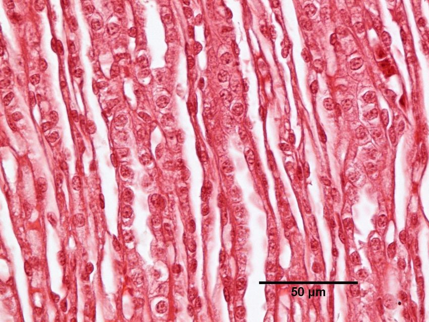
Veterinarni Medicina, 59, 2014 (2): 112–116                                                         Case Report

During necropsies of the chinchillas, specimens of       troduced, and included lethargy, reduced mobil-
the skin, stomach, small intestine (duodenum), large     ity, decreased appetite, and increased reactivity
intestine (colon), liver, kidneys, adrenal glands, and   to external stimuli. After three weeks, sporadic
lungs were collected from three animals at the end of    and sudden deaths were reported, and then ten-
the third, sixth, and eleventh weeks and after seven     dencies toward hair loss and the gnawing away of
and 10 months. The samples were fixed in 10% buff-       hair were observed. In addition, the chinchillas’
ered formalin and the paraffin sections were stained     coats became dull, loosened, and were prone to
with haematoxylin and eosin (Bancroft and Gamble         loss. After 1 month, alopecia areata progressing to
2008). During the necropsies performed in the elev-      alopecia universalis was reported in some animals
enth week, the reaction (pH) of fluid accumulated        (Figure 1). Lack of hair was also evident in locations
in the abdominal space and pleural space and pH of       that could not be self-mutilated.
feed and its precipitates were determined using the        During the sixth week of breeding, white discol-
potentiometric method. At that time, a bacterio-         orations appeared on the teeth. Two months later
logical examination of the feed was also performed.      (two months and three weeks after the introduc-
                                                         tion of the feed), the aforementioned symptoms
                                                         became more severe; hair loss in particular was
Clinical case                                            more pronounced. Polyuria was also reported; large
                                                         pools of urine and undigested light-brown stool
   A chinchilla (Chinchilla lanigera) farm with an       with mucus were visible beneath the cages together
inventory of 1330 animals reported widespread            with large amounts of crumbled feed, suggesting
deaths that resulted in the elimination of the whole     that the animals had discarded the feed instead
herd within 10 months.                                   of consuming it. Bleeding from the reproductive
   The breeding material used on this farm originat-     tract and massive abortions were also reported.
ed from an authorised breeding herd. The average         Neonates were born small, and lived only up to
body mass of adult animals was up to 600 g/animal.       one week because the mothers did not produce
The husbandry and breeding conditions on the             milk. Animal behaviour (the clamping of teeth on
farm were exemplary and complied with all vet-           the bars of the cages) indicated that the animals
erinary requirements. The building was new, well         were dying in pain. At that point, 405 deaths had
illuminated, equipped with a very good ventilation       been reported, and a change of feed was intro-
system (ground air conditioning), and had a sepa-        duced. However, deaths continued; 95 additional
rate storage room for feed and straw. The cages in       deaths were recorded in chinchillas younger than
the rooms were arranged in four-stage rows. The          six months of age. In the subsequent month, it
humidity was 40% and the temperature was 18 °C.          was concluded that the animals were unsuitable
The animals had constant access to water and were        for further breeding because of emaciation, limited
fed with feed from a single manufacturer.                mobility, and described changes in the hair coat,
   After three months of feeding, yellowish and          and 460 of them were euthanised. An additional
cream-yellow longitudinal precipitates of 1 mm in
diameter and up to 3 mm long were noted inside the
pellets; these precipitates were initially thought by
the manufacturer to be fragments of non-pelleted
cereals. An organoleptic evaluation revealed that
these precipitates saponified on the tongue to cause
numbness and were tart in taste. A glistening, yel-
low, and vitreous-transparent dust was visible in
the bags of feed; it left sediment on the hand and
was clearly visible against a dark background.

Clinical symptoms

 The first clinical symptoms in the chinchillas          Figure 1. Alopecia universalis in a chinchilla. One month
were noted two weeks after the feed had been in-         after introduction of the feed with sodium carbonate

                                                                                                              113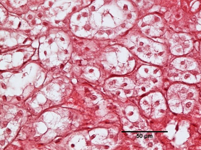
Case Report Veterinarni Medicina, 59, 2014 (2): 112–116
tine mucosa was hypertrophic and showed signs of
catarrhal inflammation. Histopathology revealed
damaged tips and shortening of the intestinal villi
in this section of the digestive tract.
The liver was enlarged, with compact texture and
sub-capsular petechiae. Microscopic examination
revealed parenchymatous degeneration, hyperae-
mia, and large areas of lipid infiltration and fatty
degeneration in the liver, together with numer-
ous small extravasations. Starting from the fourth
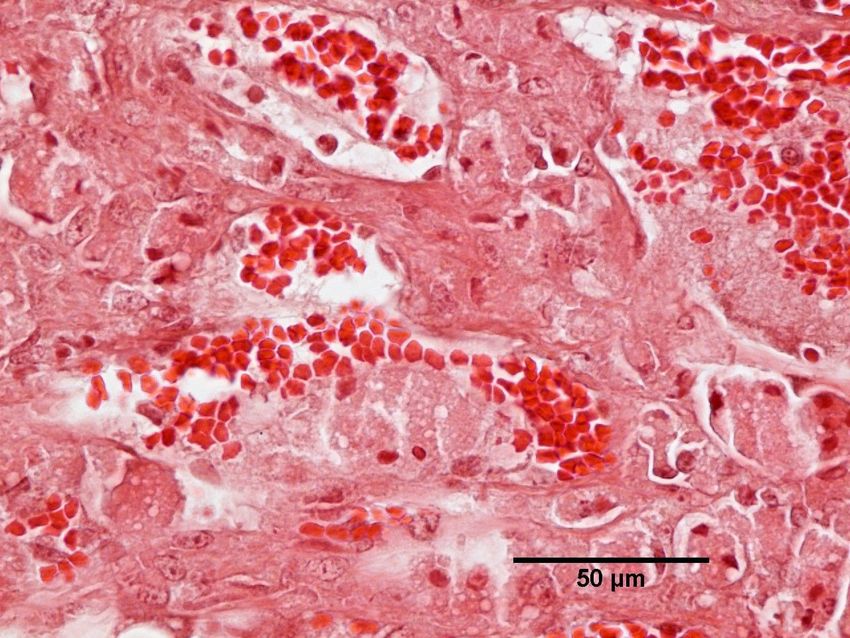
Figure 2. Haemorrhages and ulceration of the stomach month, necrotic foci were observed in animals that
mucosa (on the left) and under the adrenal gland capsule were found dead.
(on the right) Enlarged and oedematous kidneys with paren-
chymatous degeneration (Figure 3), necrotic foci, and
269 animals were euthanised 10 months after the hyperaemia with extravasations were also reported.
introduction of the aforementioned batch of feed. The adrenal glands were generally twice as large as
the physiological norm (Figure 2) and microscopy
revealed signs of hyperplasia and hypertrophy of the
Morphological lesions cortex with hyperaemia and extravasations (Figure 4)
and vacuolar degeneration (Figure 5). The urinary
Transudate with pH = 10 was found in the ab- bladder was overfilled. No lesions were noted in the
dominal space. Severe hyperaemia of the intestinal spleen or mesenteric lymph nodes.
mesentery was noted. Transudate with pH of 8.5 was noted in the pleu-
Very deep defects in the gastric mucosa devel- ral cavity, and the lungs were oedematous and hy-
oped in several dozen cases (approximately 6% of peraemic.
the herd; Figure 2). Perforation of the gastric wall Histopathological examination of the skin speci-
was sporadically observed. In addition, catarrhal mens revealed normal layers with signs of hair fol-
and sometimes haemorrhagic effusions and nu- licle atrophy.
merous erosions were observed in this section of
the digestive tract in the majority of dead animals.
Grossly, the gastric mucosa was noticeably softened Chemical and microbiological examination
in the majority of cases. The small and large in- of feed
testines were empty of contents. Occasionally, a
small volume of liquid, with greenish content was Analysis of measured feed revealed that its pH
observed in the large intestine. The small intes- ranged from 7.8 to 8.3, while the pH of precipi-
Figure 3. Chinchilla kidney with parenchymatous degen- Figure 4. Chinchilla adrenal gland with hyperaemia,
eration of the tubular cells; HE staining extravasations and necrotic foci; HE staining
114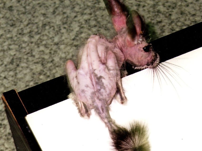
Veterinarni Medicina, 59, 2014 (2): 112–116                                                       Case Report

                                                         The lack of lesions in the oral cavity in the major-
                                                         ity of animals and initially persisting appetite did
                                                         not prompt them to refuse feed. The effects of this
                                                         substance only became apparent in the stomach
                                                         and small intestine. Sodium carbonate in a solid
                                                         form, before it was dissolved in the gastric and in-
                                                         testinal juices, had an irritating and caustic effect
                                                         on the mucosa resulting in the described lesions.
                                                         The absorption of Na+, OH–, and HCO3– into the
                                                         circulation was a toxodynamic factor with an ef-
                                                         fect on the whole body. The ions were generated
                                                         in the gastric and intestinal juices from NaOH and
                                                         Na2CO3 precipitates. These ions, present in large
                                                         amounts, were distributed throughout the body,
Figure 5. Necrotic foci and vacuolar degeneration of     and disrupted the acid-base balance of the bod-
chinchilla kidney                                        ily fluids and depleted their buffer capacity, which
                                                         led to the pH values of 8.5 to 10 (the physiological
tates extracted from the pellets was 10 to 10.5.         processes that normally take place in the gastric
The feed was also tested regarding the content of        and intestinal fluids do not occur in this pH range).
sodium in dry matter, which was 2.22% per DM.               Chinchillas have an excellently developed system
Microbiological examination did not reveal any           of water absorption in the terminal section of the
pathogenic microorganisms.                               digestive tract, and they excrete a minor volume
                                                         of urine (Alworth and Harley 2012). The excess of
                                                         sodium resulted in some disturbances in the water-
DISCUSSION AND CONCLUSIONS                               electrolyte balance, particularly an excessive excre-
                                                         tion of urine and transudation of the fluids into
  Even though chinchillas are thought to be rela-        the body cavities (pleural and peritoneal spaces).
tively resistant to infectious diseases, bacterial       Sodium derivatives caused diarrhoea by irritating
infections have become increasingly important as         the mucosa of the digestive tract. The prolonged
intensive breeding and selection for production          accumulation of NaOH and/or Na 2CO 3 particles
traits have resulted in an over-delicateness of many     resulted in a caustic effect and lysis of the tissues,
features in these animals (Crossley and Miguelez         and consequently in the perforation of the gastric
2001; Baranowski and Wojtas 2011; Swiecicka et           wall and death. The excess of Na+ also affected the
al. 2012). In the presented case, the evident lack       blood (over-coagulability) and blood vessels (hy-
of lesions in the spleen, large intestine, and lymph     peraemia). Parenchymatous degeneration devel-
nodes excluded an infectious factor as a possible        oped in the kidneys and liver. Hyperaemia of the
cause of morbidity and mortality. Furthermore, the       mucosa, petechiae, disruption of tissue integrity,
microbiological investigations did not reveal any        inflammation, and necrosis were the direct effects
bacterial agent that could contribute to the sick-       of the contact action of sodium solid derivatives ob-
ness of the herd.                                        served in the digestive tract. These lesions caused
  Chinchillas are extremely sensitive to the compo-      chronic stress, which was confirmed by hyperplasia
sition of feed mixture (Wolf et al. 2003). Symptoms      and hypertrophy of the adrenal medulla and hair
and deaths are first seen in young nurtured animals      coat mutilation (Rees 1963; Tisljar et al. 2002).
and in pregnant and lactating females.                      Because of the lack of acute symptoms of intoxi-
  Because chinchillas are herbivorous, with a pro-       cation in the chinchillas and the specific features of
longed time span of digestion (Alworth and Harley        this species, as well as the number of animals in the
2012), feed remains in their digestive tract for three   herd, it was impossible to implement any specific
to six days. In addition, chinchillas swallow pellets    emesis-based treatment in order to eliminate the
immediately, practically without grinding; there-        toxic agent from the body. The potential efficacy of
fore, the aforementioned precipitates, encapsulated      administration of organic acids (citric, acetic, and
in the feed pellets as solid matter, did not exert       ascorbic) was questionable because the diagnosis
a caustic or irritating effect on the oral mucosa.       of the toxic agent and removal of feed happened

                                                                                                           115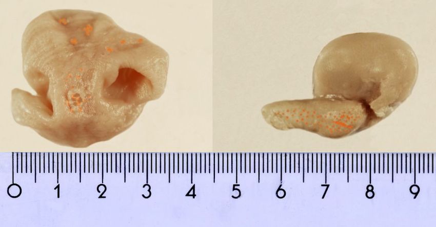
Case Report                                                          Veterinarni Medicina, 59, 2014 (2): 112–116

after a significant period of time, and injuries to the       Grant WM (1986): Toxicology of the Eye. 3rd ed. Charles
mucosa of the gastrointestinal tract were extensive.            C. Thomas Publisher, Springfield, IL. 830 pp.
The presence of sodium carbonate in feed was most             Hoefer HL (1994): Chinchillas. Veterinary Clinics of
probably due to the human factor. As a feed additive            North America: Small Animal Practice 24, 113–120.
destined for feed formula of other animal species             Jenkins JR (1992): Husbandry and common diseases of
it could have been added to the chinchilla feed by              the chinchilla (Chinchilla laniger). Journal of Small
mistake during the production process in the com-               Exotic Animal Medicine 2, 15–17.
pany. The Polish Chinchilla Breeders’ Association             Johnson W, Swanson K (1987): Final report on the safety
received similar reports regarding disturbances to              assessment of sodium sesquicarbonate, sodium bicar-
chinchilla health from other chinchilla farms in the            bonate and sodium carbonate. International Journal
country at that time.                                           of Toxicology 6,121–138.
                                                              Lewis RJ (2004): Sax’s dangerous properties of industrial
                                                                materials. 11th ed. John Wiley & Sons Inc., NJ. 3236 pp.
REFERENCES                                                    Merry CJ (1990): An introduction to chinchillas. Vet-
                                                                erinary Technician 1, 315–322.
Alworth LC, Harley SB (2012): Anatomy, physiology and         Moore RV, Greenlee HH (1975): Enterotoxemia in chin-
  behaviour. In: Suckow M, Stevens K, Wilsson R (eds.):         chillas. Laboratory Animals 9, 153–154.
  The Laboratory Rabbit, Guinea Pig, Hamster and              Rees RG (1963): Some conditions of the skin and fur of
  Other Rodents. Elsevier Inc., London. 955–965.                Chinchilla lanigera. Journal of Small Animal Practice
Bancroft JD, Gamble M (2008): Theory and Practice of            4, 213–225.
  Histological Techniques. 6th ed. Churchill Livingstone,     Swiecicka N, Zawislak J, Kubacki S, Gulda D, Drewka
  Elsevier, Philadelphia, PA. 126–133.                          M, Monkiewicz M (2012): Analysis of results of con-
Baranowski P, Wojtas J (2011): Effect of hypertrophic           formation evaluation of the standard chinchilla
  defect on the occurrence of foraminal, shape, and cri-        achieved on the breeding farm. Polish Journal of Nat-
  brosity features in the cranium and mandible of wild          ural Sciences 27, 31–39.
  and farm chinchillas (Chinchilla laniger). Bulletin of      Tisljar M, Jani D, Grabarevic Z, Simpraga B, Marinculic
  Veterinary Institute in Pulawy 55, 247–260.                   A, Pinter L, Janicki Z, Nemanic A (2002): Stress in-
Busch RH, McDonald KE, Briant JK, Morris JE, Graham             duced Cushing’s syndrome in fur-chewing chinchillas.
  TM (1983): Pathologic effects in rodents exposed to           Acta Veterinaria Hungarica 50, 133–142.
  sodium combustion products. Environmental Re-               Thomas SH, Stone CK (1994): Acute toxicity from bak-
  search 33, 138–147.                                           ing soda ingestion. American Journal of Emergency
Clarke ML, Harvey DG, Humphreys DJ (1981): Veterinary           Medicine 12, 57–59.
  Toxicology. 2nd ed. Bailliere Tindall, London. 256 pp.      Wolf P, Schroder A, Wenger A, Kamphues J (2003): The
Cousens PJ (1963): The chinchilla in veterinary practice.       nutrition of the chinchilla as a companion animal –
  Journal of Small Animal Practice 4, 199–205.                  basic data, influences and dependences. Journal of
Crossley DA, Miguelez M (2001): Skull size and cheek-           Animal Physiology and Animal Nutrition 87, 129–133.
  tooth length in wild-caught and captive-bred chinchil-
  las. Archives of Oral Biology 46, 919–928.                                                     Received: 2014–01–09
Dall J (1963): Diseases of the chinchilla. Journal of Small                     Accepted after corrections: 2014–02–26
  Animal Practice 4, 207–212.

Corresponding Author:
Joanna Wojtacka, University of Warmia and Mazury in Olsztyn, Faculty of Veterinary Medicine, Chair of Veterinary
Public Health, Michala Oczapowskiego St. 14, 10-718 Olsztyn, Poland
E-mail: joanna.wojtacka@uwm.edu.pl

116
You can also read