Solitary fibrous tumor of the central nervous system invading and penetrating the skull: A case report
←
→
Page content transcription
If your browser does not render page correctly, please read the page content below
ONCOLOGY LETTERS 25: 81, 2023

               Solitary fibrous tumor of the central nervous system
                invading and penetrating the skull: A case report
                                   QIYAN LIN, JIABIN ZHU and XIAOFENG ZHANG

                   Department of Neurosurgery, Affiliated Xiaolan Hospital, Southern Medical University,
                    Xiaolan People's Hospital of Zhongshan, Zhongshan, Guangdong 528415, P.R. China

                                 Received September 28, 2022; Accepted December 20, 2022

                                                 DOI: 10.3892/ol.2023.13667

Abstract. Solitary fibrous tumor (SFT) of the central nervous    peritoneum, liver, thyroid, mesentery, and sinuses and
system is a rare spindle cell tumor of mesenchymal origin.       orbits (2). Due to the lack of a true connective tissue compo‑
The present study reports the case of a 44‑year‑old male         nent in the central nervous system (CNS), primary SFT of
patient with SFT. Magnetic resonance imaging demonstrated        the CNS is rare, accounting for ~1% of all primary CNS
that the majority of the intracranial tumors exhibited uneven    tumors (3,4). Most CNS SFTs occur in the cranial cavity
low signals on T1‑weighted imaging (T1WI) and low mixed          and just over one‑fifth were intraspinal (5‑7). Primary spinal
signals on T2WI, and there was an enhancement on enhanced        SFT may occur at any age (5). The mean age of onset was
scanning. Furthermore, the distal part of the left occipital     40.9 years for males and 35.0 years for females (5). There
lobe exhibited hypersignals on T1WI and T2WI, and this was       was no significant difference in morbidity between males and
significantly enhanced following enhanced scanning. The          females (5). Primary spinal SFT usually occurs in the thoracic
lower part of the scalp exhibited low signals on T1WI and        spinal cord, followed by the cervical and lumbar spinal cord,
high signals on T2WI, and there was no notable enhancement       and the sacral spinal cord is rarely affected (5). Intracranial
following enhanced scanning. Magnetic resonance spectros‑        SFT occurs most commonly in adults aged 20‑70 years, with
copy demonstrated an elevated choline/creatine peak in the       similar incidence rates in males and females (8). When CNS
solid part of the tumor. Under the microscope, the tumor         SFTs occur intracranially, they are frequently extra‑axially
exhibited characteristic ‘staghorn‑shaped’ blood vessels.        located (9,10). Hemangiopericytomas (HPCs) are also
As SFT is difficult to differentially diagnose via imaging,      rare mesenchymal tumors that exhibit similar clinical,
immunohistochemical analysis of CD34, vimentin and signal        radiological and histological features to SFTs (11). The
transducer and activator of transcription 6 was performed        NGFI‑A‑binding protein (NAB2) and signal transducer and
for the definitive diagnosis of SFT. Of note, surgical resec‑    activator of transcription 6 (STAT6) gene fusion was identi‑
tion was the preferred treatment for SFT; however, due to        fied as a driver mutation of SFT (12,13). Previous pathological
the rarity of the tumor, subsequent adjuvant therapy and         findings demonstrated that SFT and HPCs contain identical
prognosis require further investigation.                         genetic abnormalities and these prompted the World Health
                                                                 Organization (WHO) to classify the two tumor types as a new
Introduction                                                     combined entity in 2016 (14). This classification described
                                                                 three grades of SFT/HPC, namely grade I, II and III. Of
Solitary fibrous tumor (SFT) is a rare spindle cell tumor of     note, the distinction between the two types was no longer
mesenchymal origin, initially reported by Klempere and           clinically significant due to the pronounced clinical and
Rabin (1) in 1931. SFT is commonly found in the medias‑          histopathological overlap. In the 2021 WHO classification of
tinum and visceral pleura; however, it also occurs in the        CNS tumors, the term ‘hemangiopericytoma’ was removed
pleura external sites, such as the head and neck, pericardium,   and replaced with SFT (15).
                                                                     A solid lesion located in the CNS distinct from fibrous
                                                                 meningioma, termed primary SFT, was initially reported by
                                                                 Carneiro et al (16) in 1996. Intracranially, SFT may occur at
                                                                 the cerebellopontine angle, spinal dura, parasagittal region,
Correspondence to: Professor Xiaofeng Zhang, Department
                                                                 meninges and the intraventricular region (17). The present
of Neurosurgery, Affiliated Xiaolan Hospital, Southern Medical
University, Xiaolan People's Hospital of Zhongshan, 65 Jucheng   article reports on a 44‑year‑old male patient with SFT. The
Avenue, Zhongshan, Guangdong 528415, P.R. China                  SFT originated from the superior sagittal sinus and not only
E‑mail: zhangfimmu@163.com                                       penetrated through the skull, but also invaded the bilateral
                                                                 occipital lobes distally. This is the first case of SFT completely
Key words: solitary fibrous tumor, central nervous system,       penetrating the skull, to the best of our knowledge. The
diagnosis                                                        imaging data, histopathological features and treatment of SFT
                                                                 were briefly reviewed and the imaging features of this case
                                                                 were discussed.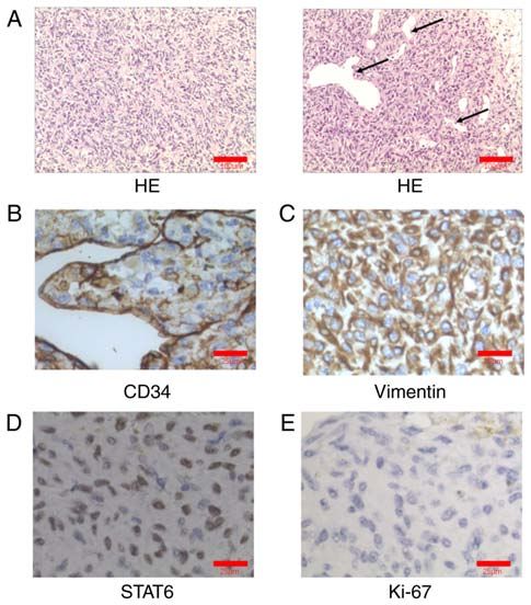
2                                         LIN et al: SOLITARY FIBROUS TUMOR OF THE CNS

Case report                                                         immunoreactivity to CD34, vimentin and STAT6 (Fig. 2B‑D).
                                                                    The Ki‑67 labeling index was ~10% with no signs of necrosis
A 44‑year‑old male patient presented at the neurology               (Fig. 2E). The tumor cells presented as weakly positive for
outpatient clinic of Xiaolan People's Hospital of Zhongshan         epithelial membrane antigen (EMA) and negativity for proges‑
(Zhongshan, China) in August 2020 due to dizziness and              terone receptor (PR) (Fig. S1B and C). Grade I SFT cells are
blurred vision for one month. Neurological examination of           fusiform, with lower cell density and higher collagen content.
the patient appeared normal; however, a visual field defect         Grade II SFT has more cells, less collagen, no specific cell
was observed below the central visual field of both eyes. Bone      arrangement and typical staghorn‑shaped blood vessels.
window computed tomography angiography (Ingenuity CT;               There are more than 4 mitotic figures per 10 HPF of grade III
Philips Medical Systems, Inc.; slice thickness, 0.8 mm; center,     SFT (14). Intracranial SFT is mainly differentiated from
450; width, 1,600) of the head demonstrated that the mass had       meningioma because they are similar in clinical presentation
invaded and penetrated the skull (Fig. 1A).                         and pathological diagnosis. The histopathological features of
    Head magnetic resonance imaging (MRI) findings                  SFT are sparse and dense areas separated by fibrous stroma,
demonstrated a mass shadow near the occipital cerebral falx,        with hemangiopericytoma branching vessels (18). The pheno‑
with an irregular shape and unclear boundaries. The mass            type of SFT is characterized by a patternless architecture
was ~64x44x64 mm in size and stretched across both sides            or a short fascicular pattern (14). SFT is histopathologically
of the cerebral falx. The mass filled the posterior portion of      characterized by alternating low‑cell and high‑cell areas and
the superior sagittal sinus to form a filling defect, invading      thick collagen bands (14). Microscopically, the meningioma
and penetrating the occipital bone, and the boundary between        cells are nested, with abundant cytoplasm and unclear cells
the mass and bilateral occipital lobe brain tissue was unclear.     (syncytioid) (19). Pseudo‑inclusions are common in menin‑
Of note, the majority of the intracranial tumors exhibited          gioma nuclei, where cells have weakly defined cell boundaries
uneven low signals on T1‑weighted imaging (T1WI) and low            (syncyti‑like) (19). In SFT, STAT6 is positive in almost all
mixed signals on T2WI, with notable enhancements following          patients, while CD34 is positive to varying degrees (20‑22).
enhanced scanning. The distal part of the left occipital lobe       However, all forms of meningiomas characteristically
demonstrated hypersignals on T1WI and T2WI, with signifi‑           expressed EMA and PR; CD34 reactivity was patchy and
cant enhancements following enhanced scanning. In addition,         weak; STAT6 was not expressed (21,22). Histopathological
the lower part of the scalp exhibited low signals on T1WI and       examination confirmed WHO grade II SFT.
high signals on T2WI, and there were not notably enhanced               The protocols of the imaging examinations, histopatholog‑
following enhanced scanning (Fig. 1B‑D).                            ical staining and immunohistochemistry (IHC) are provided
    Results of the magnetic resonance spectroscopy (MRS)            in the supplemental data.
demonstrated a multi voxel collection with no notable                   Three‑dimensional emphasis radiotherapy was performed
N‑acetylaspartic acid peak in the mass. The choline/creatine        18 days after surgery [tumor absorbed dose: Planning target
peak in the solid area was increased; however, no notable           volume (PTV)1, 60.2 Gray/28 fractions; PTV2, 54.6 Gray/28
abnormalities were found in the spectral lines of the tissues       fractions]. At the follow‑up 3 months after the surgery, the
surrounding the mass (Fig. 1E).                                     patient reported that the headache and dizziness symptoms
    A craniotomy was performed for tumor resection and              had gradually disappeared after the surgery. The patient's
follow‑up MRI demonstrated complete tumor resection                 visual field was examined using a Humphrey II 740 Visual
(Fig. 1F). During the operation, the tumor was gray and red in      Field (Carl Zeiss Meditec), indicating that the patient's visual
color with an abundant blood supply (Fig. S1A). Foci indica‑        field was significantly improved compared with that prior to
tive of previous bleeding far from the origin occurred in the       surgery. During the two‑year follow‑up, the patient experi‑
tumors in both the occipital lobe and the scalp. In addition, the   enced no recurrence of SFT.
tumor texture was uneven, with both soft and tough sections,
with an incomplete capsule and lobulated invasive growth. The       Discussion
tumor broke through the brain tissue of the occipital lobe and
the demarcation between the tumor and the brain tissue of the       The majority of intracranial SFTs are dural masses originating
occipital lobe was unclear. The occipital pia mater was mark‑       predominantly from thick collagen bands, which are produced
edly edematous. The adjacent occipital skull demonstrated           by fibroblasts, most frequently occurring in the parasagittal
osteolytic bone destruction, the tumor penetrated the occipital     sinus and spinal canal (3,23‑25). In the present study, the
bone to form a local mass under the scalp and the local scalp       patient experienced SFT originating in the superior sagittal
thickened reactively.                                               sinus, consistent with previous reports (3,23‑25).
    Pathological examination revealed that the tumor was                Symptoms of SFT vary and patients may present with
composed of alternately distributed cell‑rich areas and             several non‑specific symptoms associated with elevated intra‑
cell‑sparse areas. The tumor cells in the cell‑rich area were       cranial pressure or tumor location. These include headache,
short‑spindle or oval, with little cytoplasm and uniform nuclear    nausea, vomiting, dizziness, gait disturbance, hemiplegia,
chromatin. There was no notable atypia in the two areas. The        hearing loss and memory disturbance (26).
frequency of mitotic figures was 1/10 high‑power fields (HPF)           Sugiyama et al (27) reported on an 86‑year‑old male with
and the tumor cells were arranged in sheets and striae. These       SFT, which was located in the right parietal lobe and invaded
were hemangiopericytoma‑like, with abundant blood vessels           the parietal bone, who presented with sustained progressive
in the tumor. Thus, these were labeled ‘staghorn‑shaped blood       motor weakness in the left lower extremity for 1 month. Another
vessels’ (Fig. 2A). Tumor cells presented with diffuse strong       study reported on a 30‑year‑old male with SFT, which was
ONCOLOGY LETTERS 25: 81, 2023 3
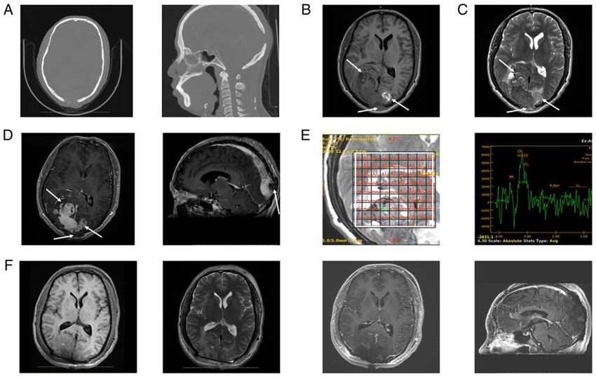
Figure 1. Preoperative and postoperative imaging profiles of the tumor. (A) The axial (left panel) and sagittal (right panel) images of the bone window of
the head computed tomography angiogram demonstrated that the mass invaded and penetrated the skull. MRI on admission demonstrated a mass shadow
near the occipital cerebral falx, with an irregular shape and unclear boundaries. The mass was ~64x44x64 mm in size. (B) The majority of the intracranial
and subcutaneous tumors demonstrated uneven low signals on T1WI, while the tumors at the far end of the left occipital lobe demonstrated high signals on
T1WI. (C) The majority of the intracranial tumors demonstrated low mixed signals on T2WI, while the tumors at the far end of the left occipital lobe and
under the scalp demonstrated high signals on T2WI. (D) The axial (left panel) demonstrated that the tumor crossed both sides of the cerebral falx. There were
unclear boundaries with the bilateral occipital lobe brain tissue. The majority of intracranial tumors and the tumors at the far end of the left occipital lobe
were significantly enhanced following enhanced scanning, while the tumors under the scalp were not significantly enhanced following enhanced scanning.
The sagittal (right panel) images of the contrast‑enhanced MRI demonstrated that tumors filled the posterior section of the superior sagittal sinus to form a
filling defect, invading and penetrating the occipital bone. (E) The voxel (left panel) and corresponding magnetic resonance spectroscopy map (right panel)
demonstrated no notable N‑acetylaspartic acid peaks in the mass collected by multivoxel, elevated choline/creatine peaks in the solid area, and no notable
abnormalities in the spectral lines of tissues around the mass. (F) Postoperative MRI demonstrated that the tumor was completely removed. The bilateral
occipital lobes surrounding the surgical area demonstrated T1WI low signal (left panel) and T2WI high signal (second left panel), and the axial (second right
panel) and sagittal (right panel) images of enhanced scanning demonstrated obvious enhancement along the edges of the surgical area. White arrows indicate
the tumor. MRI, magnetic resonance imaging; T1W1, T1‑weighted imaging; T2W1, T2‑weighted imaging.
located near the right temporal lobe and led to the thickening to occur at the base of the skull (34), sagittal sinus (35), falx
of the temporal bone of its neighbor; the patient developed left cerebri and peritentorium cerebelli (36), or near the venous
facial nerve paralysis and dysarthria, and decreased muscle sinus (37). In addition, intracranial SFT is characterized by
strength of the left upper and lower limbs (28). SFT in the extracranial tumors, which are lobular or irregular, and some
present case reported was located in the parietal occipital area may appear oval‑ or dumbbell‑shaped (38‑40). Previous
and invaded and penetrated the skull and the patient presented CT scans demonstrated high or equal density. The majority
with headache, dizziness and blurred vision. Headache and of boundaries were clear; however, a small number of the
dizziness are mainly caused by increased intracranial pres‑ boundaries with the brain tissue were not clear (27,28,40,41).
sure. Blurred vision is caused by a tumor pressing on the visual Cystic degeneration and necrosis in the tumor were common,
center. The patient of the present study had no symptoms of but there was no calcification (27,28,40,41). The density of
limb weakness, facial nerve paralysis or dysarthria. the tumor following necrosis and cystic degeneration was
The differential diagnosis of SFT via imaging is diffi‑ uneven and destruction of the skull adjacent to the tumor may
cult due to its variable signal intensity on MRI scans (29). occur (27,28,40,41). In general, SFT appears as isointense to
Differentiation from meningioma, schwannoma, neurofi‑ slightly high on T1WI and isointense on T2WI, compared
broma, metastases and lymphoma was required (30‑33). with gray matter. T1WI demonstrated isointense to slightly
Computed tomography and MRI are important imaging high signals (27,28,36) and isointense mixed signals (36)
techniques for the diagnosis of SFT. The medical imaging in the case of cystic degeneration and necrosis. In addi‑
of intracranial SFT reveals numerous characteristics and tion, T2WI demonstrated slightly high or isointense mixed
previous imaging revealed that intracranial SFT is more likely signals (27,36), and isointense mixed signals in the case of4 LIN et al: SOLITARY FIBROUS TUMOR OF THE CNS
The majority of the intracranial tumors in the patient in
the present report demonstrated uneven low signal intensity on
T1WI and low mixed signal intensity on T2WI, which differed
from the results obtained from previous reports. Due to the
intraoperative situation, it was hypothesized that the tumor
demonstrated low signal intensity on T1WI and T2WI due
to intra‑tumor hemorrhage. SFT may cause skull destruction
when adjacent to the skull, which is manifested as hyperos‑
tosis, bone erosion or bone destruction (27,28,40,41,48). To
the best of our knowledge, this is the first case of SFT that
completely penetrated the skull. The SFT signal penetrating
the skull under the scalp demonstrated a low signal on T1WI
and a high signal on T2WI, and no notable enhancements
were observed following enhanced scanning. There was
no notable enhancement of SFT under the scalp following
enhanced scanning, which was also inconsistent with the
results obtained from previous reports (36,38,40,42‑44). Thus,
the tumor was considered heterogeneous. As tumor cells were
dense, the interstitial components were relatively sparse with
few vascular components. Of note, the sub‑scalp tumor was
not enhanced in the conventional enhancement time window.
Thus, SFT under the scalp may require delayed enhancement
for accurate development. In addition, for SFT located inside
Figure 2. Histopathological and immunohistochemical features of the tumor.
(A) The tumor was composed of alternately distributed cell‑rich areas and
and outside the skull, and under the scalp, dynamic enhance‑
cell‑sparse areas (left panel). The tumor cells in the cell‑rich area were ment of multiple time windows is required in MRI to fully
short spindle‑ or oval‑shaped, with little cytoplasm and uniform nuclear display the scope and nature of the tumor, and to avoid miscal‑
chromatin (left panel). There was no notable atypia in the two areas (left culation.
panel). The frequency of mitotic figures was 1/10 high‑power fields and
the tumor cells were arranged in sheets and striae (left panel). These were
The diagnosis of SFT mainly relies on pathological exami‑
hemangiopericytoma‑like with abundant blood vessels in the tumor (right nation. Histological staining demonstrates that the tumor
panel). Thus, these were labeled ‘staghorn‑shaped blood vessels’, indicated tissue is rich in spindle‑shaped or polygonal cells. In typical
using black arrows (magnification, x100; scale bar, 100 µm; H&E staining). cases, a large number of ‘staghorn‑shaped’ blood vessels and
Immunohistochemical examination demonstrated strong expression of
(B) CD34, (C) vimentin and (D) STAT6. (E) The Ki‑67 labeling index was
collagen fibers may be observed. The tumor cells are arranged
~10% (magnification, x400; scale bar, 25 µm). A brown color in the cells in concentric circles around the blood vessels, and these may
indicated positive staining for CD34, vimentin, STAT6 and Ki‑67. STAT6, form dense or sparse areas (49). IHC staining demonstrated
signal transducer and activator of transcription 6. that CD34, vimentin and STAT6 are positive in SFT tissues,
and the Ki‑67 proliferation index is frequently indicative of
patient prognosis (50). Various studies recommended that
cystic degeneration and necrosis (36). Following enhanced high Ki‑67 (>5%) should be included as an adverse prognostic
MRI scanning, the tumor appeared significantly strength‑ parameter in assessing the prognosis of SFT of the CNS (7,51).
ened, and those with cystic degeneration demonstrated At present, CD34 is considered the most consistent marker in
heterogeneous enhancement (36,38,40,42‑44). Peritumoral SFT and positive staining is reported in 95‑100% of patients;
edema is often mild (41,44,45). however, its absence does not rule out this tumor (52,53).
The imaging findings of SFT were similar to those of STAT6 is positive in almost all patients with intracranial
meningioma and MRS may be used to distinguish SFT from SFT (22,54). STAT6 may be associated with the fusion of the
meningioma. The relative ratios of choline and myo‑inositol NAB2‑STAT6 gene caused by 12q chromosome rearrange‑
are increased in SFT compared with meningioma (40,46). ment (25). Thus, detection of STAT6 or the NAB2‑STAT6
Chen et al (47) also reported that the normalized apparent fusion gene is recommended for the diagnosis of intracranial
diffusion coefficient ratios and intratumoral susceptibility SFT (54‑56). NAB2 and STAT6 are neighbour genes local‑
signal intensity are useful for differentiating SFT/HPC from ized on the long arm of chromosome 12 and transcribed in
meningioma. In the present case reported, SFT occurred near opposite directions (57). In SFT, an intrachromosomal inver‑
the cerebral falx. STF was irregular, exhibited unclear bound‑ sion places the genes in the same orientation, which results
aries with the occipital lobe brain tissue, displayed notable in an in‑frame fusion transcribed from the NAB2 promoter,
enhancements in the intracranial section following enhanced leading to STAT6 nuclear expression that may be detected by
scanning and exhibited an elevated choline peak in the MRS IHC (14,57). The expression of STAT6 in intracranial SFT
analysis, which was consistent with previous reports (40,46). tissue was detected using IHC staining, and the NAB2‑STAT6
SFT in the present case reported was located in the distal fusion gene was accurately detected with both high specificity
part of the left occipital lobe, demonstrated high signals on and sensitivity (54‑56). STAT6 IHC is both a highly specific
T1WI and T2WI, and was significantly enhanced following and sensitive surrogate for NAB2‑STAT6 gene fusions, and
enhanced scanning. These results were also consistent with the specificity and sensitivity of nuclear STAT6 for SFT/HPCs
those previously reported (27,28,36,38,40,42‑44). were 100 and 96.6%, respectively (20). In the present study,ONCOLOGY LETTERS 25: 81, 2023 5
STAT6 expression was detected by IHC instead of detecting resection is the preferred treatment option for SFT. The
the NAB2‑STAT6 fusion gene. Different NAB2‑STAT6 indications for adjuvant therapy following surgery remain to
fusion variants may be related to clinical pathology and be elucidated. Due to the potential for recurrence, rigorous
prognosis (12,58‑60). Therefore, the lack of NAB2‑STAT6 long‑term follow‑up, including periodic imaging surveillance,
fusion gene detection was a possible limitation of the present is recommended.
report. The SFT tissue of the patient described in the present
study was positive for CD34, vimentin and STAT6, which was Acknowledgements
consistent with the results of previous reports (50,52‑54). The
patient experienced no tumor recurrence following surgery. Not applicable.
SFT is characterized by high rates of local and extracranial
metastases (61). Results of previous studies demonstrated that Funding
in patients with SFT for a prolonged period, there is a risk of
recurrence, even after 10 years of the initial resection (62‑64). No funding was received.
Therefore, patients with SFT require active treatment and
long‑term follow‑up. As the tumor described in the present Availability of data and materials
study is rare, treatment and prognosis require further inves‑
tigation. The datasets used and/or analyzed during the present study are
Yu et al (65) retrospectively studied patients treated for available from the corresponding author on reasonable request.
intracranial SFT between January 2009 and June 2019. Their
results demonstrated reduced WHO grading, and patients Authors' contributions
who underwent gross total resection and adjuvant therapy,
such as Gamma Knife surgery, exhibited prolonged progres‑ QL and XZ designed the study and drafted the manuscript. JZ
sion‑free survival (PFS) (65). Of note, the aforementioned collected and analyzed the clinical data. XZ critically revised
previous study was retrospective in nature, with a small the manuscript. All authors have read and approved the final
sample size and selection bias, leading to biased results. manuscript. QL and XZ confirm the authenticity of all the raw
Results of a multi‑center study demonstrated that postop‑ data.
erative radiotherapy, including 2‑dimensional conventional
radiotherapy, 3‑dimensional conformal radiotherapy and Ethics approval and consent to participate
intensity‑modulated radiotherapy, may significantly improve
the PFS of patients with SFT, irrespective of the surgical Not applicable.
extent and grade (61). Of note, the present study did not
investigate the effects of different radiotherapy techniques Patient consent for publication
on SFT. At present, there are no standardized treatment
guidelines for intracranial malignant SFT. Surgical resec‑ Written informed consent was obtained from the patient for
tion and postoperative radiotherapy are not effective in the publication of the present manuscript, including the medical
treatment of intracranial malignant SFT. Anlotinib, a newly data and any accompanying images.
multitargeted tyrosine kinase inhibitor with anti‑neoplastic
and anti‑angiogenic activities, inhibits tumor angiogenesis Competing interests
and proliferation (66). Anti‑angiogenesis may be a potential
option for the treatment of SFT (67‑69). Surgery, radio‑ The authors declare that they have no competing interests.
therapy and anlotinib alone are effective in the treatment of
malignant intracranial SFT (13). However, the present article References
reports one case and further research and larger randomized
controlled trials are required to verify its findings. Pazopanib, 1. Klemperer P and Rabin CB: Primary neoplasms of the pleura.
a multi‑target receptor tyrosine kinase inhibitor with potent A report of five cases. Arch Pathol 11: 385‑412, 1931.
2. Goodlad JR and Fletcher CD: Solitary fibrous tumour arising at
anti‑angiogenic properties, is approved for the treatment unusual sites: Analysis of a series. Histopathology 19: 515‑522,
of advanced renal cell carcinoma and certain subtypes of 1991.
advanced soft tissue sarcoma (70). Of note, pazopanib is 3. Wang Y, Zhang J, Liu Q, Liu F, Zhu X and Zhang J: Solitary
fibrous tumor of the pineal region with delayed ectopic intra‑
effective in treating patients with metastatic or unresectable cranial metastasis: A case report and review of the literature.
SFT (69,71). The present study demonstrated that surgical Medicine (Baltimore) 98: e15737, 2019.
resection is the optimal choice for the treatment of SFT, and 4. Shukla P, Gulwani HV, Kaur S and Shanmugasundaram D:
Reappraisal of morphological and immunohistochemical
postoperative radiotherapy may significantly improve PFS in spectrum of intracranial and spinal solitary fibrous tumors/heman‑
patients. Molecular targeted therapy, such as tyrosine kinase giopericytomas with impact on long‑term follow‑up. Indian J
inhibitors anlotinib and pazopanib, is a promising approach Cancer 55: 214‑221, 2018.
for malignant, unresectable or metastatic SFT. 5. Wang L, Yu J, Shu D, Huang B, Wang Y and Zhang L: Primary
endodermal hemangiopericytoma/solitary fibrous tumor of the
In conclusion, SFT is a rare tumor type. Due to the rarity cervical spine: A case report and literature review. BMC Surg 21:
and similarity to other more common brain tumors, SFTs 405, 2021.
exhibit a high rate of misdiagnosis following imaging. Of 6. Apra C, El Arbi A, Montero AS, Parker F and Knafo S: Spinal
solitary fibrous tumors: An original multicenter series and
note, histopathological testing is critical for differentiating systematic review of presentation, management, and prognosis.
SFT from other CNS disorders. In addition, complete tumor Cancers (Basel) 14: 2839, 2022.6 LIN et al: SOLITARY FIBROUS TUMOR OF THE CNS
7. Bisceglia M, Galliani C, Giannatempo G, Lauriola W, Bianco M, 26. Wang XQ, Zhou Q, Li ST, Liao CL, Zhang H and Zhang BY:
D'angelo V, Pizzolitto S, Vita G, Pasquinelli G, Magro G and Solitary fibrous tumors of the central nervous system: Clinical
Dor DB: Solitary fibrous tumor of the central nervous system: A features and imaging findings in 22 patients. J Comput Assist
15‑year literature survey of 220 cases (August 1996‑July 2011). Tomogr 37: 658‑665, 2013.
Adv Anat Pathol 18: 356‑392, 2011. 27. Sugiyama H, Tsutsumi S, Hashizume A, Inaba T and Ishii H:
8. Thway K, Ng W, Noujaim J, Jones RL and Fisher C: The current Are bone erosion and peripheral feeding vessels hallmarks of
status of solitary fibrous tumor: Diagnostic features, variants, intracranial solitary fibrous tumor/hemangiopericytoma? Radiol
and genetics. Int J Surg Pathol 24: 281‑292, 2016. Case Rep 17: 2702‑2707, 2022.
9. Marcó Del Pont F, Ries Centeno T, Villalonga JF, Giovannini SJM, 28. Chikasue T, Uchiyama Y, Tanoue S, Komaki S, Sugita Y and
Caffaratti G, Lorefice E and Cervio A: Results in the treatment Abe T: Intracranial solitary fibrous tumor/hemangiopericytoma
of intracranial hemangiopericytomas. Case series. Neurocirugia mimicking cystic meningioma: A case report and literature
(Astur: Engl Ed) 32: 62‑68, 2021. review. Radiol Case Rep 16: 1637‑1642, 2021.
10. Claus E, Seynaeve P, Ceuppens J, Vanneste A and Verstraete K: 29. Kim JH, Yang KH, Yoon PH and Kie JH: Solitary fibrous tumor
Intracranial solitary fibrous tumor. J Belg Soc Radiol 101: 11, of central nervous system: A case report. Brain Tumor Res
2017. Treat 3: 127‑131, 2015.
11. Zeng L, Wang Y, Wang Y, Han L, Niu H, Zhang M, Ke C, 30. Liu X, Deng J, Sun Q, Xue C, Li S, Zhou Q, Huang X, Liu H
Chen J and Lei T: Analyses of prognosis‑related factors of and Zhou J: Differentiation of intracranial solitary fibrous
intracranial solitary fibrous tumors and hemangiopericytomas tumor/hemangiopericytoma from atypical meningioma using
help understand the relationship between the two sorts of tumors. apparent diffusion coefficient histogram analysis. Neurosurg
J Neurooncol 131: 153‑161, 2017. Rev 45: 2449‑2456, 2022.
12. Barthelmeß S, Geddert H, Boltze C, Moskalev EA, Bieg M,
Sirbu H, Brors B, Wiemann S, Hartmann A, Agaimy A and 31. Yue X, Huang J, Zhu Y and Du Y: Solitary fibrous tumor/heman‑
Haller F: Solitary fibrous tumors/hemangiopericytomas with giopericytoma in the cerebellopontine angle mimicking vestibular
different variants of the NAB2‑STAT6 gene fusion are character‑ schwannoma: A case report and literature review. Medicine
ized by specific histomorphology and distinct clinicopathological (Baltimore) 99: e19651, 2020.
features. Am J Pathol 184: 1209‑1218, 2014. 32. Mondal SK, Mallick MG, Bandyopadhyay R and Mondal PK:
13. Zhang DY, Su L and Wang YW: Malignant solitary fibrous tumor Neurofibroma of kidney: An uncommon neoplasm and diag‑
in the central nervous system treated with surgery, radiotherapy nostic dilemma with solitary fibrous tumor. J Cancer Res Ther 6:
and anlotinib: A case report. World J Clin Cases 10: 631‑642, 388‑390, 2010.
2022. 33. Smith AB, Horkanyne‑Szakaly I, Schroeder JW and Rushing EJ:
14. Louis DN, Perry A, Reifenberger G, von Deimling A, From the radiologic pathology archives: Mass lesions of the
Figarella‑Branger D, Cavenee WK, Ohgaki H, Wiestler OD, dura: Beyond meningioma‑radiologic‑pathologic correlation.
Kleihues P and Ellison DW: The 2016 World Health Organization Radiographics 34: 295‑312, 2014.
classification of tumors of the central nervous system: A 34. Peng Z, Wang Y, Wang Y, Li Q, Fang Y, Fan R, Zhang H and
summary. Acta Neuropathol 131: 803‑820, 2016. Jiang W: Hemangiopericytoma/solitary fibrous tumor of the
15. Louis DN, Per ry A, Wesseling P, Brat DJ, Cree IA, cranial base: A case series and literature review. BMC Surg 22:
Figarella‑Branger D, Hawkins C, Ng HK, Pfister SM, 289, 2022.
Reifenberger G, et al: The 2021 WHO classification of tumors 35. Kalani MY, Martirosyan NL, Eschbacher JM, Nakaji P,
of the central nervous system: A summary. Neuro Oncol 23: Albuquerque FC and Spetzler RF: Large hemangiopericytoma
1231‑1251, 2021. associated with arteriovenous malformations and dural arterio‑
16. Carneiro SS, Scheithauer BW, Nascimento AG, Hirose T and venous fistulae. World Neurosurg 76: 592.e7‑e10, 2011.
Davis DH: Solitary fibrous tumor of the meninges: A lesion 36. Bai LC, Luo TY, Zhu H and Xu R: MRI features of intracranial
distinct from fibrous meningioma. A clinicopathologic and anaplastic hemangiopericytoma. Oncol Lett 13: 2945‑2948, 2017.
immunohistochemical study. Am J Clin Pathol 106: 217‑224, 37. Chen Z, Ye N, Jiang N, Yang Q, Wanggou S and Li X: Deep
1996. learning model for intracranial hemangiopericytoma and menin‑
17. Hu SW, Tsai KB, Yang SF, Lee KS and Chai CY: Unusual soli‑ gioma classification. Front Oncol 12: 839567, 2022.
tary fibrous tumors in the central nervous system: A report of two 38. Ma L, Wang L, Fang X, Zhao CH and Sun L: Diagnosis and treat‑
cases. Kaohsiung J Med Sci 21: 179‑184, 2005. ment of solitary fibrous tumor/hemangiopericytoma of central
18. Huang SC and Huang HY: Solitary fibrous tumor: An evolving nervous system. Retrospective report of 17 patients and literature
and unifying entity with unsettled issues. Histol Histopathol 34: review. Neuro Endocrinol Lett 39: 88‑94, 2018.
313‑334, 2019. 39. Yi X, Xiao D, He Y, Yin H, Gong G, Long X, Liao W, Li X, Sun L,
19. Buerki RA, Horbinski CM, Kruser T, Horowitz PM, James CD Zhang Y and Zhang B: Spinal solitary fibrous tumor/heman‑
and Lukas RV: An overview of meningiomas. Future Oncol 14: giopericytoma: A clinicopathologic and radiologic analysis of
2161‑2177, 2018. eleven cases. World Neurosurg 104: 318‑329, 2017.
20. Li Q, Zhang C and Li Z: Delayed pulmonary metastasis 40. Clarençon F, Bonneville F, Rousseau A, Galanaud D, Kujas M,
and recurrence of intracranial malignant solitary fibrous Naggara O, Cornu P and Chiras J: Intracranial solitary fibrous
tumor/hemangiopericytoma: Case report and literature review. tumor: Imaging findings. Eur J Radiol 80: 387‑394, 2011.
Oncol Lett 24: 255, 2022. 41. Al Armashi AR, Alkrekshi A, Al Zubaidi A, Somoza‑Cano FJ,
21. Perry A, Scheithauer BW and Nascimento AG: The immu‑ Hammad F, Elantably D, Patell K and Ravakhah K: Grade III
nophenotypic spectrum of meningeal hemangiopericytoma: A solitary fibrous tumor/hemangiopericytoma: An enthralling
comparison with fibrous meningioma and solitary fibrous tumor
of meninges. Am J Surg Pathol 21: 1354‑1360, 1997. intracranial tumor‑A case report and literature review. Radiol
22. Macagno N, Figarella‑Branger D, Mokthari K, Metellus P, Case Rep 17: 3792‑3796, 2022.
Jouvet A, Vasiljevic A, Loundou A and Bouvier C: Differential 42. Liu Y, Wang Q, Zhang T, Yang L and Liang WJ: MR imaging
diagnosis of meningeal SFT‑HPC and meningioma: Which of intracranial solitary fibrous tumor: A retrospective study of
immunohistochemical markers should be used? Am J Surg 7 cases. Afr Health Sci 18: 799‑806, 2018.
Pathol 40: 270‑278, 2016. 43. Zhang Z, Li Y, She L, Wang X, Yan Z, Sun S, Antony A
23. Bertero L, Anfossi V, Osella‑Abate S, Disanto MG, Mantovani C, and Zhang H: A footprint‑like intracranial solitary fibrous
Zenga F, Rudà R, Garbossa D, Soffietti R, Ricardi U, et al: tumor/hemangiopericytoma with extracranial extension and
Pathological prognostic markers in central nervous system acute intratumoral hemorrhage. J Craniofac Surg 31: e682‑e685,
solitary fibrous tumour/hemangiopericytoma: Evidence from a 2020.
small series. PLoS One 13: e0203570, 2018. 44. Zhou JL, Liu JL, Zhang J and Zhang M: Thirty‑nine cases of
24. Keraliya AR, Tirumani SH, Shinagare AB, Zaheer A and intracranial hemangiopericytoma and anaplastic hemangioperi‑
Ramaiya NH: Solitary fibrous tumors: 2016 Imaging update. cytoma: A retrospective review of MRI features and pathological
Radiol Clin North Am 54: 565‑579, 2016. findings. Eur J Radiol 81: 3504‑3510, 2012.
25. Kim BS, Kim Y, Kong DS, Nam DH, Lee JI, Suh YL and 45. He L, Li B, Song X and Yu S: Signal value difference between
Seol HJ: Clinical outcomes of intracranial solitary fibrous tumor white matter and tumor parenchyma in T1‑ and T2‑weighted
and hemangiopericytoma: Analysis according to the 2016 WHO images may help differentiating solitary fibrous tumor/heman‑
classification of central nervous system tumors. J Neurosurg 129: giopericytoma and angiomatous meningioma. Clin Neurol
1384‑1396, 2018. Neurosurg 198: 106221, 2020.ONCOLOGY LETTERS 25: 81, 2023 7
46. Ohba S, Murayama K, Nishiyama Y, Adachi K, Yamada S, Abe M, 60. Tai HC, Chuang IC, Chen TC, Li CF, Huang SC, Kao YC, Lin PC,
Hasegawa M and Hirose Y: Clinical and radiographic features for Tsai JW, Lan J, Yu SC, et al: NAB2‑STAT6 fusion types account
differentiating solitary fibrous tumor/hemangiopericytoma from for clinicopathological variations in solitary fibrous tumors. Mod
meningioma. World Neurosurg 130: e383‑e392, 2019. Pathol 28: 1324‑1335, 2015.
47. Chen T, Jiang B, Zheng Y, She D, Zhang H, Xing Z and Cao D: 61. Lee JH, Jeon SH, Park CK, Park SH, Yoon HI, Chang JH, Suh CO,
Differentiating intracranial solitary fibrous tumor/hemangioperi‑ Kang SJ, Lim DH, Kim IA, et al: The role of postoperative
cytoma from meningioma using diffusion‑weighted imaging and radiotherapy in intracranial solitary fibrous tumor/hemangio‑
susceptibility‑weighted imaging. Neuroradiology 62: 175‑184, pericytoma: A multi‑institutional retrospective study (KROG
2020. 18‑11). Cancer Res Treat 54: 65‑74, 2022.
48. Nagai Yamaki V, de Souza Godoy LF, Alencar Bandeira G, 62. Sonabend AM, Zacharia BE, Goldstein H, Br uce SS,
Tavares Lucato L, Correa Lordelo G, Fontoura Solla DJ, Santana Hershman D, Neugut AI and Bruce JN: The role for adju‑
Neville I, Jacobsen Teixeira M and Silva Paiva W: Dural‑based vant radiotherapy in the treatment of hemangiopericytoma:
lesions: Is it a meningioma? Neuroradiology 63: 1215‑1225, 2021. A surveillance, epidemiology, and end results analysis.
49. Sun LJ, Dong J, Gao F, Chen DM, Li K, Liu J, Zhang C, Tohti M J Neurosurg 120: 300‑308, 2014.
and Yang XP: Intracranial solitary fibrous tumor: Report of two 63. Vuorinen V, Sallinen P, Haapasalo H, Visakorpi T, Kallio M and
cases. Medicine (Baltimore) 98: e15327, 2019. Jääskeläinen J: Outcome of 31 intracranial haemangiopericy‑
50. Zuo Z, Zhou H, Sun Y, Mao Q, Zhang Y and Gao X: Rapidly tomas: Poor predictive value of cell proliferation indices. Acta
growing solitary fibrous tumors of the pleura: A case report and Neurochir (Wien) 138: 1399‑1408, 1996.
review of the literature. Ann Transl Med 8: 890, 2020. 64. Guthrie BL, Ebersold MJ, Scheithauer BW and Shaw EG:
51. Macagno N, Vogels R, Appay R, Colin C, Mokhtari K; French Meningeal hemangiopericytoma: Histopathological features,
CNS SFT/HPC Consortium; Dutch CNS SFT/HPC Consortium, treatment, and long‑term follow‑up of 44 cases. Neurosurgery 25:
Küsters B, Wesseling P, Figarella‑Branger D, et al: Grading 514‑522, 1989.
of meningeal solitary fibrous tumors/hemangiopericytomas: 65. Yu Y, Hu Y, Lv L, Chen C, Yin S, Jiang S and Zhou P: Clinical
analysis of the prognostic value of the marseille grading system outcomes in central nervous system solitary‑fibrous tumor/heman‑
in a cohort of 132 patients. Brain Pathol 29: 18‑27, 2019. giopericytoma: A STROBE‑compliant single‑center analysis.
52. Vogels RJ, Vlenterie M, Versleijen‑Jonkers YM, Ruijter E, World J Surg Oncol 20: 149, 2022.
Bekers EM, Verdijk MA, Link MM, Bonenkamp JJ, van der 66. Shen G, Zheng F, Ren D, Du F, Dong Q, Wang Z, Zhao F,
Graaf WT, Slootweg PJ, et al: Solitary fibrous tumor‑clinico‑ Ahmad R and Zhao J: Anlotinib: A novel multi‑targeting
pathologic, immunohistochemical and molecular analysis of tyrosine kinase inhibitor in clinical development. J Hematol
28 cases. Diagn Pathol 9: 224, 2014. Oncol 11: 120, 2018.
53. Davanzo B, Emerson RE, Lisy M, Koniaris LG and Kays JK: 67. Maruzzo M, Martin‑Liberal J, Messiou C, Miah A, Thway K,
Solitary fibrous tumor. Transl Gastroenterol Hepatol 3: 94, 2018. Alvarado R, Judson I and Benson C: Pazopanib as first line treat‑
54. Yamashita D, Suehiro S, Kohno S, Ohue S, Nakamura Y, Kouno D, ment for solitary fibrous tumours: The Royal Marsden Hospital
Ohtsuka Y, Nishikawa M, Matsumoto S, Bernstock JD, et al: experience. Clin Sarcoma Res 5: 5, 2015.
Intracranial anaplastic solitary fibrous tumor/hemangiopericy‑ 68. Ebata T, Shimoi T, Bun S, Miyake M, Yoshida A, Shimomura A,
toma: Immunohistochemical markers for definitive diagnosis. Noguchi E, Yonemori K, Shimizu C, Fujiwara Y, et al: Efficacy
Neurosurg Rev 44: 1591‑1600, 2021. and safety of pazopanib for recurrent or metastatic solitary
55. Chenhui Z, He G, Wu Z, Rong J, Ma F, Wang Z, Fang J, Gao W, fibrous tumor. Oncology 94: 340‑344, 2018.
Song H, Zhang F, et al: Intracranial solitary fibrous tumor/heman‑ 69. Martin‑Broto J, Stacchiotti S, Lopez‑Pousa A, Redondo A,
giopericytomas: A clinical analysis of a series of 17 patients. Br J Bernabeu D, de Alava E, Casali PG, Italiano A, Gutierrez A,
Neurosurg: 1‑8, 2021 (Epub ahead of print). Moura DS, et al: Pazopanib for treatment of advanced malignant
56. Sahoo N, Mohapatra D, Panigrahi S, Lenka A, Das P and dedifferentiated solitary fibrous tumour: A multicentre,
and Mohapatra SS: Intracranial solitary fibrous tumor/ single‑arm, phase 2 trial. Lancet Oncol 20: 134‑144, 2019.
hemangiopericytoma: A clinicoradiological poorly recognized 70. Schutz FA, Choueiri TK and Sternberg CN: Pazopanib: Clinical
entity-an institutional experience. Turk Neurosurg: Nov 19, 2020 development of a potent anti‑angiogenic drug. Crit Rev Oncol
(Epub ahead of print). Hematol 77: 163‑171, 2011.
57. Georgiesh T, Namløs HM, Sharma N, Lorenz S, Myklebost O, 71. Martin‑Broto J, Cruz J, Penel N, Le Cesne A, Hindi N, Luna P,
Bjerkehagen B, Meza‑Zepeda LA and Boye K: Clinical and Moura DS, Bernabeu D, de Alava E, Lopez‑Guerrero JA, et al:
molecular implications of NAB2‑STAT6 fusion variants in soli‑ Pazopanib for treatment of typical solitary fibrous tumours: A
tary fibrous tumour. Pathology 53: 713‑719, 2021. multicentre, single‑arm, phase 2 trial. Lancet Oncol 21: 456‑466,
58. Akaike K, Kurisaki‑Arakawa A, Hara K, Suehara Y, Takagi T, 2020.
Mitani K, Kaneko K, Yao T and Saito T: Distinct clinicopatho‑
logical features of NAB2‑STAT6 fusion gene variants in solitary This work is licensed under a Creative Commons
fibrous tumor with emphasis on the acquisition of highly malig‑ Attribution-NonCommercial-NoDerivatives 4.0
nant potential. Hum Pathol 46: 347‑356, 2015. International (CC BY-NC-ND 4.0) License.
59. Huang SC, Li CF, Kao YC, Chuang IC, Tai HC, Tsai JW, Yu SC,
Huang HY, Lan J, Yen SL, et al: The clinicopathological signifi‑
cance of NAB2‑STAT6 gene fusions in 52 cases of intrathoracic
solitary fibrous tumors. Cancer Med 5: 159‑168, 2016.You can also read