NLRP3 Inflammasome in Metabolic-Associated Kidney Diseases: An Update
←
→
Page content transcription
If your browser does not render page correctly, please read the page content below
REVIEW
                                                                                                                                           published: 08 July 2021
                                                                                                                                 doi: 10.3389/fimmu.2021.714340

                                          NLRP3 Inflammasome in
                                          Metabolic-Associated Kidney
                                          Diseases: An Update
                                          Wei Xiong 1, Xian-Fang Meng 2* and Chun Zhang 1*
                                          1Department of Nephrology, Union Hospital, Tongji Medical College, Huazhong University of Science and Technology,
                                          Wuhan, China, 2 Department of Neurobiology, School of Basic Medical Sciences, Tongji Medical College, Huazhong
                                          University of Science and Technology, Wuhan, China

                                          Metabolic syndrome (MS) is a group of complex metabolic disorders syndrome, which
                                          refers to the pathological state of metabolism disorder of protein, fat, carbohydrate and
                                          other substances in human body. The kidney is an important organ of metabolism, and
                                          various metabolic disorders can lead to the abnormalities in the structure and function of
                                          the kidney. The recognition of pathogenesis and treatment measures of renal damage in
                                          MS is a very important part for the renal function preserve. Inflammatory response caused
                           Edited by:     by various metabolic factors is a protective mechanism of the body, but persistent
                             Bin Yang,
               University of Leicester,   inflammation will become a harmful factor and aggravate kidney damage. Inflammasomes
                     United Kingdom       are sensors of the innate immune system that play crucial roles in initiating inflammation in
                      Reviewed by:        response to acute infections and chronic diseases. They are multiprotein complex
                 Xiong Zhong Ruan,
         University College London,
                                          composed of cytoplasmic sensors (mainly NLR family members), apoptosis-associated
                     United Kingdom       speck-like protein (ASC or PYCARD) and pro-caspase-1. After receiving exogenous and
                             Hui Cai,     endogenous stimuli, the sensors begin to assemble inflammasome and then promote the
      Emory University, United States
                                          release of inflammatory cytokines IL-1b and IL-18, resulting in a special way of cell death
                 *Correspondence:
                        Chun Zhang        named pyroptosis. In the kidney, NLRP3 inflammasome can be activated by a variety of
           drzhangchun@hust.edu.cn        pathways, which eventually leads to inflammatory infiltration, renal intrinsic cell damage
                    Xian-Fang Meng
           xfmeng@mails.tjmu.edu.cn
                                          and renal function decline. This paper reviews the function and specific regulatory
                                          mechanism of inflammasome in kidney damage caused by various metabolic disorders,
                  Specialty section:      which will provide a new therapeutic perspective and targets for kidney diseases.
        This article was submitted to
         Molecular Innate Immunity,       Keywords: NLRP3, inflammasome, kidney diseases, metabolic syndrome, innate immunity
              a section of the journal
             Frontiers in Immunology

            Received: 25 May 2021
           Accepted: 28 June 2021
                                          INTRODUCTION
           Published: 08 July 2021
                                          Metabolic syndrome (MS) is a group of complex metabolic disorders syndrome, which refers to the
                           Citation:      pathological state of metabolism disorder of protein, fat, carbohydrate and other substances in
   Xiong W, Meng X-F and Zhang C
                                          human body (1). In the 1990s, the overall prevalence of adult MS in the United States was 22%, and
     (2021) NLRP3 Inflammasome in
        Metabolic-Associated Kidney
                                          the prevalence increased with age, among which the prevalence rates of 20-29, 60-69 and over 70
                Diseases: An Update.      years old were 6.7%, 43.5% and 42%, respectively (2). By the 2000’s, the prevalence continued to
         Front. Immunol. 12:714340.       increase to 34.5% (3). The etiology of MS has not been clear, and it is considered to be the result of
   doi: 10.3389/fimmu.2021.714340         multi gene and multi environment interaction, which is closely related to genetics and immunity (4).

Frontiers in Immunology | www.frontiersin.org                                   1                                         July 2021 | Volume 12 | Article 714340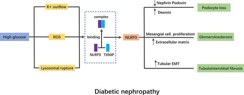
Xiong et al. NLRP3 in Kidney Diseases
The disease is affected by many environmental factors, mainly inflammatory response. NLRP3 inflammasome consists of
manifested in the high fat, high carbohydrate diet structure, low nucleotide-binding domain–like receptors (NLRs), apoptosis-
labor intensity and less exercise (4). MS includes a variety of associated speck-like protein containing caspase recruitment
metabolic disorders, including obesity, hyperglycemia, domain (ASC) and caspase protease. The structure of NLRs
hypertension, dyslipidemia, high blood viscosity, high uric mainly includes the middle nucleotide-binding and
acid, high fatty liver incidence and hyperinsulinemia (5). At oligomerization domain (NACHT), the downstream adapter
present, it is believed that the common causes of these factors are protein pyrindomain (PYD) or caspase recruitment domain
insulin resistance and hyperinsulinemia caused by obesity, (CARD), and leucine-richrepeats (LRRs). Caspase-1 is the
especially central obesity (6). MS is a risk factor for a variety of activated form of pro-caspase-1, which can cleave cytokine
diseases, such as hypertension, coronary heart disease, stroke, precursors such as interleukin (IL)-1b, IL-18 and other
chronic kidney disease (CKD), and even some cancers, including cytokine precursors, transform them into mature form, and
breast cancer, endometrial cancer, prostate cancer related to sex participate in the inflammatory reaction (14).
hormone, as well as pancreatic cancer, hepatobiliary cancer, The inflammasome is a complex composed of a variety of
colon cancer in digestive system (6–8). proteins in the cytoplasm, which integrates different damage
The kidney is an important organ of metabolism, and various stimulating signals and activates the innate immune defense
metabolic abnormalities can affect the structure and function of function (14, 15). The innate immune system recognizes
kidney. Among people with MS, the prevalence of CKD exceeds invading microorganisms and danger signals in the body
20%, which is much higher than that of the general population. through specific pattern recognition receptors (PRRs).
At the same time, among patients with CKD, the prevalence of Currently known PRRs are divided into two types, namely toll-
MS and subgroup metabolic disorders is much higher than that like receptors (TLRs) located on the cell membrane and NLRs
of non-CKD patients (9). A retrospective analysis of more than located in the cytoplasm (16). NLRP3 is the most well-studied
6,000 American adults aged over 20 years found that MS was an and most comprehensive inflammasome in the family of NLRs.
independent risk factor for CKD (10). The probability of CKD After LRRs of NLRP3 recognizes specific signal, it exposes and
and microalbuminuria was 2.6 times and 1. 9 times of that of the polymerizes the NACHT domain, and recruits ASC and pro-
normal population, and the more abnormal components of MS caspase-1 through PYD-PYD and CARD-CARD. Through the
metabolism, the greater the risk (10). A study of 75,468 Chinese cleavage of pro-caspase-1 to mature caspase-1, cytokine
people also supported the above view. The incidence of CKD precursors of IL-1b and IL-18 are cleaved into an active form
in MS patients and those without MS was 57% and and secreted out of the cell. In addition to promoting the
28%, respectively (11). The clinical manifestations of MS maturation and secretion of IL-1b and IL-18, it can also
related renal damage include glomerular hyperfiltration, mediate a special programmed cell death pyroptosis by
microalbuminuria, proteinuria, changes in renal tubular activating caspase-1, which is characterized by the formation of
function, eGFR < 60ml/(min·1.73m2), and increase of renal caspase-1-dependent plasma membrane pore size, a large
vascular resistance by ultrasonic. Insulin resistance is the number of release of inflammatory mediators and DNA
central link of MS. Insulin receptors are widely expressed in damage, and finally leads to osmotic disintegration of cells (17).
the kidney, such as podocytes, mesangial cells, endothelial cells Many endogenous and exogenous factors can stimulate the
and renal tubular epithelial cells (12). Observation of kidney production of NLRP3 inflammasome through different
tissue pathology of donor kidneys revealed that chronic mechanisms. There are clear reports about crystals or particles
pathological changes were more common in kidney tissues of (cholesterol crystals, asbestos, silica, etc.), bacterial toxins,
MS patients, manifested as varying degrees of glomerular microorganisms (viruses, bacteria and fungi), and some
sclerosis, renal tubular atrophy, renal interstitial fibrosis and vaccine adjuvants (18, 19). Given that NLRP3 inflammasome is
arteries hardening (13). an intracellular recognition receptor and the diversity of
Therefore, exploring the pathogenesis and prevention recognition substances, these activators may have a common
measures of renal damage in MS is a very important part of endogenous signal transduction molecule, however this common
the prevention and treatment of CKD. This article summarizes endogenous signal transduction molecule is not clear yet (20). At
the important role of inflammasome in renal damage caused by present, there are mainly three different modes to illustrate the
different metabolic factors, and provides a new perspective for activation mechanism of NLRP3 inflammasome. These three
the treatment of CKD in the future. modes include potassium channel open and outflow, cathepsin-B
secretion caused by lysosomal damage and rupture, and the
production of reactive oxygen species (ROS) (21). Various
microbial toxins, enzymes and extracellular ATP can activate
GENERAL INTRODUCTION OF NLRP3 ATP-P2X7 receptors, make potassium ions outflow, and activate
INFLAMMASOME NLRP3 inflammasome (22). Crystalline substances such as
silicon dioxide, antibiotics and antifungal drugs activate
Nod like receptor protein 3 (NLRP3) inflammasome is a inflammasome through ROS and cathepsin-B (23, 24).
macromolecular polyprotein complex with a molecular weight Renal inflammatory response is the immune response of the
of about 700 kDa, which has the function of regulating chronic kidney to infectious or non-infective activators. The specific
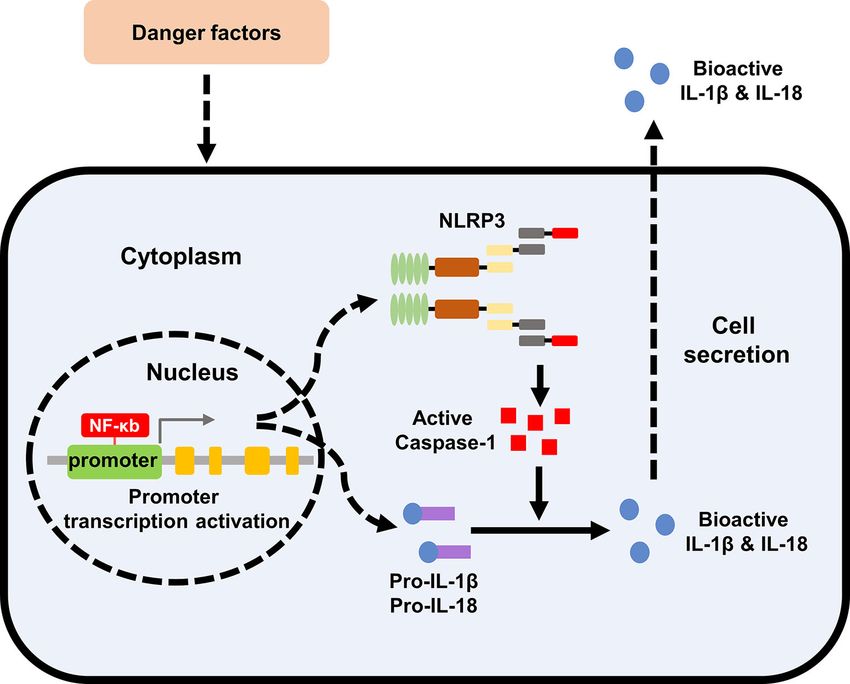
Frontiers in Immunology | www.frontiersin.org 2 July 2021 | Volume 12 | Article 714340Xiong et al. NLRP3 in Kidney Diseases expression of NLRP3 inflammasome components in kidney NLRP3 INFLAMMASOME IN METABOLIC- tissues has not yet been fully clarified. Renal mononuclear ASSOCIATED KIDNEY DISEASES phagocytes, such as dendrites and macrophages, can express the components of NLRP3 inflammasome and may induce cell Diabetic Nephropathy death by activating caspase-1 (25). At the same time, some Diabetes mellitus (DM) is a group of metabolic diseases studies have confirmed that renal tubular epithelial cells and characterized by hyperglycemia. When DM continues to progress, podocytes also activate the NLRP3-ASC-caspase-1 axis, express it often causes chronic damage to the eyes, kidneys, blood vessels, and release mature IL-1b and IL-18 (26–28). As an intracellular and feet. Among them, diabetic nephropathy (DN) is the most pattern recognition receptor, NLRP3 inflammasome plays an harmful inflammatory complication. It is also the main important role in stimulating and regulating immune microvascular of DM and the main cause of end-stage renal inflammation. The activation of NLRP3 inflammasome is disease (ESRD). Inflammatory response is the key factor for the involved in the acute and chronic inflammation of the kidneys sustainable development of DN. The activation of various by inducing the secretion of IL-1b and IL-18, leading to the inflammatory factors, such as C-reactive protein (CRP), monocyte automatic defense and inflammatory response (26, 29). NLRP3 chemoattractant protein-1 (MCP-1) and inflammasomes, promote inflammasome also participates in the occurrence and macrophage infiltration, renal tubular fibrosis, and eventually development of a variety of metabolic diseases as an important accelerate glomerulosclerosis (31). member (30). Therefore, the in-depth study on the mechanism The activation of NLRP3 inflammasome was detected in DN of NLRP3 inflammasome associated with metabolic disorders patients and diabetic mice (32). MCC950 was a selective and and kidney injury will provide new ideas and directions for the potent inhibitor of NLRP3 inflammasome, the use of which treatment of metabolic related kidney diseases. The specific improved renal function, podocyte injury and renal fibrosis in formation and activation of NLRP3 Inflammasome was shown db/db mice (33). Homozygous and hemizygous caspase-1 in Figure 1. deficiency had protective effect on db/db mice, while caspase-3 FIGURE 1 | Formation and activation of NLRP3 inflammasome. The effect of extracellular stimulating factors activates the intracellular NF-kB pathway. The activation of NF-kB pathway promotes the expression of inflammasome NLRP3, IL-1b, and IL-18. The activation of the inflammasome NLRP3 promotes the activation of caspase-1, and the activated caspase-1 promotes the maturation of IL-1b and IL-18, which are then secreted into the extracellular to exert biological effects. NLRP3, nod like receptor protein 3; IL, interleukin. Frontiers in Immunology | www.frontiersin.org 3 July 2021 | Volume 12 | Article 714340
Xiong et al.                                                                                                            NLRP3 in Kidney Diseases

deficiency had not, suggesting that caspase-3-dependent cell               role in inflammation. Treating HK-2 cells with the mtROS
death had no significant effect on the formation of DN, while              antioxidant MitoQ inhibited the dissociation of thioredoxin
caspase-1-dependent inflammatory activation played an                      (TRX) from TXNIP, and then blocked the interaction between
important role (34). Moreover, the use of a novel monoclonal              TXNIP and NLRP3, resulting in the inactivation of NLRP3
antibody of IL-1b in diabetic mice reduced renal damage                   inflammasome and the inhibition of IL-1b maturation (50).
markers, ameliorated fibrosis, and preserved the number of                 Another study confirmed that ATP-P2X4 signaling mediated the
podocytes (35). Thioredoxin-interacting protein (TXNIP) was               activation of HG-induced NLRP3 inflammasome, regulated the
a mediator of oxidative stress and has been reported to interact          secretion of IL-1b, and caused the development of tubulointerstitial
with NLRP3 inflammasome, leading to its activation (36). The               inflammation in DN (48). IRE1a was endoplasmic reticulum stress
expression of TXNIP and NLRP3 was both significantly                       (ERS)-related factor. Using IRE1a RNase specific inhibitor (STF-
increased in diabetic rats (36). Polyphenols, natural                     083010) in HG-induced NRK-52E cells inhibited the TXINP/
antioxidants, have been proved to reduce pyroptosis in DN,                NLRP3 pathway-mediated pyroptosis and renal damage,
probably due to the inhibition of TXNIP/NLRP3 pathway (37).               suggesting that ERS might also leading to the activation of
Other drugs with antioxidant function have also been confirmed             NLRP3 inflammasome (51).
to improve DN by targeting NLRP3 inflammasome, suggesting                      In general, HG stimulation can activate NLRP3
that NLRP3 inflammasome plays a crucial role in the                        inflammasome through a variety of pathways, which will lead
pathogenesis of DN (38–40).                                               to the abnormalities of intrinsic cells in kidney (Figure 2).
    Podocytes, namely glomerular visceral epithelial cells,               Current studies have proved that NLRP3 activation was
participate in stabilizing glomerular capillaries, maintaining            widespread in DN, and targeted therapy of NLRP3 played an
the function of glomerular filtration barrier, regulating                  important role in the improvement of DN.
ultrafiltration coefficient K/f and maintaining the normal
morphology of glomerular basement membrane (GBM) (41).                    Hypertension-Related Nephropathy
Studies have shown that podocyte injury plays a key role in the           Hypertension-related nephropathy is the damage of renal
pathogenesis of DN (41). High glucose (HG) activated the                  structure and function caused by primary hypertension. The
NLRP3 inflammasome in mouse podocytes, which was                           kidney can excrete excess water and sodium salt through urine,
manifested by increased protein levels of NLRP3, ASC and                  and prevent protein and blood cells from leaking out of blood
caspase-1, and the activity of caspase-1 was also significantly            vessels. High blood pressure increases the blood pressure in the
elevated (42). After the podocytes were transfected with                  blood vessels, leading to the leakage of protein into the urine,
NLRP3-small interfering RNA (siRNA), the expression of                    causing damage to the renal filter system. Long term poor control
caspase-1 and IL-1b was reduced, while the expression of the              of hypertension will cause irreversible damage to the kidney.
podocyte functional protein nephrin was significantly                      Clinical hypertension is related to kidney inflammation and
increased (43). The mechanism of NLRP3 inflammasome on                     increased circulating levels of IL-1b and IL-18, indicating that
podocytes has not been fully elucidated. It has been found that           inflammasome activity may be involve in the blood pressure
activation of NLRP3 inflammasome aggravated podocyte                       fluctuation and kidney injury (52).
autophagy and reduced nephrin expression, while NLRP3                         In mice with deoxycorticosterone acetate and saline (1K/DOCA/
silencing effectively restored podocyte autophagy and                     salt)-induced hypertension, the mRNA levels of NLRP3, ASC, pro-
alleviated podocyte injury induced by HG, suggesting that                 caspase-1 and pro-IL-1b were evaluated, as well as the protein
autophagy might participate in the regulation of NLRP3                    expression of active caspase-1 and mature IL-1b (53). ASC−/− mice
inflammasome on podocytes (44).                                            exhibited a sluggish pressor response and the treatment of NLRP3
    Glomerular mesangial cells play an important role in the              inflammasome inhibitor MCC950 reversed the hypertension in 1K/
process of glomerular injury and repair. Early DN mainly                  DOCA/salt treated mice (53). Nitric oxide (NO) inhibition and salt
manifested in the proliferation of mesangial cells, which then            overload lead to hypertension, albuminuria, glomerulosclerosis,
synthesize and secrete of a large number of mesangial matrixes,           glomerular ischemia and interstitial fibrosis. In this model,
gradually occlude the capillaries and lead to glomerulosclerosis.         allopurinol (ALLO), an NLRP3 inhibitor, significantly improved
It was found that the activation of NLRP3 also existed in                 hypertension, proteinuria and interstitial inflammation and fibrosis
glomerular mesangial cells stimulated by HG (45). Some                    (54). In a CKD model of 5/6 nephrectomy (5/6 Nx), the degree of
extracts of traditional Chinese medicine have been found to               tubulointerstitial fibrosis and proteinuria was decreased in
target NLRP3 to alleviate HG induced mesangial cell                       NLRP3−/− mice, meanwhile, the mitochondrial morphology and
proliferation (46, 47).                                                   CKD-related hypertension were also ameliorated (55). So far, some
    Renal tubular injury is one of the important determinants of          literatures have confirmed the important role of NLRP3 activation
progressive renal failure in DN. In vitro, the expression of NLRP3        in hypertension-related nephropathy, but the specific regulatory
and the release of IL-1b, IL-18 and ATP were significantly increased       mechanism still needs to be further explored.
in HK-2 cells stimulated by HG (48). Knockdown of NLRP3
resisted HG induced tubular EMT by inhibiting ROS production              Obesity-Related Nephropathy
and the phosphorylation of Smad3, p38MAPK and ERK1/2 (49).                In 1974, Weisinger et al. firstly reported that severe obesity can
The overproduction of mitochondrial ROS (mtROS) plays a key               lead to a large amount of proteinuria, and named this disease as

Frontiers in Immunology | www.frontiersin.org                         4                                    July 2021 | Volume 12 | Article 714340
Xiong et al. NLRP3 in Kidney Diseases FIGURE 2 | Mechanism of NLRP3 inflammasome in diabetic nephropathy. High glucose stimulation activates NLRP3 inflammasome mainly through K+ outflow, ROS and lysosomal rupture. TNXIP binding to NLRP3 is a pivotal mechanism of NLRP3 inflammasome activation. The activation of NLRP3 inflammasome will lead to podocyte lose, glomerulosclerosis and tubulointerstitial fibrosis. NLRP3, nod like receptor protein 3; ROS, reactive oxygen species; EMT, epithelial mesenchymal transition. obesity-related nephropathy (ORG) (56). Since then, clinical albumin to creatinine ratio and creatinine clearance rate in studies and animal experimental models have confirmed that obesity-prone (OP) rats with high protein and high fat diet obesity has a significant effect on the structure and function of (62). The expression of NLRP3 inflammasome was also the kidney (57). In recent years, although there are many studies downregulated under Coptidis Rhizoma treatment (62). on ORG, its specific pathogenesis is not fully understood. It is At present, the research on the pathogenesis and treatment of generally believed to be related to glucagon and insulin ORG is relatively lacking, and the activation of NLRP3 may be an resistance, the role of adipocytokines, inappropriate activation important link. Therefore, more extensive and in-depth research of renin-angiotensin-aldosterone system (RAAS), release of will give us a deeper understanding of ORG. inflammatory factors, lipid metabolism disorder and structural changes of kidney caused by obesity itself (58). Hyperuricemia It was found that the mRNA levels and protein expressions of Uric acid is a kind of anionic organic acid which is slightly soluble in NLRP3, ASC and caspase-1 in renal cortex of ORG mice were water. It is the end product of purine metabolism by xanthine significantly up-regulated (59). Also accompanied a significant oxidase. About 70% of uric acid in normal human body is excreted increase by P2X7R, an activation molecule of NLRP3. The through kidney, and the remaining 30% is excreted through bile treatment of P2X7R antagonist (KN−62 or A438079) reversed duct and intestine (63). The generation and excretion of uric acid in the changes of NLRP3 inflammasome components as well as healthy human body is in dynamic balance. Once this balance is attenuated podocytes injury treated by leptin (59). The broken, the generation or excretion of uric acid increase or decrease, expression of IL-1b and IL-18 levels also gradually increased in resulting in the accumulation of uric acid in the body, which will the kidney of high-fat diet (HFD) fed mice detected by lead to hyperuricemia. Fasting serum uric acid level > 420 mmol · immunohistochemistry (60). Knockdown of caspase-1 L - 1 (male) and > 360 mmol · L - 1 (female) is usually used as the expression with siRNA inhibited palmitate-induced death and diagnostic criteria of hyperuricemia. apoptosis of HK-2 cells (60). Some natural substances and Hyperuricemia can easily lead to renal hemodynamics, traditional Chinese medicine components have been shown histology and function changes, causing serious consequences to affect ORG by regulating the activation of NLRP3 such as renal tubulointerstitial inflammation, kidney stones, inflammasome. Fisetin (FIS) is a natural flavonoid, which renal fibrosis and polycystic kidney disease (64). The study of significantly attenuated HFD-induced histological changes in 266 patients with hyperuricemia found that the incidence of renal tissue samples, reduced the expression of kidney injury nephropathy was 15.11%, while the incidence of nephropathy molecule-1 (KIM-1) and altered the expression of nephrin and was only 2.19% in the population with normal serum uric acid podocin, thus improving renal insufficiency (61). In this process, level (65). Another study among 190 patients with chronic gout the expression of NLRP3 inflammasome components was also found that the incidence rate of renal damage was 86.13%, decreased, suggesting that its mechanism might be related to significantly higher than 7.14% in the control group, suggesting inflammasome (61). Coptidis Rhizoma, a classical traditional that hyperuricemia was closely related to the incidence of renal Chinese herb, reduced dyslipidemia and improved urinary damage and was another risk factor for kidney diseases (66). Frontiers in Immunology | www.frontiersin.org 5 July 2021 | Volume 12 | Article 714340
Xiong et al.                                                                                                               NLRP3 in Kidney Diseases

    At present, it is believed that hyperuricemia induced kidney              NLRP3 inflammasome activation (77). The production of ROS
injury is mainly related to hyperuricemia induced RAAS                        plays an important role in the activation of NLRP3
hyperfunction, inflammatory reaction, renal microvascular injury               inflammasome in hHcys-induced kidney injury. Nicotinamide
and so on, but the exact mechanism remains unclear. Affiliated                 adenine dinucleotide phosphate (NADPH) oxidase is considered
Bao’an Hospital of Shenzhen conducted a cohort study among                    to be the main source of superoxide in the kidney. NADPH
control, hyperuricemia and gouty nephropathy patients. The results            oxidase inhibition (NOX) reversed the upregulated protein levels
showed that the expression of the NLRP3 inflammasome in                        of NLRP3, ASC and caspase-1 stimulated by Hcys in mouse
peripheral blood mononuclear cells, and the levels of IL-1b and               podocytes (78). In vivo, NOX inhibition also protected glomeruli
IL-18 in the plasma were upregulated in the gouty nephropathy                 and podocytes from hHcys-induced damage, which was
group compared with the control and hyperuricemia groups (67). In             manifested by reduced proteinuria and glomerular sclerosis
rats with hyperuricemia and dyslipidemia induced by fructose,                 (78). TEMPOL is a recognized antioxidant. In hHcys mice, the
NLRP3 inflammasome in kidney tissues was activated, which                      treatment of TEMPOL reduced colocalization of NLRP3 with
manifested by overexpression of NLRP3, ASC and caspase-1,                     ASC, caspase-1 activation and as well as IL-1b production,
resulting in excessive production of IL-1b, IL-18, IL-6 (68). Using           suggesting the treatment inhibited the activation of NLRP3
the CRISPR/Cas9 system to functionally disrupt expression of urate            inflammasome (79). Meanwhile, the glomerular injury induced
oxidase (UOX) in Wistar rats spontaneously and persistently                   by hHcys has also been improved (79). As in other metabolic-
increased serum uric acid level compared with wild-type rats.                 associated kidney diseases mentioned above, the binding of
UOX-KO rats established increased interstitial fibrosis,                       TXNIP to NLRP3 is a key signaling mechanism in hHcys-
macrophage infiltration, increased expression of NLRP3 and                     induced kidney injury as well. Inhibition of TXNIP by
IL-1b, and activation signaling pathways associated with                      verapamil or TXNIP shRNA transfection broke the binding
autophagy, indicating that autophagy and NLRP3-dependent                      and disrupted the formation of glomerular inflammasome (80).
inflammation played crucial role in the development of                            As a crucial role in the pathogenic process of hHcys-induced
hyperuricemia induced kidney injury (69). The activation                      kidney injury, NLRP3 inflammasome has been regarded as a
of NLRP3 inflammasome has been proved to be a target of                        novel target for the treatment of glomerular injury in hHcys.
hyperuricemia induced renal injury. A large number of natural                 Many compounds with anti-inflammatory properties, such as
extracts have been found to improve renal function by inhibiting the          anandamide, DHA metabolites-resolvins, resolvin D1 (RvD1)
activity of NLRP3 inflammasome in hyperuricemia (70–73).                       and 17S-hydroxy DHA (17SHDHA), blocked podocyte injury
    Although there are a lot of epidemiological and experimental              and glomerular sclerosis during hHcys via the suppression of
research reports on the relationship between uric acid and kidney             NLRP3 inflammasome activity (81–83).
injury, the underlying pathological mechanism still needs further
research. The mechanism of hyperuricemia-induced renal injury
has a wide range of cross-talks. The activation of NLRP3
inflammasome plays a complex and important role in                             CONCLUSION
promoting the occurrence and development of renal disease.
The mechanism of its interaction with other factors still needs to            MS is a group of clinical syndromes of chronic inflammation and
be further explored.                                                          metabolic disorders caused by insulin resistance. With the
                                                                              improvement of the economic level and the spread of
Hyperhomocysteinemia                                                          unhealthy lifestyles, the prevalence of MS is on the rise
Hyperhomocysteinemia (hHcys) is a disease characterized by                    globally, especially in developing countries and regions. Recent
elevated homocysteine in the blood, which has been recognized                 studies have found that MS is an independent risk factor for
as one of the important risk factors of kidney disease. HHcys can             CKD. The pathogenesis of kidney damage caused by MS is
cause anabolism of cholesterol and triglycerides, impaired                    related to poor primary disease control, insulin resistance,
endothelial function, thrombosis, and monocyte activation                     chronic inflammation, and endothelial function damage.
(74). Hyperhomocysteinemia is present in 85% of patients with                 NLRP3 inflammasome is the sensor of the innate immune
chronic renal failure, and persists after the initiation of dialysis or       system that initiates inflammatory response to stimulations,
kidney transplantation (75).                                                  and participates in the occurrence and development of various
   In 2012, Zhang et al. firstly discovered that all the components            metabolic diseases and kidney injury (Table 1). As we mentioned
of NLRP3 inflammasome were existed in podocytes and were                       above, a variety of metabolic disorders leads to the activation of
significantly evaluated by the treatment with L-homocysteine (L-               NLRP3 inflammasome in the kidney. The activation of NLRP3
Hcys) (76). Silencing the ASC gene or inhibiting caspase-1                    inflammasome aggravates renal inflammatory infiltration and
activity could alleviate podocyte injury and improve                          tissue damage through many pathways including autophagy,
glomerulosclerosis in mice with hHcys (76). Podocin, nephrin                  inflammatory factor release and EMT. At the same time,
and desmin are critical markers of podocyte injury. Another                   factors related to tissue damage, such as ROS, autophagy
study demonstrated that in folate free (FF) diet induced hHcys                related molecules continue to stimulate the activation of
mice, NLRP3−/− mice showed increased protein level of podocin                 NLRP3 inflammasome and impair renal function. Therefore,
and nephrin but decreased expression of desmin compared to                    the activation of inflammasome and kidney injury are
wild-type mice, indicating the potential pathogenic effects of                mutually reinforcing.

Frontiers in Immunology | www.frontiersin.org                             6                                   July 2021 | Volume 12 | Article 714340
Xiong et al. NLRP3 in Kidney Diseases
TABLE 1 | The role of NLRP3 inflammasome in the kidney under different Accordingly, to explore the specific mechanism and
metabolic disorders.
important role of NLRP3 in kidney injury induced by
Metabolic factor NLRP3 Function metabolic disorders will provide new ideas and directions for
state the prevention and treatment of metabolic-associated
kidney diseases.
Hyperglycemia activate podocyte lose, glomerulosclerosis and
tubulointerstitial fibrosis
Hypertension activate hypertension, proteinuria, interstitial
inflammation and fibrosis
Obesity activate dyslipidemia, podocyte injury, cell death and AUTHOR CONTRIBUTIONS
apoptosis
Hyperuricemia activate interstitial fibrosis and macrophage CZ and X-FM conceived and designed the manuscript. WX did
infiltration literature searching, drafted the manuscript, and drew the
Hyperhomocysteinemia activate proteinuria and glomerular sclerosis
figures. CZ and X-FM reviewed and revised the article. All
authors contributed to the article and approved the
submitted version.
Because inflammasomes are intracellular recognition
receptors, scientists believe that there may be common factors
for their activation. In the kidney damage caused by different
metabolic factors, there are currently three recognized activation FUNDING
pathways, namely the outflow of potassium ions, the release of
ROS, and the rupture of lysosomes. But for each metabolic factor, This work was supported by Grants from the National Natural
specific activators are also found. For example, in DN, NLRP3 Science Foundation of China (81961138007, 81974096,
inflammasome can also be activated by endoplasmic reticulum 81770711), and program for HUST Academic Frontier Youth
stress and autophagy. Team (2017QYTD20).
REFERENCES in Living Kidney Donors. Am J Transplant (2013) 13:2342–51. doi: 10.1111/
ajt.12369
1. Schlaich M, Straznicky N, Lambert E, Lambert G. Metabolic Syndrome: A 14. Schroder K, Tschopp J. The Inflammasomes. Cell (2010) 140:821–32.
Sympathetic Disease? Lancet Diabetes Endocrinol (2015) 3:148–57. doi: 10.1016/j.cell.2010.01.040
doi: 10.1016/S2213-8587(14)70033-6 15. Lamkanfi M, Dixit VM. Inflammasomes and Their Roles in Health and
2. Ford ES, Giles WH, Dietz WH. Prevalence of the Metabolic Syndrome Disease. Annu Rev Cell Dev Biol (2012) 28:137–61. doi: 10.1146/annurev-
Among US Adults: Findings From the Third National Health and cellbio-101011-155745
Nutrition Examination Survey. JAMA (2002) 287:356–9. doi: 10.1001/ 16. Hoffman HM, Wanderer AA. Inflammasome and IL-1beta-Mediated
jama.287.3.356 Disorders. Curr Allergy Asthma Rep (2010) 10:229–35. doi: 10.1007/s11882-
3. Ford ES, Giles WH, Mokdad AH. Increasing Prevalence of the Metabolic 010-0109-z
Syndrome Among U.S. Adults. Diabetes Care (2004) 27:2444–9. doi: 10.2337/ 17. Sharma D, Kanneganti TD. The Cell Biology of Inflammasomes: Mechanisms
diacare.27.10.2444 of Inflammasome Activation and Regulation. J Cell Biol (2016) 213:617–29.
4. Grundy SM. Metabolic Syndrome: Connecting and Reconciling Cardiovascular doi: 10.1083/jcb.201602089
and Diabetes Worlds. J Am Coll Cardiol (2006) 47:1093–100. doi: 10.1016/ 18. Duewell P, Kono H, Rayner KJ, Sirois CM, Vladimer G, Bauernfeind FG, et al.
j.jacc.2005.11.046 NLRP3 Inflammasomes are Required for Atherogenesis and Activated by
5. Eckel RH, Grundy SM, Zimmet PZ. The Metabolic Syndrome. Lancet (2005) Cholesterol Crystals. Nature (2010) 464:1357–61. doi: 10.1038/nature08938
365:1415–28. doi: 10.1016/S0140-6736(05)66378-7 19. Wen H, Miao EA, Ting JP. Mechanisms of NOD-Like Receptor-Associated
6. Grundy SM. Pre-Diabetes, Metabolic Syndrome, and Cardiovascular Risk. Inflammasome Activation. Immunity (2013) 39:432–41. doi: 10.1016/
J Am Coll Cardiol (2012) 59:635–43. doi: 10.1016/j.jacc.2011.08.080 j.immuni.2013.08.037
7. Agrawal V, Shah A, Rice C, Franklin BA, McCullough PA. Impact of Treating 20. Mangan MSJ, Olhava EJ, Roush WR, Seidel HM, Glick GD, Latz E. Targeting
the Metabolic Syndrome on Chronic Kidney Disease. Nat Rev Nephrol (2009) the NLRP3 Inflammasome in Inflammatory Diseases. Nat Rev Drug Discov
5:520–8. doi: 10.1038/nrneph.2009.114 (2018) 17:588–606. doi: 10.1038/nrd.2018.97
8. Bishehsari F, Voigt RM, Keshavarzian A. Circadian Rhythms and the Gut 21. Jin C, Flavell RA. Molecular Mechanism of NLRP3 Inflammasome Activation.
Microbiota: From the Metabolic Syndrome to Cancer. Nat Rev Endocrinol J Clin Immunol (2010) 30:628–31. doi: 10.1007/s10875-010-9440-3
(2020) 16:731–9. doi: 10.1038/s41574-020-00427-4 22. Petrilli V, Papin S, Dostert C, Mayor A, Martinon F, Tschopp J. Activation of
9. Zhang X, Lerman LO. The Metabolic Syndrome and Chronic Kidney Disease. the NALP3 Inflammasome Is Triggered by Low Intracellular Potassium
Transl Res (2017) 183:14–25. doi: 10.1016/j.trsl.2016.12.004 Concentration. Cell Death Differ (2007) 14:1583–9. doi: 10.1038/sj.
10. Chen J, Muntner P, Hamm LL, Jones DW, Batuman V, Fonseca V, et al. The cdd.4402195
Metabolic Syndrome and Chronic Kidney Disease in U.S. Adults. Ann Intern 23. Zhou R, Tardivel A, Thorens B, Choi I, Tschopp J. Thioredoxin-Interacting
Med (2004) 140:167–74. doi: 10.7326/0003-4819-140-3-200402030-00007 Protein Links Oxidative Stress to Inflammasome Activation. Nat Immunol
11. Song H, Wang X, Cai Q, Ding W, Huang S, Zhuo L. Association of Metabolic (2010) 11:136–40. doi: 10.1038/ni.1831
Syndrome With Decreased Glomerular Filtration Rate Among 75,468 Chinese 24. Campden RI, Zhang Y. The Role of Lysosomal Cysteine Cathepsins in NLRP3
Adults: A Cross-Sectional Study. PloS One (2014) 9:e113450. doi: 10.1371/ Inflammasome Activation. Arch Biochem Biophys (2019) 670:32–42.
journal.pone.0113450 doi: 10.1016/j.abb.2019.02.015
12. Hale LJ, Coward RJ. The Insulin Receptor and the Kidney. Curr Opin Nephrol 25. Lichtnekert J, Kulkarni OP, Mulay SR, Rupanagudi KV, Ryu M, Allam R, et al.
Hypertens (2013) 22:100–6. doi: 10.1097/MNH.0b013e32835abb52 Anti-GBM Glomerulonephritis Involves IL-1 But Is Independent of NLRP3/
13. Ohashi Y, Thomas G, Nurko S, Stephany B, Fatica R, Chiesa A, et al. ASC Inflammasome-Mediated Activation of Caspase-1. PloS One (2011) 6:
Association of Metabolic Syndrome With Kidney Function and Histology e26778. doi: 10.1371/journal.pone.0026778
Frontiers in Immunology | www.frontiersin.org 7 July 2021 | Volume 12 | Article 714340Xiong et al. NLRP3 in Kidney Diseases
26. Komada T, Muruve DA. The Role of Inflammasomes in Kidney Disease. Nat 47. Chen F, Wei G, Xu J, Ma X, Wang Q. Naringin Ameliorates the High Glucose-
Rev Nephrol (2019) 15:501–20. doi: 10.1038/s41581-019-0158-z Induced Rat Mesangial Cell Inflammatory Reaction by Modulating the
27. Xiong W, Meng XF, Zhang C. Inflammasome Activation in Podocytes: A New NLRP3 Inflammasome. BMC Complement Altern Med (2018) 18:192.
Mechanism of Glomerular Diseases. Inflamm Res (2020) 69:731–43. doi: 10.1186/s12906-018-2257-y
doi: 10.1007/s00011-020-01354-w 48. Chen K, Zhang J, Zhang W, Zhang J, Yang J, Li K, et al. ATP-P2X4 Signaling
28. Lau A, Chung H, Komada T, Platnich JM, Sandall CF, Choudhury SR, et al. Mediates NLRP3 Inflammasome Activation: A Novel Pathway of Diabetic
Renal Immune Surveillance and Dipeptidase-1 Contribute to Contrast- Nephropathy. Int J Biochem Cell Biol (2013) 45:932–43. doi: 10.1016/
Induced Acute Kidney Injury. J Clin Invest (2018) 128:2894–913. j.biocel.2013.02.009
doi: 10.1172/JCI96640 49. Song S, Qiu D, Luo F, Wei J, Wu M, Wu H, et al. Knockdown of NLRP3
29. Chang A, Ko K, Clark MR. The Emerging Role of the Inflammasome in Alleviates High Glucose or TGFB1-Induced EMT in Human Renal Tubular
Kidney Diseases. Curr Opin Nephrol Hypertens (2014) 23:204–10. Cells. J Mol Endocrinol (2018) 61:101–13. doi: 10.1530/JME-18-0069
doi: 10.1097/01.mnh.0000444814.49755.90 50. Han Y, Xu X, Tang C, Gao P, Chen X, Xiong X, et al. Reactive Oxygen Species
30. Sharma BR, Kanneganti TD. NLRP3 Inflammasome in Cancer and Metabolic Promote Tubular Injury in Diabetic Nephropathy: The Role of the
Diseases. Nat Immunol (2021) 22:550–9. doi: 10.1038/s41590-021-00886-5 Mitochondrial Ros-Txnip-Nlrp3 Biological Axis. Redox Biol (2018) 16:32–
31. Duran-Salgado MB, Rubio-Guerra AF. Diabetic Nephropathy and 46. doi: 10.1016/j.redox.2018.02.013
Inflammation. World J Diabetes (2014) 5:393–8. doi: 10.4239/wjd.v5.i3.393 51. Ke R, Wang Y, Hong S, Xiao L. Endoplasmic Reticulum Stress Related Factor
32. Shahzad K, Bock F, Dong W, Wang H, Kopf S, Kohli S, et al. Nlrp3- IRE1alpha Regulates TXNIP/NLRP3-Mediated Pyroptosis in Diabetic
Inflammasome Activation in Non-Myeloid-Derived Cells Aggravates Nephropathy. Exp Cell Res (2020) 396:112293. doi: 10.1016/j.yexcr.
Diabetic Nephropathy. Kidney Int (2015) 87:74–84. doi: 10.1038/ki.2014.271 2020.112293
33. Zhang C, Zhu X, Li L, Ma T, Shi M, Yang Y, et al. A Small Molecule Inhibitor 52. Krishnan SM, Sobey CG, Latz E, Mansell A, Drummond GR. IL-1beta and IL-
MCC950 Ameliorates Kidney Injury in Diabetic Nephropathy by Inhibiting 18: Inflammatory Markers or Mediators of Hypertension? Br J Pharmacol
NLRP3 Inflammasome Activation. Diabetes Metab Syndr Obes (2019) (2014) 171:5589–602. doi: 10.1111/bph.12876
12:1297–309. doi: 10.2147/DMSO.S199802 53. Krishnan SM, Dowling JK, Ling YH, Diep H, Chan CT, Ferens D, et al.
34. Shahzad K, Bock F, Al-Dabet MM, Gadi I, Kohli S, Nazir S, et al. Caspase-1, Inflammasome Activity Is Essential for One Kidney/Deoxycorticosterone
But Not Caspase-3, Promotes Diabetic Nephropathy. J Am Soc Nephrol (2016) Acetate/Salt-Induced Hypertension in Mice. Br J Pharmacol (2016)
27:2270–5. doi: 10.1681/ASN.2015060676 173:752–65. doi: 10.1111/bph.13230
35. Lei Y, Devarapu SK, Motrapu M, Cohen CD, Lindenmeyer MT, Moll S, et al. 54. Zambom FFF, Oliveira KC, Foresto-Neto O, Faustino VD, Avila VF, Albino
Interleukin-1beta Inhibition for Chronic Kidney Disease in Obese Mice With AH, et al. Pathogenic Role of Innate Immunity in a Model of Chronic NO
Type 2 Diabetes. Front Immunol (2019) 10:1223. doi: 10.3389/fimmu. Inhibition Associated With Salt Overload. Am J Physiol Renal Physiol (2019)
2019.01223 317:F1058–67. doi: 10.1152/ajprenal.00251.2019
36. Samra YA, Said HS, Elsherbiny NM, Liou GI, El-Shishtawy MM, Eissa LA. 55. Gong W, Mao S, Yu J, Song J, Jia Z, Huang S, et al. NLRP3 Deletion Protects
Cepharanthine and Piperine Ameliorate Diabetic Nephropathy in Rats: Role Against Renal Fibrosis and Attenuates Mitochondrial Abnormality in Mouse
of NF-kappaB and NLRP3 Inflammasome. Life Sci (2016) 157:187–99. With 5/6 Nephrectomy. Am J Physiol Renal Physiol (2016) 310:F1081–8.
doi: 10.1016/j.lfs.2016.06.002 doi: 10.1152/ajprenal.00534.2015
37. An X, Zhang Y, Cao Y, Chen J, Qin H, Yang L. Punicalagin Protects Diabetic 56. Weisinger JR, Kempson RL, Eldridge FL, Swenson RS. The Nephrotic
Nephropathy by Inhibiting Pyroptosis Based on TXNIP/NLRP3 Pathway. Syndrome: A Complication of Massive Obesity. Ann Intern Med (1974)
Nutrients (2020) 12(5):1516. doi: 10.3390/nu12051516 81:440–7. doi: 10.7326/0003-4819-81-4-440
38. Liu YW, Hao YC, Chen YJ, Yin SY, Zhang MY, Kong L, et al. Protective Effects 57. Camara NO, Iseki K, Kramer H, Liu ZH, Sharma K. Kidney Disease and
of Sarsasapogenin Against Early Stage of Diabetic Nephropathy in Rats. Obesity: Epidemiology, Mechanisms and Treatment. Nat Rev Nephrol (2017)
Phytother Res (2018) 32:1574–82. doi: 10.1002/ptr.6088 13:181–90. doi: 10.1038/nrneph.2016.191
39. Lu M, Yin N, Liu W, Cui X, Chen S, Wang E. Curcumin Ameliorates Diabetic 58. Wahba IM, Mak RH. Obesity and Obesity-Initiated Metabolic Syndrome:
Nephropathy by Suppressing NLRP3 Inflammasome Signaling. BioMed Res Mechanistic Links to Chronic Kidney Disease. Clin J Am Soc Nephrol (2007)
Int (2017) 2017:1516985. doi: 10.1155/2017/1516985 2:550–62. doi: 10.2215/CJN.04071206
40. Sun Z, Ma Y, Chen F, Wang S, Chen B, Shi J. Artesunate Ameliorates High 59. Hou XX, Dong HR, Sun LJ, Yang M, Cheng H, Chen YP. Purinergic 2x7
Glucose-Induced Rat Glomerular Mesangial Cell Injury by Suppressing the Receptor Is Involved in the Podocyte Damage of Obesity-Related
TLR4/NF-kB/NLRP3 Inflammasome Pathway. Chem Biol Interact (2018) Glomerulopathy Via Activating Nucleotide-Binding and Oligomerization
293:11–9. doi: 10.1016/j.cbi.2018.07.011 Domain-Like Receptor Protein 3 Inflammasome. Chin Med J (Engl) (2018)
41. Mathieson PW. The Podocyte as a Target for Therapies–New and Old. Nat 131:2713–25. doi: 10.4103/0366-6999.245270
Rev Nephrol (2011) 8:52–6. doi: 10.1038/nrneph.2011.171 60. Li LC, Yang JL, Lee WC, Chen JB, Lee CT, Wang PW, et al. Palmitate
42. Liu Y, Xu Z, Ma F, Jia Y, Wang G. Knockdown of TLR4 Attenuates High Aggravates Proteinuria-Induced Cell Death and Inflammation Via CD36-
Glucose-Induced Podocyte Injury Via the NALP3/ASC/Caspase-1 Signaling Inflammasome Axis in the Proximal Tubular Cells of Obese Mice. Am J
Pathway. BioMed Pharmacother (2018) 107:1393–401. doi: 10.1016/ Physiol Renal Physiol (2018) 315:F1720–31. doi: 10.1152/ajprenal.00536.2017
j.biopha.2018.08.134 61. Ge C, Xu M, Qin Y, Gu T, Lou D, Li Q, et al. Fisetin Supplementation Prevents
43. Li J, Wang B, Zhou G, Yan X, Zhang Y. Tetrahydroxy Stilbene Glucoside High Fat Diet-Induced Diabetic Nephropathy by Repressing Insulin
Alleviates High Glucose-Induced MPC5 Podocytes Injury Through Resistance and RIP3-Regulated Inflammation. Food Funct (2019) 10:2970–
Suppression of NLRP3 Inflammasome. Am J Med Sci (2018) 355:588–96. 85. doi: 10.1039/c8fo01653d
doi: 10.1016/j.amjms.2018.03.005 62. Ren Y, Wang D, Lu F, Zou X, Xu L, Wang K, et al. Coptidis Rhizoma Inhibits
44. Hou Y, Lin S, Qiu J, Sun W, Dong M, Xiang Y, et al. NLRP3 Inflammasome NLRP3 Inflammasome Activation and Alleviates Renal Damage in Early
Negatively Regulates Podocyte Autophagy in Diabetic Nephropathy. Biochem Obesity-Related Glomerulopathy. Phytomedicine (2018) 49:52–65.
Biophys Res Commun (2020) 521:791–8. doi: 10.1016/j.bbrc.2019.10.194 doi: 10.1016/j.phymed.2018.05.019
45. Feng H, Gu J, Gou F, Huang W, Gao C, Chen G, et al. High Glucose and 63. Mandal AK, Mount DB. The Molecular Physiology of Uric Acid Homeostasis.
Lipopolysaccharide Prime NLRP3 Inflammasome Via ROS/TXNIP Pathway Annu Rev Physiol (2015) 77:323–45. doi: 10.1146/annurev-physiol-021113-
in Mesangial Cells. J Diabetes Res (2016) 2016:6973175. doi: 10.1155/2016/ 170343
6973175 64. Braga TT, Foresto-Neto O, Camara NOS. The Role of Uric Acid in
46. Wang S, Zhao X, Yang S, Chen B, Shi J. Salidroside Alleviates High Glucose- Inflammasome-Mediated Kidney Injury. Curr Opin Nephrol Hypertens
Induced Oxidative Stress and Extracellular Matrix Accumulation in Rat (2020) 29:423–31. doi: 10.1097/MNH.0000000000000619
Glomerular Mesangial Cells by the TXNIP-NLRP3 Inflammasome Pathway. 65. Toprak O, Cirit M, Esi E, Postaci N, Yesil M, Bayata S. Hyperuricemia as a
Chem Biol Interact (2017) 278:48–53. doi: 10.1016/j.cbi.2017.10.012 Risk Factor for Contrast-Induced Nephropathy in Patients With Chronic
Frontiers in Immunology | www.frontiersin.org 8 July 2021 | Volume 12 | Article 714340Xiong et al. NLRP3 in Kidney Diseases
Kidney Disease. Catheter Cardiovasc Interv (2006) 67:227–35. doi: 10.1002/ 77. Xia M, Conley SM, Li G, Li PL, Boini KM. Inhibition of
ccd.20598 Hyperhomocysteinemia-Induced Inflammasome Activation and
66. Padang C, Muirden KD, Schumacher HR, Darmawan J, Nasution AR. Glomerular Sclerosis by NLRP3 Gene Deletion. Cell Physiol Biochem
Characteristics of Chronic Gout in Northern Sulawesi, Indonesia. (2014) 34:829–41. doi: 10.1159/000363046
J Rheumatol (2006) 33:1813–7. 78. Abais JM, Zhang C, Xia M, Liu Q, Gehr TW, Boini KM, et al. NADPH
67. Zhang YZ, Sui XL, Xu YP, Gu FJ, Zhang AS, Chen JH. NLRP3 Inflammasome Oxidase-Mediated Triggering of Inflammasome Activation in Mouse
and Lipid Metabolism Analysis Based on UPLC-Q-TOF-MS in Gouty Podocytes and Glomeruli During Hyperhomocysteinemia. Antioxid Redox
Nephropathy. Int J Mol Med (2019) 44:172–84. doi: 10.3892/ijmm.2019.4176 Signal (2013) 18:1537–48. doi: 10.1089/ars.2012.4666
68. Hu QH, Zhang X, Pan Y, Li YC, Kong LD. Allopurinol, Quercetin and Rutin 79. Abais JM, Xia M, Li G, Gehr TW, Boini KM, Li PL. Contribution of
Ameliorate Renal NLRP3 Inflammasome Activation and Lipid Accumulation Endogenously Produced Reactive Oxygen Species to the Activation of
in Fructose-Fed Rats. Biochem Pharmacol (2012) 84:113–25. doi: 10.1016/ Podocyte NLRP3 Inflammasomes in Hyperhomocysteinemia. Free Radic
j.bcp.2012.03.005 Biol Med (2014) 67:211–20. doi: 10.1016/j.freeradbiomed.2013.10.009
69. Wu M, Ma Y, Chen X, Liang N, Qu S, Chen H. Hyperuricemia Causes Kidney 80. Abais JM, Xia M, Li G, Chen Y, Conley SM, Gehr TW, et al. Nod-Like
Damage by Promoting Autophagy and NLRP3-Mediated Inflammation in Receptor Protein 3 (NLRP3) Inflammasome Activation and Podocyte
Rats With Urate Oxidase Deficiency. Dis Model Mech (2021) 14(3): In jury Via Thioredoxin-Interactin g Protein (TXNIP) During
dmm048041. doi: 10.1242/dmm.048041 Hyperhomocysteinemia. J Biol Chem (2014) 289:27159–68. doi: 10.1074/
70. Cui D, Liu S, Tang M, Lu Y, Zhao M, Mao R, et al. Phloretin Ameliorates jbc.M114.567537
Hyperuricemia-Induced Chronic Renal Dysfunction Through Inhibiting 81. Li G, Xia M, Abais JM, Boini K, Li PL, Ritter JK. Protective Action of
NLRP3 Inflammasome and Uric Acid Reabsorption. Phytomedicine (2020) Anandamide and Its COX-2 Metabolite Against L-Homocysteine-Induced
66:153111. doi: 10.1016/j.phymed.2019.153111 NLRP3 Inflammasome Activation and Injury in Podocytes. J Pharmacol Exp
71. Hu J, Wu H, Wang D, Yang Z, Zhuang L, Yang N, et al. Weicao Capsule Ther (2016) 358:61–70. doi: 10.1124/jpet.116.233239
Ameliorates Renal Injury Through Increasing Autophagy and NLRP3 82. Zhang Q, Conley SM, Li G, Yuan X, Li PL. Rac1 GTPase Inhibition Blocked
Degradation in UAN Rats. Int J Biochem Cell Biol (2018) 96:1–8. Podocyte Injury and Glomerular Sclerosis During Hyperhomocysteinemia
doi: 10.1016/j.biocel.2018.01.001 Via Suppression of Nucleotide-Binding Oligomerization Domain-Like
72. Li G, Guan C, Xu L, Wang L, Yang C, Zhao L, et al. Scutellarin Ameliorates Receptor Containing Pyrin Domain 3 Inflammasome Activation. Kidney
Renal Injury Via Increasing CCN1 Expression and Suppressing NLRP3 Blood Press Res (2019) 44:513–32. doi: 10.1159/000500457
Inflammasome Activation in Hyperuricemic Mice. Front Pharmacol (2020) 83. Li G, Chen Z, Bhat OM, Zhang Q, Abais-Battad JM, Conley SM, et al. NLRP3
11:584942. doi: 10.3389/fphar.2020.584942 Inflammasome as a Novel Target for Docosahexaenoic Acid Metabolites to
73. Wang M, Zhao J, Zhang N, Chen J. Astilbin Improves Potassium Oxonate- Abrogate Glomerular Injury. J Lipid Res (2017) 58:1080–90. doi: 10.1194/
Induced Hyperuricemia and Kidney Injury Through Regulating Oxidative jlr.M072587
Stress and Inflammation Response in Mice. BioMed Pharmacother (2016)
83:975–88. doi: 10.1016/j.biopha.2016.07.025 Conflict of Interest: The authors declare that the research was conducted in the
74. Russo C, Morabito F, Luise F, Piromalli A, Battaglia L, Vinci A, et al. absence of any commercial or financial relationships that could be construed as a
Hyperhomocysteinemia Is Associated With Cognitive Impairment in potential conflict of interest.
Multiple Sclerosis. J Neurol (2008) 255:64–9. doi: 10.1007/s00415-007-0668-7
75. Ferechide D, Radulescu D. Hyperhomocysteinemia in Renal Diseases. J Med Copyright © 2021 Xiong, Meng and Zhang. This is an open-access article distributed
Life (2009) 2:53–9. under the terms of the Creative Commons Attribution License (CC BY). The use,
76. Zhang C, Boini KM, Xia M, Abais JM, Li X, Liu Q, et al. Activation of Nod- distribution or reproduction in other forums is permitted, provided the original author(s)
Like Receptor Protein 3 Inflammasomes Turns on Podocyte Injury and and the copyright owner(s) are credited and that the original publication in this journal is
Glomerular Sclerosis in Hyperhomocysteinemia. Hypertension (2012) cited, in accordance with accepted academic practice. No use, distribution or
60:154–62. doi: 10.1161/HYPERTENSIONAHA.111.189688 reproduction is permitted which does not comply with these terms.
Frontiers in Immunology | www.frontiersin.org 9 July 2021 | Volume 12 | Article 714340You can also read


