An affinity matured human monoclonal antibody targeting fusion loop epitope of dengue virus with in vivo therapeutic potency - Nature
←
→
Page content transcription
If your browser does not render page correctly, please read the page content below
www.nature.com/scientificreports
OPEN An affinity‑matured human
monoclonal antibody targeting
fusion loop epitope of dengue virus
with in vivo therapeutic potency
Tomohiro Kotaki1,2,7*, Takeshi Kurosu3, Ariadna Grinyo‑Escuer4, Edgar Davidson4,
Siti Churrotin2, Tamaki Okabayashi5, Orapim Puiprom5, Kris Cahyo Mulyatno2,
Teguh Hari Sucipto2, Benjamin J. Doranz4, Ken‑ichiro Ono6, Soegeng Soegijanto2 &
Masanori Kameoka1*
Dengue virus (DENV), from the genus flavivirus of the family flaviviridae, causes serious health
problems globally. Human monoclonal antibodies (HuMAb) can be used to elucidate the mechanisms
of neutralization and antibody-dependent enhancement (ADE) of DENV infections, leading to the
development of a vaccine or therapeutic antibodies. Here, we generated eight HuMAb clones from
an Indonesian patient infected with DENV. These HuMAbs exhibited the typical characteristics of
weak neutralizing antibodies including high cross-reactivity with other flaviviruses and targeting of
the fusion loop epitope (FLE). However, one of the HuMAbs, 3G9, exhibited strong neutralization
(NT50 < 0.1 μg/ml) and possessed a high somatic hyper-mutation rate of the variable region, indicating
affinity-maturation. Administration of this antibody significantly prolonged the survival of interferon-
α/β/γ receptor knockout C57BL/6 mice after a lethal DENV challenge. Additionally, Fc-modified 3G9
that had lost their in vitro ADE activity showed enhanced therapeutic potency in vivo and competed
strongly with an ADE-prone antibody in vitro. Taken together, the affinity-matured FLE-targeting
antibody 3G9 exhibits promising features for therapeutic application including a low NT50 value,
potential for treatment of various kinds of mosquito-borne flavivirus infection, and suppression of
ADE. This study demonstrates the therapeutic potency of affinity-matured FLE-targeting antibodies.
Many clinically-important mosquito-borne viruses, including dengue virus (DENV), Japanese encephalitis virus
(JEV), West Nile virus (WNV), and Zika virus (ZIKV) belong to the genus flavivirus of the family flaviviridae1.
Of these, DENV causes the most serious health problems worldwide, in terms of the number of patients and
fatalities. Infection with any of four serotypes of dengue virus (DENV-1 to DENV-4) causes dengue fever and
dengue hemorrhagic f ever2, or dengue and severe dengue, as classified by the World Health O rganization3. An
estimated 390 million cases of DENV infection occur annually worldwide4. Of these, 100 million people develop
symptomatic dengue and 21,000 die4,5.
One of the mechanisms hypothesized to cause a higher risk of severe dengue is the antibody-dependent
enhancement (ADE) of the infection, whereby pre-existing anti-DENV antibodies induced by the primary
acrophages6.
infection or vaccination facilitate subsequent DENV infections of Fc receptor-positive cells like m
Thus, DENV antibodies exhibit two conflicting activities: neutralization and ADE. Neutralization suppresses
viremia, resulting in protection against DENV infection, while ADE increases viremia, and this is associated
with severe dengue7. This phenomenon might have increased the risk of developing severe dengue disease among
seronegative people who have received vaccinations of CYD-TDV, which is the only licensed dengue v accine8,9.
In addition, antibodies induced by ZIKV infection are reported to enhance DENV infections by ADE in vitro,
1
Department of Public Health, Kobe University Graduate School of Health Sciences, Kobe, Japan. 2Collaborative
Research Center for Emerging and Re‑Emerging Infectious Diseases, Institute of Tropical Disease, Airlangga
University, Surabaya, Indonesia. 3Department of Virology I, National Institute of Infectious Diseases (NIID), Tokyo,
Japan. 4Integral Molecular, Inc., Philadelphia, PA, USA. 5Mahidol‑Osaka Center for Infectious Diseases (MOCID),
Faculty of Tropical Medicine, Mahidol University, Bangkok, Thailand. 6Medical & Biological Laboratories Co.,
Ltd., Tokyo, Japan. 7Present address: Department of Virology, Research Institute for Microbial Diseases, Osaka
University, Suita, Japan. *email: tkotaki@biken.osaka-u.ac.jp; mkameoka@port.kobe-u.ac.jp
Scientific Reports | (2021) 11:12987 | https://doi.org/10.1038/s41598-021-92403-9 1
Vol.:(0123456789)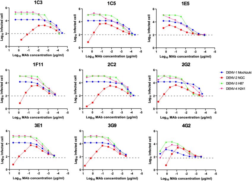
www.nature.com/scientificreports/
Hybridoma clones
1C3 1C5 1E5 1F11 2C2 2G2 3E1 3G9
Indonesian isolates
DENV-1 0.90 1.89 0.65 0.24 0.59 0.21 1.00 0.24
DENV-2 0.12 2.67 0.69 0.02 0.04 0.02 0.05 0.02
DENV-3 0.87 > 3.60 0.92 1.29 3.39 1.99 2.41 0.73
DENV-4 0.64 1.70 0.89 0.02 0.12 0.13 0.24 0.13
Prototype strains
DENV-1 3.46 3.07 > 3.86 0.84 6.17 3.33 3.48 3.45
DENV-2 0.03 0.18 0.21 0.09 0.10 0.20 0.11 0.06
DENV-3 0.38 0.34 0.38 0.11 0.30 0.44 0.62 0.12
DENV-4 0.89 0.09 0.17 0.03 0.11 0.06 0.22 < 0.04
Other flaviviruses
JEV 1.33 0.74 0.87 0.21 0.71 0.85 0.44 0.47
WNV* – – – 0.17 – – – 0.11
ZIKV* – – – 0.05 – – – 0.11
Table 1. The NT50 values of the HuMAbs. Average of two independent neutralization tests is shown (μg/ml).
*The WNV and ZIKV results were determined using SRIPs containing the prM-E genes of each virus.
and vice versa10,11. ADE complicates dengue pathogenesis, and, thus, forms an obstacle to developing a fully
effective dengue vaccine and prophylactic or therapeutic antibodies.
The DENV genome encodes three structural proteins (capsid [C], premembrane/membrane [prM/M], and
envelope [E]) and seven non-structural p roteins1. The viral particle is assembled in the lumen of the endoplasmic
reticulum, where nucleocapsid (viral RNA complexed with the C protein) is incorporated into the lipid bilayer
containing prM and E proteins1. As this immature viral particle traffics through the trans-Golgi network, a
host serine protease (furin) cleaves the prM protein from the immature virus, resulting in maturation. This
maturation step occasionally remains incomplete, resulting in a mixture of virus particles at different states of
maturity12,13. Virus particles exhibit conformational dynamics referred to viral ‘breathing’14. The maturity and
breathing of virions have an impact on the recognition of antibodies and, thereby, affect their neutralizing and
enhancing activities15,16.
The E protein is the major target of neutralizing antibodies, since it is located on the surface of a DENV
virion17. Three domains (domains I, II, and III; DI, DII and DIII) have been identified in the E protein structure18.
Each DENV particle contains 180 monomers of E protein that form 90 E-dimers19.
The neutralization and ADE activities of antibodies are determined by the epitope of the v irus20–22. Antibod-
ies targeting the fusion loop epitope (FLE) or bc loop on DII generally exhibit low levels of neutralization, high
ADE, and high cross-reactivity to fl aviviruses23–28. These antibodies are extensively induced during second-
ary DENV infections, because the epitopes are highly conserved among the flaviviruses29,30. Antibodies that
bind to E-dimers, quaternary-structure epitopes, or the hinge regions of DI-DII exhibit strong neutralization
ability by blocking viral conformational changes and membrane f usion31–33. Indeed, antibodies that recognize
complex epitopes account for much of the virus neutralizing activity that occurs in the serum of convalescing
patients31,32,34. However, many of these antibodies are serotype-specific. Antibodies that target domain III are
serotype-specific and show higher neutralizing activity than those targeting domains I-II, although domain III-
targeting antibodies are not predominantly produced in h umans32,34,35.
Human monoclonal antibodies (HuMAbs) could be useful tools for elucidating the mechanisms of neu-
tralization and ADE, information that is required for vaccine development. In addition, HuMAbs can be used
for prophylactic or therapeutic purposes. Several groups have been successful in generating HuMAbs against
DENV, using various methods31,33,36. Here, using newly developed SPYMEG cell technology, we generated eight
anti-DENV HuMAb clones from an Indonesian patient infected with DENV37,38.
Results
Hybridoma preparation and details of HuMAbs. We established eight HuMAb clones from a blood
specimen of an Indonesian patient with dengue using SPYMEG cells that belong to a human hybridoma fusion
partner cell l ine37,38. The patient was diagnosed with acute dengue fever (2 days after onset); the blood specimen
was found to be anti-DENV IgM/IgG- and NS1-positive, indicating a secondary infection. The DENV serotype
could not be determined by RT-PCR using RNA extracted from the patient serum39. All the HuMAbs were
reactive to detergent-inactivated DENV for ELISA, indicating that HuMAbs recognized the E-monomer. All
antibodies belonged to IgG1 subtype.
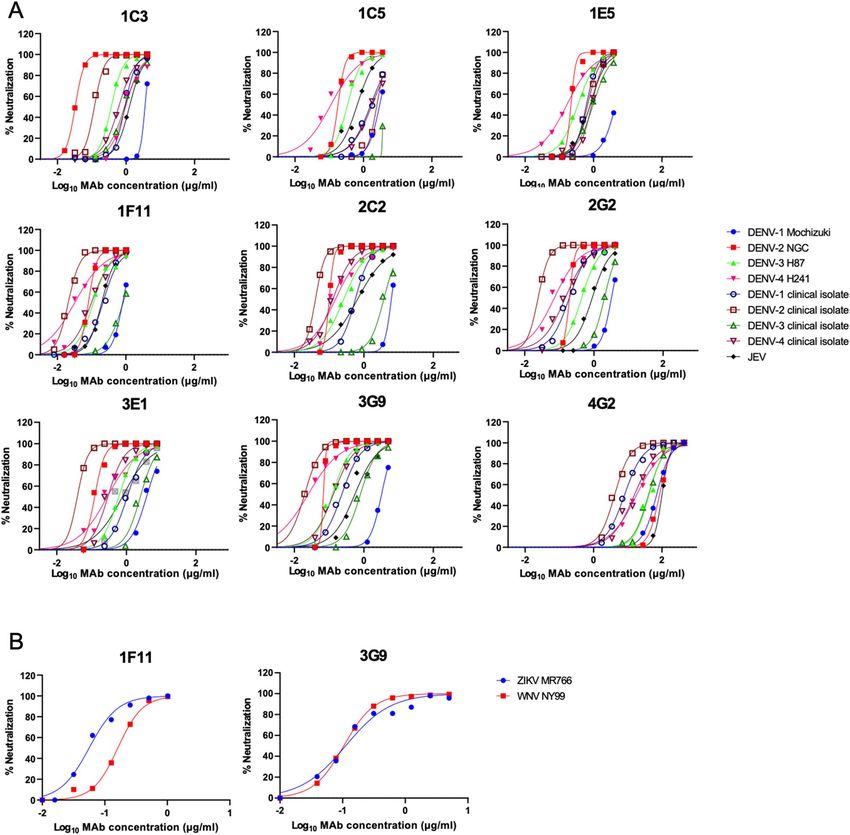
Neutralization activity. All HuMAbs exhibited neutralizing activity against all four serotypes of DENV
prototype strains and Indonesian isolates (Table 1, Fig. 1A). DENV-2, in particular, was strongly neutralized;
the NT50 was lower than 0.1 μg/ml. All of the HuMAbs also neutralized JEV. Overall, HuMAbs 1F11 and 3G9
showed stronger neutralizing activity. Thus, these two promising HuMAbs were subjected to neutralization tests
Scientific Reports | (2021) 11:12987 | https://doi.org/10.1038/s41598-021-92403-9 2
Vol:.(1234567890)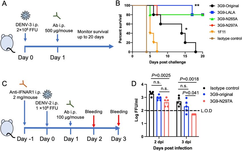
www.nature.com/scientificreports/
Figure 1. Neutralization of DENV, JEV, WNV, and ZIKV. (A) Neutralizing activity against DENVs and JEV.
(B) Neutralizing activity against SRIPs containing prM-E of WNV and ZIKV. Average of two independent
experiments is shown.
using single-round infectious particles (SRIPs) containing prM-E proteins of ZIKV and WNV, due to the una-
vailability of the infectious viruses40,41. Again, 1F11 and 3G9 neutralized ZIKV and WNV particles as efficiently
as DENV (Fig. 1B). These data indicated that these HuMAbs were broadly-neutralizing antibodies to mosquito-
borne flaviviruses.
Stage of HuMAb neutralization. The flaviviruses can be neutralized by several mechanisms including
the inhibition of receptor binding, inhibition of membrane fusion, and aggregation of virion particles42. To
determine the neutralization mechanism, a time of addition assay using Vero cells and the DENV-2 New Guinea
C (NGC) strain was carried out43. The antibodies all neutralized DENV-2 during the pre-adsorption assay but
not the post-adsorption assay (Table 2). This suggested that our HuMAbs blocked pre-adsorption steps includ-
ing viral adsorption but not post-adsorption steps, such as viral membrane fusion or conformation change.
Epitope mapping. The epitope target of an antibody influences its neutralization potency and mecha-
nism. Therefore, epitope mapping was conducted using HEK293 cells transfected with a DENV2 prM-E mutant
library, which possess a single alanine substitution at each residue of prM-E (661 mutants in total)44. Four anti-
bodies (1C3, 1F11, 3E1, and 3G9) were tested against all the mutants, while the other four antibodies (1C5, 1E5,
Scientific Reports | (2021) 11:12987 | https://doi.org/10.1038/s41598-021-92403-9 3
Vol.:(0123456789)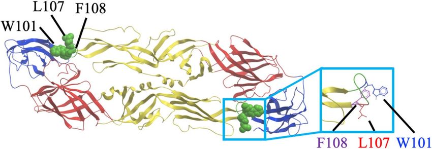
www.nature.com/scientificreports/
Hybridoma clones
1C3 1C5 1E5 1F11 2C2 2G2 3E1 3G9
Pre-adsorption 0.04 0.30 0.47 0.06 0.16 0.43 0.23 0.07
Post-adsorption > 8.3 > 7.2 > 0.95 > 2.0 > 13.9 > 8.1 > 15.1 > 5.9
Table 2. The NT50 values from the pre-adsorption and post-adsorption assays. Average of two independent
neutralization tests is shown (μg/ml).
Figure 2. Deduced epitope locations on a ribbon diagram of the DENV-2 E protein. The deduced epitope
locations were plotted on a DENV-2 E dimer, based on the data provided by the Protein Data Bank accession
number 1OAN. DI, DII, and DIII are indicated in red, yellow, and blue, respectively. The fusion loop region
(98–110) is colored in green. Mutations affecting reactivity with the HuMAbs are shown as green spheres and in
the magnified square.
Hybridoma clones
1C3 1C5 1E5 1F11 2C2 2G2 3E1 3G9
W101A
Critical W101A W101A W101A W101A W101A W101A W101A
L107A
residue L107A L107A F108A F108A F108A F108A F108A
F108A
Table 3. Critical mutations that abolished HuMAb binding.
2C2, and 2G2) were tested only against selected mutants with an FLE mutation, because of the availability of
materials. The data are shown in Supplementary Fig. S1. The critical residues that abolished the binding abil-
ity of the HuMAbs tested are presented in Fig. 2 and Table 3. The W101A mutation, a key epitope site for FLE
antibodies, was critical to the activity of all eight HuMAbs26,29,45. In addition to W101A, L107A and/or F108A
were responsible for binding. All of the critical residues were located on the fusion loop of the E protein, which
is highly conserved among the flaviviruses. These data substantiated the breadth of neutralizing ability exhibited
by the HuMAbs considered in this study. Our antibodies were typical FLE antibodies, which are dominantly
produced when a secondary DENV infection occurs.
The escape mutant analysis was attempted to further investigate the epitope, by passaging DENV-2 in the
presence of the HuMAbs in Vero cells and then sequencing the E protein gene in any surviving viruses. However,
we did not identify any mutations in the E protein of surviving viruses.
ADE activity. Targeting of the FLE is a typical characteristic of weak neutralizing and high-ADE
a ntibodies29,30. We measured ADE activity using semi-adherent K562 cells46. Although the HuMAb clones
exhibited strong neutralizing activity in Vero cells, strong ADE activity was observed in our assay system (Fig. 3).
DENV-2 was neutralized at high concentrations of the HuMAb clones, while the other strains, DENV-1, -3, and
-4, were not neutralized. These data suggested that the cloned HuMAbs could contribute to disease enhancing
activity.
Recombinant IgG construction and evaluation. Although the cloned antibodies were potent in terms
of low NT50 in Vero cells, high ADE was observed in K562 cells. However, these antibodies could potentially
be used in therapeutic applications, by modifying the effector function and disrupting the ADE activity. Thus,
one of three mutations [L234A + L235A (LALA), D265A, and N297A] was introduced to the Fc region of the
best neutralizing HuMAb, 3G9, to disrupt the interaction with Fc receptors47. We chose to test three muta-
tions, because the effect of Fc-modification during the in vivo animal experiment varied depending on the study
Scientific Reports | (2021) 11:12987 | https://doi.org/10.1038/s41598-021-92403-9 4
Vol:.(1234567890)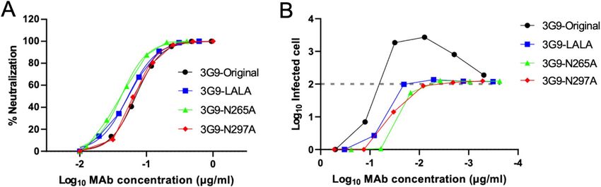
www.nature.com/scientificreports/
Figure 3. ADE activity of the HuMAbs. The DENV-2 NGC strain and K562 cells were used. Dotted lines
indicate the baseline of the infected cells in control (100 infected cells; 2.0). Each data point represents the
average obtained from two independent ADE assays.
3G9-Original 3G9-LALA 3G9-N265A 3G9-N297A
NT50 (μg/ml) 0.069 0.053 0.042 0.066
Table 4. NT50 values of Fc-modified 3G9. Average of two independent neutralization tests is shown (μg/ml).
group48–53. These three mutations are described as being the same in terms of the effect on loss of binding to Fc
receptors and should not compromise antibody neutralizing p otency54 or shorten the half-life significantly55.
However, N297A abolishes N-linked glycosylation at N-297, while the LALA and D265A mutants would be
fully glycosylated. Additionally, LALA and N297A reduce complement component 1q (C1q) binding, whereas
D265A does not compromise complement-dependent cytotoxicity, which is related to virus clearance20,47. The
difference in the mutations may cause subtle changes in the IgG phenotype, resulting in differences in in vivo
efficacy55. The recombinant Fc-modified 3G9 antibodies neutralized DENV-2 at comparable level to that of the
3G9-original in Vero cells (Table 4, Fig. 4A). As expected, the recombinant 3G9 antibodies did not show ADE
activity in K562 cells, even at a sub-neutralizing concentration (Fig. 4B).
HuMAb protection in vivo. Because the neutralization and ADE activities in vitro do not always correlate
with protection in vivo, we tested the ability of antibodies to protect against DENV infection in animal models.
We tested the ability of 3G9, 1F11 (another potent HuMAb), and Fc-modified 3G9 to protect interferon-α/β/γ
receptor knockout (IFN-α/β/γR KO) C57BL/6 mice challenged with DENV-356. DENV-3 was chosen because of
the availability of the lethal infection mouse model, in which DENV infection causes vascular leakage without
showing neurologic d isorder56. Antibodies (500 μg/mouse) were injected intraperitoneally (i.p.) after one day
of virus challenge (Fig. 5A). Mice administered with an isotype control IgG died within 4 days post infection
(dpi) (Fig. 5B). 1F11 exhibited almost no protection, while the original unmodified 3G9 significantly prolonged
survival, although all mice died within 20 dpi (p < 0.01). All three recombinant 3G9 antibodies significantly
enhanced the survival rate when compared with the original 3G9 (p < 0.05). There were no significant differences
among the three recombinant antibodies (p > 0.05).
Scientific Reports | (2021) 11:12987 | https://doi.org/10.1038/s41598-021-92403-9 5
Vol.:(0123456789)www.nature.com/scientificreports/
Figure 4. Neutralizing and ADE activities of the Fc-modified 3G9. (A) Neutralizing activity against DENV-2
NGC strain using Vero cells. Average of two independent experiments is shown. (B) ADE activity against
DENV-2 using K562 cells. Average of two independent experiments is shown.
Figure 5. In vivo efficacy test. (A) Scheme of the animal challenge experiment using IFN-α/β/γR KO C57BL/6
mice. (B) Survival of infected mice after viral challenge. A group of five mice were infected with 2.0 × 10⁶ FFU
of DENV-3. Statistical significance was analyzed using a log rank test. *P < 0.01 for the comparison between
isotype control and 3G9-original. **P < 0.05 for the comparison between 3G9-original and the Fc-modified 3G9.
There was no significant difference among the Fc-modified 3G9 (P > 0.05). (C) Scheme of the animal challenge
experiment carried out using immunocompetent BALB/c mice. (D) Viremia levels of the infected mice after the
viral challenge. A group of five mice were infected with 1.0 × 10⁶ FFU of DENV-2. The limit of detection (L.O.D)
was 100 FFU/ml. Viremia levels below the detection limit are plotted as half of the detection limit (50 FFU/
ml). Statistical significance was analyzed using a one-way ANOVA. P < 0.05 was considered significant. n.s. not
significant.
To further validate the potency of in vivo protection, another animal experiment was carried out using
DENV-2 NGC strain and immunocompetent BALB/c mice treated with mouse monoclonal antibody against
type I IFN receptor (Fig. 5C)57. Viral load was measured as a readout because this model is not a lethal infection
model. There was no significant difference between isotype control and 3G9-original, possibly due to the low
amount of administrated IgG (100 μg/mouse) (Fig. 5D). However, 3G9-N297A showed a significant reduction
Scientific Reports | (2021) 11:12987 | https://doi.org/10.1038/s41598-021-92403-9 6
Vol:.(1234567890)www.nature.com/scientificreports/
Figure 6. Competition ELISA and ADE assay. (A) Competition ELISA. Mouse monoclonal antibodies 4G2,
7F4, or 15C12 at 1 μg/ml were mixed with serially diluted 3G9 and incubated in a DENV-coated ELISA plate.
The OD relative to that of the no competition well (without 3G9) is shown. Each data represents the average of
two independent experiments. (B) Competition ADE. 100 ng/ml of 4G2 and 1:640 diluted mouse serum, which
showed the peak level of enhancement, were mixed with DENV-2 NGC and serially diluted 3G9-N297A. The
number of infected cells relative to that of the no competition wells is shown. Each data represents the average of
two independent experiments.
V region
V gene D gene J gene AA mutations CDR3
14.3%
Heavy chain IGHV3-23*02 IGHD3-16*01 IGHJ4*02 AKLFGVGDSDGY
(14/98)
7.1%
Light chain IGLV7-46*01 – IGLJ3*02 LLSYGGGRPV
(7/98)
Table 5. Immunogenetic analysis of 3G9. The closest germline genes were determined using the IMGT tool.
of viral load compared to isotype control (p < 0.01). Although 3G9-N297A did not significantly reduce viral load
at 2 dpi (P = 0.11) compared to 3G9-original, the significance was observed at 3 dpi (P = 0.041), demonstrating
faster viral clearance. These results indicated that the reduced ADE shown by these modified antibodies promotes
their effectiveness in vivo and suggests that recombinant 3G9 antibodies are promising therapeutic agents.
Competition assay using ELISA and ADE assays. Our animal experiments were infection models
without ADE. Thus, it remained unclear whether the Fc-modified 3G9 could suppress infection with ADE
in vivo. An antibody highly competitive to 4G2 (low-avidity FLE antibody) has been reported to protect mice
from lethal DENV infection with A DE58. Since we had not established an in vivo ADE infection model, com-
petition assays were conducted. Competitive ELISA showed that 3G9 strongly competed with 4G2 but not with
other mouse monoclonal antibodies (7F4 and 15C12, targeting the central part of domain II and the A strand of
domain III, respectively) (Fig. 6A)20. 3G9 inhibited 4G2 binding by more than 50% when both were applied to
the assay at the same concentration.
It has been reported that the suppression of ADE caused by pre-existing antibodies in vitro could be an
indicator of in vivo protection e fficacy58. Thus, a competitive ADE assay was performed using 4G2 and DENV2-
immunized mouse serum, which showed peak levels of ADE at 100 ng/ml and a 1:640 dilution, respectively
(Fig. 3, Fig. S2). These dilutions were used, therefore, for the subsequent competitive ADE assay. 3G9-N297A
suppressed the peak ADE caused by 4G2 and D2-immunized mouse serum by 50% at only 30 ng/ml (Fig. 6B).
A previous study indicated that an antibody that strongly reduced ADE infection (> 50%) at 1000 ng/ml offered
good therapeutic efficacy in an in vivo ADE model58. The competition intensities found in the current study
were higher than the threshold for in vivo protection quoted in the previous report, although direct comparison
is impossible.
Immunogenetic analysis. Even though 3G9 is an FLE antibody, it showed high potency in terms of NT50
values and in vivo protection. Therefore, we analyzed the sequences of the 3G9 VH and VL regions using the
IMGT tool to identify the closest VH and Vλ germline genes. The results indicated that the VH gene was derived
from IGHV3-23*02 and the Vλ gene from IGLV7-46*01 (Table 5). Somatic hyper-mutation (SHM) rates of the
VH and Vλ genes were 14.3% and 7.1%, respectively. 3G9 showed distinct VH germline gene usage and the
highest SHM rate of the VH regions than the HuMAbs generated in this study (Table S1). The high SHM rate
of the VH regions indicated the levels of affinity-maturation during the secondary i nfection50. The sequences of
HuMAbs have been added in Supplementary Table S2.
Scientific Reports | (2021) 11:12987 | https://doi.org/10.1038/s41598-021-92403-9 7
Vol.:(0123456789)www.nature.com/scientificreports/
Discussion
To date, no specific therapeutic agent against flavivirus infection has been made available. Considering the cur-
rent success of antibody therapy against respiratory syncytial virus, Ebola virus, and severe acute respiratory syn-
drome coronavirus 2 infections, antibody therapy for dengue is a promising target. DENV-neutralizing HuMAbs
have been reported using various techniques and patient b ackgrounds31–33,36,38. The objective of this study was to
establish therapeutic antibody candidates against dengue using the newly developed SPYMEG cell t echnology37,38.
Multiple experiments were attempted using blood samples from dengue patients in acute or convalescent phase
of infection. We successfully established eight HuMAb clones from a single dengue patient. The patient whose
hybridoma was successfully established was in the acute phase of secondary DENV infection, indicating that
the B cells were highly activated and IgG repertoire was abundant. The SPYMEG cell fusion method was highly
successful when the patient was in the acute infection phase, which was consistent to the results of a previous
study38. The circulating serotypes and/or genotypes of DENV vary depending on the region and year59,60. These
antibodies, therefore, could be a potent therapeutic agent for use against DENV strains circulating in Southeast
Asia, including Indonesia, which is one of the largest dengue-endemic countries.
Several groups have generated potent neutralizing HuMAbs against DENV. In general, highly potent neu-
tralizing antibodies are serotype-specific, bind to E-dimers or quaternary-structure epitopes, and inhibit both
pre- and post-attachment steps61. In contrast, weak neutralizing antibodies are highly cross-reactive, bind to
E-monomer FLEs, and inhibit the only pre-attachment step. Although all the HuMAbs generated in this study
recognized typical epitope of weak neutralizing antibodies, they showed considerably low N T50 values, especially
against DENV-2. The NT50 values were < 0.1 μg/ml, as low as those of previously reported highly potent neutral-
izing antibodies61. However, these values cannot be compared directly, because the neutralizing antibody titer
is influenced by the host cell type, degree of viral maturation, virion breathing, and other assay conditions62,63.
Thus, 4G2 (a low neutralizing FLE antibody) was used to standardize the NT50 assay. One of the HuMAb clones,
3G9, exhibited a N T50 that was 1000-fold lower than that of 4G2 against DENV-2 (Table 1). This was comparable
to the findings of a previous study on potent neutralizing a ntibodies64, corroborating the suggestion that 3G9 is
a promising neutralizing antibody.
High neutralizing potency, especially against DENV-2, is beneficial, because the only licensed dengue vac-
cine (CYD-TDV) confers low-level protection against DENV-265,66. Cross-reactive neutralization is reported to
play an important role in DENV-2 protection, while serotype-specific neutralization is key in protection against
other serotypes for those who received CYD-TDV v accination67. The HuMAbs described in this study could
compensate for the drawbacks associated with the current dengue vaccine.
3G9 also neutralized other flaviviruses, with NT50 values of around 0.1 μg/ml. ZIKV is generally less suscep-
tible to FLE antibodies due to high thermostability and less virion b reathing68,69. A potent neutralizing antibody
targeting both DENV and ZIKV could be promising, considering their co-circulation in the environment and
the occurrence of ADE in both diseases10,11. 3G9 may be applied therapeutically in the treatment of various kinds
of mosquito-borne flavivirus infections.
Other groups have reported potent neutralizing antibodies that target FLEs and closely related bc loop. For
example, 2A10G6, targeting D98, R99, and W101 motifs within the fusion loop, has a broad neutralizing capa-
bility against all four serotypes of DENV and protects mice against lethal challenges from DENV and W NV70.
71
753C12, targeting both the fusion loop and adjacent bc loop, exhibits low NT50 values . 1C19, targeting only bc
loop also exhibits broad and potent neutralization to D ENVs36. Furthermore, E60, targeting FLE, showed thera-
peutic potency in vivo . The promising results of FLE antibodies with Fc-modification were r eported53,58,73.
58,72
These reports substantiate the potential of FLE antibodies.
No viral escape mutant was obtained, even after 5 viral passages in the presence of the HuMAbs in Vero cells.
The fusion loop is highly conserved among flavivirus, and an escape mutation at this position would be lethal to
the virus. This feature also could be an advantage to the development of therapeutics.
Generally, cross-reactive non-conformational epitopes are less neutralizing, while serotype-specific conforma-
tional epitopes are highly p otent61. Cross-reactive FLE antibodies are induced exclusively at the secondary infec-
tion phase29,30. Tsai et al. reported that FLE antibodies derived from patients with a secondary infection are more
potent than that those from patients with a primary infection71. Considering the high SHM rate (14.2%) on the V
gene (Table 5), 3G9 was considerably affinity-matured, and, therefore, p otent50. In addition, as a third or fourth
DENV infection is less possible, affinity-matured FLE antibodies play an important role in viral protection74.
Although the HuMAbs investigated in this study were promising in intensity and breadth of neutralization,
strong ADE was observed. DENV-2 was slightly neutralized at a high concentration of IgG in the ADE assay,
coinciding with the high level of neutralization activity in Vero cells. Meanwhile, the other serotypes of DENV
were not neutralized. The ADE assay measures the balance of neutralizing and enhancing activities, and, thus,
no neutralizing activity was o bserved46. The HuMAb clones were subtyped as IgG1, which show relatively higher
75
levels of ADE a ctivity . These data indicate that modification of the Fc region is mandatory prior to therapeutic
application.
Fc-modified 3G9 neutralized DENV at a similar level to the original antibodies, but ADE was not observed,
even at sub-neutralizing levels in vitro. In addition, these recombinant antibodies prolonged the survival of the
infected mice and reduced viremia level, compared with the original antibody. Some groups have reported that
disrupting the Fc-interaction significantly enhances survival r ates50,52,58, while other groups have demonstrated
the attenuation of protection or no significant difference49,51. Our study demonstrated an improved survival rate
and no difference among the three recombinant antibodies. These data suggested that disrupting Fc-interactions
is promising, at least in mice, regardless of the mutations. The results indicated that Fc-modified 3G9 is a good
therapeutic candidate.
Scientific Reports | (2021) 11:12987 | https://doi.org/10.1038/s41598-021-92403-9 8
Vol:.(1234567890)www.nature.com/scientificreports/
While our data suggests the importance of eliminating the ADE function of protective antibodies, a limitation
of our study was the lack of animal experiments using an ADE model. Williams et. al. reported that the assay of
antibody competition or ability to displace low-avidity FLE antibodies and reduce ADE in vitro could enable the
prediction of in vivo therapeutic efficacy with ADE58. Our ADE competition assay data revealed that 3G9 com-
petes with 4G2 and polyclonal immunized-mouse serum (Fig. 6). The observed degree of competitiveness was
stronger than that shown previously by E60, a potent neutralizing FLE a ntibody58, although direct comparison
was impossible. An affinity-matured FLE antibody would be superior to other potent neutralizing antibodies
targeting the DI-DII hinge region or DIII, in terms of competing with pre-existing ADE-prone FLE antibodies,
which may warrant the suppression of ADE in vivo58.
Another limitation of the study is that the DENV circulating in infected humans has a unique morphology,
a higher degree of maturity, and less sensitivity to FLE antibodies than those derived from established culture
cells76,77. It is possible that neutralizing potency of 3G9 is compromised in human infection since 3G9 is an FLE
antibody. Although protection in mice may demonstrate therapeutic potency, further neutralizing and ADE tests
using DENV in human serum are warranted.
In conclusion, we have described a potent HuMAb, 3G9, which targets FLE. Fc-modified 3G9 displayed
neutralizing potency in vivo. The affinity-matured FLE antibody has several features that make it appropriate
for therapeutic application, including a low N T50 value, potential for the treatment of various kinds of mosquito-
borne flavivirus infection, competition with pre-existing ADE-prone antibodies, and less potential for viral
escape. This study reconfirms the therapeutic potency of affinity-matured FLE antibodies.
Methods
Ethical statement and patient recruitment. Blood samples were collected from patients with dengue
at a private hospital in Surabaya, Indonesia. Signed informed consent was acquired from the patients or their
parents upon collection of blood samples. This study was approved by the Ethics Committees of Airlangga Uni-
versity (Ethics Committee Approval Number: 24-934/UN3.14/PPd/2013) and Kobe University Graduate School
of Medicine (Ethics Committee Approval Number: 784). Diagnosis data were obtained from medical records.
Animal experiments using IF-α/β/γR KO C57BL/6 mice were performed at the National Institute of Infectious
Diseases in Japan (NIID), in accordance with the guidelines for the care and use of laboratory animals at the
NIID. The study was approved by the Animal Experiment Committee of the NIID (Ethics Committee Approval
Number: 115064). Trained laboratory personnel performed the anesthesia of mice via i.p. injection of a mixture
of medetomidine, midazolam, and butorphanol during viral inoculation and euthanasia by cervical dislocation.
Animal experiments using immunocompetent BALB/c mice were performed at Kobe University. The study
was approved by the Animal Experiment Committee (Ethics Committee Approval Number: P180504). Trained
laboratory personnel performed mice anesthesia via isoflurane inhalation during viral inoculation and eutha-
nasia by cervical dislocation.
All the experiments were carried out in accordance with the ARRIVE guidelines (http://www.nc3rs.org.uk/
page.asp?id=1357) and all relevant guidelines and regulations.
Hybridoma preparation. Approximately 5 ml of blood were obtained from one patient and the peripheral
blood mononuclear cells were isolated by centrifugation using a Ficoll-Paque PLUS (GE Healthcare, Uppsala,
Sweden). These cells were then fused with SPYMEG cells at a ratio of 10:1, to establish hybridomas, as described
previously37,38.
Cell lines. The SPYMEG cells and established hybridomas were maintained in Dulbecco’s modified Eagle
medium (DMEM) supplemented with 15% fetal bovine serum (FBS) and 3% BM-condimed (Sigma Aldrich, St.
Louis, MO)18. Vero cells were cultured in Eagle’s minimum essential medium (MEM) supplemented with 10%
FBS and 60 μg/ml kanamycin. C6/36 cells were cultured in MEM supplemented with 10% FBS, nonessential
amino acids, and 60 μg/ml kanamycin. HEK-293 T cells were maintained in DMEM supplemented with 10%
FBS. K562 erythroleukemia cells were cultivated in RPMI 1640 medium supplemented with 10% FBS, 100 units/
ml penicillin, and 100 μg/ml streptomycin. FreeStyle 293-F cells (Invitrogen, Gaithersburg, MD) were cultured
in FreeStyle 293 Expression Medium (Thermo Fisher Scientific, Waltham, MA).
Viruses. DENV-1 genotype I, DENV-2 cosmopolitan genotype, DENV-3 genotype I, and DENV-4 geno-
type II, which are typical isolates found in Surabaya, Indonesia, were used for the antibody characterization59,60.
Sequence information is available upon request. In addition, the Mochizuki strain of DENV-1, NGC strain of
DENV-2, H87 strain of DENV-3, H241 strain of DENV-4, and Nakayama strain of JEV were used as prototype
viruses79. The DENV-3 strain P12/08, derived from patients infected with DENV-3 in Thailand in 2008, was used
in the animal experiments56. Viruses were propagated in C6/36 cells and stored at − 80 °C until use.
Hybridoma screening by ELISA. Rabbit serum immunized with the DENV-2 NGC strain was coated
onto Nunc Maxisorp ELISA plates (Thermo Fisher Scientific, Waltham, MA). Then, the DENV-2 Indonesian
strain (inactivated with Tween 20), hybridoma culture (or DMEM as a negative control), alkaline phosphatase
(AP) conjugated anti-human IgG (Abcam, Canbridge, UK), and p-nitrophenyl phosphate (PNPP) (Nacalai-
tesque, Kyoto, Japan) were serially incubated, and the absorbance was measured at 415 nm. The wells showing
higher values than the average + 3SD of the negative controls were considered to be positive.
Scientific Reports | (2021) 11:12987 | https://doi.org/10.1038/s41598-021-92403-9 9
Vol.:(0123456789)www.nature.com/scientificreports/
Isotyping and quantification of HuMAbs. HuMAbs were isotyped using anti-human IgG1, IgG2, IgG3,
or IgG4 by ELISA (Abcam, Canbridge, UK). HuMAbs were coated onto the ELISA plates. Then, murine anti-
human IgG (anti-IgG1, IgG2, IgG3, or IgG4), AP-conjugated anti-mouse IgG, and PNPP were serially incubated,
and the absorbance was measured at 415 nm. The subclass of each HuMAb was determined following the tar-
geted identification of the first antibody’s subclass, which showed the highest value among IgG1 to IgG4. Human
IgG was quantified using a Human IgG Quantification kit (RD Biotech, Besançon, France).
Preparation of single‑round infectious particles. pCMV-JErep-fullC, a pcDNA3 plasmid containing
JEV genes encoding the whole C and all the NS proteins, was transfected into 293 T cells with a prM-E expression
plasmid of the WNV NY99 strain or ZIKV MR776 strain, to prepare SRIPs40,41. The culture media containing
the SRIPs were harvested 3 days post-transfection. The harvested SRIPs were subjected to neutralization tests.
Antibodies and immunized serum. D1-4G2 (anti-E protein, cross-reactive to the flavivirus group;
American Type Culture Collection, Manassas, VA) and JE-2D5 (anti-JEV-NS1 protein) were used to detect
virus-infected and SRIP-infected cells, respectively (see below)40. Two mouse monoclonal antibodies, 7F4 (tar-
geting the central part of DII) and 15C12 (targeting the A strand of DIII), were used for the competition ELISA20.
In addition, a mouse polyclonal antibody against DENV-2 (immunized mouse serum) was used for the
competition ADE assay. Six-week old BALB/c mice were immunized three times with 100 μg of DNA vaccine
(prM-E protein expression plasmid) intratibially, at two-week intervals78. Blood samples were collected one week
after the third immunization.
Titration of viral infectivity and neutralization test. Infective titers were determined in Vero cells on
a 96-well plate, by counting the infectious foci after immunostaining (see below) and expressed as FFU.
Neutralizing tests were performed as described p reviously78. Briefly, flat-bottom 96-well plates were seeded
with Vero cells (2 × 10⁴ cells/well). The following day, 100 FFU of virus and serially diluted antibody were mixed
and incubated for 1 h at 37 °C, followed by inoculation into the Vero cells. At 24 h post-infection, the cells were
fixed and immunostained (see below). The neutralizing antibody titer was expressed as the minimum IgG con-
centration yielding a 50% reduction in focus number ( NT50).
Immunostaining. Immunostaining was performed as described p reviously78. Briefly, infected cells were
fixed with acetone-methanol (1:1). These cells were incubated serially with the antibodies described above, bioti-
nylated anti-mouse or -human IgG, a VECTASTAIN Elite ABC kit (Vector Laboratories, Burlingame, CA), and
a VIP peroxidase substrate kit (Vector Laboratories, Burlingame, CA).
Time of addition assay. A time of addition assay was carried out, as described previously43. For the pre-
adsorption assay, approximately 100 FFU of virus were pre-incubated with serially diluted HuMAbs for 1 h at
4 °C and then inoculated onto 2 × 10⁴ Vero cells on a 96-well plate. Next, the unadsorbed viruses and excess anti-
bodies were washed out with PBS. The cells were then incubated for 24 h at 37 °C, followed by immunostaining
and focus counting.
For the post-adsorption assay, virus was added directly to the cells for 1 h at 4 °C. Then, unadsorbed virus was
removed by washing the cells with PBS three times, and bound virus was incubated with serially diluted HuMAbs
for an additional hour at 4 °C. The cells were then incubated for 24 h at 37 °C, followed by immunostaining and
focus counting. The results are expressed in the same way as for the neutralization assay.
Epitope mapping. Epitope mapping was conducted as reported p reviously44. A DENV2 (strain 16681)
prM-E expression construct was subjected to high-throughput “Shotgun Mutagenesis,” generating a compre-
hensive mutation library. Each prM-E residue of the construct was changed individually to alanine (alanine resi-
dues to serine). In total, 661 DENV2 mutants were generated (100% coverage of prM-E). HEK-293 T cells were
transfected with an expression vector for DENV-2 prM-E or its mutants, fixed with 4% paraformaldehyde, and
intracellular MAb binding was detected using a high-throughput immunofluorescence flow cytometry assay.
Antibody reactivity against each mutant protein clone was calculated relative to the reactivity of the wild-type
protein. Each raw data point was background-subtracted and normalized to the value for reactivity with wild-
type DENV2 prM-E. Mutations within clones were identified as critical to the MAb epitope if they did not sup-
port reactivity of the MAb (< 20% of the MAb’s reactivity with wild-type prM-E) but did support reactivity of
other conformation-dependent MAbs (> 70% of reactivity with wild-type).
Generation of escape mutants. The DENV-2 NGC strain was passaged 5 times in the presence of the
HuMAbs in Vero cells. The concentration of HuMAbs was increased with passages, starting from NT50 to 5 times
NT50. Surviving viruses were sequenced in the E region.
ADE assay. ADE activity was measured using semi-adherent K562 cells and expressed as the number of
infected cells46. Briefly, serial four-fold dilutions of antibody samples were incubated with 100 FFU virus for 2 h
at 37 °C in 96 well poly-L-lysine coating plates. The mixture was mixed with 1 × 10⁵ K562 cells and incubated
for a further 2 days. After immunostaining, viral foci were counted manually. The baseline of the infected cells
(without antibody) was 2.0 (100 FFU). The infected cell number fell lower than 2.0 when the virus was neutral-
ized but rose higher when ADE occurred.
Scientific Reports | (2021) 11:12987 | https://doi.org/10.1038/s41598-021-92403-9 10
Vol:.(1234567890)www.nature.com/scientificreports/
Generation of Fc‑modified recombinant antibodies. RNA was extracted from 5 × 10⁶ to 10 × 106
hybridoma cells using TRIzol reagent (Invitrogen, Gaithersburg, MD). Heavy (H)- and light (L)-chain cDNAs
containing the gene encoding the antibody-binding (Fab) region of 3G9 were amplified and sequenced, as previ-
ously reported79. Then, primer sets were designed to clone the Fab regions of H and L chains into pFUSE-hIgG1-
Fc1 and pFUSE2-CLIg-hL2, respectively (InvivoGen, San Diego, CA). Gene cloning was performed following
the manufacturer’s instructions (primer information is available upon request).
Then, three kinds of mutation [L234A/L235A(LALA), D265A, and N297A] that abolished Fc-Fc receptor
interaction were introduced into the Fc region using a site-directed mutagenesis kit, following the manufacturer’s
protocol (TOYOBO, Osaka, Japan)47.
The plasmids containing H- or L-chain genes (50 μg each) were transduced to 1 × 10⁸ 293F cells using 293fec-
tin reagent (ThermoFisher Scientific, Waltham, MA). After incubating the cells at 37 °C for 4 days, the culture
fluids were harvested and purified by protein G (GE Healthcare, Chicago, IL). The concentration of purified IgG
was calculated by measuring the absorbance at 280 nm. Purified IgG was then used in neuralization tests, ADE
assays, and animal experiments (see below).
Animal experiments. Five or six 6-week old IFN-α/β/γR KO C57BL/6 mice per group were challenged i.p.
with 2.0 × 10⁶ FFU of DENV-3 (P12/08) under anesthesia. Twenty hours post challenge, HuMAbs 1F11, 3G9,
3G9-LALA, 3G9-N265A, 3G9-N297A, or isotype control (500 μg/mouse) were injected i.p., and the mice were
observed for 20 days. Mice were euthanized for humane purposes if they showed apparent symptoms.
Five 6-week old BALB/c mice per group were injected with 2 mg of anti-type I IFN receptor monoclonal
antibody (MAR1-5A3, BioXcell, Lebanon, NH) i.p. at day 1 before initiating the viral challenge with 1.0 × 10⁶
FFU of DENV-2 (NGC) i.p. under anesthesia. Twenty hours post-challenge, HuMAb 3G9, 3G9-N297A, or iso-
type control (100 μg/mouse) were injected via the i.p route. Blood samples were collected at days 2 and 3 post
challenge. Infectious viral particles were subjected to viral titration.
Competition assays. Competition ELISA: The DENV-2 NGC strain was coated on ELISA plates, as
described above. Then, 1 μg/ml of each type of mouse monoclonal antibody (4G2, 7F4, or 15C12) was mixed
with serially diluted 3G9 (four tenfold dilutions starting at 10 μg/ml) in a separate 96-well plate, and 100 μl of the
mixture were added to each ELISA plate. The plates were then incubated with AP-conjugated anti-mouse IgG,
followed by color development with PNPP. The relative optical density (OD) was expressed as the average OD of
each sample divided by the OD of the non-competition control well (without 3G9).
Competition ADE: Serially diluted 3G9-N297A (eight twofold dilutions starting at 2000 ng/ml) solutions were
prepared in a separate 96-well plate, followed by the addition of 4G2 or DENV-2 immunized to the concentra-
tion that showed peak enhancement of DENV-2 infection in K562 cells. Then, 36 μl of the mixture were used for
the ADE assay, as described a bove46. Relative infection was expressed as the average number of infected cells in
each sample divided by the number of infected cells in the non-competition control well (without 3G9-N297A,
around 1000 cells).
Statistics. All calculations were performed using GraphPad Prism 8 (GraphPad Software Inc.).
Data availability
The datasets generated during and/or analyzed during the current study are available from the corresponding
author on reasonable request.
Received: 6 October 2020; Accepted: 8 June 2021
References
1. Pierson, T. C. & Diamond, M. S. The continued threat of emerging flaviviruses. Nat. Microbiol. 5, 796–812 (2020).
2. Halstead, S. B. Dengue. Lancet 370, 1644–1652 (2007).
3. World Health Organization (WHO). Handbook for Clinical Management of Dengue. http://www.who.int/denguecontrol/97892
41504713/en. (2012).
4. Bhatt, S. et al. The global distribution and burden of dengue. Nature 496, 504–507 (2013).
5. Thomas, S. J. & Endy, T. P. Current issues in dengue vaccination. Curr. Opin. Infect. Dis. 26, 429–434 (2013).
6. Halstead, S. B. & O’Rourke, E. J. Dengue viruses and mononuclear phagocytes. I. Infection enhancement by non-neutralizing
antibody. J. Exp. Med. 146, 201–217 (1977).
7. Vaughn, D. W. et al. Dengue viremia titer, antibody response pattern, and virus serotype correlate with disease severity. J. Infect.
Dis. 181, 2–9 (2000).
8. Hadinegoro, S. R. et al. Efficacy and long-term safety of a dengue vaccine in regions of endemic disease. N. Engl. J. Med. 373,
1195–1206 (2015).
9. Aguiar, M., Stollenwerk, N. & Halstead, S. B. The impact of the newly licensed dengue vaccine in endemic countries. PLOS Negl.
Trop. Dis. 10, e0005179 (2016).
10. Castanha, P. et al. Dengue virus-specific antibodies enhance Brazilian Zika virus infection. J. Infect. Dis. 215, 781–785 (2017).
11. Kawiecki, A. B. & Christofferson, R. C. Zika virus–induced antibody response enhances dengue virus Serotype 2 replication in vitro.
J. Infect. Dis. 214, 1357–1360 (2016).
12. Junjhon, J. et al. Influence of pr-M cleavage on the heterogeneity of extracellular dengue virus particles. J. Virol. 84, 8353–8358
(2010).
13. Pierson, T. C. & Diamond, M. S. Degrees of maturity: The complex structure and biology of flaviviruses. Curr. Opin. Virol. 2,
168–175 (2012).
14. Kuhn, R. J., Dowd, K. A., Beth Post, C. & Pierson, T. C. Shake, rattle, and roll: Impact of the dynamics of Flavivirus particles on
their interactions with the host. Virology 479–480, 508–517 (2015).
Scientific Reports | (2021) 11:12987 | https://doi.org/10.1038/s41598-021-92403-9 11
Vol.:(0123456789)www.nature.com/scientificreports/
15. Dowd, K. A., DeMaso, C. R. & Pierson, T. C. Genotypic differences in dengue virus neutralization are explained by a single amino
acid mutation that modulates virus breathing. MBio 6, e01559-e11515 (2015).
16. Tsai, W. Y. et al. Potent neutralizing human monoclonal antibodies preferentially target mature dengue virus particles: Implication
for novel strategy for dengue vaccine. J. Virol. 92, e00556-e618 (2018).
17. Roehrig, J. T. Antigenic structure of Flavivirus proteins. Adv. Virus Res. 59, 141–175 (2003).
18. Modis, Y., Ogata, S., Clements, D. & Harrison, S. C. A ligand-binding pocket in the dengue virus envelope glycoprotein. Proc. Natl
Acad. Sci. U. S. A. 100, 6986–6991 (2003).
19. Kuhn, R. J. et al. Structure of dengue virus: Implications for Flavivirus organization, maturation, and fusion. Cell 108, 717–725
(2002).
20. Yamanaka, A., Kotaki, T. & Konishi, E. A mouse monoclonal antibody against dengue virus type 1 Mochizuki strain targeting
envelope protein domain II and displaying strongly neutralizing but not enhancing activity. J. Virol. 87, 12828–12837 (2013).
21. Nelson, S. et al. Maturation of West Nile virus modulates sensitivity to antibody-mediated neutralization. PLoS Pathog. 4, e1000060
(2008).
22. Pierson, T. C. et al. The stoichiometry of antibody-mediated neutralization and enhancement of West Nile virus infection. Cell
Host Microbe 1, 135–145 (2007).
23. Crill, W. D. & Chang, G. J. Localization and characterization of flavivirus envelope glycoprotein cross-reactive epitopes. J. Virol.
78, 13975–13986 (2004).
24. Crill, W. D., Trainor, N. B. & Chang, G. J. A detailed mutagenesis study of flavivirus cross-reactive epitopes using West Nile virus-
like particles. J. Gen. Virol. 88, 1169–1174 (2007).
25. Trainor, N. B., Crill, W. D., Roberson, J. A. & Chang, G. J. Mutation analysis of the fusion domain region of St Louis encephalitis
virus envelope protein. Virology 360, 398–406 (2007).
26. Oliphant, T. et al. Antibody recognition and neutralization determinants on domains I and II of West Nile Virus envelope protein.
J. Virol. 80, 12149–12159 (2006).
27. Thompson, B. S. et al. A therapeutic antibody against west nile virus neutralizes infection by blocking fusion within endosomes.
PLoS Pathog. 5, e1000453 (2009).
28. Vogt, M. R. et al. Poorly neutralizing cross-reactive antibodies against the fusion loop of West Nile virus envelope protein protect
in vivo via Fc-{gamma} receptor and complement-dependent effector mechanisms. J. Virol. 22, 11567–11580 (2011).
29. Lin, H. E. et al. Analysis of epitopes on dengue virus envelope protein recognized by monoclonal antibodies and polyclonal human
sera by a high throughput assay. PLOS Negl. Trop. Dis. 6, e1447 (2012).
30. Lai, C. Y. et al. Antibodies to envelope glycoprotein of dengue virus during the natural course of infection are predominantly
cross-reactive and recognize epitopes containing highly conserved residues at the fusion loop of domain II. J. Virol. 82, 6631–6643
(2008).
31. Teoh, E. P. et al. The structural basis for serotype-specific neutralization of dengue virus by a human antibody. Sci. Transl. Med. 4,
139ra83 (2012).
32. de Alwis, R. et al. Identification of human neutralizing antibodies that bind to complex epitopes on dengue virions. Proc. Natl
Acad. Sci. U. S. A. 109, 7439–7444 (2012).
33. Dejnirattisai, W. et al. A new class of highly potent, broadly neutralizing antibodies isolated from viremic patients infected with
dengue virus. Nat. Immunol. 16, 170–177 (2015).
34. de Alwis, R. et al. In-depth analysisof the antibody response of individuals exposed to primary dengue virus infection. PLOS Negl.
Trop. Dis. 5, e1188 (2011).
35. Wahala, W. M. P. B., Kraus, A. A., Haymore, L. B., Accavitti-Loper, M. A. & de Silva, A. M. Dengue virus neutralization by human
immune sera: Role of envelope protein domain III-reactive antibody. Virology 392, 103–113 (2009).
36. Smith, S. A. et al. The potent and broadly neutralizing human dengue virus-specific monoclonal antibody 1C19 reveals a unique
cross-reactive epitope on the bc loop of domain II of the envelope protein. MBio 4, e00873-e1813 (2013).
37. Kubota-Koketsu, R. et al. Broad neutralizing human monoclonal antibodies against influenza virus from vaccinated healthy donors.
Biochem. Biophys. Res. Commun. 387, 180–185 (2009).
38. Setthapramote, C. et al. Human monoclonal antibodies to neutralize all dengue virus serotypes using lymphocytes from patients
at acute phase of the secondary infection. Biochem. Biophys. Res. Commun. 423, 867–872 (2012).
39. Lanciotti, R. S., Calisher, C. H., Gubler, D. J., Chang, G. J. & Vorndam, A. V. Rapid detection and typing of dengue viruses from
clinical samples by using reverse transcriptase-polymerase chain reaction. J. Clin. Microbiol. 30, 545–551 (1992).
40. Yamanaka, A., Suzuki, R. & Konishi, E. Evaluation of single-round infectious, chimeric dengue type 1 virus as an antigen for
dengue functional antibody assays. Vaccine 32, 4289–4295 (2014).
41. Yamanaka, A. et al. Utility of Japanese encephalitis virus subgenomic replicon-based single-round infectious particles as antigens
in neutralization tests for Zika virus and three other flaviviruses. J. Virol. Methods 243, 164–171 (2017).
42. Pierson, T. C., Fremont, D. H., Kuhn, R. J. & Diamond, M. S. Structural insights into the mechanisms of antibody-mediated neu-
tralization of flavivirus infection: Implications for vaccine development. Cell Host Microbe. 4, 229–238 (2008).
43. Hung, S. L. et al. Analysis of the steps involved in dengue virus entry into host cells. Virology 257, 156–167 (1999).
44. Davidson, E. & Doranz, B. J. A high-throughput shotgun mutagenesis approach to mapping B-cell antibody epitopes. Immunology
143, 13–20 (2014).
45. Costin, J. M. et al. Mechanistic study of broadly neutralizing human monoclonal antibodies against dengue virus that target the
fusion loop. J. Virol. 87, 52–66 (2013).
46. Konishi, E., Tabuchi, Y. & Yamanaka, A. A simple assay system for infection-enhancing and -neutralizing antibodies to dengue
type 2 virus using layers of semi-adherent K562 cells. J. Virol. Methods 163, 360–367 (2010).
47. Saunders, K. O. Conceptual approaches to modulating antibody effector functions and circulation half-life. Front. Immunol. 10,
1296 (2019).
48. Fibriansah, G. et al. Cryo-em structure of an antibody that neutralizes dengue virus type 2 by locking e protein dimers. Science
349, 88–91 (2015).
49. Ramadhany, R. et al. Antibody with an engineered Fc region as a therapeutic agent against dengue virus infection. Antivir. Res.
124, 61–68 (2015).
50. Xu, M. et al. A potent neutralizing antibody with therapeutic potential against all four serotypes of dengue virus. NPJ Vaccines 2,
2. https://doi.org/10.1038/s41541-016-0003-3 (2017).
51. Hu, D. et al. A broadly neutralizing germline-like human monoclonal antibody against dengue virus envelope domain III. PLOS
Pathog. 15, e1007836 (2019).
52. Balsitis, S. J. et al. Lethal antibody enhancement of dengue disease in mice is prevented by Fc modification. PLOS Pathog. 6,
e1000790 (2010).
53. Goncalvez, A. P., Engle, R. E., St Claire, M., Purcell, R. H. & Lai, C. J. Monoclonal antibody-mediated enhancement of dengue
virus infection in vitro and in vivo and strategies for prevention. Proc. Natl Acad. Sci. U. S. A. 104, 9422–9427 (2007).
54. Hessell, A. J. et al. Fc receptor but not complement binding is important in antibody protection against HIV. Nature 449, 101–104
(2007).
55. Shields, R. L. et al. High resolution mapping of the binding site on human IgG1 for Fc Gamma RI, Fc Gamma RII, Fc Gamma RIII,
and FcRn and Design of IgG1 variants with improved binding to the Fc Gamma R. J. Biol. Chem. 276, 6591–6604 (2001).
Scientific Reports | (2021) 11:12987 | https://doi.org/10.1038/s41598-021-92403-9 12
Vol:.(1234567890)You can also read