Specific cortical and subcortical alterations for reactive and proactive aggression in children and adolescents with disruptive behavior
←
→
Page content transcription
If your browser does not render page correctly, please read the page content below
Zurich Open Repository and
                                                                        Archive
                                                                        University of Zurich
                                                                        Main Library
                                                                        Strickhofstrasse 39
                                                                        CH-8057 Zurich
                                                                        www.zora.uzh.ch

 Year: 2020

     Specific cortical and subcortical alterations for reactive and proactive
        aggression in children and adolescents with disruptive behavior

   Naaijen, Jilly ; Mulder, Leandra M ; Ilbegi, Shahrzad ; de Bruijn, Sanne ; Kleine-Deters, Renee ;
   Dietrich, Andrea ; Hoekstra, Pieter J ; Marsman, Jan-Bernard C ; Aggensteiner, Pascal M ; Holz,
  Nathalie E ; Boettinger, Boris ; Baumeister, Sarah ; Banaschewski, Tobias ; Saam, Melanie C ; M E
Schulze, Ulrike ; Santosh, Paramala J ; Sagar-Ouriaghli, Ilyas ; Mastroianni, Mathilde ; Castro Fornieles,
Josefina ; Bargallo, Nuria ; Rosa, Mireia ; Arango, Celso ; Penzol, Maria J ; Werhahn, Julia E ; Walitza,
Susanne ; Brandeis, Daniel ; Glennon, Jeffrey C ; Franke, Barbara ; Zwiers, Marcel P ; Buitelaar, Jan K

Abstract: Maladaptive aggression, as present in conduct disorder (CD) and, to a lesser extent, opposi-
tional defiant disorder (ODD), has been associated with structural alterations in various brain regions,
such as ventromedial prefrontal cortex (vmPFC), anterior cingulate cortex (ACC), amygdala, insula and
ventral striatum. Although aggression can be subdivided into reactive and proactive subtypes, no neu-
roimaging studies have yet investigated if any structural brain alterations are associated with either of
the subtypes specifically. Here we investigated associations between aggression subtypes, CU traits and
ADHD symptoms in predefined regions of interest. T1-weighted magnetic resonance images were acquired
from 158 children and adolescents with disruptive behavior (ODD/CD) and 96 controls in a multi-center
study (aged 8-18). Aggression subtypes were assessed by questionnaires filled in by participants and their
parents. Cortical volume and subcortical volumes and shape were determined using Freesurfer and the
FMRIB integrated registration and segmentation tool. Associations between volumes and continuous
measures of aggression were established using multilevel linear mixed effects models. Proactive aggres-
sion was negatively associated with amygdala volume (b = -10.7, p = 0.02), while reactive aggression
was negatively associated with insula volume (b = -21.7, p = 0.01). No associations were found with CU
traits or ADHD symptomatology. Classical group comparison showed that children and adolescents with
disruptive behavior had smaller volumes than controls in (bilateral) vmPFC (p = 0.003) with modest
effect size and a reduced shape in the anterior part of the left ventral striatum (p = 0.005). Our study
showed negative associations between reactive aggression and volumes in a region involved in threat re-
sponsivity and between proactive aggression and a region linked to empathy. This provides evidence for
aggression subtype-specific alterations in brain structure which may provide useful insights for clinical
practice.

DOI: https://doi.org/10.1016/j.nicl.2020.102344

Posted at the Zurich Open Repository and Archive, University of Zurich
ZORA URL: https://doi.org/10.5167/uzh-189003
Journal Article
Published Version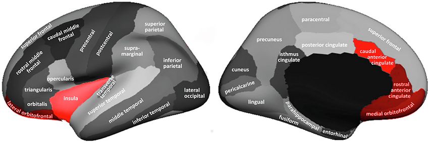
The following work is licensed under a Creative Commons: Attribution-NonCommercial-NoDerivatives
4.0 International (CC BY-NC-ND 4.0) License.

Originally published at:
Naaijen, Jilly; Mulder, Leandra M; Ilbegi, Shahrzad; de Bruijn, Sanne; Kleine-Deters, Renee; Dietrich,
Andrea; Hoekstra, Pieter J; Marsman, Jan-Bernard C; Aggensteiner, Pascal M; Holz, Nathalie E; Boet-
tinger, Boris; Baumeister, Sarah; Banaschewski, Tobias; Saam, Melanie C; M E Schulze, Ulrike; Santosh,
Paramala J; Sagar-Ouriaghli, Ilyas; Mastroianni, Mathilde; Castro Fornieles, Josefina; Bargallo, Nuria;
Rosa, Mireia; Arango, Celso; Penzol, Maria J; Werhahn, Julia E; Walitza, Susanne; Brandeis, Daniel;
Glennon, Jeffrey C; Franke, Barbara; Zwiers, Marcel P; Buitelaar, Jan K (2020). Specific cortical and
subcortical alterations for reactive and proactive aggression in children and adolescents with disruptive
behavior. NeuroImage: Clinical, 27:102344.
DOI: https://doi.org/10.1016/j.nicl.2020.102344

                                                   2
NeuroImage: Clinical 27 (2020) 102344

                                                                    Contents lists available at ScienceDirect

                                                                     NeuroImage: Clinical
                                                        journal homepage: www.elsevier.com/locate/ynicl

                                                                                                                                                                    T
Specific cortical and subcortical alterations for reactive and proactive
aggression in children and adolescents with disruptive behavior
Jilly Naaijena,b, , Leandra M Muldera,b, Shahrzad Ilbegia, Sanne de Bruijna,b,
                         ⁎

Renee Kleine-Detersc, Andrea Dietrichc, Pieter J Hoekstrac, Jan-Bernard C Marsmand,
Pascal M Aggensteinere, Nathalie E Holze, Boris Boettingere, Sarah Baumeistere,
Tobias Banaschewskie, Melanie C Saamf, Ulrike M E Schulzef, Paramala J Santoshg,h,
Ilyas Sagar-Ouriaghlig, Mathilde Mastroiannig, Josefina Castro Fornielesi, Nuria Bargalloi,j,
Mireia Rosai,j, Celso Arangok, Maria J Penzolk, Julia E Werhahnl, Susanne Walitzal,
Daniel Brandeise,l, Jeffrey C Glennona, Barbara Frankem,n, Marcel P Zwiersb, Jan K Buitelaara,o
a
  Radboud University Medical Center, Donders Institute for Brain, Cognition and Behavior, Department of Cognitive Neuroscience, Nijmegen, the Netherlands
b
  Radboud University, Donders Institute for Brain, Cognition and Behavior, Centre for Cognitive Neuroimaging, Nijmegen, the Netherlands
c
  University of Groningen, University Medical Center Groningen, Department of Child and Adolescent Psychiatry, Groningen, the Netherlands
d
  Cognitive Neuroscience Center, Department of Neuroscience, University Medical Center Groningen, Groningen, the Netherlands
e
  Department of Child and Adolescent Psychiatry and Psychotherapy, Central Institute of Mental Health, Medical Faculty Mannheim /Heidelberg University, Mannheim,
Germany
f
  Department of Child and Adolescent Psychiatry/Psychotherapy, University Hospital, University of Ulm, Germany
g
  Department of Child and Adolescent Psychiatry, King’s College London, London, UK
h
  Centre for Interventional Paediatric Psychopharmacology and Rare Diseases, South London and Maudsley NHS Foundation Trust, London, UK
i
  Child and Adolescent Psychiatry and Psychology Department, 2014SGR489, Institute Clinic of Neurosciences, Hospital Clinic of Barcelona, CIBERSAM, IDIBAPS,
Department of Medicine, University of Barcelona. Villarroel, 170, Barcelona 08036, Spain
j
  Clinic Image Diagnostic Center (CDIC), Hospital Clinic of Barcelona; Magnetic Resonance Image Core Facility, IDIBAPS, Barcelona, Spain
k
  Child and Adolescent Psychiatry Department, Hospital General Universitario Gregorio Marañón School of Medicine, Universidad Complutense, IiSGM, CIBERSAM,
Madrid, Spain
l
  Department of Child and Adolescent Psychiatry and Psychotherapy, Psychiatric Hospital, University of Zurich, Zurich, Switzerland
m
   Radboud University Medical Center, Donders Institute for Brain, Cognition and Behavior, Department of Human Genetics, Nijmegen, the Netherlands
n
  Radboud University Medical Center, Donders Institute for Brain, Cognition and Behavior, Department of Psychiatry, Nijmegen, the Netherlands
o
  Karakter Child and Adolescent Psychiatry University Center, Nijmegen, the Netherlands

A R T I C LE I N FO                                        A B S T R A C T

Keywords:                                                  Maladaptive aggression, as present in conduct disorder (CD) and, to a lesser extent, oppositional defiant disorder
Aggression subtypes                                        (ODD), has been associated with structural alterations in various brain regions, such as ventromedial prefrontal
Ventromedial prefrontal cortex                             cortex (vmPFC), anterior cingulate cortex (ACC), amygdala, insula and ventral striatum. Although aggression
Insula                                                     can be subdivided into reactive and proactive subtypes, no neuroimaging studies have yet investigated if any
Amygdala
                                                           structural brain alterations are associated with either of the subtypes specifically. Here we investigated asso-
Conduct disorder
                                                           ciations between aggression subtypes, CU traits and ADHD symptoms in predefined regions of interest.
                                                              T1-weighted magnetic resonance images were acquired from 158 children and adolescents with disruptive
                                                           behavior (ODD/CD) and 96 controls in a multi-center study (aged 8–18). Aggression subtypes were assessed by
                                                           questionnaires filled in by participants and their parents. Cortical volume and subcortical volumes and shape
                                                           were determined using Freesurfer and the FMRIB integrated registration and segmentation tool. Associations
                                                           between volumes and continuous measures of aggression were established using multilevel linear mixed effects
                                                           models.
                                                              Proactive aggression was negatively associated with amygdala volume (b = -10.7, p = 0.02), while reactive
                                                           aggression was negatively associated with insula volume (b = -21.7, p = 0.01). No associations were found with
                                                           CU traits or ADHD symptomatology. Classical group comparison showed that children and adolescents with

  ⁎
    Corresponding author at: Radboud University Medical Center, Donders Institute for Brain, Cognition and Behavior, Department of Cognitive Neuroscience, P.O.
box 9101, 6500 HB Nijmegen, the Netherlands.
    E-mail address: j.naaijen@donders.ru.nl (J. Naaijen).

https://doi.org/10.1016/j.nicl.2020.102344

Available online 11 July 2020
Received 3 February 2020; Received in revised form 10 June 2020; Accepted 7 July 2020

2213-1582/ Crown Copyright © 2020 Published by Elsevier Inc. This is an open access article under the CC BY-NC-ND license
(http://creativecommons.org/licenses/BY-NC-ND/4.0/).
J. Naaijen, et al.                                                                                                             NeuroImage: Clinical 27 (2020) 102344

                                                 disruptive behavior had smaller volumes than controls in (bilateral) vmPFC (p = 0.003) with modest effect size
                                                 and a reduced shape in the anterior part of the left ventral striatum (p = 0.005).
                                                    Our study showed negative associations between reactive aggression and volumes in a region involved in
                                                 threat responsivity and between proactive aggression and a region linked to empathy. This provides evidence for
                                                 aggression subtype-specific alterations in brain structure which may provide useful insights for clinical practice.

1. Introduction                                                                   extending to the ventrolateral prefrontal cortex (PFC)/orbitofrontal
                                                                                  cortex (OFC) and in the medial superior frontal gyrus extending to the
    Aggression, overt behavior with the intention of inflicting damage,            anterior cingulate cortex (ACC) with small-medium effect sizes (Rogers
is a behavioral trait with important roles throughout evolution in de-            and De Brito, 2016). Another meta-analysis including ODD/CD and
fense and predation. However, when expressed in humans in the wrong               ADHD studies (n = 415) reported reduced volumes of the amygdala,
context, aggression may lead to social maladjustment and crime.                   insula and frontal regions in ODD/CD as well, with greater reductions
Maladaptive aggression is commonly observed across childhood in                   in the presence of comorbid ADHD (Noordermeer et al., 2016). How-
disruptive behavioral disorders, in particular in conduct disorder (CD)           ever, other studies have not been able to find any group differences in
and to a lesser degree in oppositional defiant disorder (ODD). CD is               GM volume in these regions between participants with conduct pro-
defined as a repetitive and persistent pattern of behavior, which vio-             blems and controls or found opposite results with positive associations
lates the rights of others and major age-appropriate societal rules. ODD          between CU traits and insula volume and CD symptoms and amygdala
is characterized by a frequent and persistent pattern of irritable and            volume (Cohn et al., 2016; Holz et al., 2016).
angry mood, vindictiveness, and inappropriate and disobedient beha-                   In the present multi-center study, we investigated the association
vior toward authority figures (American Psychiatric Association, 2013).            between structural alterations and continuous measures of reactive and
Both disorders are highly comorbid with attention-deficit/hyperactivity            proactive aggression as well as CU traits in the largest sample of chil-
disorder (ADHD), which has been associated with aggressive behavior               dren/adolescents with disruptive behavior reported so far. We used pre-
as well (Saylor and Amann, 2016).                                                 selected regions of interest based on the previous meta-analyses and
    Subtyping of aggressive behavior is considered an important step              thus investigated ACC, insula, vmPFC, amygdala, and ventral striatum.
towards effective prevention and treatment strategies, as currently                We expected the vmPFC, ACC and insula volume to be associated with
available treatment options for maladaptive aggressive behavior have              reactive aggression. For the ventral striatum and amygdala, associa-
limited efficacy (Bakker et al., 2016; Waschbusch et al., 2007). A pro-             tions with proactive aggression were expected. We chose not to perform
mising subdivision, derived from animal studies, defines impulsive and             a whole brain VBM analysis to limit the number of independent tests
instrumental subtypes of aggression, also referred to as reactive and             (Focke et al., 2014), because surface-based morphometry measures
proactive aggression, respectively (Poulin and Boivin, 2000). Reactive            have been shown to be more robust across different MR scanners, as
aggression is thought to be associated with high arousal, impulsivity,            used in our multi-site design (Clarkson et al., 2011) and due to its
strong emotions and uncontrolled behavior. Animal studies have shown              higher sensitivity to capture subtle grey matter changes (Palaniyappan
that this form of aggression is mediated by a circuit that is responsive to       and Liddle, 2012; Winkler et al., 2010). We focused on analyses of the
threat (and frustration) and involves the amygdala (Haller, 2018).                volumes of these regions and subsequently investigated the shape of the
Furthermore, this circuit may be regulated by frontal cortical regions,           subcortical structures for subtler morphological changes and cortical
such as the ventromedial prefrontal cortex (vmPFC) and the anterior               thickness and surface area of the cortical areas.
cingulate cortex (ACC) (Blair, 2013). In contrast, proactive aggression is
hypothesized to be goal-directed, planned behavior associated with low            2. Methods and materials
arousal and higher levels of callous unemotional and/or psychopathic
traits. This form of aggression often goes hand in hand with impaired             2.1. Participants
stimulus-reinforcement learning (which involves the amygdala) com-
bined with impaired prediction error signaling (which involves the                    We included 277 participants (n = 176 cases and n = 101 healthy
striatum), leading to a poor understanding of the value of objects, cues          controls) aged 8–18 years who were recruited across nine sites in
and responses represented in the vmPFC as well as a lower empathy                 Europe (see supplementary material for details). Exclusion criteria for
level (Blair, 2013). Although the subdivision of reactive versus proac-           all participants were contraindications for MRI, an IQ < 80 and a
tive aggression is the most prevalent subdivision referred to in litera-          primary DSM-5 diagnosis of psychosis, bipolar disorder, major depres-
ture, it is so far not used clinically and no neuroimaging studies have           sion and/or anxiety disorder. Participants that were included as “cases”
yet investigated if any structural brain alterations are associated with          were diagnosed with conduct disorder (CD) and/or oppositional defiant
either of these aggression subtypes specifically in a clinical sample              disorder (ODD) and/or scored above the clinical cut-off for aggressive
(Yang et al., 2017). Prior research has focused on the presence or ab-            behavior and/or rule-breaking behavior as measured with the Child
sence of callous unemotional (CU) traits and/or childhood versus                  Behavior Checklist (CBCL) completed by parents (Bordin et al., 2013).
adolescent onset of CD in subtyping aggression. The strongest evidence            Within the control group no psychiatric disorders or scores within the
from such studies so far points to an involvement of the fronto-limbic-           clinical range were allowed, as determined by screening questionnaires
striatal circuitry in aggressive behavior (Blair, 2013).                          (CBCL). Participants that were using medication were at a stable dose
    Several studies have focused on structural abnormalities related to           for at least two weeks. Ethical approval for the study was obtained for
aggression, although not in consistent subgroups of disruptive dis-               all sites separately by local ethics committees. After description of the
orders. Almost all of these performed voxel-based morphometry (VBM)               study written informed consent was given by the participants and/or
analyses of grey matter associating these measures to either conduct              their parents.
problems, conduct disorder or CU traits (De Brito et al., 2009; Budhiraja
et al., 2017; Cohn et al., 2016; Fairchild et al., 2011). A recent meta-          2.2. Phenotypic information
analysis of thirteen VBM studies included almost 400 participants (aged
9–21 years) with conduct problems and showed that individuals with                   Clinical diagnoses of ODD, CD and possible comorbid ADHD were
conduct disorder/problems compared with controls had smaller grey                 confirmed by structured diagnostic interviews with both child and
matter (GM) volumes in the left amygdala, in the bilateral insula                 parents using the Kiddie Schedule for Affective Disorders and

                                                                              2
J. Naaijen, et al. NeuroImage: Clinical 27 (2020) 102344
Schizophrenia (K-SADS; Kaufman et al., 1997). Participants were ad- participants with externalizing disorders are more prone to motion
ministered a screening-module, followed, if needed, by application of artefacts, we used a rating system that has been described and thor-
disorder-specific modules. Aggressive/disruptive behavior was mea- oughly applied to an MRI data-set of children with ADHD and/or CD
sured by the aggressive behavior and rule-breaking behavior sub-scales before (Backhausen et al., 2016). Segmentation was visually inspected
of the CBCL (Bordin et al., 2013). The Reactive Proactive Aggression for all Freesurfer and FSL-first output for all scans. Scans of 18 cases and
Questionnaire (RPQ; Raine et al., 2006) completed by participants 5 healthy controls were excluded due to anxiety in the scanner (scan-
themselves was used to subtype aggressive behavior. The presence of session aborted) or due to poor data quality, which was based on ratings
CU traits was assessed by the Inventory of Callous Unemotional Traits of image sharpness, ringing, contrast to noise ratio of the subcortical
(ICU; Kimonis et al., 2008) completed by parents. A continuous mea- structures and of GM/WM. Total GM volume, total brain volume (TBV),
sure for ADHD symptoms was derived from the K-SADS by summing the amygdala and ventral striatum volumes are compared across in- and
number of inattention and hyperactivity/impulsivity symptoms. IQ was excluded participants in Figure S1.
estimated from four subtests (vocabulary, similarities, block design and
picture completion) of the Wechsler Intelligence Scale for Children III
or IV (Wechsler, 2002). Information about use of medication was col- 2.5. Statistical analyses
lected via parental report on the measurement day.
Statistical analyses were performed with the R statistical program (R
Team, 2013). Group distributions in sex were tested with Pearson’s chi-
2.3. MR acquisition and processing
squared test. Group differences in continuous demographic measures
were assessed with one-way analyses of variance (ANOVAs) or Kruskal-
MRI data-sets were acquired on 3 T scanners across nine different
Wallis rank sum tests when assumptions of homogeneity of variance
sites in Europe (see Table 1 for the scan parameters and Table S2 for
and normality of distributions were violated.
Vendor specifications). T1-weighted images were processed with the
We investigated whether volume of the ventral striatum, amygdala,
FMRIB Software Library (FSL; Smith et al., 2004) for subcortical volume
ACC, vmPFC and insula were related to continuous measures of ag-
and shapes and with Freesurfer v5.3.0 (http://surfer.nmr.mgh.harvard.
gression and to CU-traits by using linear mixed effects models in R with
edu) for measures of cortical volumes and cortical thickness (CT) and
a maximum likelihood fit (lme4 package; (Bates et al., 2015)) with
surface area (SA; Dale et al., 1999; Desikan et al., 2006). Subcortical
effect sizes being presented as “r”. For the associations between vo-
segmentation was performed with the automated FMRIB integrated
lumes and continuous measures of aggression, assumptions for line-
registration and segmentation tool (FIRST; Patenaude et al., 2011)
arity, homogeneity of variance and normality of residuals were met.
which included affine registration to MNI-space followed by a seg-
Hemisphere was used as within-subjects’ factor and the respective
mentation procedure integrating both shape and intensity information
continuous measure as between-subject’s variable of interest. Age, sex
for accurate segmentation of subcortical structures, including the bi-
and TBV were added as possible confounders of non-interest and par-
lateral ventral striatum and amygdala. Volumes of these respective
ticipant as random factor to account for within subject variability across
regions were extracted for statistical analysis. Vertex analysis was
hemispheres. Since reactive and proactive aggression are highly cor-
performed with FIRST_utils to determine shape. A multivariate Gaus-
related (r = 0.69 in this sample), any significant associations between
sian model of the location and intensity variation of the vertex was used
either of them with volumes were controlled for the effect of the other.
to generate surface meshes. Localized shape differences using the 3D-
Due to the skewed distribution of reactive and proactive aggression
coordinates of the corresponding vertices with vertex-wise F-statistics
scores in the control group, we additionally repeated analyses with
were calculated after alignment to the average shape of the cohort and
significant results in the cases only group and investigated whether
removal of global scaling (useRigidAlign and useScale). Cortical re-
diagnostic group affected the results by adding this to the model.
construction was performed in FreeSurfer using the Desikan-Killiany
We also investigated traditional cases-control differences in volume
atlas (see Fig. 1). CT was calculated for each vertex on the reconstructed
and shape, and post-hoc CT and SA of the same regions to better
cortical sheet and was defined as the closest distance between the grey/
compare our results to previous studies (with a larger sample), using
white matter boundary and the GM/CSF boundary (Fischl and Dale,
the same linear mixed effects model replacing the continuous measure
2000). SA was measured at the geometric middle of the inner and outer
of aggression with diagnostic status. As part of the supplemental ma-
cortical surfaces. CT, SA and volume of the ACC, vmPFC and insula
terial we additionally report whole-brain analyses results for these case-
were extracted from the parcellations and segmentations using the
control comparisons. All p-values of the continuous measures and di-
‘mri_segstats’ function.
agnostic status on (sub)cortical volumes only are corrected for multiple
comparisons using the false discovery rate (FDR) of q < 0.05. Analyses
2.4. Quality control of thickness and surface area were considered post-hoc tests and were
not corrected for multiple comparisons. Possible effects of scan-site on
T1-weighted images and segmentation were all visually inspected these analyses are shown in the supplementary material.
and evaluated by an experienced rater (JN). Since images of Statistical shape analyses based on the vertex-wise F-statistic of the
Table 1
Scan parameters for the T1-scan across the different sites.
Scanner Site TR*/TE/T1 (ms) Flip angle Field of view Matrix RL/AP/slices Voxel size (mm) Acceleration factor
Siemens Nijmegen 2300/2.98/900 9 256 212/256/176 1.0 × 1.0 × 1.2 2
Mannheim 2300/2.96/900 9 256 212/256/176 1.0 × 1.0 × 1.2 2
Ulm 2300/2.96/900 9 256 212/256/176 1.0 × 1.0 × 1.2 2
Barcelona 2300/2.98/900 9 256 212/256/176 1.0 × 1.0 × 1.2 2
Madrid 2300/2.98/900 9 256 212/256/176 1.0 × 1.0 × 1.2 2
Rome 2300/2.86/900 9 256 212/256/176 1.0 × 1.0 × 1.2 2
Philips Groningen 6.69/3.11/900 8 270 256/232/170 1.0 × .1.0 × 1.0 1.8
Zurich 6.69/3.11/900 9 270 256/232/170 1.0 × 1.0 × 1.0 1.8
GE London 7.31/3.02/400 11 270 256/256/196 1.0 × 1.0 × 1.2 1.75
* As provided by the manufacturer.
3J. Naaijen, et al. NeuroImage: Clinical 27 (2020) 102344
Fig. 1. Regions included in the cortical volume analyses. ACC consisted of rostral and caudal anterior cingulate cortex; vmPFC consisted of lateral- and medial
orbitofrontal cortex (Boes et al., 2009).
bilateral ventral striatum and amygdala structures were performed aggression (all p-values > 0.05). There was no effect of CU-traits on
using FSL randomize (Winkler et al., 2014) with 5000 random per- any of the cortical or subcortical volumes (all p-values > 0.05). There
mutations and threshold-free cluster enhancement (Smith and Nichols, was also no effect of any of the continuous measures of aggression on
2009). Bonferroni corrections were used for multiple comparisons ventral striatum or amygdala shape.
corrections for testing the shape of multiple structures The effect of covariates and the model outputs are described in the
(pcorrected = 0.01). For illustrative purposes, we also performed classical Supplementary Material (See Table S3 for the proactive aggression
vertex analysis containing vectors displaying the direction of shape model and Table S4 for the reactive aggression model).
alterations.
3. Results
Table 2
3.1. Demographics Demographic and clinical characteristics (n = 254).
Case Control Test statistic p-value
Due to the exclusion of 23 participants, our final sample consisted of (n = 158) n = 96)
254 participants (n = 158 cases, 130 male; and n = 96 controls, 55
male). Out of the 158 cases, 59 were diagnosed with ODD, 11 with CD N N
Sex m/f 130/28 55/41 χ2 = 17.60 < 0.001
and 42 with both ODD and CD. The other 46 participants were included
Mean (SD) Mean
as “case” based on a CBCL aggression and/or rule-breaking behavior (SD)
subscale T-score of ≥ 70. Among the cases, 44 participants were di- Age 13.0 (2.8) 13.5 (2.6) K-Wχ2 = 2.33 0.13
a
agnosed with comorbid ADHD. Table 2 provides a summary of the IQ 99.8 (11.3) 106.5 F = 21.41 < 0.001
(10.6)
demographic and clinical information. Table S1 provides demographic
Reactive aggression 12.0 (4.6) 5.8 (3.4) K-Wχ2 = 93.55 < 0.001
information across the different scan-sites. Proactive aggression 4.6 (4.4) 0.8 (1.4) K-Wχ2 = 79.42 < 0.001
ICU total score 33.4 (9.9) 21.0 (8.5) K-Wχ2 = 77.97 < 0.001
b
3.2. Continuous measures ODD symptom 3.7 (2.4) –
counts
b
CD symptom counts 1.8 (2.4) –
We found an effect of proactive aggression on amygdala volume, b
ADHD
where an increase in proactive aggression was associated with smaller - Inattention 3.4 (3.0) –
amygdala volume (b = -10.7, t(348.3) = -2.27, p = 0.02, r = 0.14) in - Hyperactivity/ 3.0 (2.9) –
our entire sample. This association remained present after controlling impulsivity
CBCL Aggression T- 64.7 (25.9) 46.2 K-Wχ2 = 72.19 < 0.001
for reactive aggression (b = -14.1, t(302.7) = -2.29, p = 0.02, score (17.2)
r = 0.14). However, when investigating cases only (n = 158), this CBCL Rule-breaking 61.7 (17.0) 46.3 2
K-W χ = 72.53 < 0.001
association became non-significant (b = -9.37, t(219.5) = -1.67, T-score (16.7)
c
p = 0.09, r = 0.11). Medication use
- Stimulants 66 –
Reactive aggression was associated with the volume of the insula
- Antipsychotics 37 –
(b = -21.7, t(360.5) = -2.51, p = 0.01 r = 0.13), where more reactive - Antidepressants 4 –
aggression was associated with smaller volumes, also when controlled - dOther 7 –
for the effect of proactive aggression (b = -26.5, t(309.9) = -2.35,
p = 0.02, r = 0.13; see Fig. 2). The associations were also present when ADHD, attention-deficit/hyperactivity disorder; CBCL, Child Behavior
investigating the cases only (n = 158; b = –32.60, t(218.7) = -2.53, Checklist; CD, conduct disorder; ICU, Inventory of Callous Unemotional Traits;
K-W, Kruskal-Wallis; m/f, male/female; ODD, oppositional defiant disorder; SD,
p = 0.01, r = 0.17). Adding diagnostic group to the model did not
standard deviation;
change the effect of proactive aggression on amygdala volume (b = -
a IQ estimated from a subset of the Wechsler Intelligence Scale for Children III
11.7, t(331.1) = -2.29, p = 0.02) or the effect of reactive aggression on (Wechsler, 2002).
insula volume (b = –22.82, t(337.2) = -2.27, p = 0.02). b As measured with the K-SADS (Kaufman et al., 1997).
No effect of hemisphere or any interactions between hemisphere cMedication use was determined by parental report.
and reactive aggression were observed. When investigating CT and SA d Other medications included mood-stabilizers (Lithium), anti-epileptic medi-
of the insula separately, we did not find an association with reactive cation and benzodiazepines.
4J. Naaijen, et al. NeuroImage: Clinical 27 (2020) 102344
Fig. 2. Negative association between proactive aggression and total amygdala volume (A) and between reactive aggression and total insula volume (B). The thick
black line represents associations across the entire sample. For illustrative purposes the associations for cases (dark-grey) and controls (light-grey) are shown as well.
3.3. Case-control comparisons difference in one anterior cluster of voxels (voxel-coordinates: x = 102,
y = 143, z = 69, size = 28 voxels). This difference reflects a regional
In our case-control comparisons we found that cases showed a sig- decreased shape of the anterior part of the left striatum in cases com-
nificantly smaller volume of the vmPFC than controls (b = 572.4, t pared to controls. No shape differences between cases and controls were
(305.6) = -3.02, p = 0.003, r = 0.17). No other group differences were found for the amygdala.
found regarding volume, CT or SA in any of the regions (all p-va-
lues > 0.05). See Table S5 of the model output and the supplemental
text for the effects of covariates. 4. Discussion
Shape analyses of the subcortical regions revealed differences in the
left (but not right) ventral striatum (Fig. 3), showing an overall inward The current study investigated whether structural brain alterations
position of the vertices (corrected p-value of 0.005) with the largest in regions of interest chosen on the basis of two meta-analyses were
differentially associated with reactive and proactive subtypes of
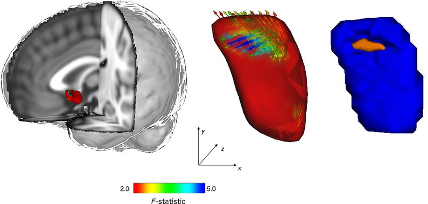
Fig. 3. Vertex analyses of shape alterations in the left ventral striatum. Left panel shows the anatomical location of the area and the local areas exhibiting shape
change (alterations in color). The middle panel shows shape changes in the ODD/CD participants compared with controls with vector directions. Inward direction
represents relative inward position of the vertices, pointing to regional decreased shapes. The color of the surface and the arrows indicate the Pillai’s trace F-statistic
in the middle panel. The right panel shows the results analyzed with the vertex-wise F-statistic. The region in orange corresponds to the anterior part of the ventral
striatum shown to be smaller in cases than in controls (which resembles the dark blue arrows in the middle panel).
5J. Naaijen, et al. NeuroImage: Clinical 27 (2020) 102344
aggression. Our main finding is that we could confirm these differential participants with aggression scores in the clinical range on the CBCL
associations by observing reactive aggression to be associated with who did not fulfil all diagnostic criteria for ODD and/or CD. Especially
smaller insula volume and proactive aggression with smaller amygdala the CD group was relatively small. In addition, the amount of ODD and
volume, although not with the other regions of interest and with rela- CD symptoms in our cases group were relatively small. This may have
tively small effect sizes. These associations, however, survived ex- caused heterogeneity in our cases, thus reducing symptom severity and
tensive controlling for possible confounding variables. No effects of CU may be reflected in our more pronounced findings of reduced volume in
traits were found in any of the regions. In addition, cases showed areas that are less specific for CU traits and lack of empathy types of
smaller volume of the vmPFC compared with healthy controls and aggression (ACC, vmPFC and striatum). However, reducing the sample
differences regarding shape of the left ventral striatum. Participants to only cases with a clinical diagnosis did not change our results.
with disruptive behavior only showed subtle differences with controls. Second, the male/female ratio was different for cases and controls
However, small neuroanatomic abnormalities may nevertheless have which is inherent to the ratio of males/females that are diagnosed with
implications for behavior (Hoogman et al., 2017); here we showed a these types of disorders. There was, however, no effect of sex on any of
first association with aggression subtypes, implicating effects on func- our results. The variance induced by the multi-site setup may further
tioning. have diluted some of our findings. However, investigating in multiple
Aggression and conduct problems have been associated with several centers also allowed us to have a larger sample size compared to many
neurocognitive dysfunctions moderated by the presence or absence of previous studies and facilitated in generalizing our findings. Our sample
psychopathic or CU traits (Blair et al., 2014). Higher levels of conduct of controls showed very little variation in aggressive behaviour, pos-
problems or CU-traits (often associated with lower empathy) has been sibly reducing our power for dimensional analyses. However, in-
linked to reduced amygdala response to fear, sadness and pain (Blair vestigating only cases as well as the full sample resulted in the same
et al., 2004; Jones et al., 2009), and to reduced amygdala volume associations between amygdala volume and proactive aggression (al-
(Cardinale et al., 2019; Fairchild et al., 2011, 2013; Pardini et al., though non-significant) and insula volume and reactive aggression. The
2014). However, we did not find an association between amygdala two measures of aggression are further highly associated with each
volume and CU-traits, but with proactive aggression, another severe other, but controlling the effect of one for the effect of the other did not
form of aggression often associated with high CU-traits. Despite pre- change our results.
vious evidence for proactive aggression involving the striatum and In conclusion, the current study showed a negative relation between
vmPFC (Yang et al., 2017), related to impaired prediction error sig- proactive aggression and amygdala volume and between reactive ag-
naling and impaired decision making, we did not find an association gression and insula volume (after controlling for several confounding
between proactive aggression and the volumes of these regions in our variables) and a decreased volume of the vmPFC in children and ado-
sample. lescents with disruptive behaviour compared with controls. Our find-
Decision making and empathy are highly dependent on each other, ings support the idea of subtype-specific impairments in aggression,
where learning and representing the valence of objects and actions is where different brain regions are involved in empathy, threat response
critical in deciding whether to respond empathically, and is related to and decision making which are in turn more associated with either
dysfunction of not only striatum, amygdala and vmPFC, but also insula proactive or reactive aggression. This may have implications for de-
(Fanning et al., 2017; O’Doherty, 2012). In the current study, we found signing targeted intervention strategies, which needs to be further ex-
a negative relation between reactive aggression and insula volume, a plored in future studies.
region assumed to be associated with responses to threat and frustra-
tion. Our result is in line with studies that report the amygdala to be Acknowledgments
associated with fear and sadness while the insula is more responsive to
angry faces (Fusar-Poli et al., 2009; Passamonti et al., 2010). A reduced This project has received funding from the European Union’s
insula volume in association with more reactive aggression may explain Seventh Framework Programme for research, technological develop-
a deficit in responding to threatening stimuli. Deficits in the insula have ment and demonstration under grant agreement no 602805
also been associated with poor decision making by relating outcome (Aggressotype) and 603016 (MATRICS). This work reflects only the
information (reward/punishment) wrongly to responding which in turn authors’ views and the European union is not liable for any use that may
increases conduct problems (Blair, 2013; Dambacher et al., 2013; Frick be made of the information contained herein. We gratefully acknowl-
and White, 2008). edge and thank all the participants and their families for their en-
Our findings of reduced vmPFC volume in disruptive behaviour are thusiastic participation in the study. The authors would also like to
in accordance with the idea of reduced empathy, as this region is as- thank all PhD students, post-docs and research assistants for their in-
sociated with responses to distress cues (Dawel et al., 2012). The al- volvement in data-collection.
terations in the shape of the left ventral striatum has not been reported
in aggression before and localizes a possible shape difference to the Conflict of interest
anterior part of the ventral striatum, although no volume differences
were reported. Our differential findings of associations between T Banaschewski served in an advisory or consultancy role for
amygdala and insula volume and respectively proactive and reactive Actelion, Hexal Pharma, Lilly, Medice, NovartisOxford outcomes, PCM
aggression and group differences in vmPFC and ventral striatum reflect scientific, Shire and Viforpharma. He received conference support or
possible state (diagnosis) versus trait (continuous measures) effects speaker’s fee by Medice, Novartis and Shire. He is/has been involved in
which need replication in future studies that include both types of clinical trials conducted by Shire & Viforpharma. The present work is
analyses. unrelated to the grants and relationships noted earlier. U. Schulze re-
We did not find any association between the volume of any of our ceived a speaker’s fee from Shire and serves as an unpayed ethics ad-
regions of interest and the severity of CU traits. This may be due to the visor in two EU-funded projects which are not related to the present
relatively low levels of CU traits in our sample compared to other stu- work. C Arango has been a consultant to or has received honoraria or
dies; the group of children and adolescents in our study seem to show grants from Acadia, Ambrosseti, Caja Navarra, CIBERSAM, Fundación
more reactive (impulsive) forms of aggression. Alicia Koplowitz, Forum, Instituto de Salud Carlos III, Gedeon Richter,
Important strengths of the current study are the investigation of Janssen Cilag, Lundbeck, Merck, Ministerio de Ciencia e Innovación,
reactive and proactive aggression in a large clinical sample in relation Ministerio de Sanidad, Ministerio de Economía y Competitividad,
to structural brain alterations that was achieved because of a multi-site Mutua Madrileña, Otsuka, Roche, Servier, Shire, Schering Plough,
design. There were, however, also some limitations. First, we included Sumitomo Dainippon Pharma, Sunovio and Takeda. D Brandeis serves
6J. Naaijen, et al. NeuroImage: Clinical 27 (2020) 102344
as an unpaid scientific advisor for an EU-funded Neurofeedback trial Fairchild, G., Passamonti, L., Hurford, G., Hagan, C.C., Von Dem Hagen, E.A.H., Van
unrelated to the present work. JC Glennon has acted as a consultant for Goozen, S.H.M., Goodyer, I.M., et al., 2011. Brain structure abnormalities in early-
onset and adolescent-onset conduct disorder. Am. J. Psychiatry 168 (6), 624–633.
Boehringer Ingelheim GmbH. B Franke received an educational Fanning, J.R., Keedy, S., Berman, M.E., Lee, R., Coccaro, E.F., 2017. Neural Correlates of
speaking fee from Shire and Medice. JK Buitelaar has been consultant Aggressive Behavior in Real Time: a Review of fMRI Studies of Laboratory Reactive
to/member of advisory board of and/or speaker for Janssen Cilag BV, Aggression. Retrieved from. Current Behavioral Neuroscience Reports 4 (2),
138–150. http://link.springer.com/10.1007/s40473-017-0115-8.
Eli Lilly, Bristol-Myer Squibb, Shering Plough, UCB, Shire, Novartis and Fischl, B., Dale, A.M., 2000. Measuring the thickness of the human cerebral cortex from
Servier. He is not an employee of any of these companies, nor a stock magnetic resonance images. Proc. Natl. Acad. Sci. 97 (20), 11050–11055.
shareholder of any of these companies. He has no other financial or Focke, N.K., Trost, S., Paulus, W., Falkai, P., Gruber, O., 2014. Do manual and voxel-based
morphometry measure the same? A proof of concept study. Frontiers. Psychiatry 5
material support, including expert testimony, patents, and royalties. (APR), 1–7.
The other authors do not report any biomedical financial interests or Frick, Paul J., White, Stuart F., 2008. Research Review: The importance of callous-un-
potential conflicts of interest. emotional traits for developmental models of aggressive and antisocial behavior. J
Child Psychol & Psychiat 49 (4), 359–375. https://doi.org/10.1111/j.1469-7610.
2007.01862.x.
Appendix A. Supplementary data Fusar-Poli, P., Placentino, A., Carletti, F., Landi, P., Allen, P., Surguladze, S., Benedetti, F.,
et al., 2009. Functional atlas of emotional faces processing: A voxel-based meta-
Supplementary data to this article can be found online at https:// analysis of 105 functional magnetic resonance imaging studies. Journal of Psychiatry
and Neuroscience.
doi.org/10.1016/j.nicl.2020.102344. Haller, J., 2018. The role of central and medial amygdala in normal and abnormal ag-
gression: A review of classical approaches. Neurosci. Biobehav. Rev.
References Holz, N.E., Boecker-Schlier, R., Buchmann, A.F., Blomeyer, D., Jennen-Steinmetz, C.,
Baumeister, S., Plichta, M.M., et al., 2016. Ventral striatum and amygdala activity as
convergence sites for early adversity and conduct disorder. Social Cognitive and
American Psychiatric Association, 2013. Diagnostic and statistical manual of mental Affective Neuroscience 12, nsw120 Retrieved from https://academic.oup.com/scan/
disorders (5th ed). Author, Washington, DC. article-lookup/doi/10.1093/scan/nsw120.
Backhausen, L.L., Herting, M.M., Buse, J., Roessner, V., Smolka, M.N., Vetter, N.C., 2016. Hoogman, Martine, Buitelaar, Jan K, Faraone, Stephen V, Shaw, Philip, Franke, Barbara,
Quality control of structural MRI images applied using FreeSurfer-a hands-on 2017. Subcortical brain volume differences in participants with attention deficit
workflow to rate motion artifacts. Front. Neurosci. 10 (DEC), 1–10. hyperactivity disorder in children and adults – Authors' reply. The Lancet Psychiatry
Bakker, M.J., Greven, C.U., Buitelaar, J.K., Glennon, J.C., 2016. Practitioner Review: 4 (6), 440–441. https://doi.org/10.1016/S2215-0366(17)30200-6.
Psychological treatments for children and adolescents with conduct disorder pro- Jones, A.P., Laurens, K.R., Herba, C.M., Barker, G.J., Viding, E., 2009. Amygdala hy-
blems - a systematic review and meta-analysis. Retrieved from. J. Child Psychol. poactivity to fearful faces in boys with conduct problems and callous-unemotional
Psychiatry 1, 4–18. http://doi.wiley.com/10.1111/jcpp.12590. traits. Am. J. Psychiatry 166 (1), 95–102.
Bates, D., Mächler, M., Bolker, B.M., Walker, S.C., 2015. Fitting linear mixed-effects Kaufman, J., Birmaher, B., Brent, D., Rao, U., Flynn, C., Moreci, P., Williamson, D., et al.,
models using lme4. J. Stat. Softw. 1997. Schedule for Affective Disorders and Schizophrenia for School-Age Children
Blair, R.J.R., Mitchell, D.G.V., Peschardt, K.S., Colledge, E., Leonard, R.A., Shine, J.H., Present and Lifetime version (K-SADS-PL): Initial reliability and validity data. J. Am.
Murray, L.K., et al., 2004. Reduced sensitivity to others’ fearful expressions in psy- Acad. Child Adolesc. Psychiatry 36, 980–988.
chopathic individuals. Personality Individ. Differ. 37 (6), 1111–1122. Kimonis, E.R., Frick, P.J., Skeem, J.L., Marsee, M.A., Cruise, K., Munoz, L.C., Aucoin, K.J.,
Blair, R. James R., 2013. The neurobiology of psychopathic traits in youths. Nat. Rev. et al., 2008. Assessing callous-unemotional traits in adolescent offenders: Validation
Neurosci. 14 (11), 786–799 Retrieved from http://www.nature.com/doifinder/ of the Inventory of Callous-Unemotional Traits. Int. J. Law Psychiatry 31 (3),
10.1038/nrn3577. 241–252.
Blair, R.J.R., Leibenluft, E., Pine, D.S., 2014. Conduct Disorder and Callous-Unemotional Noordermeer, S.D.S., Luman, M., Oosterlaan, J., 2016. A Systematic Review and Meta-
Traits in Youth. N. Engl. J. Med. 371 (23), 2207–2216 Retrieved from http:// analysis of Neuroimaging in Oppositional Defiant Disorder (ODD) and Conduct
www.nejm.org/doi/10.1056/NEJMra1315612. Disorder (CD) Taking Attention-Deficit Hyperactivity Disorder (ADHD) Into Account.
Boes, A.D., Bechara, A., Tranel, D., Anderson, S.W., Richman, L., Nopoulos, P., 2009. Neuropsychol. Rev. 26 (1), 44–72.
Right ventromedial prefrontal cortex: A neuroanatomical correlate of impulse control O’Doherty, J.P., 2012. Beyond simple reinforcement learning: The computational neu-
in boys. Social Cognitive and Affective Neuroscience 4 (1), 1–9. robiology of reward-learning and valuation. Eur. J. Neurosci. 35 (7), 987–990.
Bordin, I.A., Rocha, M.M., Paula, C.S., Teixeira, M.C.T.V., Achenbach, T.M., Rescorla, Palaniyappan, L., Liddle, P.F., 2012. Differential effects of surface area, gyrification and
L.a., Silvares, E.F.M., 2013. Child Behavior Checklist (CBCL), Youth Self-Report (YSR) cortical thickness on voxel based morphometric deficits in schizophrenia.
and Teacher’s Report Form(TRF): an overview of the development of the original and NeuroImage.
Brazilian versions. Cadernos de saúde pública 29 (1), 13–28. Pardini, D.A., Raine, A., Erickson, K., Loeber, R., 2014. Lower amygdala volume in men is
De Brito, S.A., Mechelli, A., Wilke, M., Laurens, K.R., Jones, A.P., Barker, G.J., Hodgins, associated with childhood aggression, early psychopathic traits, and future violence.
S., et al., 2009. Size matters: Increased grey matter in boys with conduct problems Elsevier. Retrieved from. Biol. Psychiatry 75 (1), 73–80. https://doi.org/10.1016/j.
and callousunemotional traits. Brain 132 (4), 843–852. biopsych.2013.04.003.
Budhiraja, Meenal, Savic, Ivanka, Lindner, Philip, Jokinen, Jussi, Tiihonen, Jari, Hodgins, Passamonti, L., Fairchild, G., Goodyer, I.M., Hurford, G., Hagan, C.C., Rowe, J.B., Calder,
Sheilagh, 2017. Brain structure abnormalities in young women who presented con- A.J., 2010. Neural abnormalities in early-onset and adolescence-onset conduct dis-
duct disorder in childhood/adolescence. Cogn Affect Behav Neurosci 17 (4), order. Arch. Gen. Psychiatry 67 (7), 729–738.
869–885. https://doi.org/10.3758/s13415-017-0519-7. Patenaude, B., Smith, S.M., Kennedy, D.N., Jenkinson, M., 2011. A Bayesian model of
Cardinale, E.M., O’Connell, K., Robertson, E.L., Meena, L.B., Breeden, A.L., Lozier, L.M., shape and appearance for subcortical brain segmentation. NeuroImage 56 (3),
VanMeter, J.W., et al., 2019. Callous and uncaring traits are associated with reduc- 907–922.
tions in amygdala volume among youths with varying levels of conduct problems. Poulin, F., Boivin, M., 2000. Reactive and proactive aggression: Evidence of a two-factor
Psychol. Med. 49 (9), 1449–1458. model. Psychol. Assess. 12 (2), 115–122.
Clarkson, M.J., Cardoso, M.J., Ridgway, G.R., Modat, M., Leung, K.K., Rohrer, J.D., Fox, Raine, A., Dodge, K., Loeber, R., Gatzke-Kopp, L., Lynam, D., Reynolds, C., Stouthamer-
N.C., et al., 2011. A comparison of voxel and surface based cortical thickness esti- Loeber, M., et al., 2006. The reactive-proactive aggression questionnaire: Differential
mation methods. NeuroImage 57 (3), 856–865. correlates of reactive and proactive aggression in adolescent boys. Aggressive
Cohn, M.D., Viding, E., McCrory, E., Pape, L., van den Brink, W., Doreleijers, T.A.H., Behavior 32 (2), 159–171.
Veltman, D.J., et al., 2016. Regional grey matter volume and concentration in at-risk Rogers, J.C., De Brito, S.A., 2016. Cortical and Subcortical Gray Matter Volume in Youths
adolescents: Untangling associations with callous-unemotional traits and conduct With Conduct Problems. Retrieved from. JAMA Psychiatry 73 (1), 64. http://
disorder symptoms. Elsevier. Retrieved from. Psychiatry Research - Neuroimaging archpsyc.jamanetwork.com/article.aspx?doi=10.1001/jamapsychiatry.2015.2423.
254, 180–187. https://doi.org/10.1016/j.pscychresns.2016.07.003. Saylor, K.E., Amann, B.H., 2016. Impulsive Aggression as a Comorbidity of Attention-
Dale, A.M., Fischl, B., Sereno, M.I., 1999. Cortical surface-based analysis: I. Segmentation Deficit/Hyperactivity Disorder in Children and Adolescents. Retrieved from. J. Child
and surface reconstruction. NeuroImage 9 (2), 179–194. Adolesc. Psychopharmacol. 26 (1), 19–25. http://online.liebertpub.com/doi/10.
Dambacher, F., Sack, A.T., Lobbestael, J., Arntz, A., Brugman, S., Schuhmann, T., 2013. 1089/cap.2015.0126.
Out of control: Evidence for anterior insula involvement in motor impulsivity and Smith, S.M., Jenkinson, M., Woolrich, M.W., Beckmann, C.F., Behrens, T.E.J., Johansen-
reactive aggression. Social Cognitive and Affective Neuroscience 10 (4), 508–516. Berg, H., Bannister, P.R., et al., 2004. Advances in functional and structural MR
Dawel, A., O’Kearney, R., McKone, E., Palermo, R., 2012. Not just fear and sadness: Meta- image analysis and implementation as FSL. NeuroImage Vol, 23).
analytic evidence of pervasive emotion recognition deficits for facial and vocal ex- Smith, S.M., Nichols, T.E., 2009. Threshold-free cluster enhancement: Addressing pro-
pressions in psychopathy. Neurosci. Biobehav. Rev. blems of smoothing, threshold dependence and localisation in cluster inference.
Desikan, R.S., Ségonne, F., Fischl, B., Quinn, B.T., Dickerson, B.C., Blacker, D., Buckner, NeuroImage 44 (1), 83–98.
R.L., et al., 2006. An automated labeling system for subdividing the human cerebral Team, R., 2013. R Development Core Team. R: A Language and Environment for Statistical.
cortex on MRI scans into gyral based regions of interest. NeuroImage 31 (3), Computing 55, 275–286.
968–980. Waschbusch, D.A., Carrey, N.J., Willoughby, M.T., King, S., Andrade, B.F., 2007. Effects
Fairchild, G., Hagan, C.C., Walsh, N.D., Passamonti, L., Calder, A.J., Goodyer, I.M., 2013. of methylphenidate and behavior modification on the social and academic behavior
Brain structure abnormalities in adolescent girls with conduct disorder. J. Child of children with disruptive behavior disorders: The moderating role of callous/un-
Psychol. Psychiatry 54 (1), 86–95. emotional traits. Retrieved from. Journal of Clinical Child and Adolescent
7J. Naaijen, et al. NeuroImage: Clinical 27 (2020) 102344
Psychology 36 (4), 629–644. 2009.12.028.
Wechsler, D., 2002. WISC-III Handleiding. The Psychological Corporation, London. Winkler, A.M., Ridgway, G.R., Webster, M.A., Smith, S.M., Nichols, T.E., 2014.
Winkler, Anderson M., Kochunov, Peter, Blangero, John, Almasy, Laura, Zilles, Karl, Fox, Permutation inference for the general linear model. NeuroImage 92, 381–397.
Peter T., Duggirala, Ravindranath, Glahn, David C., 2010. Cortical thickness or grey Yang, Y., Joshi, S.H., Jahanshad, N., Thompson, P.M., Baker, L.A., 2017. Neural correlates
matter volume? The importance of selecting the phenotype for imaging genetics of proactive and reactive aggression in adolescent twins. Aggressive Behavior 43 (3),
studies. NeuroImage 53 (3), 1135–1146. https://doi.org/10.1016/j.neuroimage. 230–240.
8You can also read