Stress Echo Comprehensive Software (SECS) for state-of-art ABCDE protocol - Dialnet
←
→
Page content transcription
If your browser does not render page correctly, please read the page content below
SPECIAL ARTICLE

Stress Echo Comprehensive Software (SECS) for state-of-art ABCDE
protocol
Software integral de eco estrés para protocolo ABDCE de última generación

MARCO PATERNI1, CLARA CARPEGGIANI1, QUIRINO CIAMPI2, RODOLFO CITRO3, FRANCESCO ANTONINI-CANTERIN4, PAOLO COLONNA5,
EUGENIO PICANO1, ON BEHALF OF THE STRESS-ECHO 2020 STUDY GROUP OF THE ITALIAN SOCIETY OF ECHOCARDIOGRAPHY AND
CARDIOVASCULAR IMAGING (SIECVI)

ABSTRACT

Background: Several specialized softwares are commercially available for the elective storage of stress echo (SE) data. State-of-the-
art SE is based upon novel parameters in addition to regional wall motion.
Objective: To develop a novel software for SE data storage and reporting.
Methods: We developed the prototype of a SE Comprehensive Software (SECS) with a minimum data set eventually allowing stand-
ardized collection of data. The software runs with medium-low performance computers as well as with the most popular operating
systems (Windows, MAC OS and Linux). The export functions towards widely accepted formats allow easy data sharing. The soft-
ware is able to generate a customized report which can be expanded in PDF and comma-separated values formats.
Results: The program prototype data entry requires < 2 min per study. The main pages focus on the 5 steps of ABCDE-SE: step A
(regional wall motion); step B (B-lines with 4-site simplified scan); step C (contractile reserve with force derived from systolic blood
pressure and end-systolic volume); step D (Doppler-based coronary flow velocity reserve in left anterior descending coronary artery);
step E (EKG-based chronotropic reserve measured as peak/rest heart rate). The final page graphically summarizes the ABCDE in-
formation in a risk prediction model (cardiac death rate per year, from low risk < 1% to high risk > 3 %).
Conclusion: SECS may provide a suitable infrastructure for an advanced clinical and research application, with simple graphic for-
mat and convenient reporting option. It may represent a trade-off between exhaustive information required by scientific standards
and smooth workflow priority of busy, high volume, clinically-driven activities. Large scale validation and adaptation from users’
feedback is necessary prior to dissemination on demand.
Key words: Echocardiography, Stress – Software

RESUMEN

Introducción: Existen diversos softwares especializados en el mercado para el almacenamiento electivo de datos de eco estrés (EE).
El EE de última generación incorpora nuevos parámetros además de la motilidad parietal.
Objetivo: Desarrollar un nuevo software para el almacenamiento de datos e informe de EE.
Métodos: Desarrollamos el prototipo de Software Integral de EE (SIEE) con un conjunto mínimo de datos que permite la eventual
recolección estandarizada de datos. El software corre en computadoras con capacidad de trabajo mediana-baja y con los sistemas
operativos más usados (Windows, MAC OS y Linux). Las funciones de exportación hacia formatos altamente aceptados permiten
compartir los datos fácilmente. El software es capaz de generar un informe personalizado que se puede expandir en PDF y en forma-
tos de valores separados por comas.
Resultados: El ingreso de datos en el programa prototipo requiere menos de 2 minutos por estudio. Las páginas principales se
concentran en las 5 fases ABCDE del EE: fase A (motilidad parietal regional); fase B (líneas B con escaneo simplificado de 4 sitios);
fase C (reserva contráctil con fuerza derivada de la presión arterial sistólica y volumen de fin de sístole; fase D (Doppler de reserva
coronaria de la arteria descendente anterior); y fase E (reserva cronotrópica derivada del electrocardiograma medida como la razón
de frecuencia cardíaca pico/reposo). La última página resume la información ABCDE en un modelo de predicción de riesgo (tasa de
muerte cardiovascular anual, abarcando desde riesgo bajo 3%.
Conclusión: El SIEE puede proporcionar una infraestructura adecuada para una aplicación clínica y de investigación avanzada,
con un formato gráfico simple y opción de informe satisfactoria. Puede representar una solución intermedia entre la información
exhaustiva requerida por los estándares científicos y la prioridad de un flujo de trabajo fluido de actividades relacionadas a la clínica
con gran volumen de pacientes. Su validación en gran escala y la adaptación de acuerdo a la opinión de los usuarios es necesaria
antes de su difusión a demanda.

Palabras clave: Ecocardiografía de Estrés - Programas informáticos

REV ARGENT CARDIOL 2019;87:457-462. http://dx.doi.org/10.7775/rac.v87.i6.16647

Received: 06/09/2019 – Accepted: 08/19/2019

Address for reprints: Eugenio Picano, MD, PhD - Tel: +39050 3152246 - Fax: +390503152374 - E-mail: picano@ifc.cnr.it. Coauthors’ email: marco.
paterni@ifc.cnr.it, qciampi@gmail.com,claracarpeggiani@gmail.com,rodolfocitro@gmail.com, antonini.canterin@gmail.com, colonna@tiscali.it
1
  CNR, Institute of Clinical Physiology, Biomedicine Department, Pisa, Italy
2
  Cardiology Division, Fatebenefratelli Hospital, Benevento, Italy
3
  San Leonardo Hospital, Salerno and Italian Society of Echocardiography and Cardiovascular Imaging
4
  Highly Specialized Rehabilitation Hospital Motta di Livenza, Cardiac Prevention and Rehabilitation Unit, Treviso, and Italian Society of Echocar-
diography and Cardiovascular Imaging
5
  University Hospital, Bari, and Italian Society of Echocardiography and Cardiovascular Imaging
458                                                               ARGENTINE JOURNAL OF CARDIOLOGY / VOL 87 Nº 6 / DECEMBER 2019

INTRODUCTION                                                         RESULTS
Progress in science is made easier by sharing data.                  The program prototype has been developed as a plat-
This has been successfully achieved in several dis-                  form for testing among selected users. The data en-
ciplines (from genomics to laboratory medicine) by                   try requires < 2 min per patient. The software runs
adopting uniform approaches to acquisition, archiving                with medium-low performance computers, and with
and reporting, eventually leading to standardization,                the most popular operating systems such as Windows,
traceability and harmonization of data. (1) The same                 MAC OS and Linux, in order to reach most users. The
principle may apply to stress echocardiography (SE).                 export functions towards widely accepted formats al-
Ideally, the same technique of acquisition, the same                 low easy data sharing. The software is able to gener-
set of parameters and the same clinical meaning at-                  ate a customized report which can be expanded in
tributed to observed findings should be part of image                PDF and comma-separated value formats. .
acquisition, interpretation and reporting (2, 3). SE is                  A simple graphical user interface opens different
rapidly evolving and increasingly applied within (4, 5)              windows to enter clinical data and SE information.
and beyond CAD. (6, 7) In the contemporary ABCDE                     With simple input functions including demographics,
protocol, SE has been enriched by the assessment not                 age and gender, the user selects from a predetermined
only of step A (conventional regional wall motion ab-                menu variables related to type of disease and type of
normalities, still the time-honored cornerstone of di-               parameter. The opening includes core clinical infor-
agnosis and risk stratification), but also of additional             mation. Subsequent pages are devoted to step A of re-
step B (lung water by B-lines), step C (left ventricular             gional wall motion (Figure 1), step B of B-lines by lung
contractile reserve based on Force), step D (Doppler                 ultrasound (with simplified 4-site scan (Figure 2), step
based assessment of coronary microcirculation), and                  C of left ventricular contractile reserve (Figure 3), step
non-imaging step E (EKG-based heart rate reserve)                    D of coronary flow velocity reserve in left anterior de-
(8, 9). Each step focuses on a different pathophysi-                 scending coronary artery (Figure 4), and step E of heart
ological target and window of clinical vulnerability                 rate reserve by EKG (Figure 5). For each step, a table
of the patient: epicardial obstructive coronary artery               reports normal ranges and abnormal values, always
stenosis in step A (10 ), lung congestion in step B                  depicted in a color code from mild (yellow), moderate
(11-15), myocardial fibrosis and/or necrosis in step C               (orange) or severe (red) abnormal response. In the final
(16,17), coronary microcirculation in step D (18-21)                 page (Figure 6), the same information is provided in a
and cardiac autonomic function in step E (22). They                  graphic format with the same color-code representing
all have shown incremental value over simple region-                 the rest-stress response for every specific parameter,
al wall motion abnormalities in predicting outcome.                  with the corresponding value of risk for hard events
(15,17-19) Yet the methodology, reporting and clinical               derived from available evidences.
meaning attributed to these findings is far from being
standardized.
                                                                     DISCUSSION
    We present the prototype of a Stress Echo Compre-
                                                                     SECS implementation in the SE2020 study network
hensive Software (SECS), part of the SE2020 study
                                                                     will have a potential beneficial impact on the clinical,
promoted by the Italian National Research Council
                                                                     scientific and communication interface of the daily
and endorsed by the Italian Society of Echocardiogra-
                                                                     clinical activities taking place in SE labs.
phy and Cardiovascular Imaging. (23) In this project,
cardiologists team-worked with information technol-
ogy experts over the years to produce a unified data                 The clinical benefit
format for SE data storage and reporting, instrumen-                 The implementation of a dedicated software plays a
tal to the implementation and dissemination of the SE                crucial role for the development of a standardized,
2020 study.                                                          uniform format for storage and reporting of clinical
                                                                     and echo data. Any software developed in an echocar-
                                                                     diography environment should improve quality and
METHODS
The SE 2020 study was started in 2016 and now networks               efficiency, and the easiest place to start is a minimal
50 accredited SE labs of 16 countries targeting the recruit-         set of standardized measurements tailored to the in-
ment of 10,000 patients within the year 2020, in a variety           dications for the examination. Depending on the re-
of different diseases, from coronary artery disease to heart         sults, the software indicates whether the findings are
failure, from cardiomyopathy to congenital heart disease.            normal, also proposing a grading of the abnormal re-
To allow a standardized, flexible, and omnivorous compre-            sponse as mild, moderate and severe. As always hap-
hensive data entry, SECS prototype was developed, with a             pens in the information technology field, the program
general data entry (patient-, disease- and stress-specific) and
                                                                     is continuously and periodically updated on the basis
project-tailored pages.
    From the integration of selected inputted data, some clini-      of the users’ feedback and upgrading of knowledge,
cal risk scores are automatically calculated by the program.         including the one generated within the SE2020 study.
They include the pre-test likelihood in patients with suspect-       For instance, the ABCDE format is upgraded in val-
ed coronary artery disease on the basis of age, gender and type      vular heart disease or hypertrophic cardiomyopathy
of angina (absent, typical or atypical) or dyspnea. (8)              with the F and G steps, with step F describing regur-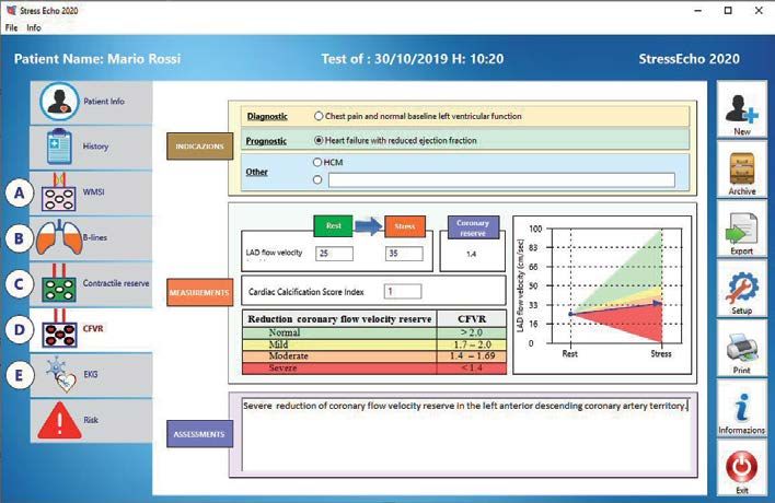
ABCDE STRESS ECHO SOFTWARE / Marco Paterni et al. 459
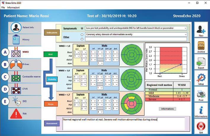
Fig. 1. Computerized case re-
port form for regional wall
motion analysis (step A). In
case of normal resting func-
tion, viability stage (middle
panel) is not contemplated.
A normal wall motion is pres-
ent at rest (all segments cod-
ed in green). At peak stress,
severe regional wall motion
abnormalities (coded in red,
dyskinesia; orange, akine-
sia; and yellow, hypokinesia)
develop. The grading of the
response is reported in tabu-
lar (right upper panel) and
graphic (left lower panel)
format, with normal range
values in green and abnor-
mal values in yellow (mild
degree), orange (moderate
degree) and red (severe de-
gree).
WMSI: Wall motion score index. CFVR: Coronary flow velocity reserve. EKG: Electrocardiogram. Ant: Anterior.
Inf: Inferior. Inf Lat: Inferolateral . Ant Lat: Anterolateral.
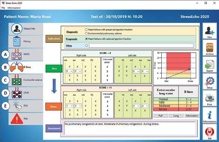
Fig. 2. Computerized case
report form for lung B-lines
with simplified 4-site scan
(step B). The grading of the
response is reported in tabu-
lar (right upper panel) and
graphic (left lower panel)
format, with the same col-
or-code as in Figure 2 (from
green, normal, to red, severe
abnormality).
WMSI: Wall motion score index. CFVR: Coronary flow velocity reserve. EKG: Electrocardiogram. MA: Mid-
axillary. AA: Anterior axillary. MC: Mid-clavicular. PS: Parasternal.
gitant flows (for instance in mitral insufficiency) and for clinical data storage and scientific data archiving,
step G for gradients (valvular and intraventricular). avoiding the loss of time and accuracy inherent to du-
In these patients, functional characterization, risk plicate entry of data. In addition, the data are frozen
stratification and therapy are best obtained with the at the time of data entry in the centralized data bank,
ABCDEFG approach. with the possibility of periodic follow-up updates. This
can only increase the quality of the data, inputted in a
The scientific benefit central data bank by researchers not involved in data
The need of expanding evidence-based practice in the acquisition and analyzed by biostatisticians unaware
various fields of SE must be fed by large scale data of patient identity.
acquired in specific subsets with an immaculate meth-
odology. With the proposed software platform, all The communication benefit
centers are gently forced to seek and store the same Communication is of the utmost importance for an ef-
data set for any given pathology, with a single input fective use of a novel or established method, and the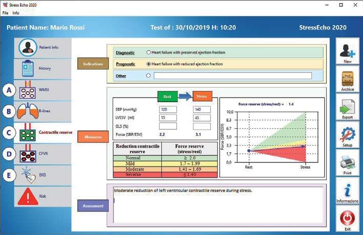
460                                                                     ARGENTINE JOURNAL OF CARDIOLOGY / VOL 87 Nº 6 / DECEMBER 2019

                                                                                                                  Fig. 3. Computerized case
                                                                                                                  report form for left ventricu-
                                                                                                                  lar contractile reserve (step
                                                                                                                  C). The left ventricular force
                                                                                                                  values are derived from the
                                                                                                                  raw data of resting and peak
                                                                                                                  systolic blood pressure and
                                                                                                                  left ventricular end-systolic
                                                                                                                  volume. The grading of the
                                                                                                                  response is reported in tabu-
                                                                                                                  lar (right upper panel) and
                                                                                                                  graphic (left lower panel)
                                                                                                                  format, with the same col-
                                                                                                                  or-code as in Figure 2 (from
                                                                                                                  green, normal, to red, severe
                                                                                                                  abnormality)

WMSI: Wall motion score index. CFVR: Coronary flow velocity reserve. EKG: Electrocardiogram. SBP: Systolic
blood pressure. LVESV: Left ventricular end-systolic volume. GLS: Global longitudinal strain. ESV: End-systolic
volume.

                                                                                                                  Fig. 4. Computerized case re-
                                                                                                                  port form for coronary flow
                                                                                                                  velocity reserve step D). The
                                                                                                                  coronary flow reserve values
                                                                                                                  are derived from the raw
                                                                                                                  data of resting and peak cor-
                                                                                                                  onary diastolic flow velocity.
                                                                                                                  The grading of the response
                                                                                                                  is reported in tabular (right
                                                                                                                  upper panel) and graphic
                                                                                                                  (left lower panel) format,
                                                                                                                  with the same color-code as
                                                                                                                  in Figure 2 (from green, nor-
                                                                                                                  mal, to red, severe abnormal-
                                                                                                                  ity).

WMSI: Wall motion score index. CFVR: Coronary flow velocity reserve. EKG: Electrocardiogram. HCM: Hyper-
trophic cardiomyopathy. LAD: Left anterior descending artery.

quality of reporting is quintessential to ensure accu-                      and criteria of interpretation of new parameters or old,
racy and consistency. With SECS dissemination, the                          established parameters applied to new pathologies. The
same format of reporting might be adopted by all labo-                      dictionary of the SE Esperanto is a common software,
ratories with the information in words coupled with a                       which should share some basic features: user-friendly
tabular and an image side, which may help the refer-                        (clear and informative, requiring few minutes for data
ring physician and the patient to capture at a glance                       archiving); specific for disease and for type of echocar-
the essence of the report, also avoiding some descrip-                      diographic parameter (since we know that what is good
tive, clinically elusive reporting which plagues SE eve-                    and important for a valvular patient is not necessar-
ryday life. As a consequence, there is today a methodo-                     ily relevant for an ischemic patient); with an intuitive
logical Tower of Babel with each laboratory applying                        graphic interface; and suitable for data merging and
its own approaches, ways of archiving and reporting,                        data analysis without further inputted data processing.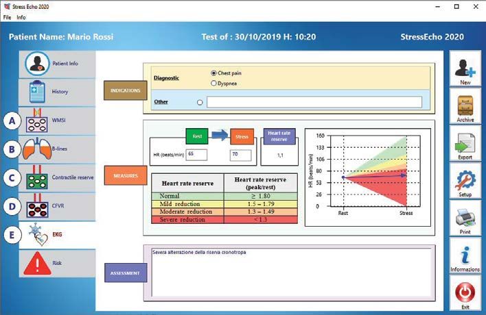
ABCDE STRESS ECHO SOFTWARE / Marco Paterni et al.                                                                                          461

Fig. 5. Computerized case re-
port form for chronotropic
reserve (step E). The data of
heart rate are shown at rest
and peak stress. The grading
of the response is reported in
tabular (right upper panel)
and graphic (left lower pan-
el) format, with the same col-
or-code (from green, normal,
to red, severe abnormality)

                                    WMSI: Wall motion score index. CFVR: Coronary flow velocity reserve. EKG: Electrocardiogram. HR: Heart rate.

Fig. 6. Risk stratification on
the basis of A-B-C-D-E param-
eters, from lowest risk (all pa-
rameters negative) to highest
risk (all parameters positive).

                                    WMSI: Wall motion score index. CFVR: Coronary flow velocity reserve. EKG: Electrocardiogram.

Study limitations                                                        eye-catching graphic format and convenient reporting
The software only focuses on SE, and a similar ap-                       option. It may represent a trade-off between gather-
proach might be helpful in other fields, such as resting                 ing comprehensive information required by scientific
transthoracic and transesophageal or pediatric echocar-                  standards and smooth workflow priority of busy, high
diography. The structure of the database implies that                    volume, clinically-driven activities. This prototype
some history and clinical information must be added                      may now undergo validation, implementation and
to complete the report. This is essential for the scien-                 later diffusion, as already done with a software for
tific use of the program, but it can be an extra-time for                user-friendly calculation of radiological risk for car-
the workflow of a busy lab not interested in systematic                  diovascular examinations, and have a bedside tool to
storage and scientific handling of accumulated data.                     for a simple assessment of the risk-benefit balance
                                                                         of cardiovascular imaging examinations (25), as now
Clinical implications                                                    recommended by major scientific cardiology societies.
SECS may provide a suitable infrastructure for the                       (26,27)
SE 2020 multicenter study, with intuitive interface,                         The overarching aim is to build the next genera-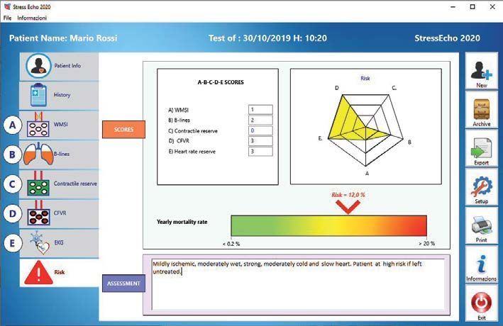
462                                                                       ARGENTINE JOURNAL OF CARDIOLOGY / VOL 87 Nº 6 / DECEMBER 2019

tion stress echo lab without walls, with common and                          Cardiov Imaging 2019; 12:e008564. http://doi.org/dfjw
                                                                             11. Picano E, Pellikka PA. Ultrasound of extravascular lung water: a
shared approach to different diseases by different
                                                                             new standard for pulmonary congestion. Eur Heart J 2016;14:2091-
labs, all speaking the same language replacing cur-                          104. http://doi.org/f8wbv4
rent deregulation which makes communication often                            12. Picano E, Scali MC, Ciampi Q, Lichtenstein D. Lung ultrasound
difficult.                                                                   for the cardiologist. JACC CV Imaging 2018;12:381-90. http://doi.
                                                                             org/dfjx
                                                                             13. Scali MC, Zagatina A, Simova I, Zhuravskaya N, Ciampi Q,
Authors’ contribution and acknowledgments                                    Paterni Met, al. B-lines with Lung Ultrasound: The Optimal
Marco Paterni is the computer scientist who devel-                           Scan Technique at Rest and During Stress . Ultrasound Med Biol
                                                                             2017;43:2558-66. http://doi.org/gck4z8
oped the software and modified it according to the                           14. Scali MC, Cortigiani L, Simionuc A, Gregori D, Marzilli M, Pi-
users’ criticism and suggestions. Quirino Ciampi                             cano E Exercise-induced B-lines identify worse functional and prog-
contributed to the development of the software and                           nostic stage in heart failure patients with depressed left ventricular
                                                                             function. Eur J Heart Fail 2017;19:1468-78. http://doi.org/f9qch4
its assessment in the initial, pre-dissemination, beta-
                                                                             15. Scali MC, Zagatina A, Ciampi Q, Cortigiani L, D’Andrea A, Djord-
testing phase; he also revised the manuscript with                           jevic-Dikic A. The functional meaning of B-profile during Stress
critically intellectual contribution. Clara Carpeggiani,                     Lung ultrasound. JACC Cardiovasc Imaging 2019;12:928-30. http://
Rodolfo Citro, Francesco Antonini-Canterin and Pao-                          doi.org/dfjz
                                                                             16. Bombardini T, Gherardi S, Arpesella G, Maccherini M, Serra
lo Colonna revised the manuscript for critically intel-                      W, Magnani G et al. Favorable short-term outcome of transplanted
lectual content. Eugenio Picano had the original idea                        hearts selected from marginal donors by pharmacological stress
and drafted the manuscript.                                                  echocardiography. J Am Soc Echocardiogr 2011;24:353-62. http://
                                                                             doi.org/b2bbcw
Conflicts of interest                                                        17. Cortigiani L, Huqi A, Ciampi Q, Bombardini T, Bovenzi F, Picano
None declared.                                                               E, et al. Integration of Wall Motion, Coronary Flow Velocity, and Left
                                                                             Ventricular Contractile Reserve in a Single Test: Prognostic Value
   (See authors’ conflicts of interest forms on the website/
                                                                             of Vasodilator Stress Echocardiography in Patients with Diabetes. J
Supplementary material).                                                     Am Soc Echocardiogr 2018;31:692-701. http://doi.org/gdpq83
                                                                             18. Lowenstein J, Tiano C, Marquez G, Presti C, Quiroz C. Simulta-
                                                                             neous analysis of wall motion and coronary flow reserve of the left
REFERENCES                                                                   anterior descending coronary artery by transthoracic Doppler echo-
                                                                             cardiography during dipyridamole stress. J Am Soc Echocardiogr
1. Greenberg N. Update on current concepts and meanings in labo-             2003;16:607-13. http://doi.org/d2g25c
ratory medicine --Standardization, traceability and harmonization.           19. Lowenstein J. The assessment of coronary flow reserve should
Clin Chim Acta 2014;432:49-54. http://doi.org/f59k8m                         be an integral part of stress echo. Agonist. Rev Argent Cardiol
2. PPellikka PA, Arruda-Olson A, Chaudhry FA, Chen MH, Marshall              2010;7:432-5.
JE, Porter TR, et al. Guidelines for Performance, Interpretation, and        20. Lowenstein JA, Caniggia C, Rousse G, Amor M, Sánchez ME,
Application of Stress Echocardiography in Ischemic Heart Disease:            Alasia D, et al. Coronary flow velocity reserve during pharmacologic
From the American Society of Echocardiography                                stress echocardiography with normal contractility adds important
[published online ahead of print, 2019 Nov 15]. J Am Soc Echocar-            prognostic value in diabetic and nondiabetic patients. J Am Soc
diogr 2019;S0894-7317(19)30825-9. http://doi.org/dhm3                        Echocardiogr 2014;27:1113-9. http://doi.org/bkgj
3. Sicari R, Nihoyannopoulos P, Evangelista A, Kasprzak J, Lancel-           21. Ciampi Q, Zagatina A, Cortigiani L, Gaibazzi N, Borguezan Da-
lotti P, Poldermans D, et al; European Association of Echocardiogra-         ros C , Zhuravskaya N, et al. Functional, Coronary Anatomic and
phy Stress echocardiography expert consensus statement: European             Prognostic Correlates of Coronary Flow Velocity Reserve during
Association of Echocardiography (EAE) (a registered branch of the            Stress Echocardiography. J Am Coll Cardiol 2019; 74:2280-93. http://
ESC). Eur J Echocardiogr 2008;9:415-37. http://doi.org/dvxvq8                doi.org/dfj2
4. Picano E. Stress echocardiography: from pathophysiological toy            22. Cortigiani L, Carpeggiani C, Landi P, Raciti M, Bovenzi F, Picano
to diagnostic tool. Point of view. Circulation 1992;85:1604-12. http://      E. Usefulness of blunted heart rate reserve as an imaging-indepen-
doi.org/bkgp                                                                 dent prognostic predictor during dipyridamole-echocardiography
5. Knuuti J, Wijns W, Saraste A, Capodanno D, Barbato E, Funck-              test. Am J Cardiol 2019;124: 972-7. http://doi.org/dfj3
Brentano C. 2019 ESC Guidelines for the diagnosis and management             23. Picano E, Ciampi Q, Citro R, D’Andrea A, Scali MC, Cortigiani
of chronic coronary syndromes. Eur Heart J 2019; Eur Heart J 2019.           L, et al.Stress echo 2020: The international Stress Echo study in
pii: ehz425.                                                                 ischemic and non-ischemic heart disease. Cardiovasc Ultrasound
6. Picano E, Pellikka PA Stress echo applications beyond coronary            2017;15-3.
artery disease. Eur Heart J. 2014;35:1033-40. http://doi.org/bkgr            24. Pellikka PA, Douglas PS, Miller JG, Abraham TP, Baumann R,
7. Lancellotti P, Pellikka PA, Budts W, Chaudry F, Donal E, Dulgh-           Buxton DB, et al. American Society of Echocardiography Cardiovas-
eru R, et a. The Clinical Use of Stress Echocardiography in Non-             cular Technology and Research Summit: a roadmap for 2020. J Am
Ischaemic Heart Disease: Recommendations from the European                   Soc Echocardiogr 2013;6:325-37. http://doi.org/bkgf
Association of Cardiovascular Imaging and the American Society of            25. Carpeggiani C, Paterni M, Caramella D, Vano E, Semelka R, Pi-
Echocardiography. J Am Soc Echocardiogr 2017;30:101-38. http://              cano E, et al. A novel tool for user-friendly estimation of natural,
doi.org/dfjt                                                                 diagnostic and professional radiation risk: Radio-Risk software. Eur
8. Picano E, Ciampi Q, Wierzbowska-Drabik K, Urluescu ML, Mor-               J Radiol. 2012;81:3563-7. http://doi.org/dtvj48
rone D, Carpeggiani C. The new clinical standard of integrated qua-          26. Picano E, Vañó E, Rehani MM, Cuocolo A, Mont L, Bodi V, et
druple stress echocardiography with ABCD protocol. Cardiov Ultra-            al. The appropriate and justified use of medical radiation in cardio-
sound 2018;16:22. http://doi.org/cv9d                                        vascular imaging: a position document of the ESC Associations of
9. Picano E, Scali MC. Stress echo, carotid arteries and more:               Cardiovascular Imaging, Percutaneous Cardiovascular Interven-
Its versatility for our imaging times. JACC Cardiovasc Imaging               tions and Electrophysiology. Eur Heart J. 2014;35:665-72. http://doi.
2018;11:181-3. http://doi.org/dfjv                                           org/bkgq
10. Cortigiani L, Urluescu M, Coltelli M, Carpeggiani C, Bovenzi F,          27. 2018 ACC/HRS/NASCI/SCAI/SCCT Expert Consensus Docu-
Picano E. Apparent declining prognostic value of a negative stress           ment on Optimal Use of Ionizing Radiation in Cardiovascular Im-
echocardiography based on regional wall motion abnormalities in              aging: Best Practices for Safety and Effectiveness: A Report of the
patients with normal resting left ventricular function due to the            American College of Cardiology Task Force on Expert Consensus
changing referral profile of the population under study. Circulation         Decision Pathways. J Am Coll Cardiol 2018;71:e283-e351.
You can also read