Structural and functional plasticity in the somatosensory cortex of chronic stroke patients
←
→
Page content transcription
If your browser does not render page correctly, please read the page content below
doi:10.1093/brain/awl214                                                                                                                      Brain (2006), 129, 2722–2733

Structural and functional plasticity in the
somatosensory cortex of chronic stroke patients
Judith D. Schaechter,1,2 Christopher I. Moore,3 Brendan D. Connell,1,2
Bruce R. Rosen1,2 and Rick M. Dijkhuizen1,2,4
1
 MGH/MIT/HMS Athinoula A. Martinos Center for Biomedical Imaging, Charlestown, 2Department of Radiology,
Harvard Medical School, Boston, 3Department of Brain and Cognitive Sciences and McGovern Institute for Brain Research,
Massachusetts Institute of Technology, Cambridge, MA, USA and 4Image Sciences Institute, University Medical Center
Utrecht, Utrecht, The Netherlands
Correspondence to: Judith D. Schaechter, PhD, MGH/MIT/HMS Athinoula A. Martinos Center for Biomedical Imaging,

                                                                                                                                                                                              Downloaded from http://brain.oxfordjournals.org/ by Brandon Brock on January 14, 2013
13th Street, Building 149, Room 2301, Charlestown, MA 02129, USA
E-mail: judith@nmr.mgh.harvard.edu

Animal studies have demonstrated that motor recovery after hemiparetic stroke is associated with functional
and structural brain plasticity. While studies in stroke patients have revealed functional plasticity in sensori-
motor cortical areas in association with motor recovery, corresponding structural plasticity has not been
shown. We sought to test the hypothesis that chronic hemiparetic stroke patients exhibit structural plasticity
in the same sensorimotor cortical areas that exhibit functional plasticity. Functional MRI during unilateral
tactile stimulation and structural MRI was conducted in chronic stroke patients and normal subjects. Using
recently developed computational methods for high-resolution analysis of MRI data, we evaluated for between-
group differences in functional activation responses, and cortical thickness of areas that showed an enhanced
activation response in the patients. We found a significant (P < 0.005) increase in the activation response in areas
of the ventral postcentral gyrus (POG) in the patients relative to controls. These same ventral POG areas
showed a significant (P < 0.05) increase in cortical thickness in the patients. Control cortical areas did not show a
significant between-group difference in thickness or activation response. These results provide the first evi-
dence of structural plasticity co-localized with areas exhibiting functional plasticity in the human brain after
stroke.

Keywords: stroke; MRI; cortical thickness; plasticity; postcentral gyrus

Abbreviations: BOLD = blood oxygenation level-dependent; JHFT = Jebson Hand Function Test; POG = postcentral gyrus;
PRG = precentral gyrus
Received December 26, 2005. Revised June 5, 2006. Accepted July 19, 2006. Advance Access publication August 18, 2006.

Introduction
Hemiparesis is the most common acute deficit after stroke.                                      (Kleim et al., 2002), and in the somatosensory cortex after
While most patients experience partial recovery of motor                                        altered somatosensory experience (Hickmott and Steen,
function, stroke remains a leading cause of chronic disability                                  2005). Collectively, these studies have led to the hypothesis
in modern society. Motor recovery after experimental stroke                                     that coupled functional and structural plasticity in the brain
in animals has been associated with reorganized neural                                          underlies post-stroke motor recovery. Studies in stroke
activity (i.e. functional plasticity) in sensorimotor cortical                                  patients have observed functional plasticity during the pro-
areas (Nudo and Milliken, 1996; Dijkhuizen et al., 2001)                                        cess of motor recovery after stroke, similar to that described
that also exhibit morphological changes (i.e. structural plas-                                  in animal stroke models (Schaechter, 2004; Ward, 2005).
ticity) (Jones and Schallert, 1992; Stroemer et al., 1995; Li                                   However, to our knowledge no study has examined whether
et al., 1998; Carmichael et al., 2001; Wei et al., 2001; Zhang                                  structural plasticity occurs in concert with functional plasti-
et al., 2002). Further, co-localized functional and structural                                  city in the human brain after stroke.
plasticity has been observed in the rat motor cortex after                                         Recent developments in computational methods for
prolonged exercise (Swain et al., 2003) and motor learning                                      analysing     magnetic resonance images have enabled
# The Author (2006). Published by Oxford University Press on behalf of the Guarantors of Brain. All rights reserved. For Permissions, please email: journals.permissions@oxfordjournals.org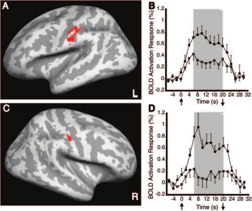
Structural plasticity in stroke patients                                                            Brain (2006), 129, 2722–2733              2723

high-resolution functional and structural analysis of the                   or impaired) status of their right upper limb acutely after stroke,
human brain. These methods permit highly accurate locali-                   based on data extracted from the initial stroke hospitalization
zation of cortical activation (Dale et al., 1999; Fischl et al.,            medical records; and (ii) premorbid hand dominance using the
1999a, b) and precise measurement of cortical thickness                     Edinburgh Inventory laterality quotient (LQ) scale [range =
                                                                            "100% (strongly left-handed) to 100% (strongly right-handed)]
(Fischl and Dale, 2000; Han et al., 2006). Using these com-
                                                                            (Oldfield, 1971), evaluated by asking the patients to recall their
putational methods following combined functional and                        hand preference prior to stroke. At the time of enrolment, all
structural MRI, we tested the hypothesis that chronic hemi-                 patients had completed standard in-patient and out-patient post-
paretic stroke patients exhibit structural plasticity in the same           stroke rehabilitation. In addition, three patients (#2, 5 and 7) had
sensorimotor cortical areas that exhibit functional plasticity.             received supplemental out-patient therapy aimed at improving
Our examination of functional and structural changes in                     right upper limb function as participants of other research studies
patients widens the lens towards a complete understanding                   during the chronic stroke phase. These additional therapies were
of the restorative mechanisms occurring in the human brain                  completed at least 2 months prior to participation in the current
after stroke, which will ultimately serve in the development of             study (range 2–49 months).
new post-stroke interventions that maximize motor recovery.                    Nine control subjects with no history of stroke and a normal
                                                                            neurological examination were also enrolled. These subjects were
Material and methods

                                                                                                                                                       Downloaded from http://brain.oxfordjournals.org/ by Brandon Brock on January 14, 2013
                                                                            relatively well matched to the stroke patients with regard to age
Nine patients with a chronic stroke, former in-patients of the              (mean = 60 years, SD = 12 years; range 36–72 years), gender
Spaulding Rehabilitation Hospital, were enrolled (Table 1). Entry           (2 females, 7 males) and hand dominance (LQ mean = 81%,
criteria included (i) an ischaemic stroke incurred >1 year earlier that     SD = 21%) (Oldfield, 1971).
spared the precentral gyrus (PRG) and postcentral gyrus (POG)                  All subjects provided written informed consent in accordance
based on MRI; (ii) stroke restricted to the left hemisphere, a criterion    with the Human Subjects Committee of the Partners Institutional
applied to increase patient homogeneity because brain plasticity after      Review Board.
stroke may differ depending on hemisphere (Zemke et al., 2003); (iii)
acute loss of right-hand strength to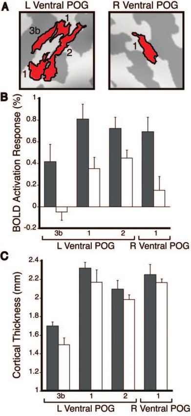
2724       Brain (2006), 129, 2722–2733                                                                                   J. D. Schaechter et al.

applied, based on prior recommendations (Stern, 1992). JHFT times           of Alice software (Hayden Image Processing Solutions, Denver, CO,
of the right hand were normalized, by per cent, to that of the left         USA) that was used to determine the location and volume of
hand (left time/right time · 100). Patients unable to perform               the lesion based on the T2-weighted images acquired from the
the JHFT subtests with the affected hand were given a score of              patients. Lesion volume was determined by an experienced neuro-
0%. A composite motor function score for each patient was deter-            radiologist who manually outlined the T2 abnormality slice by slice.
mined by entering all patients’ motor function data into a principal           Visual inspection of the two sets of T1-weighted MPRAGE
component analysis using MATLAB (The Mathworks, v6.5.1). The                structural images from each subject revealed no motion artefacts.
first principal component, which accounts for the greatest percen-          The two structural volumes from each subject were motion-
tage of the variability within the data, was taken as the score repre-      corrected using a 6-parameter (3 translations, 3 rotations) rigid-
senting motor outcome for each patient. We examined relationships           body transformation, intensity-normalized, then averaged to create
between the motor outcome scores and the MRI-based measures of              a single structural volume with high contrast-to-noise. Cortical sur-
functional and structural plasticity across the patients.                   faces from each subject were reconstructed using an automated
    Tactile sensitivity was tested at the glabrous surface of the middle    procedure that involved white matter segmentation, then tessellation
phalanx of the right and left D3 using Semmes–Weinstein monofila-           of the identified grey/white matter boundary and pial surfaces, as
ments. Testing was conducted using an adaptive psychophysical algo-         described in detail previously (Dale et al., 1999). Visual inspection of
rithm (Simpson, 1989) for 40 trials per digit, with the tactile detection   the reconstructed surfaces in the central sulcus region (within our

                                                                                                                                                       Downloaded from http://brain.oxfordjournals.org/ by Brandon Brock on January 14, 2013
threshold determined as the force perceived with 80% accuracy.              primary region-of-interest) on each subject’s structural volume
                                                                            revealed no abnormalities (data not shown).
MRI                                                                            The cortical surface model from each subject was spatially
MRIs were acquired using a 3T Siemens Allegra magnetic resonance            normalized to a spherical surface template using a procedure that
scanner and a quadrature head coil. With the subject supine, cush-          optimally aligns major cortical sulci and gyri (Fischl et al., 1999a).
ions were packed around the head to limit its motion, and splints           Cortical surface models from all study subjects were averaged to
were used to immobilize the forearms (in pronation) and hands               provide a group-average cortical surface model that best accounts
(digits in extension) off the abdominal area. The splints were fab-         for the topological variability of the subjects in this study (Fischl
ricated with an opening to permit access to the glabrous surface of         et al., 1999b).
the middle phalanyx of D3. Two structural volumes were collected               We tested whether the spatial alignment of each subject’s cortical
using a T1-weighted MPRAGE sequence [repetition time (TR) =                 surface model to the spherical surface template was accurate even in
7 ms; echo time (TE) = 3 ms; flip angle (a) = 7# ; field-of-view            brains spatially distorted by a stroke. Visual inspection revealed
(FOV) = 256 · 256 mm; matrix size = 192 · 256; effective slice              accurate mapping of the template’s central sulcus region to each
thickness = 1.33 mm]. In stroke patients, T2-weighted turbo spin-           subject’s cortical surface model, including surface models from
echo images (TR = 10 s; TE = 65 ms; a = 120# ; FOV = 210 · 210 mm;          patients with a large stroke (data not shown). Further, there were
matrix size = 256 · 256; slice thickness = 5 mm; interslice gap =           no significant (P > 0.05, two-tailed, unpaired Student’s t-test)
1 mm; number of slices = 20) were collected. Blood oxygenation              between-group differences in the per cent overlap of the template’s
level-dependent (BOLD) functional images were collected using a             central sulcus region to the central sulcus on the left (lesioned hemi-
T2*-weighted gradient-echo, echo planar imaging sequence (TR =              sphere of patients) or right cortical surface models (left—patients:
2 s; TE = 30 ms; a = 90# ; FOV = 200 · 200 mm; matrix size = 64 · 64)       63 6 2%, controls: 65 6 2%; right—patients: 74 6 2%, controls:
with slices (thickness = 5 mm; interslice gap = 1 mm; number = 20;          75 6 2%). These findings exclude misalignment of the cortical
number of acquisitions/slice = 130) parallel to the anterior                surface models across subjects as responsible for the between-
commissure–posterior commissure line. Functional images were                group differences in the BOLD activation response and cortical
collected during periods of passive tactile stimulation (6 · 20 s)          thickness found in this study (Results section).
alternating with periods of no stimulation (7 · 20 s). We opted                Thickness across the cortical mantle was computed in the struc-
to employ passive tactile stimulation, rather than an active motor          tural volume for each subject using an automated procedure that
task, because the former allowed us to probe sensorimotor func-             measures the shortest distance between vertices of the grey/white
tional representations (Moore et al., 2000) of the affected hand in         matter boundary and pial surfaces, as described previously (Fischl
patients who represent a wide range of residual motor impairment.           and Dale, 2000). As this method uses signal intensity and continuity
Punctate tactile stimulation was delivered manually by an investi-          information, the thickness measurements can achieve an accuracy of
gator over the glabrous surface of the middle phalanyx of D3 by             at least an order of magnitude greater than the voxel dimensions
means of a Semmes–Weinstein monofilament (60 g), paced at 3 Hz              of the raw MRI data used to generate the cortical surface reconstruc-
by a computer-generated (MacStim, version 2.6) metronome pre-               tions. This automated method has been shown to be highly reliable
sented to the investigator only. Four functional runs were collected        (Han et al., 2006), and has been validated histologically and manu-
during unilateral D3 stimulation (two right D3, two left D3) to each        ally in pathological and aged human brains (Rosas et al., 2002;
subject. The side of stimulation during collection of the first             Kuperberg et al., 2003; Salat et al., 2004). The thickness measure-
functional run was randomized across subjects, and the side of              ments from each subject were spatially normalized into the spherical
stimulation during subsequent functional runs in each subject alter-        surface-based coordinate system (Fischl et al., 1999a).
nated between sides. Subjects were instructed to keep their eyes               Functional images from each subject were co-registered with their
closed, their entire body relaxed and to pay close attention to the         structural volume using a rigid-body transformation. The functional
tactile sensation at their digit.                                           data were motion-corrected using a 6-parameter (3 translations,
                                                                            3 rotations) rigid-body transformation, intensity-normalized and
Image analysis                                                              spatially smoothed using a 3D Gaussian kernel [full-width at half
FreeSurfer software (http://surfer.nmr.mgh.harvard.edu) was used            maximum (FWHM) = 3 mm]. Each subject’s functional data from
for all functional and structural image analysis, with the exception        replicate runs collected during unilateral D3 tactile stimulation were
Structural plasticity in stroke patients Brain (2006), 129, 2722–2733 2725
concatenated, and the average signal intensity and standard devia- structural plasticity co-localizes with areas of functional plasticity in
tion of the residual error were estimated at each voxel for each chronic stroke patients by evaluating the mean BOLD activation
condition. These voxel-based signal intensity maps were spatially response (6–20 s post-stimulation onset) and mean cortical thick-
normalized into the spherical surface-based coordinate system ness in these secondary regions-of-interest.
(Fischl et al., 1999a). Between-group differences in cortical thickness in our function-
In order to test the hypothesis that structural plasticity co- ally defined regions-of-interest could potentially result from differ-
localizes with functional plasticity in sensorimotor cortical areas ences in the gradient of T1 signal intensity in tissue bordering the
in stroke patients, we empirically identified regions-of-interest exhi- cortex, which could cause the surface reconstruction algorithm to
biting functional plasticity in the patients. Functionally defined incorrectly localize the grey/white matter boundary and pial sur-
regions-of-interest were determined as follows. A general linear faces. To address this possibility, we tested for between-group dif-
model was fitted at each vertex across the cortical surface. The ferences in change in mean T1 signal intensity of tissue 10% above
general linear model included a low-frequency drift term, as well and below the normalized distance between the two reconstructed
as motion-correction parameters as nuisance regressors. The hae- surfaces of our functionally defined regions-of-interest using two-
modynamic response to unilateral D3 stimulation was modelled as a tailed, unpaired Student’s t-tests. We found no significant (P > 0.05)
gamma function (delta = 2.25 s, tau = 1.25 s) (Dale and Buckner, differences in signal intensity change across the grey/white matter
1999) convolved with a boxcar function. The difference in the BOLD boundary or pial surfaces for our functionally defined regions-of-
Downloaded from http://brain.oxfordjournals.org/ by Brandon Brock on January 14, 2013
activation response between the patient and control groups was interest in the patients relative to controls (data not shown). This
estimated at each vertex, with subject regarded as a fixed effect. A finding excludes differential localization of the grey/white matter
fixed-effects model was used at this stage of the analysis because boundary and pial surfaces in the structural volume as responsible
the vertex-to-vertex correspondence in the D3 functional fields for the between-group differences in cortical thickness we observed
might be relatively low due to differences in sensorimotor experience in our functionally defined regions-of-interest (Results section).
and/or stroke across the subjects. The between-group difference To test the selectivity of structural changes co-localized with areas
maps were corrected for multiple comparisons by controlling the of functional plasticity, we also evaluated mean cortical thickness
false discovery rate (Genovese et al., 2002) to 1%, and the minimum (and mean BOLD activation response to contralateral D3 tactile
cluster surface area to 50 mm2. Surviving clusters of the difference stimulation) in two sets of control cortical areas. The first set of
maps became candidate regions-of-interest. In order to guard control cortical areas was selected as being topographically related to
against our functionally defined regions-of-interest being driven our functionally defined regions-of-interest, while respecting known
by outliers, we then tested more stringently for between-group dif- anatomical and physiological distinctions. Each topographically
ferences in the BOLD activation response in the candidate regions- defined control region-of-interest was a circle with a surface area
of-interest using a random-effects model. The raw time-course of that approximated the mean surface area of our functionally defined
the BOLD activation response (in per cent signal change from base- regions-of-interest. The second set of control cortical areas was
line) was averaged across all vertices of each region-of-interest for commonly activated in the patients and controls in response to
each subject. The average time-course was used to compute the tactile stimulation to the right (affected side of patients) D3. The
mean BOLD activation response occurring from the completion functionally defined control regions-of-interest were determined by
of the rising phase (6 s after stimulation onset) to the end of the estimating the average BOLD activation response across all the sub-
stimulation (20 s after stimulation onset). For each candidate jects at each vertex, with subject regarded as a random effect. The
region-of-interest, the mean BOLD activation response from all average activation map was thresholded by imposing a probability
subjects was entered into a two-tailed, unpaired Student’s t-test level of 0.001 at each vertex and a minimum cluster surface area
to test for between-group differences in functional activation. of 50 mm2. We excluded from the thresholded, average activation
Finally, regions-of-interest that exhibited a between-group differ- map the functionally defined regions-of-interest that exhibited a
ence in the mean BOLD activation response at an alpha level of between-group difference in the BOLD activation response. The
0.05 became the functionally defined regions-of-interest used in resultant activation map was subdivided based on known anatomical
further analyses. and physiological distinctions to achieve functionally defined
We observed that the functionally defined regions-of-interest control regions-of-interest.
traversed distinct somatosensory cortical areas (Results section)
based on prior anatomical and physiological studies in monkeys
and humans (Kaas, 1983; Jones, 1986; Brodmann, 1994; Geyer Statistical analysis
et al., 1999; Grefkes et al., 2001). Accordingly, our primary regions- StatView (version 4.5) was used for statistical procedures. Between-
of-interest were subdivided into areas corresponding to putative area group differences in sensorimotor function were analysed using the
3b (residing in the posterior wall of the central sulcus), putative area two-tailed, unpaired Student’s t-test, with the exception of one
1 (residing in the crown of the POG) and putative area 2 (residing in motor function measure for which the Mann–Whitney U-test was
the anterior wall of the postcentral sulcus). Neurons of area 3b, applied because the scores from the controls were not normally
considered the primary somatosensory cortex based on anatomical distributed. Separate two-way, mixed-model analysis of variance
and physiological criteria, have been shown to possess discrete tactile (ANOVA), with group (patients, controls) as the between-subjects
receptive fields. Area 1 neurons have been shown to possess tactile factor and cortical area as the within-subjects factor, was used to
receptive fields that are more complex than those in area 3b. Area 2 test for main and interaction effects of the factors on activation
neurons have been shown to be responsive to tactile and proprio- response and cortical thickness. We point out that the level of sig-
ceptive stimulation and have complex receptive field properties. nificance of a main effect of group on activation response in the
These secondary regions-of-interest were delineated on the group- cortical areas is inflated because the functionally defined regions-of-
average cortical surface model to best account for topograph- interest were explicitly selected because they exhibited significant
ical variability across the subjects. We tested our hypothesis that between-group differences in activation response. On the other2726 Brain (2006), 129, 2722–2733 J. D. Schaechter et al.
hand, the level of significance of a main effect of group on cortical The patients also exhibited a range of tactile sensitivity at
thickness is valid because the cortical areas were selected indepen- the time of study enrolment (Table 2). The tactile detection
dent of the thickness measurements. Relationships between activa- threshold of the right D3 of two patients (#6 and 9) was
tion response and cortical thickness in functionally defined and outside the tactile detection threshold range of the right
control regions-of-interest in the patients and controls were eval-
D3 of the control subjects (0.008–0.160 g). However, even
uated using the Pearson correlation coefficient, uncorrected for
multiple comparisons. In patients, additional relationships between
with consideration given to these two patients, tactile detec-
motor outcome, using the first principal component of the motor tion thresholds were not significantly different between
function scores, versus thickness and versus activation were groups for either the right D3 (patients: 0.36 6 0.21 g,
evaluated using the Pearson correlation coefficient (uncorrected). controls: 0.09 6 0.02; P = 0.22, Student’s t-test) or left D3
A difference in activation response between patients with good ver- (patients: 0.10 6 0.02 g, controls: 0.08 6 0.02; P = 0.51,
sus poor motor outcome (dichotomized based on the first principal Student’s t-test). Further, tactile sensitivity of the right D3
component of the motor function scores) was tested using the of all patients was an order of magnitude greater than the
Mann–Whitney U-test. Alpha was set to 0.05 for all statistical strength of the tactile stimulation delivered during func-
tests. Results are expressed as the mean 6 SEM. tional MRI (60 g), reflecting our enrolment criterion that
this stimulus be detectable. This result rules out the possibi-
Downloaded from http://brain.oxfordjournals.org/ by Brandon Brock on January 14, 2013
Results lity that the observed between-group differences in the BOLD
Sensorimotor function activation response to the tactile stimulation (see below) were
Review of patient medical records revealed that acutely after due to lack of stimulus detection in the patient group.
stroke the right (affected) hand had no muscle power in all
nine enrolled patients, and had impaired somatosensation
Functional MRI
in three of the nine patients (Table 1).
Figure 1 shows a typical BOLD activation response pattern
At the time of study enrolment, formal testing revealed
to unilateral tactile stimulation to D3 in a control subject. The
that the chronic stroke patients exhibited a range of upper
tactile stimulation produced marked activation responses in
limb motor function (Table 2). All but one patient (#8) could
the cortex contralateral to the stimulated hand, including the
grip with the right (affected) hand, indicating that eight of
hand region of the PRG and POG, as well as ventral POG,
the nine patients had made recovery of motor function of
secondary somatosensory cortex and supplementary motor
the right hand since the acute stroke. On a group basis,
area. Activation responses in the ipsilateral cortex were
however, the patients had residual motor impairments of
confined largely to the ventral POG and secondary somato-
the right upper limb. Normalized grip strength was signifi-
sensory cortex.
cantly reduced in the patients (patients: 50 6 11%; controls:
Between-group difference analysis revealed that the
113 6 5%; P < 0.0001, Student’s t-test). Scores on the FMSS
patients exhibited a significant increase in the BOLD activa-
were significantly lower in the patients (patients: 42 6 7;
tion response, as compared with controls, to stimulation of
controls: 66 6 0; P < 0.01, Mann–Whitney U-test). The
the right (affected side of patients) D3 in a region of the left
normalized time to perform functional tasks of the JHFT
(ipsilesional in patients) ventral POG (304 mm2; patients:
was significantly longer in the patients (patients: 49 6 14%;
0.68 6 0.12%; controls: 0.32 6 0.07%; P < 0.025, Student’s
controls: 108 6 3%; P = 0.001, Student’s t-test). The first
t-test; Fig. 2A and B). This ventral POG region did not
principal component of the motor function measures from
intersect the stroke cavity in any patient. Patients also
all of the patients accounted for 95.4% of the variability,
exhibited a significant increase in the activation response,
and was taken to reflect motor outcome of each patient.
as compared with controls, to stimulation of the left
(unaffected side of patients) D3 in a region of the right
Table 2 Sensorimotor function of the right (affected)
(contralesional in patients) ventral POG (94 mm2; patients:
upper limb in the chronic stroke patients
0.69 6 0.13%; controls: 0.15 6 0.13%; P < 0.01, Student’s
Patient Grip FMSS JHFT Motor Tactile t-test; Fig. 2C and D). There was no cortical region in which
strength score (% L) outcome detection right or left D3 stimulation elicited a significantly stronger
(% L) (1st PC) threshold (g)
activation response in the controls as compared with the
1 89 66 122 85 0.160 patients.
2 91 66 89 62 0.008 The ventral POG regions that exhibited a between-group
3 53 48 62 14 0.160
difference in the BOLD activation response were further
4 75 60 86 48 0.160
5 60 53 31 3 0.160 subdivided into areas corresponding to 3b (primary soma-
6 8 11 0 "72 2.000 tosensory cortex) and 1 and 2 (secondary somatosensory
7 28 33 30 "30 0.040 cortices), based on recognized anatomical and physiological
8 0 6 0 "78 0.160 distinctions (Kaas, 1983; Jones, 1986; Brodmann, 1994;
9 43 36 21 "26 0.400 Geyer et al., 1999; Grefkes et al., 2001). Accordingly, the
An entry of 0 indicates that the patient was unable to perform the left ventral POG region was divided into putative areas 3b,
test with the affected hand. L, left hand; PC, principal component. 1 and 2, whereas the right ventral POG region was identifiedStructural plasticity in stroke patients Brain (2006), 129, 2722–2733 2727
Downloaded from http://brain.oxfordjournals.org/ by Brandon Brock on January 14, 2013
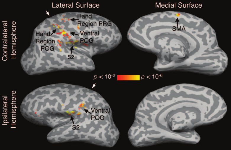
Fig. 1 Statistical activation maps obtained from functional MRI during tactile stimulation to the left D3 of a normal subject. The BOLD
activation response pattern is typical for that observed in controls. The activation maps (P < 0.01, corrected) are displayed on the model of
the subject’s inflated cortical surfaces. Light grey areas of a cortical surface indicate gyri; dark grey areas indicate sulci. White arrowheads
point to the central sulcus. S2, secondary somatosensory cortex; SMA, supplementary motor area.
as putative area 1 (Fig. 3A). The mean BOLD activation were regional differences in cortical thickness across the
response in putative areas 3b, 1 and 2 was estimated in ventral POG areas. More importantly, they indicate that cor-
the patients and controls (Fig. 3B). An ANOVA used to tical thickness of the ventral POG areas was greater in the
test effects on the activation responses in the ventral patients as compared with controls.
POG areas detected a significant main effect of group In order to examine whether the significant between-group
[F(1,64) = 13.03, P < 0.005]; we note that this significance difference in thickness observed in the ventral POG areas was
level is inflated because the ventral POG areas were sub- restricted to cortical areas that also exhibited an enhanced
components of functionally defined regions-of-interest that BOLD activation response, we performed analyses on two sets
were explicitly selected because they exhibited significant of four control cortical areas—topographically defined and
between-group differences in activation. This ANOVA functionally defined. The topographically defined control
also detected a significant main effect of cortical area cortical areas were located in the (i) anterior wall of the
[F(3,64) = 8.12, P < 0.0005], but no significant interaction central sulcus (putative area 4) (Rademacher et al., 2001)
between the two factors. These results indicate that the in the ventral PRG of the left (ipsilesional in patients) hemi-
chronic stroke patients exhibited an enhanced activation sphere; (ii) crown of the hand region POG (putative area 1) of
response in the identified ventral POG areas. The data further the left hemisphere; (iii) left calcarine sulcus (putative area
show that while the magnitude of the activation response 17); and (iv) anterior wall of the central sulcus (putative area
differed across the cortical areas, this was not affected by 4) in the ventral PRG of the right (contralesional in patients)
subject group. hemisphere. The functionally defined control cortical areas
were located in the hand region of the left POG in the (i)
fundus of the central sulcus (putative area 3a) (Geyer et al.,
Structural MRI 1999); (ii) posterior wall of the central sulcus (putative
We next examined whether the chronic stroke patients area 3b); (iii) crown of the POG (putative area 1); and
exhibited structural changes in the ventral POG areas that (iv) anterior wall of the postcentral sulcus (putative area 2).
exhibited an enhanced BOLD activation response. Figure 3C Figure 4A shows the mean cortical thickness in the two
shows the mean cortical thickness of the ventral POG areas. sets of control cortical areas. An ANOVA conducted to test
An ANOVA conducted to test whether cortical thickness of for effects on thickness across the topographically defined
the ventral POG areas differed between groups and across control cortical areas did not detect a significant main effect
areas detected significant main effects of group [F(1,64) = of group [F(1,64) = 0.11, P = 0.75], nor a significant inter-
4.69, P < 0.05] and area [F(3,64) = 28.39, P < 0.0001], with no action effect, but did detect a significant main effect of area
significant interaction effect. This result indicates that there [F(3,64) = 101.56, P < 0.0001]. A separate ANOVA conducted2728 Brain (2006), 129, 2722–2733 J. D. Schaechter et al.
Downloaded from http://brain.oxfordjournals.org/ by Brandon Brock on January 14, 2013
Fig. 2 Between-group differences in BOLD activation response to
unilateral tactile stimulation to D3. Difference maps are displayed
on the group-average, inflated cortical surface model. (A) Area in
the left (ipsilesional in patients) ventral POG with a greater
activation response in patients as compared with controls during
right (affected side of patients) D3 stimulation. (B) Time-course of
the BOLD activation response across the left, ventral POG
area (red in A). The mean activation response was significantly
(P < 0.025, Student’s t-test) greater in patients relative to controls
during the 6–20 s stimulation interval (grey bar). (C) Area in the
right (contralesional in patients) ventral POG with a greater
activation response in patients as compared with controls during
left (unaffected side of patients) D3 stimulation. (D) Time-course
of the BOLD activation response in the right, ventral POG area
(red in C). The mean activation response was significantly
(P < 0.01, Student’s t-test) greater in patients relative to controls
during the 6–20 s stimulation interval. Filled squares, patients; open
squares, controls; upward arrow, stimulation onset; downward
arrow, stimulation end.
to test for effects on thickness across the functionally defined
control cortical areas also did not detect a significant main
effect of group [F(1,64) = 0.348, P = 0.56], nor a significant
interaction effect, but detected a significant main effect of Fig. 3 Cortical thickness and BOLD activation response in putative
area [F(3,64) = 24.495, P < 0.0001]. areas of the ventral POG. (A) Delineation of putative areas 3b,
1 and 2 of the left (L) and right (R) ventral POG area. (B)
Figure 4B shows the mean BOLD activation response in the Magnitude of the activation response in putative areas of the L
two sets of control cortical areas. An ANOVA conducted to ventral POG during right (affected side of patients) D3 stimulation,
test for effects on activation across the topographically and of the R ventral POG during left D3 stimulation, in the patients
defined control cortical areas did not detect a significant and controls. A two-way, mixed-model ANOVA detected
main effect of group [F(1,64) = 0.09, P = 0.77], nor a sig- significant main effects of group [F(1,64) = 13.03, P < 0.005] and
cortical area [F(3,64) = 8.12, P < 0.0005], with no significant
nificant interaction effect, but did detect a significant main interaction between these two factors. (C) Thickness of the
effect of area [F(3,64) = 9.65, P < 0.0001]. Similarly, an putative ventral POG areas in the patients and controls. A two-
ANOVA conducted to test for effects across the functionally way, mixed-model ANOVA testing effects across the ventral POG
defined control cortical areas did not detect a significant main areas detected significant main effects of group [F(1,64) = 4.69,
effect of group [F(1,64) = 0.09, P = 0.76], nor a significant P < 0.05] and area [F(3,64) = 28.39, P < 0.0001], with no significant
interaction. Grey bars, patients; white bars, controls.
interaction effect, but did detect a significant main effect of
area [F(3,64) = 11.77, P < 0.0001].
These analyses revealed no significant between-group in cortical thickness observed in the ventral POG areas of the
difference in thickness in control cortical areas that also patients relative to the control subjects was not generalized
showed no significant between-group difference in the BOLD across the cortex, but was restricted to areas also exhibiting
activation response. These findings indicate that the increase increases in the activation response.Structural plasticity in stroke patients Brain (2006), 129, 2722–2733 2729
Fig. 5 Plot of relationship between cortical thickness and activation
response in putative area 3b of the left (ipsilesional) ventral POG in
Downloaded from http://brain.oxfordjournals.org/ by Brandon Brock on January 14, 2013
the stroke patients.
In patients, putative area 3b of the left (ipsilesional) ventral
POG exhibited a significant positive correlation between
thickness and activation (r = 0.75, P < 0.025, uncorrected,
Pearson correlation; Fig. 5). No other ventral POG area or
control cortical area exhibited a significant correlation
between thickness and activation in the patients or controls.
We also tested in the patients for relationships between
motor outcome (first principal component of the motor
function scores) and plasticity (activation response, cortical
thickness). No significant correlation between these measures
was detected. However, there was a non-significant trend
toward an increase in the activation response in the left (ipsi-
lesional) ventral POG during right (affected) D3 stimulation
in patients with good motor outcome as compared with those
with poor motor outcome (good: 0.81 6 0.18%, n = 5; poor:
0.50 6 0.12%, n = 4; P = 0.086, Mann–Whitney U-test).
Fig. 4 Thickness and BOLD activation response in topographically
defined and functionally defined control cortical areas. Topogra- Discussion
phically defined control areas were located in putative area 4 of the
left (L) and right (R) ventral PRG, putative area 1 in the hand region The adult human brain responds adaptively to experience and
of the L POG, and putative area 17 of the L calcarine sulcus. injury (Buonomano and Merzenich, 1998). Several studies
Functionally defined control areas were located in the hand region have shown that recovery of motor function in patients
of the L POG. (A) Thickness in the two sets of control cortical after stroke is associated with functional reorganization in
areas. Separate two-way, mixed-model ANOVAs did not detect the brain (Schaechter, 2004; Ward, 2005). The present study
significant main effects of group, nor interaction effects, but did
detect significant main effects of area [topographically defined: provides the first demonstration of structural plasticity co-
F(3,64) = 101.56, P < 0.0001; functionally defined: F(3,64) = 24.495, localized with functional plasticity in the human brain after
P < 0.0001]. (B) Magnitude of the activation response in the two stroke.
sets of control cortical areas during tactile stimulation to the
contralateral D3. Separate two-way, mixed-model ANOVAs did
not detect significant main effects of group, nor interaction effects, Structural plasticity
but did detect significant main effects of area [topographically We found that thickness of the cortical mantle of ventral POG
defined: F(3,64) = 9.65, P < 0.0001; functionally defined: F(3,64) = areas, corresponding to areas 3b, 1 and 2, was significantly
11.77, P < 0.0001]. Grey bars, patients; white bars, controls. greater in the chronic stroke patients relative to the control
subjects. Two related results strengthen this finding. Firstly,
Correlation analyses we did not find a significant increase in thickness in control
In the patients and controls, we examined the correlation cortical areas defined either topographically or functionally,
between cortical thickness and the BOLD activation response suggesting that an increase in thickness was not a generalized
in each ventral POG area, and each control cortical area. finding across the cortical mantle of chronic stroke patients.2730 Brain (2006), 129, 2722–2733 J. D. Schaechter et al.
Secondly, we detected significant thickness differences across reason why patients with varied lesion sites should exhibit
distinct ventral POG and control cortical areas delineated functional plasticity in the ventral POG is unclear. However,
based on known anatomical and functional criteria, which it is notable that the ventral POG was activated in our control
is consistent with previous reports of gyral regions of neo- subjects, consistent with previous neuroimaging studies
cortex being thicker than sulcal regions (Fischl and Dale, (Burton et al., 1997; Burton et al., 1999). This finding suggests
2000), and the anterior wall of the central sulcus being thicker that the ventral POG might normally play a role in higher-
than the posterior wall (Meyer et al., 1996). The magnitude of order processing of somatosensory information. After hemi-
the cortical thickness increase we observed in the ventral POG paretic stroke, it is possible that activity in the ventral POG
areas was relatively small, ranging from !4 to 13%, yet is increases to support sensorimotor function. Ventral areas of
comparable with the increases in cortical thickness others the ipsilesional sensorimotor cortex have previously been
have found in rat motor cortex after motor skill learning reported to undergo functional reorganization in patients
(Anderson et al., 2002; Kleim et al., 2002), in rat visual cortex with subcortical or cortical stroke (Weiller et al., 1993;
after rearing in an enriched environment (Diamond et al., Zemke et al., 2003; Cramer and Crafton, 2006). Further,
1966) and in cortices of humans with specific mental disor- functional reorganization involving the ipsilesional POG
ders (Rauch et al., 2004; Thompson et al., 2005). Our finding associated with the stroke-affected hand has been evidenced
Downloaded from http://brain.oxfordjournals.org/ by Brandon Brock on January 14, 2013
is also consistent with the increased grey matter volume previously as a partial (Pineiro et al., 2001; Calautti et al.,
found in the sensorimotor cortices of professional musicians 2003) or complete (Cramer et al., 2000; Jang et al., 2005) shift
with extensive motor training relative to amateur musicians in the sensorimotor cortex activation toward the POG.
and non-musicians (Gaser and Schlaug, 2003). These pre- The functional change we observed in the ipsilesional ven-
vious findings, in conjunction with our result, suggest that tral POG in the patients was associated with motor recovery
the human sensorimotor cortex can undergo structural of the affected hand from the time of the acute stroke to study
changes in response to sensorimotor experience and brain enrolment in eight of the nine patients. However, we found
injury. only a non-significant trend toward an increased BOLD acti-
Given the spatial resolution and signal-to-noise of the vation response in the ipsilesional ventral POG in patients
current structural MRI data collected using a 3 T MRI scan- with good as compared with poor motor outcome of the
ner, the microscopic substrate of the increase in cortical affected upper limb. Therefore, the enhanced activation
thickness in the ventral POG areas was not discernible. response we observed in the ipsilesional POG was associated,
However, previous studies suggest that neural and non- but not directly correlated, with motor recovery in our
neural elements may have contributed to the cortical thick- patients. This result is similar to the lack of correlation
ness increase we observed. In rats, an increase in cortical between the extent of posterior shift in activation toward
thickness has been shown to co-localize with an increase the POG and the level of motor recovery reported in earlier
in the number of synapses per neuron in the motor cortex studies (Pineiro et al., 2001; Calautti et al., 2003). While there
after motor training (Kleim et al., 2002), and with an increase is growing evidence that motor recovery after stroke is related
in glial number in the visual cortex after environmental to functional reorganization of the brain (Ward, 2005), our
enrichment (Diamond et al., 1966). Experimental stroke in ability to detect a significant relationship between our indices
rats has been shown to induce synaptogenesis (Stroemer et al., of functional plasticity and motor outcome may have been
1995), angiogenesis (Wei et al., 2001; Zhang et al., 2002), as limited by several factors, such as differences among the
well as increased neuronal sprouting (Li et al., 1998; patients in lesion site and post-stroke rehabilitation. Further,
Carmichael et al., 2001) and dendritic arborization (Jones the cross-sectional design of our study limited our correlation
and Schallert, 1992). Further, it has been recently shown analyses to motor function measures acquired at a single
that experimental stroke induces sequential waves of neuro- session late after stroke, rather than multiple measures
nal growth-promoting genes (Carmichael et al., 2005). Col- acquired over time during the recovery process. Conse-
lectively, these findings lead us to speculate that the cortical quently, our study was well designed to detect persistent
thickness increase we observed in the chronic stroke patients functional changes common to stroke patients, but less
was associated with growth of neural elements as well as well designed to identify functional plasticity mechanisms
several non-neural elements that provide metabolic and tightly linked to the process of motor recovery after stroke.
structural support to the new neural elements. The observed lack of a significant relationship between motor
outcome and functional activation might also reflect our use
of passive tactile stimulation, rather than an active motor
Functional plasticity task, during functional MRI. Use of the passive task permitted
Activation was increased in the ipsilesional POG ventral to us to enrol patients who represent a broad range of residual
the hand region in response to tactile stimulation to the motor deficit. However, functional plasticity related to tactile
affected D3 of the stroke patients as compared with controls. stimulation in patients may relate only indirectly to motor
The location of this functional change was found in patients outcome late after stroke. Further research is required to
whose stroke location, while left-sided and sparing the PRG elucidate the precise relationship between functional plasti-
and POG in all cases, was otherwise fairly heterogeneous. The city in sensorimotor cortices, operating in conjunction withStructural plasticity in stroke patients Brain (2006), 129, 2722–2733 2731
structural plasticity, and the complex dynamics of reacquisi- ventral POG area or control cortical area exhibited a signifi-
tion of motor function after stroke. cant correlation between thickness and activation in the
We also observed enhanced activation in the contralesional patients or controls suggests that these cortical parameters
ventral POG associated with the unaffected hand of patients. are not generally related. It is unclear why in the patients
Though less commonly studied than functional plasticity ventral POG areas other than putative area 3b did not exhibit
associated with the stroke-affected limb, evidence of reorga- a significant relationship between thickness and activation.
nization associated with the unaffected upper limb has been One factor may relate to putative area 3b of the ventral POG
observed previously in patients after unilateral stroke being more closely linked to the hand region area 3b (primary
(Cramer et al., 1997b; Luft et al., 2004). Among the possible somatosensory cortex) with neurovascular activity strongly
mechanisms to account for reorganization of the unaffected connected to thalamic input, whereas the other ventral POG
hand representation in the contralesional somatosensory areas may be more closely associated with the higher-order
cortex is a use-dependent change (Elbert and Rockstroh, somatosensory cortices with neurovascular activity reflecting
2004) resulting from increased compensatory use of the a greater degree of cortico-cortical communication. An incre-
unaffected upper limb. This interpretation is supported by mental change in the neural and non-neural elements asso-
studies in rats that showed increased use of the non-impaired ciated with a cortical thickness increase in higher-order
Downloaded from http://brain.oxfordjournals.org/ by Brandon Brock on January 14, 2013
limb after a unilateral sensorimotor cortex lesion that was somatosensory cortices may relate poorly to their neurovas-
associated with structural changes in the contralesional cular response because of extensive cortico-cortical
sensorimotor cortex (Jones and Schallert, 1994). Another communication.
explanation is a transcallosal impact of reorganization within In conclusion, the findings of this study highlight the
the ipsilesional somatosensory cortex. Direct and indirect capacity of the adult human brain for structural and func-
callosal connections between hand representations within tional plasticity in association with motor recovery after
sensorimotor cortices of the two hemispheres have been stroke. These findings were made possible by the application
demonstrated in animals (Killackey et al., 1983; Krubitzer of recently developed computational methods for high-
et al., 1998). resolution analysis of cortical activation patterns and cortical
thickness from magnetic resonance images. Such findings
expand our understanding of the spectrum of changes occur-
Co-localized structural and ring in the brain after stroke in humans that may mediate
functional plasticity restoration of function. It is hoped that an understanding of
Ventral POG areas of the chronic stroke patients exhibited the full complement of restorative brain mechanisms will lead
evidence of structural and functional plasticity. An increase in to the development of new therapeutic interventions that
synaptic and capillary density may have contributed to the promote recovery after stroke in patients.
parallel increase in cortical thickness and BOLD activation
response in these areas. Co-localized structural and Acknowledgements
functional plasticity has been observed previously in sensori- This study was supported by grants from the American Heart
motor cortical areas of animals in response to manipulations Association—New England Affiliate (to J.D.S. and R.M.D.),
of sensorimotor experience (Kleim et al., 2002; Swain et al., American Health Assistance Foundation (to J.D.S.), National
2003; Hickmott and Steen, 2005), and has been implicated Institutes of Health K23-HD044425 (to J.D.S.), Royal
in motor recovery after experimental stroke (Jones and Netherlands Academy of Arts and Sciences (to R.M.D.),
Schallert, 1992; Stroemer et al., 1995; Nudo and Milliken, NCRR (P41-RR14075) and the MIND Institute. We thank
1996; Li et al., 1998; Carmichael et al., 2001; Dijkhuizen Mingwang Zhu, MD, PhD, for calculating lesion volumes,
et al., 2001; Wei et al., 2001; Zhang et al., 2002). Our results Katherine Perdue for assistance in data analysis, as well
suggest that reorganization of the human brain after as Doug Greve, PhD and Bruce Fischl, PhD, for helpful
stroke may also involve coupled structural and functional discussions.
remodelling.
Among the ventral POG areas in which we observed co- References
localized structural and functional plasticity (ipsilesional Anderson BJ, Eckburg PB, Relucio KI. Alterations in the thickness of motor
putative areas 3b, 1 and 2; contralesional putative area 1), cortical subregions after motor-skill learning and exercise. Learn Mem
2002; 9: 1–9.
putative area 3b exhibited a significant linear correlation
Brodmann K. Localisation in the cerebral cortex. Garey LJ, translator.
between cortical thickness and BOLD activation response London: Smith-Gordon (original work published 1909); 1994.
in the patients. The BOLD activation response has been Buonomano DV, Merzenich MM. Cortical plasticity: from synapses to maps.
shown to scale roughly linearly with the extent of local neural Annu Rev Neurosci 1998; 21: 149–86.
processing (Logothetis et al., 2001) and cerebral perfusion Burton H, MacLeod AM, Videen TO, Raichle ME. Multiple foci in parietal
(Mandeville and Rosen, 2002). Therefore, the linear increase and frontal cortex activated by rubbing embossed grating patterns across
fingerpads: a positron emission tomography study in humans. Cereb
in the BOLD activation response observed in putative area 3b Cortex 1997; 7: 3–17.
might have been due to an increase in stimulation-induced Burton H, Abend NS, MacLeod AM, Sinclair RJ, Snyder AZ, Raichle ME.
synaptic activity and capillary perfusion. That no other Tactile attention tasks enhance activation in somatosensory regions of2732 Brain (2006), 129, 2722–2733 J. D. Schaechter et al.
parietal cortex: a positron emission tomography study. Cereb Cortex 1999; Jones EG. Connectivity of the primate sensory-motor cortex. In: Peters A,
9: 662–74. Jones EG, editors. Cerebral cortex. New York: Plenum Press; 1986: p 113–84.
Calautti C, Leroy F, Guincestre JY, Baron JC. Displacement of primary Jones TA, Schallert T. Overgrowth and pruning of dendrites in adult rats
sensorimotor cortex activation after subcortical stroke: a longitudinal PET recovering from neocortical damage. Brain Res 1992; 581: 156–60.
study with clinical correlation. Neuroimage 2003; 19: 1650–4. Jones TA, Schallert T. Use-dependent growth of pyramidal neurons after
Carmichael ST, Wei L, Rovainen CM, Woolsey TA. New patterns of neocortical damage. J Neurosci 1994; 14: 2140–52.
intracortical projections after focal cortical stroke. Neurobiol Dis 2001; Kaas JH. What, if anything, is SI? Organization of first somatosensory area of
8: 910–22. cortex Physiol Rev 1983; 63: 206–31.
Carmichael ST, Archibeque I, Luke L, Nolan T, Momiy J, Li S. Growth- Killackey HP, Gould HJ 3rd, Cusick CG, Pons TP, Kaas JH. The relation of
associated gene expression after stroke: evidence for a growth-promoting corpus callosum connections to architectonic fields and body surface maps
region in peri-infarct cortex. Exp Neurol 2005; 193: 291–311. in sensorimotor cortex of new and old world monkeys. J Comp Neurol
Cramer SC, Crafton KR. Somatotopy and movement representation sites 1983; 219: 384–419.
following cortical stroke. Exp Brain Res 2006; 168: 25–32. Kleim JA, Barbay S, Cooper NR, Hogg TM, Reidel CN, Remple MS, et al.
Cramer SC, Nelles G, Schaechter JD, Kaplan JD, Finklestein SP. Computer- Motor learning-dependent synaptogenesis is localized to functionally
ized measurement of motor performance after stroke. Stroke 1997a; reorganized motor cortex. Neurobiol Learn Mem 2002; 77: 63–77.
28: 2162–8. Krubitzer L, Clarey JC, Tweedale R, Calford MB. Interhemispheric
Cramer SC, Nelles G, Benson RR, Kaplan JD, Parker RA, Kwong KK, et al. connections of somatosensory cortex in the flying fox. J Comp Neurol
A functional MRI study of subjects recovered from hemiparetic stroke. 1998; 402: 538–59.
Downloaded from http://brain.oxfordjournals.org/ by Brandon Brock on January 14, 2013
Stroke 1997b; 28: 2518–27. Kuperberg GR, Broome MR, McGuire PK, David AS, Eddy M, Ozawa F, et al.
Cramer SC, Moore CI, Finklestein SP, Rosen BR. A pilot study of somatotopic Regionally localized thinning of the cerebral cortex in schizophrenia.
mapping after cortical infarct. Stroke 2000; 31: 668–71. Arch Gen Psychiatry 2003; 60: 878–88.
Dale AM, Buckner RL. Selective averaging of rapidly presented individual Li Y, Jiang N, Powers C, Chopp M. Neuronal damage and plasticity identified
trials using fMRI. Hum Brain Mapp 1999; 5: 329–40. by microtubule-associated protein 2, growth-associated protein 43, and
Dale AM, Fischl B, Sereno MI. Cortical surface-based analysis. I: cyclin D1 immunoreactivity after focal cerebral ischemia in rats. Stroke
Segmentation and surface reconstruction. Neuroimage 1999; 9: 179–94. 1998; 29: 1972–80.
Diamond MC, Law F, Rhodes H, Lindner B, Rosenzweig MR, Krech D, et al. Logothetis NK, Pauls J, Augath M, Trinath T, Oeltermann A. Neuro-
Increases in cortical depth and glia numbers in rats subjected to enriched physiological investigation of the basis of the fMRI signal. Nature 2001;
environment. J Comp Neurol 1966; 128: 117–26. 412: 150–7.
Dijkhuizen RM, Ren J, Mandeville JB, Wu O, Ozdag FM, Moskowitz MA, et al. Luft AR, Waller S, Forrester L, Smith GV, Whitall J, Macko RF, et al. Lesion
Functional magnetic resonance imaging of reorganization in rat brain location alters brain activation in chronically impaired stroke survivors.
after stroke. Proc Natl Acad Sci USA 2001; 98: 12766–71. Neuroimage 2004; 21: 924–35.
Elbert T, Rockstroh B. Reorganization of human cerebral cortex: the Mandeville JB, Rosen BR. Functional MRI. In: Toga AW, Mazziotta JC,
range of changes following use and injury. Neuroscientist 2004; 10: editors. Brain mapping: the methods. San Diego: Academic Press; 2002:
129–41. p 315–49.
Fischl B, Dale AM. Measuring the thickness of the human cerebral cortex from Medical Research Council (Great Britian). Aids to the examination of the
magnetic resonance images. Proc Natl Acad Sci USA 2000; 97: 11050–5. peripheral nervous system. London: H M Stationery Office; 1976.
Fischl B, Sereno MI, Dale AM. Cortical surface-based analysis. II: Inflation, Meyer JR, Roychowdhury S, Russell EJ, Callahan C, Gitelman D,
flattening, and a surface-based coordinate system. Neuroimage 1999a; Mesulam MM. Location of the central sulcus via cortical thickness of
9: 196–207. the precentral and postcentral gyri on MR. AJNR Am J Neuroradiol 1996;
Fischl B, Sereno MI, Tootell RBH, Dale AM. High-resolution intersubject 17: 1699–706.
averaging and a coordinate system for the cortical surface. Hum Brain Moore CI, Stern CE, Corkin S, Fischl B, Gray AC, Rosen BR, et al. Segregation
Mapp 1999b; 8: 272–84. of somatosensory activation in the human rolandic cortex using fMRI.
Fugl-Meyer AR, Jaasko L, Leyman I, Olsson S, Steglind S. The post-stroke J Neurophysiol 2000; 84: 558–69.
hemiplegic patient: a method for the evaluation of physical performance. Nudo RJ, Milliken G. Reorganization of movement representations in
Scand J Rehabil Med 1975; 7: 13–31. primary motor cortex following focal ischemic infarcts in adult squirrel
Gaser C, Schlaug G. Brain structures differ between musicians and non- monkeys. J Neurophysiol 1996; 75: 2144–9.
musicians. J Neurosci 2003; 23: 9240–5. Oldfield RC. The assessment and analysis of handedness: the Edinburgh
Genovese CR, Lazar NA, Nichols T. Thresholding of statistical maps in Inventory. Neuropsychologia 1971; 9: 97–113.
functional neuroimaging using the false discovery rate. Neuroimage 2002; Pineiro R, Pendlebury S, Johansen-Berg H, Matthews PM. Functional
15: 870–8. MRI detects posterior shifts in primary sensorimotor cortex activation
Geyer S, Schleicher A, Zilles K. Areas 3a, 3b, and 1 of human primary after troke: evidence of local adaptive reorganization? Stroke 2001;
somatosensory cortex. 1. Microstructural organization and interindividual 32: 1134–9.
variability. Neuroimage 1999; 10: 63–83. Rademacher J, Burgel U, Geyer S, Schormann T, Schleicher A, Freund HJ, et al.
Grefkes C, Geyer S, Schormann T, Roland P, Zilles K. Human somatosensory Variability and asymmetry in the human precentral motor system.
area 2: observer–independent cytoarchitectonic mapping, interindividual A cytoarchitectonic and myeloarchitectonic brain mapping study. Brain
variability, and population map. Neuroimage 2001; 14: 617–31. 2001; 124: 2232–58.
Han X, Jovicich J, Salat D, van der Kouwe A, Quinn B, Czanner S, et al. Rauch SL, Wright CI, Martis B, Busa E, McMullin KG, Shin LM, et al.
Reliability of MRI-derived measurements of human cerebral cortical A magnetic resonance imaging study of cortical thickness in animal phobia.
thickness: the effects of field strength, scanner upgrade and manufacturer. Biol Psychiatry 2004; 55: 946–52.
Neuroimage, 2006; 32: 180–94. Rosas HD, Liu AK, Hersch S, Glessner M, Ferrante RJ, Salat DH, et al.
Hickmott PW, Steen PA. Large-scale changes in dendritic structure Regional and progressive thinning of the cortical ribbon in Huntington’s
during reorganization of adult somatosensory cortex. Nat Neurosci disease. Neurology 2002; 58: 695–701.
2005; 8: 140–2. Salat DH, Buckner RL, Snyder AZ, Greve DN, Desikan RS, Busa E, et al.
Jang SH, Ahn SH, Yang DS, Lee DK, Kim DK, Son SM. Cortical Thinning of the cerebral cortex in aging. Cereb Cortex 2004; 14:
reorganization of hand motor function to primary sensory cortex in 721–30.
hemiparetic patients with a primary motor cortex infarct. Arch Phys Med Schaechter JD. Motor rehabilitation and brain plasticity after hemiparetic
Rehabil 2005; 86: 1706–8. stroke. [Review]. Prog Neurobiol 2004; 73: 61–72.You can also read