Targeting the ALS/FTD-associated A-DNA kink with anthracene-based metal complex causes DNA backbone straightening and groove contraction
←
→
Page content transcription
If your browser does not render page correctly, please read the page content below
9526–9538 Nucleic Acids Research, 2021, Vol. 49, No. 16                                                                          Published online 9 April 2021
https://doi.org/10.1093/nar/gkab227

Targeting the ALS/FTD-associated A-DNA kink with
anthracene-based metal complex causes DNA
backbone straightening and groove contraction
Cyong-Ru Jhan1,† , Roshan Satange2,3,† , Shun-Ching Wang3,† , Jing-Yi Zeng3 ,
Yih-Chern Horng4 , Peng Jin 5 , Stephen Neidle 6 and Ming-Hon Hou 1,2,3,*
1
 Department of Life Sciences, National Chung-Hsing University, Taichung 402, Taiwan, 2 Ph.D. Program in Medical
Biotechnology, National Chung Hsing University, Taichung 402, Taiwan, 3 Institute of Genomics and Bioinformatics;

                                                                                                                                                                  Downloaded from https://academic.oup.com/nar/article/49/16/9526/6219111 by guest on 15 October 2021
National Chung Hsing University, Taichung 402, Taiwan, 4 Department of Chemistry, National Changhua University of
Education, Changhua 50058, Taiwan, 5 Department of Human Genetics, Emory University School of Medicine,
Atlanta, GA 30322, USA and 6 The School of Pharmacy, University College London, London, WC1N 1AX, United
Kingdom

Received January 22, 2021; Revised March 16, 2021; Editorial Decision March 17, 2021; Accepted March 26, 2021

ABSTRACT                                                                                 INTRODUCTION
The use of a small molecule compound to reduce                                           Abnormal expansion of the GGGGCC (G4C2) hexanu-
toxic repeat RNA transcripts or their translated aber-                                   cleotide DNA repeat within the C9orf72 intron is asso-
rant proteins to target repeat-expanded RNA/DNA                                          ciated with two neurodegenerative disorders: amyotrophic
with a G4C2 motif is a promising strategy to treat                                       lateral sclerosis (ALS) and frontotemporal dementia (FTD)
C9orf72-linked disorders. In this study, the crystal                                     (1,2). Generally, healthy individuals carry 20–30 hexanu-
                                                                                         cleotide repeat units, while in patients with ALS/FTD, this
structures of DNA and RNA–DNA hybrid duplexes
                                                                                         is expanded to between 800 and >4000 repeats. The pro-
with the -GGGCCG- region as a G4C2 repeat motif                                          posed pathologic mechanisms of ALS and FTD consist of
were solved. Unusual groove widening and sharper                                         loss-of-function of the C9orf72 protein and a toxic gain-of-
bending of the G4C2 DNA duplex A-DNA confor-                                             function from C9orf72 repeat RNA or dipeptide repeat pro-
mation with B-form characteristics inside was ob-                                        teins (3,4). Many studies have suggested that the expansion
served. The G4C2 RNA–DNA hybrid duplex adopts                                            of G4C2 hexanucleotide repeats may be attributed to the
a more typical rigid A form structure. Detailed struc-                                   slippage of DNA strands, forming either quadruplex struc-
tural analysis revealed that the G4C2 repeat motif of                                    tures that are stabilized by G-quartets during DNA replica-
the DNA duplex exhibits a hydration shell and greater                                    tion or possibly hairpins stabilized by duplex (5–8). Expan-
flexibility and serves as a ‘hot-spot’ for binding                                       sion of the hexanucleotide repeat present in the non-coding
                                                                                         region of C9orf72 leads to reduced levels of the C9orf72
of the anthracene-based nickel complex, NiII (Chro)2
                                                                                         protein (9–11). Furthermore, expanded hexanucleotide re-
(Chro = Chromomycin A3). In addition to the origi-
                                                                                         peats are transcribed, and the resulting RNA forms nu-
nal GGCC recognition site, NiII (Chro)2 has extended                                     clear foci that segregate various RNA-binding proteins to
specificity and binds the flanked G:C base pairs of                                      cause neurodegeneration (12–14). The final proposed mech-
the GGCC core, resulting in minor groove contrac-                                        anism involves G4C2 repeat-associated non-ATG transla-
tion and straightening of the DNA backbone. We have                                      tion, which may produce toxic dipeptide proteins (15) (Fig-
also shown that Chro-metal complexes inhibit neu-                                        ure 1). Targeting RNA structures of G4C2 hexanucleotide
ronal toxicity and suppresses locomotor deficits in                                      repeats is one of the most widely explored strategies for drug
a Drosophila model of C9orf72-associated ALS. The                                        design against ALS and FTD. Several groups have identi-
approach represents a new direction for drug discov-                                     fied small-molecule ligands and antisense oligonucleotides
ery against ALS and FTD diseases by targeting G4C2                                       that specifically bind to the expanded G4C2 repeat hairpin
                                                                                         RNA to reduce toxic RNA foci and dipeptide proteins to
repeat motif DNA.
                                                                                         improve ALS/FTD-associated defects (11,16–20). We show
                                                                                         here that targeting repeat-specific DNA-containing struc-
                                                                                         tures, including DNA duplexes and DNA–RNA hybrid du-

* To   whom correspondence should be addressed. Tel: +886 4 2284 0338 (Ext 7011); Fax: +886 4 2285 9329; Email: mhho@nchu.edu.tw
†
    The authors wish it to be known that, in their opinion, the first three authors should be regarded as Joint First Authors.

C The Author(s) 2021. Published by Oxford University Press on behalf of Nucleic Acids Research.
This is an Open Access article distributed under the terms of the Creative Commons Attribution-NonCommercial License
(http://creativecommons.org/licenses/by-nc/4.0/), which permits non-commercial re-use, distribution, and reproduction in any medium, provided the original work
is properly cited. For commercial re-use, please contact journals.permissions@oup.com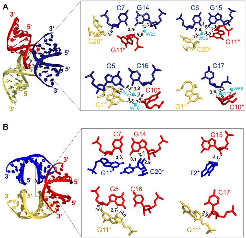
Nucleic Acids Research, 2021, Vol. 49, No. 16 9527
Downloaded from https://academic.oup.com/nar/article/49/16/9526/6219111 by guest on 15 October 2021
Figure 1. Schematic representation of mechanisms of ALS/FTD pathogenesis induced by abnormal G4C2 repeat expansions on the chromosome 9 open
reading frame 72 (C9orf72). The hexanucleotide repeat expansion resulted in different types of abnormal secondary structures such as R-loop, hairpins or
quadruplexes that interfere with cellular processes such as transcription, replication and DNA repair. The signature -GGGCCG- sequence corresponds to
the G4C2 motif is highlighted by a red colour box.
plexes, may serve as an alternative approach to G4C2 hex- gel electrophoresis (PAGE) from MDBio, Inc. (New Taipei
anucleotide repeat-associated ALS/FTD (21,22). City, Taiwan), and Genomics (New Taipei City, Taiwan).
In this study, the crystal structures of the non- RNA oligomers were purified using high performance liq-
self-complementary DNA duplex, d(GTGGGCCGAC)/ uid chromatography from MDBio, Inc. Oligonucleotides
(GTCGGCCCAC) and the analogous RNA–DNA hybrid were prepared in RNase-free water by heating at 95◦ C
duplex, r(GUGGGCCGAC)/ d(GTCGGCCCAC) were for 5 min followed by slow cooling to room tempera-
determined since the central -GGGCCG- region (G4C2 ture (-0.5◦ C/min) to allow duplex formation. To determine
repeat motif) is a core G4C2 repeat sequence. Both du- oligonucleotide concentrations, absorbance measurements
plex structures showed global similarity, including A-DNA- were performed using a JASCO V-630 spectrophotome-
like characteristics, albeit with local structure differences, ter (JASCO International Co. Ltd., Tokyo, Japan) with a
such as a wider groove and sharper bending angle in the quartz cuvette (1 cm path length) set at 260 nm. The concen-
DNA duplex. Moreover, a detailed analysis of crystal pack- tration of oligonucleotides have been calculated using Beer’s
ing revealed that the central -GGGCCG- region of the law with the extinction coefficients calculated according to
DNA duplex has greater flexibility and a hydration shell tabulated values of monomer and dimer extinction coeffi-
that might serve as ‘hot-spots’ for accommodating small cients, with reasonable assumptions (23). Stock solutions of
molecule binding, compared to the RNA–DNA hybrid ligands (1 mM) used for melting temperature assays were
duplex. Several GC-selective DNA-binding agents includ- prepared either in ddH2 O or in dimethyl sulfoxide (DMSO,
ing anthracene-, anthracycline- and acridine-based com- ≥99.9%).
pounds, a DNA cross-linker compound and polypeptide
antibiotics were screened, and it was found that metal–
anthracene complexes, NiII (Chro)2 and CoII (Chro)2 , pre- UV spectroscopy
ferred to bind and stabilize the G4C2 repeat motif com- UV melting curves were collected using a JASCO V-630
pared to other DNA binding ligands. The crystal structure spectrophotometer. The duplex DNA used for melting tem-
of NiII (Chro)2 -d(TTGGGCCGAA)2 was then solved to in- perature analysis was dissolved in buffer containing 50 mM
vestigate the structural basis behind this preference. Impor- NaCl and 50 mM sodium cacodylate (pH 7.3). A final con-
tantly, we show that NiII (Chro)2 and CoII (Chro)2 can sup- centration of 3 M of the duplex sequences was denatured
press G4C2 repeat-induced neuronal toxicity in vivo using at 95◦ C for 5 min and cooled on ice for 30 min for annealing.
a Drosophila model of G4C2 repeat expansion. These find- Ligands were added to oligonucleotide solution at a fixed
ings highlight the feasibility of utilizing the G4C2 repeat du- molar ratio of 1:1 and incubated for 1 h at 4◦ C to allow com-
plex as a target and serve as a basis for the design of novel plex formation. A 650 l sample was added to the quartz
sequence-specific ligands against ALS/FTD diseases. cuvette with a 1 cm path length and covered with a layer
of silicone oil. The sample was then equilibrated at 4◦ C for
MATERIALS AND METHODS 10 min in a spectrophotometer. UV absorbance versus. tem-
perature profiles were acquired by changing the temperature
Oligonucleotides and sample preparation
from 4 to 95◦ C at a rate of 1◦ C min−1 and recording the ab-
All chemicals used in this study were purchased from Sigma sorbance at 260 nm every 0.5 min. The Tm (temperature cor-
Chemical Co. (St. Louis, MO, USA). DNA oligomers were responding to half-dissociation of the DNA duplexes) was
commercially synthesized and purified by polyacrylamide determined from polynomial fitting of the observed curves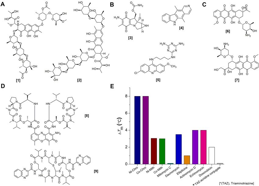
9528 Nucleic Acids Research, 2021, Vol. 49, No. 16
using Varian\Cary WinUV thermal application software v in PHENIX (version 1.10.1) and Refmac5 in CCP4i (Oxon,
3.00 (Agilent, Santa Clara, CA, USA). UK); the crystallographic and refinement statistics of the
structures are listed in Supplementary Table S1 (25,26). The
final 2Fo – Fc electron density maps were created using the
Preparation of crystals
fast Fourier transform in CCP4i and PyMOL (version 4.5),
For crystallization, PAGE-purified oligonucleotides were which was used to draw graphical representations of the re-
annealed by heating at 95◦ C for 5 min and then cooled fined structures.
slowly to room temperature (-0.5◦ C/min). Crystalliza-
tion trials were performed using the sitting-drop vapour-
DNA parameter analysis
diffusion method, and crystals were obtained at 4◦ C after
2–7 days. To grow G4C2 DNA crystals, 1 mM DNA duplex A standard A-DNA structure with the same sequence
solution was mixed with buffer containing 50 mM 2-(N- as G4C2 DNA and G4C2 RNA–DNA hybrid DNA
morpholino)ethanesulfonic acid (pH 6.5), 100 mM lithium duplexes was generated using Discovery Studio Client
Downloaded from https://academic.oup.com/nar/article/49/16/9526/6219111 by guest on 15 October 2021
chloride, 10 mM manganese (II) chloride tetrahydrate and software (version-19.1.0.18287) (Dassault Systems Biovia
2.6 M sodium malonate dibasic monohydrate at a ratio of Corp, Vélizy-Villacoublay, France) for comparison. DNA
1:3, and equilibrated against 200 l solution. Small, colour- structural parameters, including helix, base and base-pair
less, rhombic crystals appeared after 3 days of crystalliza- parameters were analysed with the Web 3DNA 2.0 server
tion and grew to a maximum size after 1–2 weeks. To obtain and the CURVES+ program (27,28). RMSDs were calcu-
G4C2 RNA–DNA hybrid crystals, equimolar concentra- lated in PyMOL and van der Waals interactions were mon-
tions (0.25 mM) of DNA and RNA oligonucleotides were itored using LigPlot+ (29). DNA parameter values for typ-
mixed in a solution containing 5 mM magnesium sulphate ical A- and B-DNA were used for comparison (30).
hydrate, 50 mM Tris hydrochloride (pH 8.5) and 2.9 M 1,6-
hexanediol. Square-shaped, colourless crystals appeared af-
MD simulation protocol
ter 1 week of crystallization. The NiII (Chro)2 –DNA com-
plex crystals were obtained from a solution of 1 mM DNA Structures of metalII (Chro)2 –DNA complexes obtained
duplex, 3 mM Chro, 6 mM NiSO4 , 40 mM sodium cacody- from the crystal structures were used for MD simulations.
late (pH 7), 2.5 mM spermine and 6% polyethylene glycol These were performed using the Discovery Studio software
(PEG) 400, and equilibrated against 500 l of 6% PEG 400. v19.1.0.18287 and the CHARMm force field (31). A 1000
Yellow-coloured, needle-shaped crystals were grown after step smart-minimizer algorithm was used, and the models
4–7 days. Each condition produced several crystals, from were immersed in an orthorhombic water box built based on
which a single crystal was used to collect the diffraction a modified TIP3P system (32). The dimensions of the water
data. box were selected such that the distances of non-hydrogen
atoms in a nucleic acid duplex were at least 7 Å from the
edge of the box. The total charge on each system under
X-ray data collection, phasing and structure refinement
study was neutralized by randomly adding sodium ions.
X-ray diffraction data from a single crystal of each of G4C2 The system was then subjected to 5000-step Steepest De-
DNA, G4C2 RNA–DNA hybrid and the NiII (Chro)2 -DNA scent (SD) and 10 000-step Conjugate Gradient (CG) min-
complex were collected at the synchrotron radiation facil- imizations followed by 20 ps heating from 50 to 300 K with
ity of the National Synchrotron Radiation Research Cen- position constraints imposed on the nucleic acid duplexes.
ter, Taiwan. Diffraction data integration and reduction were The following 20 ps equilibration run was performed with
performed using the HKL-2000 package (24). Phases for position constraints on nucleic acid duplexes. The produc-
the G4C2 DNA structure were determined in the P31 21 tion run was carried out with an NPT ensemble with peri-
space group by molecular replacement using Phaser MR odic boundary conditions. During the entire simulation, all
in a python-based hierarchical environment for integrated covalent bonds involving hydrogen were constrained using
xtallography (PHENIX) (version 1.10.1) and using a mod- the SHAKE algorithm, where the time step was set to 2 fs.
elled A-DNA structure with WinCoot (version 0.8.9.1) as The particle mesh Ewald method was used for treating long-
a template (25). The G4C2 DNA structure was then used range electrostatic interactions (33). The production simu-
to determine phases for the G4C2 RNA–DNA hybrid in lations was extended up to 500 ps for the NiII (Chro)2 –DNA
the P21 21 21 space group, using a molecular replacement and MgII (Chro)2 –DNA complex. RMSF values were anal-
protocol. The phases for the NiII (Chro)2 –DNA complex ysed using the ‘Analyse Trajectory’ function in Discovery
structure were determined by molecular replacement in Studio software v19.1.0.18287. Final graphs were plotted
the P65 22 space group with Phaser MR in the PHENIX using Origin Pro8 v8.0721 (OriginLab Corp., Northamp-
suite using the partial structure of the [Mg2+ -(Chro)2 ]– ton, MA, USA) (34).
d(TTGGCCAA)2 complex (PDB ID: 1VAQ) as a starting
template. The resulting well-defined electron density maps
Drosophila experiments
were used to build initial models using the molecular graph-
ics programs MIFit and crystallographic object-oriented The following fly lines (gmr-GAL4, UAS-GGGGCC30 -
toolkit (COOT). Initial models of the G4C2 DNA, G4C2 EGFP), (elav-GAL4, UAS-GGGGCC3 -EGFP), (elav-
RNA–DNA hybrid duplexes and the NiII (Chro)2 –DNA GAL4, UAS-GGGGCC30 -EGFP), (ok371-GAL4, UAS-
complex were rebuilt and refined with COOT and PHENIX. GGGGCC3 -EGFP), (ok371-GAL4, UAS-GGGGCC3 -
Structure refinements were performed using phenix.refine EGFP) and (gmr-GAL4, UAS-CGG90 -EGFP) have been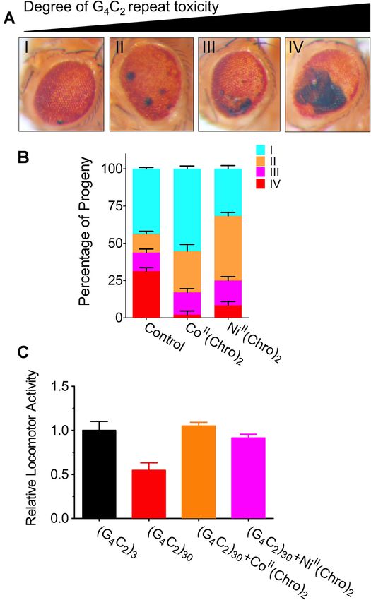
Nucleic Acids Research, 2021, Vol. 49, No. 16 9529
generated in previously published work (14). All Drosophila same sequence. The all-atom root-mean-square deviation
lines were maintained, and crosses were performed in stan- (RMSD) of ideal A-form DNA to the G4C2 DNA and
dard medium at 25◦ C (35). NiII (Chro)2 and CoII (Chro)2 G4C2 RNA-DNA hybrid duplexes was 1.3 and 1.6 Å,
were dissolved in DMSO (Sigma), and then added to green respectively. Superimposing the G4C2 DNA duplex with
coloured food (Kroger), an indicator for better mixture that of the hybrid duplex resulted in an all-atom RMSD of
with fly food, which was administered to the fly food. The 1.4 Å. These results suggested that the three structures were
same amount of DMSO was added to the fly food as an globally identical, albeit with significant local differences
internal control. The progeny were collected and aged (Figure 2C). The crystal structure of the G4C2 DNA
to 7 days. The screen was performed by scoring the eye duplex is remarkable in that the structure exists as a hybrid
phenotype, which was visualized using light microscopy. A-form with some B-form character, having average twist
For the Drosophila activity assay, equal amounts of males angles and rise steps of 35◦ and 3.3 Å, respectively, in the
and females were individually placed in Drosophila Activity central d(GGCC) core (Figure 2D), which is characteristic
Monitoring System (TriKinetics Inc, Waltham, MA, USA) of a B-DNA structure and looking down the centre of the
Downloaded from https://academic.oup.com/nar/article/49/16/9526/6219111 by guest on 15 October 2021
testing chambers that had been capped with regular food helix, it has a B-like crowded appearance. Interestingly, the
at one end. The flies were grown on a 12-h:12-h light: dark GpG and CpG side steps of the d(GGGCCG) core in the
cycle at 25◦ C for 4 weeks. Locomotor data were collected G4C2 DNA duplex have helical twists of 25◦ and 26◦ re-
in the light cycle at 25◦ C. Locomotor activity averages of spectively, an unwinding of ∼10◦ from B-DNA. Compared
each day during the fourth week were calculated. Data to the G4C2 DNA duplex, significant unwinding of base
were assayed in eight flies from each group at a time. pairs is also observed in the GpC and CpC steps of the
central d(GGCC) core for the G4C2 RNA–DNA hybrid
RESULTS duplex, with lower twist angles of 32◦ and 29◦ , respectively.
Local differences in translational parameters were also
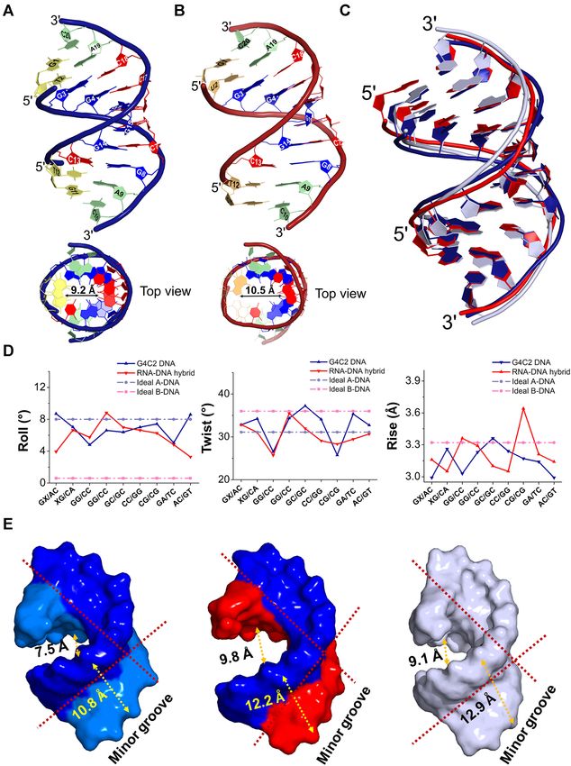
The crystal structures of the ALS/FTD-associated G4C2 re- observed in the central core of both duplexes. Values of
peat DNA motif and RNA–DNA hybrid duplexes reveal that the stagger parameter for the DNA duplex are close to
these duplexes adopt A-DNA-like conformations those for B-DNA (Supplementary Figure S2). In contrast,
To provide insight into higher-order structure for- the stagger values of the G4C2 RNA–DNA hybrid were
mation of the G4C2 motif, the crystal structures of negative and are closer to those for A-DNA. In order to
d(GTGGGCCGAC)/d(GTCGGCCCAC) (G4C2 DNA) better understand the effect of the central GGGCCG core
and r(GUGGGCCGAC)/d(GTCGGCCCAC) (G4C2 on major and minor groove geometry, average widths of
RNA-DNA hybrid) were solved at resolutions of 1.58 the major and minor grooves were calculated as interstrand
Å and 1.78 Å, respectively. The signature (GGGCCG)n P–P distances using Web 3DNA 2.0 analysis (Figure 2E)
sequence corresponds to the G4C2 motif, which comprises (27). Both structures have the minor groove wider than the
a central d(GGCC) motif flanked by two G:C base pairs major groove in the central core, which is closer to typical
embedded in the DNA. The structures include all atoms in A-DNA. It is notable that the G4C2 DNA duplex is bent to
the final refinement with no obvious indication of disorder, a greater extent than that of the G4C2 RNA–DNA hybrid,
as shown by the clear electron density map (Supplementary exposing more minor groove surface area. We suggest that
Figure S1). The oligonucleotides self-assemble into a this feature could act as a hot spot and may be important
single anti-parallel duplex for the G4C2 DNA and G4C2 for DNA recognition by various types of ligands.
RNA–DNA hybrid duplex structures in each asymmetric
unit. Duplexes were numbered from G1 to C10 in one
Crystal packing, a hydration network, and the flexibility of
strand and G11 to C20 in the complementary strand
the G4C2 DNA duplex minor groove have created hot spots
(Figure 2A,B). The crystal structures of both duplexes
for ligand binding
exhibit A-type-like DNA duplex structures with similar
widths at the major and minor grooves, which are also The ‘hot-spot’ feature for ligand binding has been further
revealed in backbone conformational angles and helical confirmed in the crystal packing of the G4C2 DNA du-
parameters (36). For example, the average glycosyl torsion plex, which exhibits a familiar ‘base-pair into minor groove’
angles ( ) of the DNA and RNA–DNA hybrid duplexes type packing that has been previously reported for DNA
have values of -161◦ and -152◦ respectively, with the sugar duplexes (Supplementary Figure S3) (37). In this crystal
pucker for all bases being well preserved in the C3 -endo packing environment, one end of an asymmetric unit du-
conformation (Supplementary Table S2). The roll angles plex interacts with the bases of other duplexes into the mi-
in the G4C2 motif pairs of the G4C2 DNA and G4C2 nor groove. Interestingly, the asymmetric unit duplex in
RNA–DNA hybrid duplexes were approximately 4–8◦ , G4C2 DNA showed direct interactions with the central G4-
which resulted in a clear kink of the DNA structures C17, G5-C16, C6-G15 and C7-G14 base-pair core region
toward the major groove. The average helical twist values through hydrogen bonding, water-mediated H-bonds and
of the G4C2 DNA and G4C2 RNA–DNA hybrid duplexes van der Waals interactions (Figure 3A). Terminal G1* and
were calculated to be 33◦ and 31◦ , respectively. This average C10* bases form H-bonding and extensive water-mediated
twist value corresponds to unwinding angles from B-DNA interactions with the G5-C16 and C17 bases. Addition-
of 3◦ and 5◦ , respectively (36). To compare the crystal ally, residues G11* and C20* from another asymmetric unit
structures, a uniform model of the ideal A-form was built formed direct hydrogen bonds and water-mediated con-
using the Discovery studio client package v19.1 with the tacts with the C6-G15 and C7-G14 base pairs. In con-
9530 Nucleic Acids Research, 2021, Vol. 49, No. 16

                                                                                                                                                               Downloaded from https://academic.oup.com/nar/article/49/16/9526/6219111 by guest on 15 October 2021
Figure 2. Overall structure features of the G4C2 motif-containing DNA and RNA-DNA hybrid duplexes in comparison with typical A-form duplex.
A crystal structure overview of (A) d(GTGGGCCGAC)/ d(GTCGGCCCAC) DNA duplex (G4C2 DNA) represented in density blue cartoon and (B)
r(GUGGGCCGAC)/d(GTCGGCCCAC) hybrid duplex (G4C2 RNA–DNA hybrid) represented in red cartoon as viewed from the side. The two duplexes
are numbered from G1 to C10 in one strand and G11 to C20 in the complementary strand. Central -GGGCCG- core residues are shown in blue and red
stick representation while the terminal residues are coloured in green and yellow sticks. The top view of each duplex is shown below with the inner diameter
of central hole. (C) Superimposition of crystal structures of G4C2 DNA duplex (density blue), G4C2 RNA–DNA hybrid (red) and a typical A-form DNA
duplex (light-blue) showing the overlap between these structures. (D) Prominent variations in the roll (◦ ), twist (◦ ) and rise (Å) DNA parameters are
observed in the crystal structure of G4C2 DNA (blue) and G4C2 RNA-DNA hybrid (red) duplexes compared to typical A- and B-form DNA duplexes.
(E) Surface representation of minor and major grooves of G4C2 DNA duplex, G4C2 RNA–DNA hybrid and a typical A-DNA duplex showing bending of
backbone. The DNA alone model is represented by a blue surface, RNA–DNA hybrid as red (RNA strand) and blue (DNA strand) surface and a typical
A-form DNA in light-blue surface representation.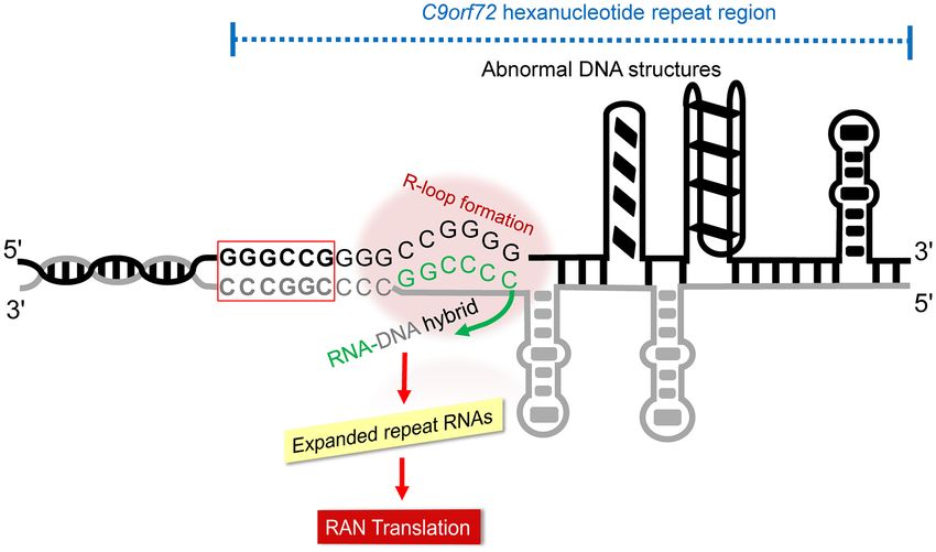
Nucleic Acids Research, 2021, Vol. 49, No. 16 9531

                                                                                                                                                            Downloaded from https://academic.oup.com/nar/article/49/16/9526/6219111 by guest on 15 October 2021
Figure 3. The detailed crystal packing shows a familiar ‘base-pair into minor groove’ type of packing in both G4C2 DNA and RNA–DNA hybrid duplexes.
(A) The two-fold symmetry of G4C2 DNA shows strong crystal contacts with the central G4-C17, G5-C16, C6-G15 and C7-G14 base-pairs which resulted
into the ‘end-to-central’ type of packing environment. The DNA duplex is shown in density blue cartoon representation while the asymmetric unit duplexes
are shown by red and yellow cartoons. (B) The two-fold symmetry of the G4C2 RNA–DNA hybrid duplex shows a distinct crystal packing environment
where the duplexes in an asymmetric unit are oriented away from the central core. The RNA–DNA hybrid duplex is shown in red colour while the
asymmetric unit duplexes are shown in blue and yellow colours. The residues in an asymmetric unit are highlighted with an asterisk (*) sign. The hydrogen
bonds and water-mediated contacts are shown in black dashed lines and the water molecules are shown as cyan spheres. Note that some symmetry related
duplexes are not shown for clarity.

trast, the crystal packing interactions in the G4C2 RNA–                       MetalII (Chro)2 complexes specifically stabilized the G4C2
DNA hybrid were oriented away from the central core (Fig-                      DNA duplex and not the RNA–DNA hybrid
ure 3B). Although the residues in the G4C2 RNA–DNA
hybrid showed hydrogen-bonding interactions, there were                        To determine the stabilization effects and selectivity to-
no water-mediated interactions. The total number of crys-                      wards the G4C2 repeats, several well-known DNA bind-
tal packing interactions in the G4C2 DNA duplex, RNA–                          ing compounds with G4C2 repeat-containing DNA duplex
DNA hybrid and other typical A-DNA-like structures is                          selectivity were screened at a fixed compound:DNA stoi-
given in Supplementary Table S3. Intriguingly, among these                     chiometry of 1:1 (Figure 4A–D). By measuring the differ-
structures, only the G4C2 DNA duplex showed a pecu-                            ence in melting temperature (Tm ) of the G4C2 repeat se-
liar ‘end-to-central’ type packing environment. This type                      quence in the presence of these compounds, it was found
of crystal packing has arisen in the G4C2 DNA duplex                           that the anthracene-based metal complexes, NiII (Chro)2
because the DNA is more bent at the central core (the                          and CoII (Chro)2 best stabilized the G4C2 repeat DNA mo-
sum of roll angles is, ∼32.2◦ ), which exposes the residues                    tif duplex with Tm value of 8.0◦ C, while the other com-
in the central core to the minor groove for terminal base                      pounds had a Tm of
9532 Nucleic Acids Research, 2021, Vol. 49, No. 16
Downloaded from https://academic.oup.com/nar/article/49/16/9526/6219111 by guest on 15 October 2021
Figure 4. Chemical structures of the DNA binding ligands use for melting temperature assay with G4C2 DNA duplex. Equimolar concentrations (1:1
ratio of DNA:ligand) of DNA binding compounds including (A) anthracene-based chromomycin A3 [1], mithramycin [2]; (B) aziridine-based mitomycin
C [3], heterotetracyclic ellipticine [4] and acridine-based triaminotriazine-acridine conjugate [5]; (C) anthracycline-based daunorubicin [6] and doxorubicin
[7]; (D) cyclic peptide antibiotics actinomycin D [8] and echinomycin [9] are used for melting temperature assay. (E) Change in DNA melting temperatures
(Tm ) for G4C2 DNA duplex in the presence of NiII (chromomycin A3)2 (Ni-Chro), CoII (chromomycin A3)2 (Co-Chro) complex, NiII (mithramycin)2 (Ni-
Mith), CoII (mithramycin)2 (Co-Mith) complexes, mitomycin C, daunomycin, ellipticine, actinomycin D, echinomycin, doxorubicin and a triaminotriazine-
acridine conjugate.
CoII (Chro)2 are able to distinguish between the specific as observed in the crystal packing. Although the two com-
structural differences of G4C2 DNA and hybrid duplexes plexes in this structure are significantly different, the central
with the same sequence. GGGCCG tetranucleotide core, upon NiII (Chro)2 binding,
adopts A-DNA-like characteristics, as reflected by the
glycosyl torsion angle values that are in the range -156◦ to
The NiII (Chro)2 complex induces conformational changes in
-179◦ with C3 -endo sugar pucker conformations for most
the G4C2 DNA duplex
of the residues and show a high degree of similarity, with an
To understand the structural basis for the pref- RMSD of 0.5 Å (Supplementary Figure S7, Supplementary
erence of NiII (Chro)2 (Figure 5A) for G4C2 Table S4). This is very distinct from the previously-reported
DNA, the crystal structure of the NiII (Chro)2 - MgII (Chro)2 -d(TTGGCCAA)2 complex structure [Protein
d(TTGGGCCGAA/TTCGGCCCAA) complex was Data Bank (PDB): 1VAQ], which adopts a B-like character
solved at 2.95 Å resolution (Supplementary Figure S6). with a pronounced kink at the central 4-bp segment (41).
One asymmetric unit contains two independent Chro-DNA The overall DNA structure of the complexes preserves
complexes that are packed through van der Waals contacts right-handed helical features with significant unwinding of
between two Chro moieties, as shown in Figure 5B; these base pairs at the GpG and CpC steps of the GGCC central
complexes are labelled CPX1 and CPX2, respectively. There core. This is in contrast to the structure of G4C2 DNA,
are significant differences between the conformations of which shows unwinding at the side steps of the GGGCCG
CPX1 and CPX2, as indicated by the 3.2 Å RMSD between duplex core. Upon ligand binding to the central core of
them. CPX2 has two terminal adenine base pairs (A9 and the DNA duplex, the roll angles among the GGGCCG
A10) extruding out of the duplex structure that are further core steps are lower than those of the G4C2 DNA alone
stabilized by intermolecular triplet hydrogen bonding structure, which causes straightening of the DNA duplex
between (A:T):T bases from the two independent strands, towards the minor groove and a consequential contractionNucleic Acids Research, 2021, Vol. 49, No. 16 9533
Downloaded from https://academic.oup.com/nar/article/49/16/9526/6219111 by guest on 15 October 2021
Figure 5. Structural overview of the NiII (Chro)2 -d(TTGGGCCGAA)2 complex. (A) Chemical structure of the anthracene-based chromomycin A3 (Chro)
dimer formation in the presence of Nickel(II) ions (NiII (Chro)2 ). (B) Each asymmetric unit contains two independent complexes, CPX1 and CPX2 as
shown in light teal and pink cartoons, respectively. Two Chro dimers are represented by green and yellow sticks while NiII ions are shown as red spheres.
(C) Superimposition of the overall structures of G4C2 DNA duplex (blue) and CPX1 of NiII (Chro)2 -DNA (pink) shows the straightening of a DNA
backbone and narrowing of minor groove due to NiII (Chro)2 binding. The two structures thus exhibit significant variations in the roll (◦ ) DNA parameter,
a comparison shown by blue (G4C2 DNA) and pink lines (NiII (Chro)2 -d(TTGGGCCGAA)2 , CPX1), respectively.
of the minor groove (Figure 5C). The average minor-groove DNA molecules abutting their terminal base pairs onto the
width for each complex is 9.3 Å at the base pairs of the minor groove of other DNA duplexes in the native G4C2
GGGCCG duplex core, whereas the major groove widens DNA structure. The binding of NiII (Chro)2 to the central
up to 17.2 Å in each complex, which differs substantially GGCC sequence shows specific intermolecular hydrogen-
from the minor and major groove widths of the G4C2 bonding interactions between O8 of the anthracene and the
DNA alone structure, at 10.8 and 7.5 Å, respectively. N2 atom of the G5 (G15) base in CPX1 and CPX2 (Sup-
The structure of NiII (Chro)2 shows that the NiII metal plementary Figure S10A, B). Likewise, the two EO1 oxygen
ion is tetrahedrally coordinated to the O1 and O9 atoms of atoms of the trisaccharide E ring are hydrogen bonded with
the two anthracenes and the A-B disaccharide and C-D-E the N2 atoms of the G4 and G14 bases. Unlike previous ob-
trisaccharide moieties attached to the anthracenes extended servations, a dihedral angle of 71◦ between two anthracene
in opposite directions in the dimer conformation (Supple- monomers positions the O3 of the B ring in an orientation
mentary Figure S8). The anthracene moieties of the Chro that enables it to form hydrogen bonds with OP1 of base
dimer is sandwiched and mutually stacked between the C- G15 in CPX1 and 2 (Supplementary Table S5). Similarly,
D glycosidic linkage of the other Chro monomer and the O3 of the B ring also forms a hydrogen bond with OP1 of
ribose of C6, bringing the backbone of the DNA duplex to- G5 in CPX1, while the distance between O3 and OP1 of
wards the ligand direction in order to stabilize the ligand– G5 in CPX2 is slightly greater than the cut-off value as-
DNA complex (Supplementary Figure S9). This is similar signed to the hydrogen bonds (Supplementary Figure S10A,
to the packing interaction resulting from the native G4C2 B). In addition, the terminal intermolecular (A:T):T triplet9534 Nucleic Acids Research, 2021, Vol. 49, No. 16
base pair adds overall stabilization of the complex structure
(Supplementary Figure S10C). The coordination of N7 of
guanines G5 and G15 with NiII ions in both complexes fur-
ther stabilizes the duplexes (Supplementary Figure S11).
In addition to these interactions in the central GGCC
core, Chro also shows extended specificity for the flanking
G:C base pairs of the central GGCC region in the G4C2
motif, as suggested by MD simulations of 500 ps for both
complexes. The dynamic behaviour of these individual base
pairs in both complexes was determined in terms of RMSF
values. The RMSF plot indicates a similar residue fluctu-
ation profile for the central GGGCCG motif with an av-
erage RMSF of 1.1 Å, indicating higher stability due to
Downloaded from https://academic.oup.com/nar/article/49/16/9526/6219111 by guest on 15 October 2021
(Chro)2 binding (Supplementary Figure S12A). The ter-
minal residues T1:A20 and T2:A19 show extreme fluctua-
tions, suggesting that Chro binding did not affect the ter-
minal residues. Interestingly, this specificity was not ob-
served in the previously reported structure of MgII (Chro)2 -
d(TTGGCCAA) (PDB ID- 1VAQ), where the flanking base
pair is A:T (Supplementary Figure S12B). This suggests
that the NiII (Chro)2 complex has extended specificity due
to the indirect binding effects of the flanking G3:C18 and
G8:C13 base pairs in the G4C2 sequence. Larger stagger
and propeller twist values might cause these base pairs to
interact with NiII (Chro)2 (Supplementary Figure S13). The
base stacking interactions and steric clashes between Chro
sugars might also allow Chro to stabilize the overall struc-
ture. In addition, LigPlot+ analysis shows that the E ring
of the trisaccharide moiety forms a close van der Waals
contact with C1 of the deoxyribose sugar of C18 and G18
from CPX1 and CPX2, respectively (Supplementary Figure
S14). The unusually low value of the base-pair rise results
in increased stacking of the flanked G3:C18 and G8:C13
pairs in the G4C2 complexes, as indicated by the hydropho-
bic interactions. Overall, these interactions strengthen the
Figure 6. Effects of metalII (Chro)2 on G4C2 repeat toxicity in the
specific recognition of the GGGCCG repeat motif by the Drosophila model. (A) The expression of (G4 C2 )30 driven by the Gal4
NiII (Chro)2 complex. driver causes neuronal toxicity in the fly eye that is classified into four
categories (I = normal; II = slight, 30%) as shown under the light microscope. (B) NiII (Chro)2 and
MetalII (Chro)2 suppressed G4C2 repeat toxicity in a CoII (Chro)2 inhibits the neuronal toxicity caused by expanded G4C2 re-
Drosophila model of G4C2 repeat expansion peats in the fly eye. (C) NiII (Chro)2 and CoII (Chro)2 suppresses the lo-
comotor deficits caused by the expression G4C2 repeats in ok371-GAL4,
Given the specific recognition of G4C2 repeats by UAS-GGGGCC30 -EGFP and ok371-GAL4, UAS-GGGGCC3 -EGFP fly
NiII (Chro)2 and CoII (Chro)2 G4C2 repeat toxicity was models. Data were assayed in eight flies from each group at a time and the
tested using a previously established Drosophila model graphs were plotted with GraphPad Prism software (version 9.0.0).
of G4C2 repeats (14). In this model, 30 G4C2 repeats
are expressed using a tissue- or cell type-specific GAL4 expressed in motor neurons using ok371-GAL4, and the
driver and their expression leads to neuronal cell death locomotor activity of the following flies, ok371-GAL4,
and locomotor deficits. NiII (Chro)2 and CoII (Chro)2 were UAS-GGGGCC30 -EGFP and ok371-GAL4, and UAS-
mixed with fly food at different concentrations and tested GGGGCC3 -EGFP was determined. Both NiII (Chro)2 and
in gmr-GAL4 and UAS-GGGGCC30 -EGFP strains. As CoII (Chro)2 suppressed the locomotor deficits caused by
described previously (14), the neuronal toxicity caused the expression of G4C2 repeats (Figure 6C).
by 30 G4C2 repeats is classified into four categories (I =
normal; II = slight, < 5%; III = moderate, 5%–30%; IV =
DISCUSSION
severe > 30%) (Figure 6A). At 500 nM, both NiII (Chro)2
and CoII (Chro)2 inhibited the neuronal toxicity caused Many studies support a gain of toxicity in which the
by the expanded G4C2 repeats in the fly eye (Figure 6B), C9orf72 repeat RNAs play a crucial role in ALS/FTD
with the CoII (Chro)2 complex almost completely sup- pathogenesis (42–44). Consequently, reducing toxic repeat
pressing the most severe toxicity. To further determine the RNA transcripts or their translated aberrant proteins by
impact of NiII (Chro)2 and CoII (Chro)2 on motor neurons targeting C9orf72 repeat RNAs with small molecules is
expressing the G4C2 repeat that were most affected in a promising strategy, and the most advanced in terms of
patients, both normal and expanded G4C2 repeats were eventual clinical development (20,45,46). However, target-Nucleic Acids Research, 2021, Vol. 49, No. 16 9535
ing RNA molecules may possess several challenges; most other A-form DNA duplexes containing the central GGCC
of the target RNAs are expressed at a low level with a motif (Figure 7C and D) (58). In one of the A-DNA du-
short half-life and RNA conformational dynamics in cells, plex structures, the end-to-side type packing environment
making it difficult for small molecule ligands to preferen- interacts two base pairs away from the central GpC step
tially recognize their target. However, the development of (Figure 7E) (59). The crystal packing interactions in G4C2
small molecules to modulate repeat expansions and related DNA are mainly stabilised through direct hydrogen bond-
instabilities or inhibit the production of toxic transcripts ing, water-mediated interactions, and van der Waals con-
and proteins by targeting specific repeat DNA-containing tacts. Interestingly, the G4C2 RNA–DNA hybrid possesses
duplexes has been shown as another potential therapeutic an extra hydroxyl moiety on the RNA strand, which re-
approach against neurological diseases (47–49). For exam- sults in a greater number of hydrogen-bonded interactions
ple, Nguyen et al. have reported several triaminotriazine- at the crystal packing interface. Other A-DNA duplexes
based small molecule ligands that inhibit abnormal expan- showed less hydrogen bonding and van der Waals interac-
sion of CTG repeats associated with myotonic dystrophy tions at the crystal packing interface (Supplementary Table
Downloaded from https://academic.oup.com/nar/article/49/16/9526/6219111 by guest on 15 October 2021
type 1 pathobiology by inhibiting transcription through S3). The G4C2 DNA duplex is more bendable and flexible
DNA binding, as well as by binding to the toxic RNA to in- than that of the G4C2 RNA–DNA hybrid, which has more
hibit aberrant protein binding (50). Siboni et al. have shown surface area of the minor groove exposed to accommodate
that actinomycin D is able to reduce CUG repeat-associated cogent ligands, such as proteins or small molecules, to in-
toxic RNA in in vitro and in vivo models by inhibiting the teract with the minor groove and make it an ideal target for
transcription of CTG repeat expansion (51). Therefore, un- appropriately-shaped small molecule binding.
derstanding the structural details of sequence-specific bind- The extensive molecular dynamics and simulations stud-
ing modes in repeat-associated DNA including the G4C2 ies by Sagui et al. reported that the G4C2 related sequences
DNA duplex and G4C2 RNA–DNA hybrid might provide from different reading frames formed various secondary
useful leads for designing small molecules targeting these structures containing G:G mismatches in the sense and C:C
special sequences. In the current study, two relevant crystal mismatches in the antisense strand (7,8). We report here
structures of the G4C2 repeat motif containing DNA and that the anthracene-based metal complexes (NiII (Chro)2
RNA–DNA hybrid duplexes were solved. These show that and CoII (Chro)2 ) selectively target the G4C2 repeat DNA
the G4C2 DNA duplex is more flexible and exhibits an A- motif duplexes, which provides the GGCC and CCGG
type-like DNA duplex, albeit with a number of base mor- tracts and preferred recognition site for these compounds
phology parameter characteristics typical of B-DNA, such (60). Therefore, we analysed the stabilizing effects of NiII -
as twist and base-pair rise steps. (Chro)2 on the G4C2 repeat related motifs while hair-
This structure resembles an intermediate in the A-B tran- pin structure formation that contains the central GGCC
sition with a prominent kink of over 32◦ , a water shell in the tract flanked by two G:C base-pairs, two G:G mismatch
minor groove, indicating a hot-spot for toxic RNA tran- pairs, one G:C base-pair and one G:G mismatch pair for
scription (52). Previous studies have shown assemblies of the sense strand, and the central CCGG tract flanked by
structured water molecules with cyclic and linear water ar- two C:C mismatch pairs for the antisense strand (Supple-
rangements that are observed in the major groove and mi- mentary Figure S15A). The melting temperature analysis
nor groove of B-DNA, respectively (39,53). However, an A- suggested that the NiII -(Chro)2 complex can firmly sta-
type double helix usually forms when DNA is dehydrated, bilize all the sense G4C2 repeat related motifs with the
but is also found in DNA/RNA hybrids (54). Compared to GGCC tract while the Chro complex had minor stabiliz-
most studied mixed DNA/RNA hybrids that are found in ing effects on the antisense G4C2 repeat motif with the
the A form, the G4C2 RNA–DNA hybrid duplex is more CCGG tract (Supplementary Figure S15B). However, in
rigid, although it is also in a typical A form; the DNA strand contrast to the same ligand-free DNA structure, the bind-
adopts a conformation close to that of duplex RNA, with ing of NiII (Chro)2 to the GGGCCG sequence showed in-
C3 -endo sugar puckers and a typical A-type helical back- creased additional specificity through hydrophobic inter-
bone conformation (55–57). The crystal packing interac- actions between NiII (Chro)2 and the flanked G:C base
tions for the current G4C2 DNA and G4C2 RNA–DNA pairs of the central GGCC tract. It was proposed that
hybrid were analysed and compared to other A-form DNA the binding of NiII (Chro)2 to the GGGCCG sequence in-
duplexes. The crystal packing interactions of G4C2 DNA duced contraction of the minor groove and straighten-
and G4C2 RNA-DNA hybrid duplexes shows a general ing of the DNA backbone by the induced fit paradigm.
‘base-pair into the minor groove’ type packing for the A- Previously, the recognition of NiII (Chro)2 of CCG trinu-
form duplexes. This crystal packing environment may be cleotide repeats had been identified as following the classic
further categorised into two types for DNA and RNA– induced-fit mechanism by triggering large-scale DNA de-
DNA hybrid duplexes. In the first type, the G4C2 DNA du- formations, with the cytosine extruded out from the helix
plex shows ‘end-to-centre’ type packing interactions, where (61). The binding of the NiII (Chro)2 complex to the nascent
one end of a symmetry-related duplex formed strong inter- G4C2 expanded strand might therefore inhibit toxic tran-
molecular interactions with the central G5 and C6 bases script formation in the resulting abnormal G4C2 expan-
of the DNA (Figure 7A). Second, the G4C2 RNA–DNA sion sequence. Drosophila is a widely used organism for the
hybrid duplex showed ‘end-to-side’ type packing, in which modelling of various neurological diseases, such as Parkin-
symmetry-related duplexes interacted with G4 and C7 bases son’s and Huntington’s diseases and ALS/FTD (62,63).
that are one step away from the central G5pC6 step bases Therefore, a Drosophila model was used that expressed
(Figure 7B). This type of packing has also been found in up to 30 G4C2 repeats, in order to evaluate the possi-9536 Nucleic Acids Research, 2021, Vol. 49, No. 16
Downloaded from https://academic.oup.com/nar/article/49/16/9526/6219111 by guest on 15 October 2021
Figure 7. Schematic representation of ‘base-pair into minor groove’ type crystal packing interactions in different A-form DNA duplex structures. (A)
The G4C2 DNA duplex showed ‘end-to-center’ type of packing interactions where the ends of DNA duplexes from different symmetry unit interact with
the central GGCC residue core in the DNA. (B) G4C2 RNA-DNA hybrid showed ‘end-to-side’ type of packing environment where the interactions of
different duplexes from crystal symmetry are away from the central GGCC core. (C–E) Other typical A-DNA duplexes showed ‘end-to-side’ type of crystal
packing interactions. Arrows indicate symmetry related DNA duplexes. The DNA bases are colored as adenine-orange, thymine-brown, cytosine-green
and guanine-blue. The respective PDB IDs for each structure are given in bold, red letters.
bility of anthracene-metal complexes targeting ALS/FTD are also thankful to Dr. Shue-Shing Chen, Biophysics Core
repeat-related sequences. These results demonstrate that Facility, Institute of Molecular Biology, Academia Sinica
NiII (Chro)2 and CoII (Chro)2 inhibit neuronal toxicity and and the staffs of Technology Commons, College of Life Sci-
suppress locomotor deficits caused by the expression of ence, NTU.
G4C2 repeats in the fly model and raises the possibility of
using a DNA-binding compound to treat G4C2-related dis-
ease pathogenesis. FUNDING
In summary, it has been demonstrated that the G4C2 mo- Ministry of Science and Technology, Taiwan [109–2628-M-
tifs of C9orf72 repeat-expanded DNA provide an appro- 005–001-MY4, 109–2311-B-005–007-MY3 to M.-H.H.].
priate structural environment with considerable flexibility, Funding for open access charge: Ministry of Science and
serving as hot spots for selective drug binding. It is no- Technology, Taiwan.
table that of the various structurally diverse DNA-binding Conflict of interest statement. None declared.
compounds evaluated for their ability to stabilize the re-
peat motif, only the two anthracene-based metal complexes
showed substantial effects. At this point even though it is REFERENCES
not yet possible to definitively state whether the biological 1. DeJesus-Hernandez,M., Mackenzie,I.R., Boeve,BF., Boxer,A.L.,
responses observed here are due to the metal complexes in- Baker,M., Rutherford,N.J., Nicholson,A.M., Finch,NiCA.,
teracting with hairpin, duplex or quadruplex forms of the Flynn,H., Adamson,J. et al. (2011) Expanded GGGGCC
hexanucleotide repeat in noncoding region of C9ORF72 causes
G4C2 repeat expansion, we suggest that this has revealed an chromosome 9p-linked FTD and ALS. Neuron, 72, 245–256.
alternative drug discovery model, directly targeting G4C2 2. Renton,AE., Majounie,E., Waite,A., Simón-Sánchez,J., Rollinson,S.,
repeat DNAs, that could increase the repertoire of lead Gibbs,J.R., Schymick,JC., Laaksovirta,H., van Swieten,J.C.,
compounds for developing new therapeutic regimes against Myllykangas,L. et al. (2011) A hexanucleotide repeat expansion in
C9ORF72 is the cause of chromosome 9p21-linked ALS-FTD.
ALS and FTD diseases. Neuron, 72, 257–268.
3. Babić,L., Župunski,M.V., Kirincich,J., Smilović,D., Hortobágyi,T.,
DATA AVAILABILITY Hof,P.R. and Šimić,G. (2019) Molecular mechanisms of
neurodegeneration related to C9orf72 hexanucleotide repeat
The atomic coordinates and structure factors for expansion. Behav. Neurol., 2019, 2909168.
the reported crystal structures have been deposited 4. Jiang,J. and Ravits,J. (2019) Pathogenic mechanisms and therapy
in the PDB under the accession numbers 6L75 for development for C9orf72 amyotrophic lateral
d(GTGGGCCGAC/GTCGGCCCAC) (G4C2 DNA du- sclerosis/frontotemporal dementia. Neurotherapeutics, 16, 1115–1132.
5. Šket,P., Pohleven,J., Kovanda,A., Štalekar,M., Župunski,V.,
plex), 7BPV for r(GUGGGCCGAC)/d(GTCGGCCCAC) Zalar,M., Plavec,J. and Rogelj,B. (2015) Characterization of DNA
(G4C2 RNA-DNA hybrid duplex) and 6L76 for the G-quadruplex species forming from C9ORF72 G4C2-expanded
NiII (Chro)2 -d(TTGGGCCGAA/TTCGGCCCAA) com- repeats associated with amyotrophic lateral sclerosis and
plex. frontotemporal lobar degeneration. Neurobiol. Aging, 36, 1091–1096.
6. Brčić,J. and Plavec,J. (2018) NMR structure of a G-quadruplex
formed by four d(G4C2) repeats: insights into structural
SUPPLEMENTARY DATA polymorphism. Nucleic Acids Res., 46, 11605–11617.
7. Zhang,Y., Roland,C. and Sagui,C. (2017) Structure and dynamics of
Supplementary Data are available at NAR Online. DNA and RNA double helices obtained from the GGGGCC and
CCCCGG hexanucleotide repeats that are the hallmark of
ACKNOWLEDGEMENTS C9FTD/ALS diseases. ACS Chem. Neurosci., 8, 578–591.
8. Zhang,Y., Roland,C. and Sagui,C. (2018) Structural and dynamical
We sincerely thank the National Synchrotron Radiation Re- characterization of DNA and RNA quadruplexes obtained from the
search Center (Taiwan) staff for X-ray data collection. We GGGGCC and GGGCCT hexanucleotide repeats associated withNucleic Acids Research, 2021, Vol. 49, No. 16 9537
C9FTD/ALS and SCA36 diseases. ACS Chem. Neurosci., 9, 26. Murshudov,G.N., Vagin,A.A. and Dodson,E.J. (1997) Refinement of
1104–1117. macromolecular structures by the maximum-likelihood method. Acta
9. Waite,A.J., Bäumer,D., East,S., Neal,J., Morris,H.R., Ansorge,O. Crystallograph. Section D, Biol. Crystallograph., 53, 240–255.
and Blake,D.J. (2014) Reduced C9orf72 protein levels in frontal 27. Li,S., Olson,W.K. and Lu,X.-J. (2019) Web 3DNA 2.0 for the
cortex of amyotrophic lateral sclerosis and frontotemporal analysis, visualization, and modeling of 3D nucleic acid structures.
degeneration brain with the C9ORF72 hexanucleotide repeat Nucleic Acids Res., 47, W26–W34.
expansion. Neurobiol. Aging, 35, 1779. 28. Blanchet,C., Pasi,M., Zakrzewska,K. and Lavery,R. (2011)
10. Frick,P., Sellier,C., Mackenzie,I.R.A., Cheng,C.-Y., CURVES+ web server for analyzing and visualizing the helical,
Tahraoui-Bories,J., Martinat,C., Pasterkamp,R.J., Prudlo,J., backbone and groove parameters of nucleic acid structures. Nucleic
Edbauer,D., Oulad-Abdelghani,M. et al. (2018) Novel antibodies Acids Res., 39, W68–W73.
reveal presynaptic localization of C9orf72 protein and reduced 29. Laskowski,R.A. and Swindells,M.B. (2011) LigPlot+: multiple
protein levels in C9orf72 mutation carriers. Acta Neuropathol. ligand–protein interaction diagrams for drug discovery. J. Chem. Inf.
Commun., 6, 72. Model., 51, 2778–2786.
11. Su,Z., Zhang,Y., Gendron,TF., Bauer,PO., Chew,J., Yang,W.-Y., 30. Olson,W.K., Bansal,M., Burley,S.K., Dickerson,R.E., Gerstein,M.,
Fostvedt,E., Jansen-West,K., Belzil,VV., Desaro,P. et al. (2014) Harvey,S.C., Heinemann,U., Lu,X.-J., Neidle,S., Shakked,Z. et al.
Discovery of a biomarker and lead small molecules to target (2001) A standard reference frame for the description of nucleic acid
Downloaded from https://academic.oup.com/nar/article/49/16/9526/6219111 by guest on 15 October 2021
r(GGGGCC)-associated defects in c9FTD/ALS. Neuron, 83, base-pair geometry. J. Mol. Biol., 313, 229–237.
1043–1050. 31. Brooks,B.R., Bruccoleri,R.E., Olafson,B.D., States,D.J.,
12. Barker,H.V., Niblock,M., Lee,Y.-B., Shaw,C.E. and Gallo,J.-M. Swaminathan,S. and Karplus,M. (1983) CHARMM: a program for
(2017) RNA Misprocessing in C9orf72-Linked Neurodegeneration. macromolecular energy, minimization, and dynamics calculations. J.
Front. Cell Neurosci., 11, 195. Comput. Chem., 4, 187–217.
13. Lee,Y.-B., Chen,H.-J., Peres,JN., Gomez-Deza,J., Attig,J., 32. Jorgensen,W.L., Chandrasekhar,J., Madura,J.D., Impey,R.W. and
Štalekar,M., Troakes,C., Nishimura,A.L., Scotter,E.L., Vance,C. Klein,M.L. (1983) Comparison of simple potential functions for
et al. (2013) Hexanucleotide repeats in ALS/FTD form simulating liquid water. J. Chem. Phys., 79, 926–935.
length-dependent RNA foci, sequester RNA binding proteins, and 33. Darden,T., York,D. and Pedersen,L. (1993) Particle mesh Ewald: An
are neurotoxic. Cell Rep., 5, 1178–1186. N·log(N) method for Ewald sums in large systems. J. Chem. Phys., 98,
14. Xu,Z., Poidevin,M., Li,X., Li,Y., Shu,L., Nelson,D.L., Li,H., 10089–10092.
Hales,C.M., Gearing,M., Wingo,T.S. et al. (2013) Expanded 34. May,R.A. and Stevenson,K.J. (2009) Software Review of Origin 8. J.
GGGGCC repeat RNA associated with amyotrophic lateral sclerosis Am. Chem. Soc., 131, 872–872.
and frontotemporal dementia causes neurodegeneration. Proc. Natl. 35. Jin,P., Zarnescu,D.C., Zhang,F., Pearson,C.E., Lucchesi,J.C.,
Acad. Sci., 110, 7778–7783. Moses,K. and Warren,S.T. (2003) RNA-mediated neurodegeneration
15. Nguyen,L., Cleary,J.D. and Ranum,L.P.W. (2019) Repeat-associated caused by the fragile X premutation rCGG repeats in Drosophila.
non-ATG translation: molecular mechanisms and contribution to Neuron, 39, 739–747.
neurological disease. Annu. Rev. Neurosci., 42, 227–247. 36. Neidle,S. (2008) In: Principles of Nucleic Acid Structure. Academic
16. Zamiri,B., Reddy,K., Macgregor,R.B. Jr and Pearson,C.E. (2014) Press, NY, pp. 38–80.
TMPyP4 porphyrin distorts RNA G-quadruplex structures of the 37. Wang,A.H.-J. and Teng,M.K. (1988) Crystallization and crystal
disease-associated r(GGGGCC)n repeat of the C9orf72 gene and packing analysis of DNA oligonucleotides. J. Cryst. Growth, 90,
blocks interaction of RNA-binding proteins. J. Biol. Chem., 289, 295–310.
4653–4659. 38. Liu,Y., Kumar,A., Depauw,S., Nhili,R., David-Cordonnier,M.-H.,
17. Wang,Z.-F., Ursu,A., Childs-Disney,J.L., Guertler,R., Yang,W.-Y., Lee,M.P., Ismail,M.A., Farahat,A.A., Say,M., Chackal-Catoen,S.
Bernat,V., Rzuczek,S.G., Fuerst,R., Zhang,Y.-J., Gendron,T.F. et al. et al. (2011) Water-mediated binding of agents that target the DNA
(2019) The hairpin form of r(G4C2)exp in c9ALS/FTD is minor groove. J. Am. Chem. Soc., 133, 10171–10183.
repeat-associated non-ATG translated and a target for bioactive small 39. Wei,D., Wilson,W.D. and Neidle,S. (2013) Small-molecule binding to
molecules. Cell Chem. Biol., 26, 179–190. the DNA minor groove is mediated by a conserved water cluster. J.
18. Donnelly,CJ., Zhang,P.-W., Pham,JT., Haeusler,AR., Mistry,NA., Am. Chem. Soc., 135, 1369–1377.
Vidensky,S., Daley,E.L., Poth,EM., Hoover,B., Fines,D.M. et al. 40. Hsu,C.-W., Chuang,S.-M., Wu,W.-L. and Hou,M.-H. (2012) The
(2013) RNA toxicity from the ALS/FTD C9ORF72 expansion is crucial role of divalent metal ions in the DNA-acting efficacy and
mitigated by antisense intervention. Neuron, 80, 415–428. inhibition of the transcription of dimeric chromomycin A3. PLoS
19. Ursu,A., Wang,K.W., Bush,J.A., Choudhary,S., Chen,J.L., One, 7, e43792.
Baisden,J.T., Zhang,Y.-J., Gendron,T.F., Petrucelli,L., Yildirim,I. 41. Hou,M.H., Robinson,H., Gao,Y.G. and Wang,A.H.J. (2004) Crystal
et al. (2020) Structural features of small molecules targeting the RNA structure of the [Mg2+-(chromomycin A3)2]–d(TTGGCCAA)2
repeat expansion that causes genetically defined ALS/FTD. ACS complex reveals GGCC binding specificity of the drug dimer chelated
Chem. Biol., 15, 3112–3123. by a metal ion. Nucleic Acids Res., 32, 2214–2222.
20. Simone,R., Balendra,R., Moens,T.G., Preza,E., Wilson,K.M., 42. Kumar,V., Hasan,G.M. and Hassan,M.I. (2017) Unraveling the role
Heslegrave,A., Woodling,N.S., Niccoli,T., Gilbert-Jaramillo,J., of RNA mediated toxicity of C9orf72 repeats in C9-FTD/ALS.
Abdelkarim,S. et al. (2018) G-quadruplex-binding small molecules Front. Neurosci., 11, 711–711.
ameliorate C9orf72 FTD/ALS pathology in vitro and in vivo. EMBO 43. Reddy,K., Zamiri,B., Stanley,S.Y., Macgregor,R.B. Jr and
Mol. Med., 10, 22–31. Pearson,C.E. (2013) The disease-associated r(GGGGCC)n repeat
21. Lu,Y., Dohno,C. and Nakatani,K. (2020) Recognition of expanded from the C9orf72 gene forms tract length-dependent uni- and
GGGGCC hexanucleotide repeat by synthetic ligand through multimolecular RNA G-quadruplex structures. J. Biol. Chem., 288,
interhelical binding. Biochem. Biophys. Res. Commun., 531, 56–61. 9860–9866.
22. Shibata,T., Murakami,E. and Nakatani,K. (2018) 44. Fratta,P., Mizielinska,S., Nicoll,A.J., Zloh,M., Fisher,E.M.C.,
1,3-Di(quinolin-2-yl)guanidine binds to GGCCCC hexanucleotide Parkinson,G. and Isaacs,A.M. (2012) C9orf72 hexanucleotide repeat
repeat DNA in C9ORF72. Bioorg. Med. Chem. Lett., 28, 2364–2368. associated with amyotrophic lateral sclerosis and frontotemporal
23. Murphy,J.H. and Trapane,T.L. (1996) Concentration and extinction dementia forms RNA G-quadruplexes. Sci. Rep., 2, 1016.
coefficient determination for oligonucleotides and analogs using a 45. Jiang,J., Zhu,Q., Gendron,TF., Saberi,S., McAlonis-Downes,M.,
general phosphate analysis. Anal. Biochem., 240, 273–282. Seelman,A., Stauffer,JE., Jafar-nejad,P., Drenner,K., Schulte,D. et al.
24. Otwinowski,Z. and Minor,W. (1997) In: Methods in Enzymology. (2016) Gain of toxicity from ALS/FTD-linked repeat expansions in
Academic Press, Vol. 276, pp. 307–326. C9ORF72 is alleviated by antisense oligonucleotides targeting
25. Adams,P.D., Afonine,P.V., Bunkoczi,G., Chen,V.B., Davis,I.W., GGGGCC-containing RNAs. Neuron, 90, 535–550.
Echols,N., Headd,J.J., Hung,L.W., Kapral,G.J., 46. Meyer,S.M., Williams,C.C., Akahori,Y., Tanaka,T., Aikawa,H.,
Grosse-Kunstleve,R.W. et al. (2010) PHENIX: a comprehensive Tong,Y., Childs-Disney,J.L. and Disney,M.D. (2020) Small molecule
Python-based system for macromolecular structure solution. Acta recognition of disease-relevant RNA structures. Chem. Soc. Rev., 49,
Crystallograph. Section D, Biol. Crystallograph., 66, 213–221. 7167–7199.You can also read