Computational Approaches: An Underutilized Tool in the Quest to Elucidate Radical SAM Dynamics
←
→
Page content transcription
If your browser does not render page correctly, please read the page content below
molecules
Review
Computational Approaches: An Underutilized Tool in the
Quest to Elucidate Radical SAM Dynamics
Tamra C. Blue              and Katherine M. Davis *

                                          Department of Chemistry, Emory University, 1515 Dickey Dr, Atlanta, GA 30322, USA; tcblue@emory.edu
                                          * Correspondence: katherine.davis@emory.edu

                                          Abstract: Enzymes are biological catalysts whose dynamics enable their reactivity. Visualizing
                                          conformational changes, in particular, is technically challenging, and little is known about these
                                          crucial atomic motions. This is especially problematic for understanding the functional diversity
                                          associated with the radical S-adenosyl-L-methionine (SAM) superfamily whose members share a
                                          common radical mechanism but ultimately catalyze a broad range of challenging reactions. Compu-
                                          tational chemistry approaches provide a readily accessible alternative to exploring the time-resolved
                                          behavior of these enzymes that is not limited by experimental logistics. Here, we review the appli-
                                          cation of molecular docking, molecular dynamics, and density functional theory, as well as hybrid
                                          quantum mechanics/molecular mechanics methods to the study of these enzymes, with a focus on
                                          understanding the mechanistic dynamics associated with turnover.

                                          Keywords: radical SAM enzymes; dynamics; computational; DFT; MD; docking; QM/MM


                                          1. Introduction
Citation: Blue, T.C.; Davis, K.M.
Computational Approaches: An
                                                Carefully choreographed structural dynamics underlie enzymatic activity. However,
Underutilized Tool in the Quest to
                                          these motions are often small in magnitude, notoriously difficult to visualize, and remain
Elucidate Radical SAM Dynamics.           largely unexplored. Short reaction timescales and/or a lack of intermediate state accumula-
Molecules 2021, 26, 2590. https://        tion complicate data collection. Furthermore, structural biology methods typically rely on
doi.org/10.3390/molecules26092590         cryogenic temperatures that limit study to stable states or those with lifetimes long enough
                                          for freeze trapping. Recent advancements in time-resolved crystallography show great
Academic Editor: Rafik Karaman            promise for monitoring atomic changes as catalysis occurs, but there are limitations [1,2].
                                          Many membrane proteins, as well as large or flexible molecules, for example, are difficult to
Received: 30 March 2021                   crystallize [3]. Likewise, in order to study dynamical properties, the macromolecule must
Accepted: 26 April 2021                   be biologically active in the crystalline state, the lattice must support turnover, and near
Published: 29 April 2021                  simultaneous reaction initiation of all constituent molecules must be achieved. While the
                                          conformational dynamics of enzymes that rely on radical-based chemistry are of particular
Publisher’s Note: MDPI stays neutral      interest due to their ability to catalyze challenging transformations during the biosynthesis
with regard to jurisdictional claims in   of many natural products and cofactors, these systems are often reactive to molecular
published maps and institutional affil-   oxygen, which similarly complicates handling.
iations.                                        This is especially true for the >300,000 members of the radical S-adenosyl-L-methionine
                                          (rSAM) superfamily [4]. These enzymes catalyze a diverse array of reactions that include
                                          C–C bond formation, decarboxylation, sulfur insertion, and methylation [5]. They are
                                          bioinformatically classified via the presence of a canonical CX3CX2C motif, the thiols
Copyright: © 2021 by the authors.         of which bind a catalytically-active [4Fe–4S] cluster. Homolytic cleavage of SAM bound
Licensee MDPI, Basel, Switzerland.        bidentate to the unique iron of this cluster yields methionine and a 50 -deoxyadenosyl
This article is an open access article    radical (dAdo•; Figure 1A) [5,6]. Most often rSAM enzymes employ this potent radical to
distributed under the terms and           abstract a substrate hydrogen, thereby enabling the enzymes’ impressive scope of reactivity.
conditions of the Creative Commons        Although they comprise one of the largest known enzyme superfamilies, characterization
Attribution (CC BY) license (https://
                                          of rSAM enzymes has been hindered by their recalcitrant nature. Computational chemistry
creativecommons.org/licenses/by/
                                          methods provide a readily accessible alternative to studying the mechanisms and atomic
4.0/).

Molecules 2021, 26, 2590. https://doi.org/10.3390/molecules26092590                                     https://www.mdpi.com/journal/molecules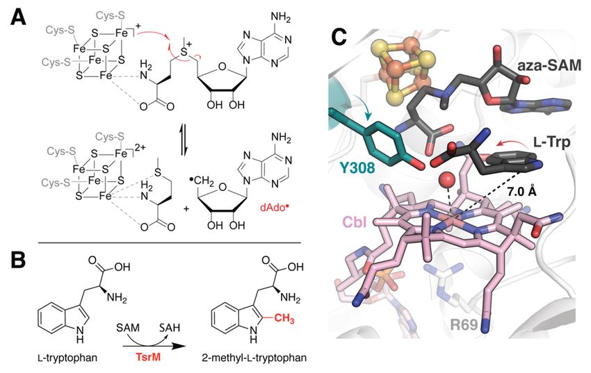
FOR PEER REVIEW 2 of 15
Molecules 2021, 26, 2590
characterization of rSAM enzymes has been hindered by their recalcitrant nature. Com‐2 of 14
putational chemistry methods provide a readily accessible alternative to studying the
mechanisms and atomic motions that facilitate rSAM catalysis while overcoming the tech‐
motions that facilitate rSAM catalysis while overcoming the technical limitations associated
nical limitations associated with time‐resolved structure determination of these enzymes.
with time-resolved structure determination of these enzymes.
Figure 1. Substrate binding in TsrM. (A) Shared reaction of rSAM enzymes. (B) Reaction catalyzed by TsrM, which while
Figure 1. Substrate binding in TsrM. (A) Shared reaction of rSAM enzymes. (B) Reaction catalyzed
annotated as a rSAM enzyme, does not appear to generate dAdo•. (C) The active site structure of TsrM (PDB accession code:
by TsrM,
6WTF) depictswhich while
the target annotated
carbon as a rSAM
in an unproductive enzyme,
position. doessimulations
Docking not appear to generate
suggest dAdo•.
an alternate (C)binding
flipped The
active site structure of TsrM (PDB accession code: 6WTF) depicts the target carbon in an unpro‐
mode for Trp in the presence of MeCbl. This proposal helps to rationalize observed reorientation of a nearby Tyr.
ductive position. Docking simulations suggest an alternate flipped binding mode for Trp in the
Computational
presence of MeCbl. This proposal approaches
helps to have
rationalize played reorientation
observed an integral role
of in definingTyr.
a nearby the rSAM su-
perfamily, since its classification in 2001, when bioinformatics was used to survey and
categorize the original ~600 members [7]. Computational chemistry methods that lever-
Computational approaches have played an integral role in defining the rSAM super‐
age our understanding of classical and quantum mechanics to analyze the fundamental
family, since its classification in 2001,
properties of atoms, when bioinformatics
molecules, was used
and chemical reactions, to survey
however, have been and cate‐
sorely under-
gorize the original utilized
~600 members [7].ofComputational
in the study chemistry
these fascinating enzymes. methods
Here, that leverage
we summarize ourbeen
work that has
performedand
understanding of classical to apply both abmechanics
quantum initio and semi-empirical calculations
to analyze the to the study
fundamental of rSAM en-
properties
zyme dynamics with a focus on docking (D), molecular dynamics (MD), density functional
of atoms, molecules, and chemical reactions, however, have been sorely underutilized in
theory (DFT) and hybrid quantum mechanics/molecular mechanics (QM/MM) methods
the study of these(Table
fascinating
1). Areasenzymes.
of untappedHere, wearesummarize
potential workwith
highlighted along thatpotential
has been per‐
pitfalls.
formed to apply both ab initio and semi‐empirical calculations to the study of rSAM en‐
zyme dynamics with2. Molecular
a focus Docking
on docking (D), molecular dynamics (MD), density func‐
tional theory (DFT) and hybridmost
Some of the critical conformational
quantum changes that an
mechanics/molecular enzyme undergoes
mechanics (QM/MM) are those
associated with substrate binding. Accordingly, visualization of reactant complexes can
methods (Table 1). Areas of untapped potential are highlighted along with potential pit‐
provide unprecedented insight into the structural basis for reactivity. Binding of a peptidyl
falls. analog for the protein substrate pyruvate formate-lyase (PFL) by the radical SAM activating
enzyme (PFL-AE) is accompanied by rearrangement of an active site beta strand, which
2. Molecular Docking
partially loses secondary structure to provide contacts with the peptide [8]. Complexation
with the PFL fragment further induces changes to side chain conformations that stabilize
Some of the most critical conformational
the cosubstrate changes
SAM, a correlation that
that may antoenzyme
help undergoes cleavage.
prevent unproductive are thoseMore
associated with substrate
generally,binding. Accordingly,
loop motions visualization
that occlude solvent of reactant
access to the active site complexes can
appear to be relatively
provide unprecedented insight into the structural basis for reactivity. Binding of a pep‐ the
widespread amongst members of the superfamily [9,10]. Despite a shared mechanism,
tidyl analog for thevast substrate
protein diversity pyruvate
substrate of rSAM enzymes precludes (PFL)
formate‐lyase a commonby substrate-binding
the radical SAM motif,
and conformational changes associated with ligand binding are often subtle. The structural
activating enzyme (PFL‐AE) is accompanied by rearrangement of an active site beta
strand, which partially loses secondary structure to provide contacts with the peptide [8].
Complexation with the PFL fragment further induces changes to side chain conformations
that stabilize the cosubstrate SAM, a correlation that may help to prevent unproductive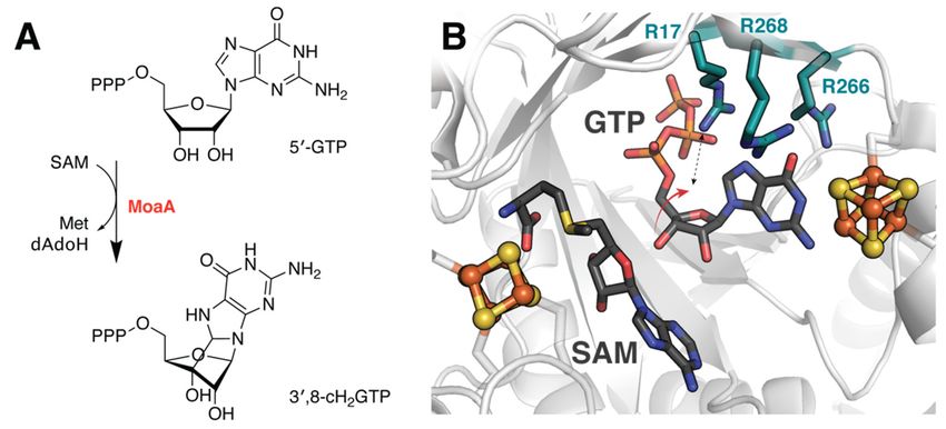
Molecules 2021, 26, 2590                                                                                            3 of 14

                           dynamics that facilitate catalytic initiation must therefore be analyzed on a case-by-case
                           basis. Molecular docking provides a broadly accessibly tool for evaluating this behavior
                           that is particularly advantageous when substrate identity is unknown, the reactant complex
                           is unstable, or co-crystallization is challenging.
                                 Docking methods iteratively explore ligand conformations and orientations to predict
                           the most likely complex formed with a given macromolecular target. In their simplest form,
                           these approaches rely on the principle of shape complementarity. Resultant models are
                           then assessed using sophisticated scoring systems which may consider physicochemical
                           properties such as solvent effects, electrostatic interactions, and hydrophobic contacts,
                           among other things, to provide an estimate of binding affinity [11]. Such simulations
                           applied to rSAM enzymes have refined crystallographically-determined substrate binding
                           modes, suggested potential dynamics associated with protein–protein complex forma-
                           tion, identified mechanistically relevant active site residues, and been instrumental in the
                           evaluation of proposed substrate candidates. Relevant examples are described below.

                           2.1. Correcting for the Wrong Form of Cobalamin in TsrM
                                 Incorporation of physiologically relevant cofactors is crucial for the accurate interpre-
                           tation of structural models. However, this can be difficult to achieve, especially in systems
                           whose cofactors require assembly, are reactive to molecular oxygen, or sensitive to light.
                           Cobalamin-dependent rSAM enzymes unfortunately share all of these traits. Co-expression
                           of cellular machinery for Fe/S-cluster assembly along with careful handling under anoxic
                           conditions can secure the canonical [4Fe-4S] cluster. The incorporation and instability of
                           methylcobalamin (MeCbl), on the other hand, has proven more problematic, and recently
                           emerged as the primary impediment to structural characterization of a true reactant com-
                           plex for tryptophan 2C methyltransferase (TsrM), annotated as a rSAM enzyme [12]. TsrM
                           catalyzes the methylation of L-tryptophan (Trp) at the C2 position during the biosynthe-
                           sis of the macrocyclic peptide antibiotic thiostrepton A (Figure 1B) [13]. Experimental
                           structure elucidation of the TsrM•Trp complex depicts an awkwardly oriented substrate
                           molecule, the C2 position of which is too distant (7 Å) for methyl transfer from the cobalt
                           ion (Figure 1C). To account for this inconsistency, the authors suggest that the observed
                           binding mode is likely due to degradation of the MeCbl cofactor to aquocobalamin during
                           crystallization and/or data collection [12]. They utilized the Glide docking program from
                           the Schrödinger software suite to test this hypothesis following manual replacement of the
                           observed cofactor by MeCbl (Table 1) [14]. While the top-scoring pose recapitulates the
                           binding conformation observed in the crystal structure, it is followed closely by a flipped
                           conformation in which the C2 position of Trp lies approximately 3.1 Å from the methyl
                           group of the cobalamin cofactor. In addition to providing a more catalytically relevant
                           model for substate binding, this simulation helps to justify the rearrangement of an active
                           site tyrosine (Y308) upon substrate binding (Figure 1C). The observed conformational
                           change places the Tyr hydroxyl group proximal to the Trp C2. As no other suitable amino
                           acids are nearby, it seems likely that Y308 serves as the active site base in the reaction.
                           However, this must be verified experimentally.

                           2.2. Predicting a Structural Framework for PFL Activation
                                Although the most common applications of molecular docking involve small-molecule
                           ligands such as Tyr, it is possible to employ docking approaches for determining protein–
                           protein interactions [15]. Such macromolecular complexes are notoriously difficult to
                           crystallize, and no rSAM enzyme has been structurally characterized bound to its protein
                           substrate to date. Instead, truncated peptide mimics have been employed to investigate the
                           structural basis for reactivity in both the anaerobic sulfatase-maturating enzyme (anSME)
                           from Clostridium perfringens and PFL-AE, introduced above [16]. The latter abstracts a
                           H atom from PFL to generate a stable glycyl radical which is used in the conversion of
                           pyruvate and coenzyme A to formate and acetyl-CoA (Figure 2A) [17]. Vey et al. expanded
                           upon their crystallographically obtained model using ZDOCK to predict binding to a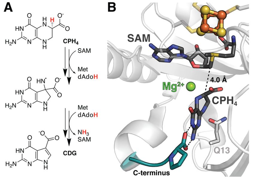
Molecules 2021, 26, x FOR PEER REVIEW 4 of 15
Molecules 2021, 26, 2590 4 of 14
conversion of pyruvate and coenzyme A to formate and acetyl‐CoA (Figure 2A) [17]. Vey
et al. expanded upon their crystallographically obtained model using ZDOCK to predict
binding
48-residueto afragment
48‐residue
of fragment of PFL that
PFL that contains the contains
catalytic the catalytic
glycine glycine
residue and residue and is
is proposed to
proposed
contact thetoactivating
contact the activating
enzyme enzyme
(Table (Table
1) [8,18]. Their1)results
[8,18].not
Their
onlyresults
accountnotforonly
twoaccount
patches
for two patches
of sequence of sequenceon
conservation conservation
the surfaceon of the surfacebut
PFL-AE, of PFL‐AE,
further led buttofurther led to the
the proposal of
proposal
concertedofconformational
concerted conformational
changes in the changes in the two
two proteins. proteins.
Namely, theyNamely, they that
hypothesize hypoth‐
loop
esize
motionthatin loop motion
PFL-AE in in
occurs PFL‐AE
tandemoccurs in tandem
with hinged with
motion ofhinged motion of the radical‐
the radical-containing domain
containing domain binding
in PFL to facilitate in PFL toandfacilitate
protectbinding and post
the radical protect the radical
formation post2B).
(Figure formation
It should(Fig‐
be
noted
ure 2B).that dockingbesimulations
It should noted that do not perfectly
docking duplicate
simulations do nottheperfectly
interactions observed
duplicate thefor the
inter‐
peptideobserved
actions mimic. Itfor
is unclear whether
the peptide mimic.theseIt discrepancies are expected
is unclear whether based on differences
these discrepancies are ex‐
in KM or
pected whether
based this is a function
on differences in KM orof rigid
whetherbody modeling.
this Computational
is a function of rigid bodyadvancements
modeling.
over the past decade
Computational have made
advancements over incorporating
the past decadeligand flexibility
have made more accessible
incorporating and
ligand flex‐
subsequent studies, such as those investigating the rSAM enzyme
ibility more accessible and subsequent studies, such as those investigating the rSAM en‐SuiB, have benefited
from this
zyme SuiB,consideration
have benefited[19].
from this consideration [19].
Figure
Figure2.2.Large‐scale
Large-scalestructural rearrangements
structural rearrangementsof of
PFL andand
PFL its activating enzyme.
its activating enzyme.(A) Reaction
(A) Reaction
catalyzed
catalyzed by
by PFL‐AE.
PFL-AE. (B)(B) Docking studies suggest hinged
hinged motion
motion of of the
theradical
radicaldomain
domainininPFL
PFL(blue
(blue cartoon,
cartoon, PDB PDB accession
accession code:code: 2PFL)
2PFL) together
together withwith loop
loop motions
motions in PFL‐AE
in PFL-AE (PDB
(PDB accession
accession code:
code:
3CB8,3CB8,
mobilemobile
loop loop
region region highlighted
highlighted on surface)
on surface) facilitate
facilitate substrate
substrate positioning.
positioning. The
The outline
outline of
of PFL is predominantly shown as a solid grey shape to help distinguish the radical domain. The
PFL is predominantly shown as a solid grey shape to help distinguish the radical domain. The
catalytic glycine residue is rendered in yellow spheres for emphasis. Note the complex depicted
catalytic glycine residue is rendered in yellow spheres for emphasis. Note the complex depicted
here is not the original docking model and was generated manually for visualization of described
here is not the results.
computational original docking model and was generated manually for visualization of described
computational results.
2.3.
2.3.Elucidating
Elucidatingthe theTransition
Transition to to Ordered
Ordered Product
Product inin SuiB
SuiB
SuiB catalyzes the formation of an unusual
SuiB catalyzes the formation of an unusual C–C crosslink C–C crosslink between
between lysine
lysine andand trypto‐
tryptophan
phan during the biosynthesis of a macrocyclic peptide natural product
during the biosynthesis of a macrocyclic peptide natural product (Figure 3A) [20]. Unfortu- (Figure 3A) [20].
Unfortunately,
nately, structural structural characterization
characterization of the enzymeof theinenzyme
complexinwith complex with its ribosomally
its ribosomally synthesized
synthesized
peptide substrate, SuiA, was insufficient to visualize the core region of thecore
peptide substrate, SuiA, was insufficient to visualize the peptideregion of the
containing
peptide containing the sites of posttranslational modification, perhaps
the sites of posttranslational modification, perhaps due to unregulated turnover [19]. The due to unregulated
turnover [19]. Theturned
authors therefore authors therefore turned
to molecular dockingtoapproaches
molecularcarried
docking outapproaches
using Rosetta carried out
to extrap-
using Rosetta to extrapolate possible binding modes for the core sequence
olate possible binding modes for the core sequence within the active site (Table 1) [21]. Unlike within the ac‐
tive
withsite
TsrM(Table
and1)PFL-AE,
[21]. Unlikethesewith TsrM and
simulations PFL‐AE,athese
employed varietysimulations employed
of constraints that nota vari‐
only
ety of constraints
enforced linkage to thatthenot only enforced linkage
crystallographically to the
observed crystallographically
portion of SuiA, but further observed por‐
restricted
tion of SuiA,between
the distance but further the restricted
location ofthe thedistance between
dAdo• radical and thethelocation
lysine Cβ,of the
from dAdo•
which radical
the H
and
atom is abstracted. Furthermore, the FastRelax algorithm sampled backbone and side algo‐
the lysine Cβ, from which the H atom is abstracted. Furthermore, the FastRelax chain
rithm sampledof
conformations backbone and side and
both the substrate chain conformations
nearby of both the
active site residues substrate
to account for and nearby
the inherent
active site of
flexibility residues to account
the molecules [22]. for the no
While inherent
consensusflexibility of the for
was reached molecules [22]. While
the uncyclized no
peptide,
consensus
four possible was
low reached for the uncyclized
energy conformations peptide, four
were identified for thepossible
product, low energy confor‐
suggesting that the
mations
unstructuredweresubstrate
identified core forbecomes
the product,
ordered suggesting that theofunstructured
upon installation the crosslink. substrate core
The associated
becomes ordered the
models highlight upon installation of side
rearrangement the crosslink.
chains in theTheactive
associated models highlight
site to accommodate the
product.
rearrangement
In particular, E319 of side
moves chains
such in the
that itsactive
gamma site to accommodate
carboxyl group could, product.
in theory, In serve
particular,
as the
E319
activemoves such
site base that its
(Figure 3A).gamma carboxyl group
The hypothesis that thiscould, in theory,
titratable groupserve as thefor
is crucial active site
turnover
was later confirmed biochemically [23].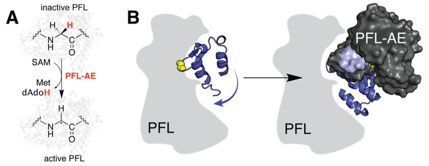
Molecules 2021, 26, x FOR PEER REVIEW 5 of 15
Molecules 2021, 26, 2590 5 of 14
base (Figure 3A). The hypothesis that this titratable group is crucial for turnover was later
confirmed biochemically [23].
Docking
Figure3.3.Docking
Figure studies
studies help
help to identify
to identify catalytic
catalytic residues
residues in both
in both SuiBSuiB and viperin.
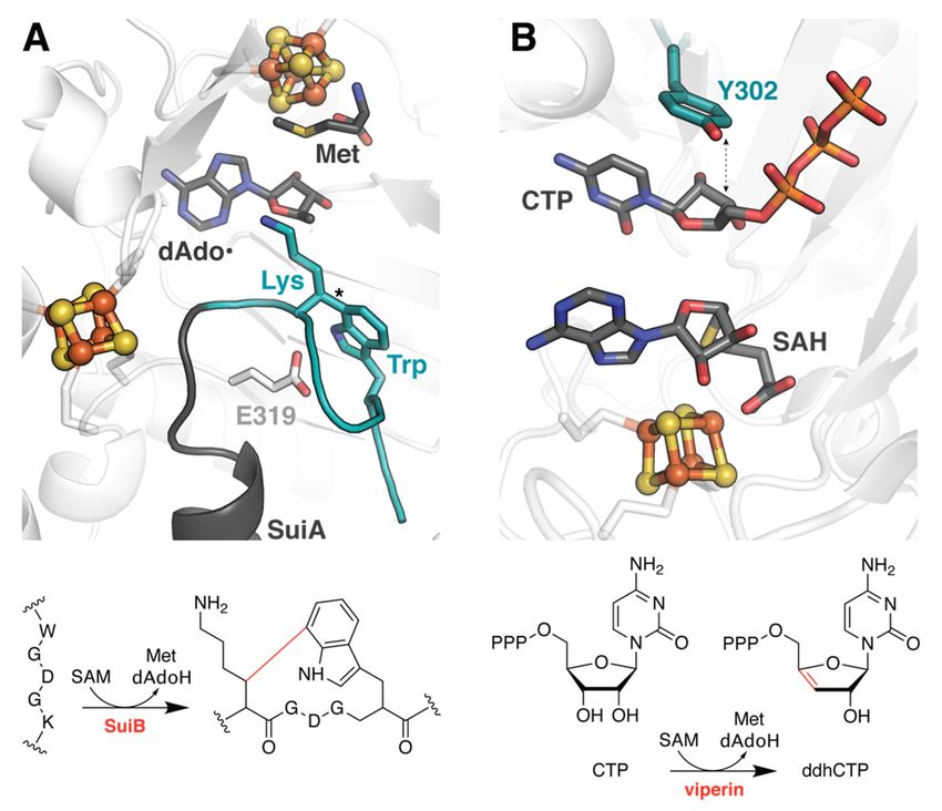
and viperin. (A) SuiB
(A) SuiB
catalyzes a C–C crosslink, marked with an asterisk in the structure, to cyclize the core
catalyzes a C–C crosslink, marked with an asterisk in the structure, to cyclize the core region region of aof a
ribosomally
ribosomally synthesized peptide, SuiA. Docking methods were used to simulate the portion ofthe
synthesized peptide, SuiA. Docking methods were used to simulate the portion of the
peptide
peptidethat
thatwas
wasdisordered
disorderedin inthe
thecrystal
crystalstructure
structure (PDB
(PDB accession
accession code:
code: 5V1Q),
5V1Q), shown
shown inin teal.
teal. The
The putative
putative active
active site site
base,base,
E319,E319, is drawn
is drawn in stick
in stick representation.
representation. (B) Together,
(B) Together, dockingdocking
and MD and MD
studies
studies have helped to elucidate the function of viperin (PDB accession code: 6Q2P) and support a
have helped to elucidate the function of viperin (PDB accession code: 6Q2P) and support a PCET
PCET role for a conserved active site Tyr.
role for a conserved active site Tyr.
2.4.
2.4.Assessing
Assessingthe theSubstrate
SubstrateScope
ScopeofofViperin
ViperinandandQueE
QueE
InInaamore
moretraditional
traditionalapplication
applicationanalogous
analogoustotoitsitsuse
useininthe
thefield
fieldofofdrug
drugdiscovery,
discovery,
molecular docking has also been used to evaluate proposed
molecular docking has also been used to evaluate proposed substrates of the rSAM substrates of the rSAM en‐
zyme viperin (virus inhibitory protein, endoplasmic reticulum‐associated,
enzyme viperin (virus inhibitory protein, endoplasmic reticulum-associated, interferon- interferon‐in‐
ducible;
inducible;Table 1). Viperin
Table orthologs
1). Viperin orthologsare ubiquitous across
are ubiquitous all domains
across of life,ofincluding
all domains in
life, includ-
humans, and haveand
ing in humans, been demonstrated
have to inhibit to
been demonstrated viral replication
inhibit [24,25]. The[24,25].
viral replication molecularThe
mechanism
molecular underlying
mechanismthis antiviral activity,
underlying however,
this antiviral was only
activity, recentlywas
however, determined [26].
only recently
From numerous
determined [26].promising candidates,
From numerous including
promising those supported
candidates, includingbythose computational
supported
docking [27,28], the mammalian
by computational enzyme
docking [27,28], thewas ultimatelyenzyme
mammalian shown to wascatalyze the conversion
ultimately shown to
ofcatalyze
cytidinethe triphosphate
conversion(CTP) to the RNA‐chain
of cytidine triphosphate terminator the RNA-chain terminator 30 -
(CTP) to3′‐deoxy‐3′,4′‐didehydro‐CTP
0 0
deoxy-3 ,4as-didehydro-CTP
(ddhCTP), shown in Figure(ddhCTP), as shown in
3B [26]. Subsequent Figuremodels
docking 3B [26].helped
Subsequent docking
to investigate
models
the role ofhelped to investigate
an active site tyrosinethein role
a key ofproton‐coupled
an active site tyrosine
electronintransfer
a key proton-coupled
(PCET) step of
electron
this reactiontransfer (PCET) step
and provided of thisto
evidence reaction and provided
substantiate evidence
the divergent to substantiate
substrate promiscuity the
ofdivergent substrate
fungal viperin frompromiscuity of fungal
Thielavia terrestris (TtRSAD2) [29]. Thielavia
viperin from Molecular terrestris
dynamics (TtRSAD2)
simulations[29].
Molecular
extended dynamics
these studies simulations extended these
to elucidate associated studies to dynamics
conformational elucidate andassociated confor-
are described
mational
below. dynamics and are described below.
InInfact,
fact,molecular
moleculardocking
docking is is often
often used
used as as a preparative
a preparative step
step for for downstream
downstream MDMD or
or QM/MM
QM/MM analysis,
analysis, and aand a similar
similar evaluation
evaluation of substrate
of substrate scope
scope was was performed
performed for
for 7‐car‐
7-carboxy-7-deazaguanine
boxy‐7‐deazaguanine synthase
synthase (QueE)
(QueE) fromB.B. multivorans
from multivorans withwithanan eyeeye toward engi-
toward
neering alternative reactions [30]. QueE catalyzes the Mg2+ -dependent rearrangement
of 6-carboxytetrahydropterin (CPH4 ) to 7-carboxy-7-deazaguanine (CDG), the basic
building block for more than 30 natural products in addition to the tRNA base queuosin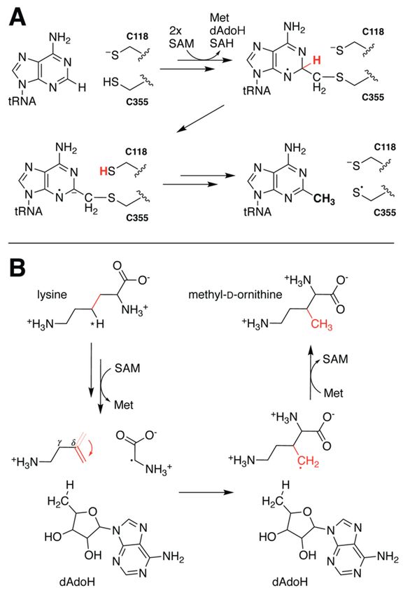
Molecules 2021, 26, x FOR PEER REVIEW 6 of 15
Molecules 2021, 26, 2590 6 of 14
engineering alternative reactions [30]. QueE catalyzes the Mg2+‐dependent rearrangement
of 6‐carboxytetrahydropterin (CPH4) to 7‐carboxy‐7‐deazaguanine (CDG), the basic build‐
ing block for more than 30 natural products in addition to the tRNA base queuosin (Figure
4A) [31].4A)
(Figure Docking approaches
[31]. Docking performedperformed
approaches in tandemin with DFT calculations
tandem of radical sta‐
with DFT calculations of
bilization energies demonstrated
radical stabilization that either less
energies demonstrated stabilization
that either less or the ability to
stabilization or persist in a
the ability
non‐preferred
to persist in a conformation
non-preferredisconformation
required for ligands to serve
is required as alternative
for ligands to servesubstrates of
as alterna-
this
tive enzyme. Asofwith
substrates thisviperin,
enzyme. these studies
As with were enhanced
viperin, by MD
these studies simulations
were enhanceddetailed
by MD
below.
simulations detailed below.
Figure 4. Subtle
Subtle structural
structural features
features of
of the
theQueE
QueEreactant
reactantcomplex
complexare
arecrucial
crucialfor
forenzyme
enzymereactivity.
reactivity.
(A) Reaction scheme for QueE. (B) A series of docking, MD, DFT, and QM/MM studies
(A) Reaction scheme for QueE. (B) A series of docking, MD, DFT, and QM/MM studies have have
demonstrated
demonstrated thethe importance
importanceofofdivalent
divalention
ionidentity
identityand
andsubstrate
substratedeformation
deformationtotoboth
bothstabilize
stabilize
the reactant complex and facilitate catalysis. Decreased flexibility in the C‐terminal tail was also
the reactant complex and facilitate catalysis. Decreased flexibility in the C-terminal tail was also
observed following reaction initiation in QueE (PDB accession codes: 4NJI).
observed following reaction initiation in QueE (PDB accession codes: 4NJI).
3.
3. Molecular
Molecular Dynamics
Dynamics
Although
Although molecular
molecular docking
docking can
can certainly
certainly inform
inform our our understanding
understanding of of enzyme
enzyme dy‐ dy-
namics, ititisislimited
limitedtoto providing
providing static snapshots
static snapshots of ligand‐bound
of ligand-bound complexes. This isThis
complexes. prob‐
is
lematic, as enzymes are constantly in motion, their atomic rearrangements
problematic, as enzymes are constantly in motion, their atomic rearrangements a con- a concerted
dance
certed precisely tailoredtailored
dance precisely to propeltothe reaction
propel the forward. For the computational
reaction forward. prediction
For the computational
and analysis
prediction andofanalysis
these time‐dependent conformational
of these time-dependent changes,changes,
conformational we turn weto molecular dy‐
turn to molec-
namics. MD utilizes
ular dynamics. MDclassical
utilizes mechanics approaches,
classical mechanics based on Newton’s
approaches, based onlaws, to simulate
Newton’s laws,
atomic motion.
to simulate The motion.
atomic forces acting
The on eachacting
forces atom onare each
mostatom
commonly calculated
are most commonly using mo‐
calcu-
lecular mechanics
lated using molecular(MM) force fields
mechanics that force
(MM) approximate
fields that atoms as chargedatoms
approximate spheres, and the
as charged
spheres, and
covalent bonds thebetween
covalent thembondsasbetween
springsthem[32]. as springs
These [32]. Thesereduce
assumptions assumptions reduce
the computa‐
the computational cost associated with ab initio calculations on
tional cost associated with ab initio calculations on macromolecular systems. Resultantmacromolecular systems.
Resultant
MD MD trajectories
trajectories can then can then be
be used used tothe
to assess assess the magnitude
magnitude of structural
of structural changes changes
(root‐
(root-mean-square
mean‐square deviation deviation [RMSD]),
[RMSD]), the the propensity
propensity forfor a given
a given residueororregion
residue region toto move
(root-mean-square
(root‐mean‐square fluctuation
fluctuation[RMSF]),
[RMSF]),and andthethe
evolution
evolution of hydrogen
of hydrogenbonding networks,
bonding net‐
among among
works, other thingsother [33].
things Despite its utility,
[33]. Despite few groups
its utility, have applied
few groups MD forMD
have applied thefor
study
the
of rSAM enzymes, and those that have primarily focused on
study of rSAM enzymes, and those that have primarily focused on better understanding better understanding the
requirements
the requirements for substrate
for substratebinding. In fact,
binding. In one
fact,of theoffirst
one theMD firststudies on a member
MD studies of the
on a member
rSAM superfamily essentially utilized the disproportionately powerful
of the rSAM superfamily essentially utilized the disproportionately powerful technique technique for the
purposes
for of docking
the purposes possiblepossible
of docking substrate candidates
substrate to viperin.
candidates to viperin.
3.1. The Flexible Binding Pocket of Fungal Viperin
3.1. The Flexible Binding Pocket of Fungal Viperin
Although all viperin orthologs tested thus far appear to catalyze the conversion of CTP
Although all viperin orthologs tested thus far appear to catalyze the conversion of
to ddhCTP, substantial experimental evidence points to the existence of additional alternate
CTP to ddhCTP, substantial experimental evidence points to the existence of additional
reactivities in both fungal- and archaeal-derived enzymes [29,34]. Prior to identification
of CTP as the primary substrate, Chakravarti et al. observed apparent modification of
isopentenyl pyrophosphate (IPP; Table 1) [34]. To evaluate whether there was a structuralMolecules 2021, 26, 2590 7 of 14
basis for IPP binding to the enzymes, they manually placed the ligand in the active site of a
crystallographically characterized mouse viperin model and performed successive rounds
of MD equilibration. The resultant complex identified numerous side chain rearrangements
that could enable interactions with the terminal phosphate of IPP, consistent with the
authors’ biochemical analysis of substrate scope. Perhaps more compelling in hindsight,
however, is the simulated orientation of IPP, which places the hypothesized substrate
radical on C3 near the putative catalytic tyrosine, identified later via docking models with
CTP (Figure 3B) [29]. Such proximity not only supports the proposed mechanism for IPP
reactivity, but further highlights the significance of the active site tyrosyl.
MD simulations following docking of uridine triphosphate (UTP), another putative
fungal viperin substrate that has been identified biochemically, to TtRSAD2 reportedly indicate
significant flexibility in the loops responsible for binding the ribose moiety (Table 1) [29].
This conformational elasticity is in agreement with electron paramagnetic resonance data
demonstrating that interaction with the sugar induces detectable structural changes likely
important for facilitating catalysis. Furthermore, the position of the uracil moiety fluctuates
within the binding pocket consistent with an insensitivity to the nucleobase identity and
perhaps the origin of the enzyme’s intriguing promiscuity.
3.2. Substrate Stabilization in QueE
MD simulations have likewise been used to assess the stability of alternative substrates
within the active site of QueE post-docking (Table 1). This analysis was critical due
to the electrostatic effects of a catalytic Mg2+ ion that stabilize a bent and energetically
unfavorable configuration of the substrate vital for reactivity (Figure 4B) [35,36]. Given
this requirement, docking models with appropriate positioning and radical stabilization
energies (RSEs) are still insufficient to validate a given molecule as a substrate. Suess
et al. demonstrated that MD trajectories of seemingly suitable protein–ligand complexes in
the absence of Mg2+ showed rapid dissociation, reiterating the importance of the ion for
complex stability [30]. Retention times of the natural substrate CPH4 were also reduced
upon replacement of Mg2+ by other ions, including Ca2+ and Na+ , likely because they fail to
maintain the same hydrogen bonding network. It should be noted that the incorporation of
MD to perform such equilibrium studies following any docking procedure is good practice.
Finally, MD was applied to gain insight into the behavior of a key catalytic intermediate.
The resultant data indicate increased rigidity of the C-terminal tail along with a tighter
hydrogen bond to the substrate upon evolution from the reactant complex (Figure 4B). Such
subtle changes in atomic position and reduced capacity for motion, leading to reduced
loop flexibility, are difficult to visualize experimentally, but can provide crucial insights
into enzymatic turnover.
4. Density Functional Theory
The computational approaches described thus far have examined conformational
dynamics that, while crucial, cannot solely account for enzymatic reactivity. Modulation of
active site electronic structure must occur in tandem to effect turnover. Molecular mechan-
ics approximations, however, are unable to elucidate these changes. Furthermore, methods
dependent on such approximations are ill equipped to accurately evaluate proposed inter-
mediate states based on the total energy. Ab initio or pseudo ab initio quantum mechanical
(QM) methods can overcome these limitations and enable the properties of multi-electron
systems to be determined. DFT, for example, allows one to calculate electronic properties
by correlating the ground state energy with the spatial density of electrons [37]. This
powerful strategy reduces the computational cost compared to wavefunction-based meth-
ods. DFT can provide insight into the thermodynamic and kinetic properties of a given
system, including reaction energies, charge/spin distributions, and vibrational frequencies,
in addition to molecular geometries [37,38]. Perhaps unsurprisingly, it is the most common
computational method used to analyze the behavior of rSAM enzymes. Herein, we will
focus on a subset of those studies that address the dynamics associated with catalysis.Molecules 2021, 26, 2590 8 of 14
4.1. Probing the Basis for SAM Cleavage in PFL-AE
Early electron nuclear double resonance (ENDOR) studies of PFL-AE observed orbital
overlap between the SAM sulfonium and the canonical Fe/S cluster, thereby providing
the first evidence to suggest that SAM binds directly to the unique Fe in radical SAM
enzymes [39,40]. To further explore the nature of this interaction and the role of the
cluster in SAM cleavage, Dey et al. used DFT calculations to assess the composition of
the molecular orbitals and evaluate potential energy surfaces (Table 1) [41]. Resultant
data were used to rationalize an increase in the sulfur X-ray absorption pre-edge intensity
upon SAM binding and validate hypotheses that this feature arises due to backbonding
between the cluster and the S-C50 σ * orbital of SAM [42]. Calculations suggest that both
reduction of the cluster and precise positioning of SAM enhance this effect to facilitate
homolytic bond cleavage. Intriguingly, recent experimental studies indicate that thermally
induced conformational changes are likely responsible for regioselectivity of the S-C50
bond [43]. The analysis of potential energy surfaces by Dey et al. also identified active site
polarity as a key component to modulating the activation energy required for reductive
SAM cleavage [41]. Namely, a decrease in polarity is correlated with a lower barrier. The
authors therefore propose that substrate binding may trigger generation of dAdo• by
decreasing solvent accessibility to the active site, which would lower the dielectric constant
in the medium. Such a gating mechanism may also help to mitigate unproductive cleavage
in the absence of substrate.
4.2. Analyzing the Effect of Ion Complexation to QueE
While DFT was used to assess the energetics associated with the characteristic radical
SAM cleavage reaction in PFL-AE, a similar approach can be used to elucidate the basis
for primary substrate modifications. This is of particular interest for enzymes that use
radicals to perform selective modifications, as radical reactivity is difficult to control. DFT,
towards this end, has been employed in the study of QueE from B. multivorans (Table 1) [36].
Calculated radical stabilization and rearrangement energies associated with the modifica-
tion of CPH4 support the QM/MM-derived hypothesis that electrostatic interactions with
Mg2+ induce a strained substrate conformation important for catalysis (Figure 4B) [35].
Obtained results further indicate that the metal ion not only promotes deviation from
substrate planarity, but must also maintain this energetically unfavorable conformation
upon generation of the radical via H-atom abstraction in order to lower the barrier for
rearrangement. This effect is diminished upon complexation with other divalent ions, in
agreement with slower kinetics and reduced activity observed experimentally. Although
distortion of the substrate destabilizes the radical, thereby increasing the probability of
undesirable off-pathway reactions, the presence of Mg2+ tunes the energy landscape to
preclude unproductive ring opening.
4.3. Determining Transition State Dynamics in MoaA
Enforcement of a nominally unfavorable substrate conformation to overcome other-
wise high activation barriers was also discovered in the rSAM enzyme MoaA, which cy-
clizes guanosine triphosphate (GTP) to generate 30 ,8-cyclo-7,8-dihydro-GTP (30 ,8-cH2 GTP)
during biosynthesis of the essential molybdenum cofactor, Moco (Figure 5A) [44]. DFT-
based analyses reveal that constraining both the nucleobase and triphosphate moiety,
consistent with observed hydrogen bonding interactions between active site constituents,
facilitates deformation of the substrate (Table 1) [45]. The resultant molecular arrangement
decreases the activation energy of cyclization. However, it does not fully account for the ex-
perimentally derived kinetics. Instead, the presence of three arginine residues that line the
substrate binding pocket lower the barrier to within range of estimated activation energies
(Figure 5B). Interactions with these residues, however, were only observed in geometry
optimizations of the transition state complex which depict the 30 -OH of the ribose pivoting
to form a hydrogen bond with Arg17. It seems likely that this dynamic behavior is crucial
to allow the reactant to pass through the transition state. Associated DFT calculationsexperimentally derived kinetics. Instead, the presence of three arginine residues that line
the substrate binding pocket lower the barrier to within range of estimated activation en‐
ergies (Figure 5B). Interactions with these residues, however, were only observed in ge‐
ometry optimizations of the transition state complex which depict the 3′‐OH of the ribose
Molecules 2021, 26, 2590 9 of 14
pivoting to form a hydrogen bond with Arg17. It seems likely that this dynamic behavior
is crucial to allow the reactant to pass through the transition state. Associated DFT calcu‐
lations that demonstrate comparable thermodynamic free energies for the reactant and
that demonstrate comparable thermodynamic free energies for the reactant and product
product complexes were also proposed to support involvement of the C‐terminal auxil‐
complexes were also proposed to support involvement of the C-terminal auxiliary cluster
iary cluster in dynamic redox
in dynamic processes
redox processesassociated withMoaA
associated with MoaA catalysis.
catalysis.
Figure 5. Active site residues facilitate MoaA catalysis. (A) Reaction catalyzed by MoaA. (B) DFT studies indicate that
Figure 5. Active site residues facilitate MoaA catalysis. (A) Reaction catalyzed by MoaA. (B) DFT
arginine residues lining the MoaA substrate binding pocket play a role in modulating reaction energetics. These calculations
studies indicate that arginine residues lining the MoaA substrate binding pocket play a role in
further suggests that reorientation of the ribose moiety enables a key interaction with Arg17 to stabilize the transition state.
modulating reaction energetics. These calculations further suggests that reorientation of the ribose
Structure shown from PDB accession code 2FB3, excluding SAM, which was aligned from 1TV8.
moiety enables a key interaction with Arg17 to stabilize the transition state. Structure shown from
PDB accession code 4.4.
2FB3, excluding
Beyond SAM, which was aligned from 1TV8.
Dynamics
Although the examples presented above highlight the importance of structural changes
4.4. Beyond Dynamics
to effect catalysis, DFT has also been applied to evaluate proposed mechanistic path-
ways [46–48], assist
Although the examples in the analysis
presented aboveof highlight
spectroscopic dataimportance
the [49], and compare the thermody-
of structural
namic reaction profiles of enzymes within the superfamily [50].
changes to effect catalysis, DFT has also been applied to evaluate proposed mechanistic
pathways [46–48],5.assist
Hybrid inMethods:
the analysis of spectroscopic
Quantum data [49],
Mechanics/Molecular and compare the ther‐
Mechanics
modynamic reaction profiles
Quantumofmechanical
enzymesapproaches
within thesuch
superfamily [50].
as DFT are incredibly powerful, but they are
impractical on a large scale due to their high computational cost. However, consideration
5. Hybrid Methods: of only a subset Mechanics/Molecular
Quantum of atoms in the active site can be equally problematic as this neglects the
Mechanics
influence of the broader protein environment on reaction energetics. Hybrid QM/MM
Quantum mechanical approaches
methods attempt such as
to overcome DFT
these are incredibly
obstacles powerful,
by utilizing but they
more accurate are cal-
ab initio
impractical on a large scaletodue
culations to their
simulate high computational
important electronic structure cost. However,
features, consideration
while handling the rest of
of only a subset of atoms in the active site can be equally problematic as this neglectsathe
the molecule classically. Approximating interactions between most atoms with simple
potential energy function dramatically increases computational tractability. Practically,
influence of the broader protein environment on reaction energetics. Hybrid QM/MM
this means assigning different regions of the protein to QM or MM treatment, as well as
methods attempt to overcome
evaluating the these obstacles
electrostatic effectby
of utilizing moreon
the MM region accurate
the QM ab initio calcula‐
components [51,52]. It
tions to simulate important
should be noted electronic structure
that a short features,
MD simulation whileperformed
is typically handling the rest of
to equilibrate the
molecules
priorApproximating
molecule classically. to initiating QM/MM calculations,
interactions and QM regions
between are mostwith
most atoms oftenarestricted
simple topo‐active
site features that directly impact reactivity. As a frame of reference,
tential energy function dramatically increases computational tractability. Practically, this studies of QueE isolated
only the CPH4 substrate, dAdo•, and six active site residues, along with the key Mg2+ ion
means assigning different
and a series regions of thewaters
of associated proteinfor to QM ormechanical
quantum MM treatment, as well
consideration as Results
[35]. eval‐ of
uating the electrostatic effect
these and of other
select the MM QM/MMregion on the QM
calculations components
aimed at elucidating[51,52]. It should
the dynamic changes
be noted that a short
thatMD simulation
facilitate radical SAMis typically performed
enzyme catalysis to equilibrate
are described below. molecules prior
to initiating QM/MM calculations, and QM regions are most often restricted to active site
5.1. Coordinated Changes to the Active Site in QueE
features that directly impact reactivity. As a frame of reference, studies of QueE isolated
We have already discussed the results of QM/MM methods applied to QueE in
only the CPH4 substrate, dAdo•,
passing to and six
contextualize active
studies site residues,
employing along withtechniques.
other computational the key Mg 2+ ion
In particular,
and a series of associated
this hybridwaters
approachforhelped
quantum mechanical
to clarify consideration
the role of Mg [35]. Results
2+ , as well as eliminate of
ring openingMolecules 2021, 26, 2590 10 of 14
as a pathway to ring contraction during the production of CDG (Table 1). The discovery
that the metal ion did not serve a catalytic function, but rather provided key interactions
with substrate that are important for generating the reactive complex, provided the basis
for subsequent studies. Zhu and Liu also observed reorientation of several active site
residues in the absence of substrate/product [35]. Simulations depict the formation of an
additional hydrogen bond between two glutamate residues (E15 and E116) that the authors
propose forms part of a proton transfer pathway required to regenerate the active state
following dissociation of the CDG product. Likewise, a nearby glutamine (Q13) side chain
rotates substantially, suggesting a potential role in controlling product release (Figure 4B).
It remains to be seen whether these dynamics are physiologically relevant. However, a
more recent structure of the orthologous QueE from B. subtilis, solved in the absence of
substrate, does show similar rotation of the analogous residue, perhaps supporting this
hypothesis [53].
5.2. Proton Transfer Precedes Methyl Transfer in RlmN
Hybrid methods have also been used to determine the order of events during catalysis
of the rSAM methyltransferase RlmN. This putative ‘housekeeping’ enzyme modifies
key adenosine nucleosides of both transfer and ribosomal RNA and has been intensively
studied in part due to its high homology with other members of the superfamily known to
confer antibiotic resistance [54]. Unlike most SAM-dependent methyltransferases, RlmN
utilizes SAM as both a methyl donor and a source of dAdo•. The methyl group is first
transferred to an active site cysteine via an SN 2 mechanism. It is then appended to the
RNA substrate by means of radical addition following reductive cleavage of the second
SAM molecule (Figure 6A) [55]. To address discrepancies between the starting model of
earlier DFT calculations [56] and a subsequent structure of a crosslinked RlmN•tRNA
complex [57], Zhao et al. applied QM/MM methods to refine the proposed mechanism
(Table 1) [58]. By evaluating the energy barriers and potential energy surfaces for different
reaction pathways, the authors were able to assign deprotonation of the target carbon
as the rate-limiting step. They further concluded that this step precedes cleavage of the
crosslinked intermediate state complex to generate a more complete picture of mechanistic
dynamics (Figure 6A).
5.3. Rotation of Intermediate Crucial for PylB Reactivity
A similar study investigated the mechanism of methylornithine synthase (PylB),
known to catalyze the rearrangement of lysine to methyl-D-ornithine during the biosynthe-
sis of the 22nd amino acid pyrrolysine [59]. Earlier structure solution of PylB in complex
with product appeared to support a fragmentation–recombination mechanism in which
hydrogen atom abstraction from lysine Cγ fractures the molecule generating a glycyl
radical and aminobutene that later reunite [60]. QM/MM calculations performed on a
lysine docking model enabled investigation of the proposed reaction pathway energetics
(Table 1) [61]. Although the study primarily focused on evaluating energy barriers for
the assessment of mechanistic feasibility, the authors did observe unanticipated dynamics
of the aminobutene intermediate. Rotation along the original lysine Cγ-Cδ bond, while
not explicitly required for recombination, was observed to facilitate return of the proton
from dAdoH. Such a reorientation dramatically reduces the distance between the newly
formed Cβ radical and the C50 of dAdoH in preparation for re-abstraction of the previously
transferred hydrogen (Figure 6B). To the best of our knowledge this rotation has yet to be
experimentally validated, but computational evidence for such behavior highlights once
more the importance of dynamics in enzyme reactivity.previously transferred hydrogen (Figure 6B). To the best of our knowledge this
Molecules 2021, 26, 2590 has yet to be experimentally validated, but computational evidence11 for
of 14 such b
highlights once more the importance of dynamics in enzyme reactivity.
Figure 6.6.Mechanistic
Figure Mechanisticdynamics of RlmN
dynamics and PylB
of RlmN elucidated
and by hybrid computational
PylB elucidated methods.
by hybrid computational me
(A) QM/MM calculations of RlmN indicate that deprotonation of the target carbon likely precedes
(A) QM/MM calculations of RlmN indicate that deprotonation of the target carbon likely p
cleavage of the tRNA-Cys crosslink. (B) Study of the PylB mechanism via hybrid methods led to the
cleavage of the tRNA‐Cys crosslink. (B) Study of the PylB mechanism via hybrid methods
proposal that reorientation of the aminobutene intermediate (indicated by the red arrow) prior to
the proposal that reorientation of the aminobutene intermediate (indicated by the red arro
recombination facilitates re-abstraction of the H atom from dAdoH.
to recombination facilitates re‐abstraction of the H atom from dAdoH.
Table 1. Summary of computational approaches applied to rSAM enzymes discussed herein.
Table 1. Summary of computational approaches applied to rSAM enzymes discussed here
Enzyme Organism PDB Code Computational Method/s
Enzyme Organism
Kitasatospora PDB Code Computational Metho
TsrM 6WTE, 6WTF D [12]
cheerisanensis
TsrM Kitasatospora cheerisanensis 6WTE, 6WTF D [12]
D [8]
PFL-AE Escherichia coli 3C8F, 3CB8 D [8]
DFT [41]
PFL‐AE
SuiB
Escherichia coli
Streptococcus suis 5V1Q
3C8F, 3CB8 D [19] DFT [41]
Mus musculus D [27–29]
Viperin
SuiB Streptococcus 5VSL, 5VSM, 6B4C
Trichoderma virens suis 5V1Q MD [29,34] D [19]
QueE Bacillus multivorans
Mus musculus 4NJI, 4NJK D,
5VSL, 5VSM, MD, DFT, QM/MM [30,35,36]
D [27–29]
Viperin
MoaA Staphylococcus aureus 1TV8, 2FB3 DFT [45]
HydE Trichoderma
Thermotoga maritimavirens 3CIW 6B4C DFT [50] MD [29,34]
QueE
RlmN Bacillus multivorans
Escherichia coli 5HR6 4NJI, 4NJK DFT,D, MD, DFT,
QM/MM [56,58] QM/MM [30
PylB Methanosarcina barkeri 3T7V QM/MM [61]
MoaA Staphylococcus aureus 1TV8, 2FB3 DFT [45]
HydE Thermotoga maritima 3CIW DFT [50]
RlmN Escherichia coli 5HR6 DFT, QM/MM [56,5
PylB Methanosarcina barkeri 3T7V QM/MM [61]Molecules 2021, 26, 2590 12 of 14
6. Conclusions
In conclusion, the case studies presented here provide an overview of molecular
docking, MD, DFT and hybrid methods applied to the study of rSAM enzymes to date.
Experimental approaches are inherently unable to observe transition state species, and the
short-lived nature of catalytic intermediates likewise complicates their characterization.
Computational methods, by contrast, provide a unique opportunity to elucidate the atomic
and electronic changes that facilitate catalysis irrespective of kinetics. Despite their appli-
cation to the systems described herein, computational methods have been underutilized
in the study of rSAM enzymes, due in part to complications associated with implemen-
tation. At the most basic level, the feasibility of these studies relies on the availability of
reliable starting models, which are currently lacking for the majority of this superfamily.
Furthermore, computational treatment of transition metal ions and organic radicals is often
challenging. Of particular note are the poor performance of DFT and other QM methods
applied to transition metal chemistry, as well as ambiguity in the parametrization of Fe/S
clusters for docking/MD simulations.
Nonetheless, there is great potential. This is especially true for molecular dynamics
approaches, which have the ability to provide insight into large-scale motions necessary
for turnover, but have primarily been implemented for pseudo-docking simulations. Such
methods would likely be especially useful in the study of multi-modular rSAM enzymes
that contain auxiliary domains beyond the canonical triose-phosphate isomerase (TIM)
or modified TIM-barrel fold characteristic of the superfamily. Indeed, most constituent
members are comprised of two or more structural domains that contribute to their func-
tional diversity, often by binding additional cofactors, or providing crucial interactions
with substrate [62]. As we continue to learn more about this fascinating superfamily, the
opportunities to apply computational approaches to generate and investigate experimental
hypotheses will only continue to grow. Careful application of these methods together with
biochemical validation position them as powerful predictors of rSAM enzyme dynamics.
Funding: Research in the corresponding author’s laboratory on radical SAM enzymes is supported
by the National Institutes of Health, Pathway to Independence Award (4R00GM129460).
Institutional Review Board Statement: Not applicable.
Informed Consent Statement: Not applicable.
Data Availability Statement: Not applicable.
Conflicts of Interest: The authors declare no conflict of interest.
References
1. Schmidt, M. Time-resolved macromolecular crystallography at pulsed X-ray sources. Int. J. Mol. Sci. 2019, 20, 1401. [CrossRef]
[PubMed]
2. Chapman, H.N. Structure determination using X-ray free-electron laser pulses. Methods Mol. Biol. 2017, 1607, 295–324. [CrossRef]
3. Holcomb, J.; Spellmon, N.; Zhang, Y.; Doughan, M.; Li, C.; Yang, Z. Protein crystallization: Eluding the bottleneck of X-ray
crystallography. AIMS Biophys. 2017, 4, 557–575. [CrossRef] [PubMed]
4. Akiva, E.; Brown, S.; Almonacid, D.E.; Barber, A.E., 2nd; Custer, A.F.; Hicks, M.A.; Huang, C.C.; Lauck, F.; Mashiyama, S.T.;
Meng, E.C.; et al. The Structure–Function Linkage Database. Nucleic Acids Res. 2013, 42, D521–D530. [CrossRef] [PubMed]
5. Broderick, J.B.; Duffus, B.R.; Duschene, K.S.; Shepard, E.M. Radical S-adenosylmethionine enzymes. Chem. Rev. 2014, 114,
4229–4317. [CrossRef] [PubMed]
6. Frey, P.A.; Booker, S.J. Radical mechanisms of S-adenosylmethionine-dependent enzymes. Adv. Protein Chem. 2001, 58, 1–45.
[CrossRef] [PubMed]
7. Sofia, H.J.; Chen, G.; Hetzler, B.G.; Reyes-Spindola, J.F.; Miller, N.E. Radical SAM, a novel protein superfamily linking unresolved
steps in familiar biosynthetic pathways with radical mechanisms: Functional characterization using new analysis and information
visualization methods. Nucleic Acids Res. 2001, 29, 1097–1106. [CrossRef] [PubMed]
8. Vey, J.L.; Yang, J.; Li, M.; Broderick, W.E.; Broderick, J.B.; Drennan, C.L. Structural basis for glycyl radical formation by pyruvate
formate-lyase activating enzyme. Proc. Natl. Acad. Sci. USA 2008, 105, 16137–16141. [CrossRef]
9. Dowling, D.P.; Vey, J.L.; Croft, A.K.; Drennan, C.L. Structural diversity in the AdoMet radical enzyme superfamily. Biochim.
Biophys. Acta (BBA) Proteins Proteom. 2012, 1824, 1178–1195. [CrossRef] [PubMed]You can also read


