The Dynamic Instability of the Aneuploid Genome - Frontiers
←
→
Page content transcription
If your browser does not render page correctly, please read the page content below
MINI REVIEW
                                                                                                                                          published: 21 February 2022
                                                                                                                                        doi: 10.3389/fcell.2022.838928

                                           The Dynamic Instability of the
                                           Aneuploid Genome
                                           Lorenza Garribba 1* and Stefano Santaguida 1,2*
                                           1
                                             Department of Experimental Oncology at IEO, European Institute of Oncology IRCCS, Milan, Italy, 2Department of Oncology and
                                           Hemato-Oncology, University of Milan, Milan, Italy

                                           Proper partitioning of replicated sister chromatids at each mitosis is crucial for maintaining
                                           cell homeostasis. Errors in this process lead to aneuploidy, a condition in which daughter
                                           cells harbor genome imbalances. Importantly, aneuploid cells often experience DNA
                                           damage, which in turn could drive genome instability. This might be the product of
                                           DNA damage accumulation in micronuclei and/or a consequence of aneuploidy-induced
                                           replication stress in S-phase. Although high levels of genome instability are associated with
                                           cell cycle arrest, they can also confer a proliferative advantage in some circumstances and
                                           fuel tumor growth. Here, we review the main consequences of chromosome segregation
                                           errors on genome stability, with a special focus on the bidirectional relationship between
                                           aneuploidy and DNA damage. Also, we discuss recent findings showing how increased
                          Edited by:       genome instability can provide a proliferation improvement under specific conditions,
               Karen Wing Yee Yuen,        including chemotherapeutic treatments.
  The University of Hong Kong, Hong
                    Kong SAR, China        Keywords: aneuploidy, cancer, genome instability, mitotic errors, chromosomal instability
                        Reviewed by:
                       Ying Wai Chan,
 The University of Hong Kong, Hong
                                           INTRODUCTION
                    Kong SAR, China
                   Zuzana Storchova,
                                           Cell division is the most hazardous stage of the cell cycle, in which a mother cell must accomplish the
University of Kaiserslautern, Germany
                                           delicate task to generate two identical daughter cells. This is achieved during mitosis, when
                  *Correspondence:
                                           chromosomes are segregated between the daughter cells after being duplicated in S phase. The
                    Lorenza Garribba
               lorenza.garribba@ieo.it
                                           mechanism underlying chromosome segregation is based on the attachment of sister chromatids to
                  Stefano Santaguida       the mitotic spindle. Central to this is the proper assembly of a specialized pool of proteins, collectively
            stefano.santaguida@ieo.it      known as the kinetochore, on centromeric regions of each sister chromatid and subsequent binding
                                           to microtubules. Kinetochores built on sister chromatids should bind to microtubules emanating
                    Specialty section:     from opposite poles to generate amphitelic attachments and to be faithfully segregated into the two
         This article was submitted to     daughter cells (Santaguida and Musacchio, 2009). The fidelity of chromosome segregation is ensured
             Cell Growth and Division,     by the spindle assembly checkpoint (SAC), which serves to delay the metaphase-anaphase transition
               a section of the journal    until all chromosomes are properly attached to the spindle. When all faulty attachments have been
  Frontiers in Cell and Developmental
                                           converted into amphitelic and the unattached kinetochores have been properly bound to the spindle,
                               Biology
                                           the SAC is silenced and cells can progress into the cell cycle (London and Biggins, 2014). Importantly,
      Received: 18 December 2021
                                           erroneous kinetochore-microtubule attachments lead to chromosome segregation errors and thus to
       Accepted: 02 February 2022
                                           the generation of aneuploid cells, i.e., cells with an abnormal number of chromosomes. Aneuploidy
       Published: 21 February 2022
                                           represents a major cause of spontaneous abortions and mental retardation, and is strongly associated
                             Citation:
                                           with cancer (Santaguida and Amon, 2015; Ben-David and Amon, 2020).
 Garribba L and Santaguida S (2022)
        The Dynamic Instability of the
                                              There are several origins of unfaithful chromosome segregation, which can be classified in “pre-
                 Aneuploid Genome.         mitotic” and “mitotic” (Burrell et al., 2013). The former includes abnormal DNA structures
    Front. Cell Dev. Biol. 10:838928.      generated by faulty DNA repair and replication that had occurred before mitosis. The latter
      doi: 10.3389/fcell.2022.838928       comprises a variety of mitotic defects, including incorrect kinetochore-microtubule attachments

Frontiers in Cell and Developmental Biology | www.frontiersin.org                  1                                       February 2022 | Volume 10 | Article 838928
Garribba and Santaguida Aneuploidy Triggers Genome Instability
(mentioned above) (Levine and Holland, 2018), aberrant SAC become accessible to cytoplasmatic endonucleases such as
function (Levine and Holland, 2018), altered microtubule TREX1 and get cleaved, exposing ssDNA and facilitating their
dynamics (Bakhoum et al., 2009) (e.g., increased stability of fragmentation (Maciejowski et al., 2015). Alternatively, they can
the attachments), mitotic spindle aberrations (e.g., multipolar get stretched by the actomyosin-dependent mechanical force,
spindle) (Basto et al., 2008; Maiato and Logarinho, 2014) and which also promotes local chromosome fragmentation (Umbreit
cohesion defects (Barber et al., 2008). Although the mechanisms et al., 2020). In both cases, due to defective replication of the
leading to chromosome mis-segregation can vary, its outcome is broken ends, the outcome will be the accumulation of additional
the generation of a progeny with an unbalanced karyotype, which, DNA damage. This results in complex chromosome
in a classical view, harbors segmental aneuploidies in case of pre- rearrangements that are compatible with chromothripsis
mitotic defects and whole-chromosome aneuploidies in case of (Maciejowski et al., 2015; Umbreit et al., 2020), a phenomenon
mitotic defects. However, what has become evident over the last in which one or a few chromosomes in a cancer cell harbor several
decade is that segregation errors originating from defects of the clustered rearrangements (Forment et al., 2012).
mitotic machinery not only can lead to gain or loss of entire Beside lagging chromosomes, micronuclei are also associated
chromosomes but also to structural chromosomal aberrations with DNA damage. Micronuclei originate from genetic material
(Janssen et al., 2011; Levine and Holland, 2018). This can be that had been erroneously segregated and become separated from
explained by the fact that such events often lead to DNA damage the daughter cell chromatin masses forming a separate
(Levine and Holland, 2018). compartment. They can contain whole chromosomes or
In this review, we will first discuss how chromosome chromosomal fragments, depending on the nature of the
segregation errors can trigger genomic instability, then will missegregation event (Thompson and Compton, 2011).
focus on the aneuploid status and its association with Increasing evidence indicates that micronuclei are
replication stress and the subsequent genomic instability. dysfunctional structures, as processes such as DNA replication,
Lastly, we will elaborate on the consequences of genomic transcription, DNA damage repair and nuclear-protein
instability on aneuploid cell proliferation, including the localization exhibit functional defects (Hoffelder et al., 2004;
implications for aneuploid cancer cell physiology. Terradas et al., 2009; Xu et al., 2011; Crasta et al., 2012;
Terradas et al., 2012). After mitosis, the genetic material
contained in the MN can be re-incorporated into the primary
CHROMOSOME SEGREGATION ERRORS nucleus of the daughter cells at a significant high frequency
LEAD TO GENOMIC INSTABILITY (Crasta et al., 2012; Soto et al., 2018). This might precipitate
cells in a state in which mutations arisen from faulty
Two common by-products of cell division errors are lagging micronuclear DNA metabolism can be potentially transferred
chromosomes in anaphase and generation of micronuclei in the from the MN to the genome.
following G1, which both can be associated with DNA damage. In It is noteworthy that micronuclei are not simple by-products
fact, when chromosomes lag behind the main DNA masses for a of missegregation events, but could play an active role in
long time and fail to clear the spindle midzone prior to triggering and fueling genomic instability (Terradas et al.,
completion of cytokinesis, they become trapped in the 2016). Indeed, they often accumulate high levels of DNA
cleavage furrow and could be broken by physical forces damage, which is due to several reasons. First, abnormal
(Janssen et al., 2011). Since the trapped genetic material stains replication in micronuclei can directly lead to DNA breaks, as
positive for γH2AX and MDC1 and cells treated with cytokinesis shown by the accumulation of γH2AX foci in the G2 phase
inhibitors such as blebbistatin display less γH2AX and 53BP1 (Crasta et al., 2012). Second, if a cell harboring a MN enters
foci, it can be concluded that DNA damage occurs during mitosis with micronuclear DNA still undergoing DNA
cytokinesis. Daughter cells that had inherited broken replication, chromosomes will compact prematurely and
chromosomes activate a DNA damage response that is typical chromosome pulverization will occur (Crasta et al., 2012).
of cells dealing with double-stranded DNA breaks (DSBs), as Third, the ruptured membrane that is commonly present in
shown by the activation of ATM/Chk2 and p53 11. Since micronuclei (Géraud et al., 1989) leads to increased torsional
inhibition of non-homologous end joining (NHEJ) prevents stress and favors chromosome fragmentation (Liu et al., 2018;
the resolution of 53BP1 foci, it can be argued that DNA Vietri et al., 2020). The combined effect of the factors listed above
damage induced by chromosome segregation errors is at least is that DNA in micronuclei can generate a wide spectrum of
partially repaired by NHEJ. This explains why some of the chromosome rearrangements. By using an elegant approach
daughter cells harbor unbalanced chromosomal translocations based on live-cell imaging and single-cell genome sequencing,
(Janssen et al., 2011). the Pellman group has shown that some of the events occurring in
Alternatively, work from the de Lange and Pellman groups has micronuclei recapitulate chromothripsis (Hatch and Hetzer,
shown that lagging chromosomes (including the dicentric ones, 2015; Zhang et al., 2015). This has been recently further
derived from telomere fusion events and suspected to trigger characterized by Ly and co-authors, who have been able to
genomic instability in cancer cells (Artandi and DePinho, 2010)) dissect the exact categories of genomic rearrangements derived
might not get broken during mitosis, but, instead, form long from a single chromosome missegregation event (Ly et al., 2019).
chromatin bridges between the daughter cells (Martin and Importantly, the fact that the micronuclear membrane is often
Santaguida, 2020). During the first interphase, they can ruptured leads to spillage of micronuclear DNA into the cytosol
Frontiers in Cell and Developmental Biology | www.frontiersin.org 2 February 2022 | Volume 10 | Article 838928Garribba and Santaguida Aneuploidy Triggers Genome Instability
(MacKenzie et al., 2017). Thus, the DNA becomes accessible to repair and mitosis are at the basis of the genome instability
the cytosolic nucleic acid sensor cGAS, which gets activated and associated with aneuploidy (Holland and Cleveland, 2012;
generates the cyclic dinucleotide cyclic GMP-AMP (cGAMP). In Chunduri and Storchová, 2019). For example, regarding DNA
turn, cGAMP triggers a type I interferon response via STING replication, it has been demonstrated by the Storchova lab that
(stimulator of interferon genes), activating an immune cells harboring extra chromosomes exhibit reduced levels of
surveillance mechanism. This establishes a direct link between MCM2-7 proteins, which are essential for the process of DNA
the micronuclei -and therefore genomic instability- and innate synthesis (Passerini et al., 2016). In line with this, analysis of DNA
immune responses (Harding et al., 2017; MacKenzie et al., 2017). replication dynamics in aneuploid cells has revealed that they
It was already known that STING activation can lead to NF-kB experience replication stress in S phase. In fact, aneuploid cells
pathway activation (Abe and Barber, 2014), but recently it has display reduced DNA replication fork rate, increased fork stalling
been observed that the activation of STING and non-canonical and prolonged S phase duration (Santaguida et al., 2017). All this
NF-kB pathway can mediate metastasis in a tumor cell- is indicative of replication stress, which is further confirmed by a
autonomous fashion (Bakhoum et al., 2018). In conclusion, high sensitivity of aneuploid cells to replication stress inducing
micronuclei can not only exacerbate genomic instability of the agents such as aphidicolin (Passerini et al., 2016). Importantly,
cell from which they were generated from, but also favor tumor exacerbation of this replication stress can represent a successful
evolution, and should therefore be considered as highly strategy to specifically hit cancer cells, which are very often
dangerous structures. aneuploid. For example, induction of replication fork
asymmetry via exposure to a PARG inhibitor can selectively
kill a subset of ovarian cancer cells (Pillay et al., 2019).
ANEUPLOIDY IS ASSOCIATED WITH It is well established that faulty DNA replication can trigger
INCREASING GENOME INSTABILITY genome instability not only by generating DNA damage in S
phase but also by challenging the fidelity of chromosome
As previously mentioned, chromosome segregation errors are segregation in mitosis. Interestingly, the mechanisms by which
associated with genome instability and lead to the generation of this occurs also include stabilization of mitotic spindle
aneuploid cells. Increasing evidence both in yeast and in higher microtubules, which favors premature centriole disengagement
eukaryotes indicates that the aneuploid status is characterized by and generates transient multipolar spindle (Böhly et al., 2019;
additional genome instability. Work from the Amon lab has Wilhelm et al., 2019). In line with the impact of replication stress
revealed that missegregation of a single chromosome is sufficient on genome stability, Burrell, McClelland and co-authors have
to induce genome instability in yeast (Torres et al., 2007; Sheltzer found that replication stress is what triggers structural and
et al., 2011). In fact, the analysis of 13 aneuploid budding yeast numerical chromosomal instability (CIN) in most colorectal
strains has shown that gain of single chromosomes leads to cancers, thus challenging the classical view that only mitotic
chromosome loss, defective mitotic recombination and DNA defects can lead to numerical aneuploidy (Burrell et al., 2013).
damage repair (Sheltzer et al., 2011). As a result, aneuploid In conclusion, aberrant DNA replication appears to be the main
strains often enter mitosis in the presence of unrepaired DNA, factor contributing to a further increase in aneuploid cell genome
which can trigger chromosomal translocations (Blank et al., instability. It is likely that both genomic imbalances caused by the
2015). Similar observations were also made by Zhu and co- aneuploid status and previously-mentioned replication problems
authors, who observed that aneuploid yeast strains generated in micronuclei contribute to the replication defects displayed by
by sporulation of triploid or pentaploid yeast also exhibit aneuploid cells. Collectively, they lead to the accumulation of
chromosomal instability (Zhu et al., 2012). additional DNA damage (Santaguida et al., 2017), thus triggering
In line with the data obtained in yeast, work in Chinese further genome instability in aneuploid cells.
hamster embryo and human cells indicates that aneuploidy is
associated with genome instability also in higher eukaryotes (Li
et al., 1997; Nawata et al., 2011; Nicholson et al., 2015). Indeed, CONSEQUENCES OF GENOMIC
HE35 cells with an extra copy of chromosome 8 display an INSTABILITY ON ANEUPLOID CELL
increase in structural chromosomal aberrations (Nawata et al.,
2011). Also, a systematic comparison between trisomic and FITNESS
diploid human cells (both untransformed amniotic fibroblasts As previously mentioned, karyotype imbalances have important
and colorectal cancer cells DLD1) has uncovered that aneuploidy consequences on cellular transcriptome and proteome. More in
is associated with increased frequency of anaphase lagging details, there is a direct dosage effect on the expression of genes
chromosomes and cytokinesis failure (Nicholson et al., 2015). present on aneuploid chromosomes, i.e., RNA expression from
Altogether, this shows that chromosome missegregation the gained chromosomes is proportional to chromosome copy
events promote additional genome instability. This is likely to number both in yeast and in higher eukaryotes (Torres et al.,
be due to the major impact of karyotype abnormalities on cellular 2007; Williams et al., 2008; Pavelka et al., 2010) -with some
transcriptome and proteome (Gao et al., 2007; Stingele et al., exceptions (Rancati et al., 2008; Letourneau et al., 2014).
2012; Donnelly et al., 2014; Dürrbaum et al., 2014; Santaguida Similarly, changes in protein abundance tend to roughly scale
et al., 2015). More specifically, imbalances in the levels of factors with changes in DNA copy number in yeast (Pavelka et al., 2010;
critical for fundamental processes such as DNA replication, DNA Sheltzer et al., 2011; MacKenzie et al., 2017). However, this is not
Frontiers in Cell and Developmental Biology | www.frontiersin.org 3 February 2022 | Volume 10 | Article 838928Garribba and Santaguida Aneuploidy Triggers Genome Instability
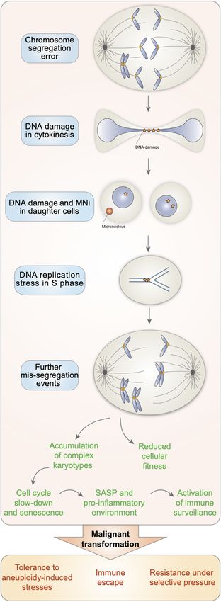
FIGURE 1 | Altogether, this triggers further missegregation events in the
subsequent cell cycles and thus the accumulation of cells with complex
karyotypes, known for displaying reduced cellular fitness, entering
senescence and displaying a senescence associated secretory phenotype
(SASP). However, in the context of cancer, the increasing genome instability
associated with aneuploidy can confer a proliferative advantage. This would
allow them to survive and provide a strong advantage in the presence of
selective pressures, such as during chemotherapy.
always the case: when translation rate was assessed in 12 disomic
yeast strains, it was found that around 20% of proteins was
synthesized at a lower rate than predicted based on copy number
changes. These proteins were mostly components of multi-
subunit complexes (Dephoure et al., 2014). Similarly, in
human aneuploid cells, the abundance of this type of proteins
and protein kinases was found to be reduced toward diploid levels
(Stingele et al., 2012). Collectively, this could indicate that either
some genes on aneuploid chromosomes are not translated
efficiently or their products are not stable. The Amon lab has
proposed that this “dosage compensation” occurs via the
activation of proteolytic pathways, so that cells can
compensate for abnormal protein stoichiometry (Torres et al.,
2008; Sheltzer et al., 2011; Pfau and Amon, 2012). And this was
also confirmed for multimolecular complexes (McShane et al.,
2016).
Given the deep impact of karyotype abnormalities on cell
physiology, it is not surprising that aneuploidy is detrimental for
cellular fitness (Santaguida and Amon, 2015; Zhu et al., 2018).
This is evident not only in budding and fission yeast, where
aneuploid strains proliferate at a slower rate than controls (Torres
et al., 2007; Holland and Cleveland, 2012), but also in mouse,
where trisomy of chromosome 1, 13, 16 or 19 is associated with
proliferation defects in MEFs (Williams et al., 2008). In humans,
all monosomies are lethal and only three autosomal trisomies are
viable: chromosome 13, 18 and 21, which are the poorest gene-
containing chromosomes (Holland and Cleveland, 2012). Only
individuals with trisomy 21 survive until adulthood and their cells
are well known to proliferate at a slower rate than age-matched
diploid cells (McCoy et al., 1983). Instead, aneuploidies of sex
chromosomes are better tolerated than aneuploidies of
autosomes, probably because the Y chromosome encodes for a
few genes only and only one X chromosome is active in adult cells
regardless of how many copies are present. It is important to
highlight that, beside gain of single chromosomes, sometimes
cells can acquire more complex karyotype abnormalities. When
aneuploidy becomes severe, cells do not simply slowdown in the
cell cycle, but activate p53 and undergo consequent cell cycle
arrest (Santaguida et al., 2017).
Intriguingly, although aneuploidy is usually deleterious for cell
physiology, some human tissues such as brain and liver naturally
contain aneuploid cells (Pack et al., 2005; Duncan et al., 2012;
Santaguida and Amon, 2015). Although the biological
FIGURE 1 | Aneuploid cells are characterized by increasing genome
significance of aneuploidy in these contexts has not been
instability. Chromosome segregation errors lead to the generation of elucidated yet, it can be speculated that aneuploidy could
aneuploid cells with DNA damage. When attempting to duplicate their allow liver cells, for example, to adapt to nutritional and
genome in S phase, aneuploid cells experience DNA replication stress. noxious stresses (Holland and Cleveland, 2012). This seems to
(Continued )
be particularly relevant to cancer cells, which are found to be very
Frontiers in Cell and Developmental Biology | www.frontiersin.org 4 February 2022 | Volume 10 | Article 838928Garribba and Santaguida Aneuploidy Triggers Genome Instability
often aneuploid (Vasudevan et al., 2021). In fact, recent work has that has been recently pioneered by the Medema and Storchova
shown that abnormal chromosome number in the context of labs (Chunduri et al., 2021; Hintzen et al., 2021). Further, it
cancer can promote resistance to chemotherapy (Salgueiro et al., remains unclear what are the exact mechanisms by which the
2020; Ippolito et al., 2021; Lukow et al., 2021), in line with the aneuploid status is associated with increasing genome instability.
observation that highly aneuploid tumors correlate with poorer Although aneuploid cells usually proliferate at a slower rate than
patient outcomes (Davoli et al., 2017; Vasudevan et al., 2020). In euploid counterparts and eventually cease to divide, some cells
details, chromosome missegregation events cause increased with karyotype imbalances can keep cycling. It would be
karyotypic heterogeneity that can be utilized by cancer cells to particularly interesting to assess the levels of genome
find the correct karyotypic landscape and thus survive under instability in this cycling sub-population of cells and
selective pressures such as chemotherapy. In conclusion, under investigate by which mechanisms they can cope with it and
stress-free conditions, aneuploidy causes decreased proliferation, remain capable to proliferate. Current available methodologies
while under suboptimal conditions aneuploid cells might grow do not allow for the recovery of a population of cycling aneuploid
better. This provides an explanation for the apparent paradox of cells suitable for further analyses (since those methodologies
cancer cells, which are very often aneuploid and at the same time employ spindle poisons to separate the cycling counterparts
are characterized by increased proliferation. (Santaguida et al., 2017; Wang et al., 2018; Wang et al.,
One final aspect that deserves to be discussed is the role that 2021)). Therefore, in the future it would be very important to
aneuploidy-induced CIN plays in tumorigenesis (Funk et al., 2016; develop new strategies to separate arrested and cycling aneuploid
van Jaarsveld and Kops, 2016). In the previous section, we mentioned cells. Since DNA replication seems to be a major contributing
that aneuploidy is frequently associated with increased genome factor to aneuploidy-induced genome instability, we speculate
instability. An increased DNA mutation rate can favor the onset that the efficiency of DNA replication and DNA repair processes
of genetic alterations that drive cellular growth and transformation would be different between arrested and cycling aneuploid cells,
(Holland and Cleveland, 2012). Recent studies in mouse have and that this would be the key factor in determining the fate of
demonstrated that CIN can favor the expansion of cells with cells harboring chromosome imbalances. Gaining this knowledge
clonal chromosomal abnormalities, which act as tumor-initiating will uncover novel dependencies of aneuploid cells with the
cells (Levine et al., 2017; Shoshani et al., 2021; Trakala et al., 2021). exciting possibility to exploit them to selectively eradicate
This is the case of chromosome 15, on which the oncogene Myc is aneuploid tumors.
located, and was found to be gained with high prevalence in a mouse
model of T-cell lymphoma (Shoshani et al., 2021; Trakala et al., 2021).
Interestingly, expression of human MYC from chromosome 6 leads AUTHOR CONTRIBUTIONS
to karyotype changes, namely gain of chromosome 6. Under this
condition, chromosome 15 is still frequently gained, unless the Rad21 All authors listed have made a substantial, direct, and intellectual
gene is deleted from it (Trakala et al., 2021). This shows that clonal contribution to the work and approved it for publication.
selection is guided by chromosomal location and identity of
specific genes.
FUNDING
CONCLUDING REMARKS Work in the Santaguida lab is supported by grants from the
Italian Association for Cancer Research (MFAG 2018—ID. 21665
In this review, we have discussed how chromosome segregation project), Ricerca Finalizzata (GR-2018-12367077), Fondazione
errors induce DNA damage and how this exacerbates genome Cariplo, the Rita-Levi Montalcini program from MIUR and the
instability in the resulting aneuploid cells, a condition that has Italian Ministry of Health with Ricerca Corrente and 5 × 1000
recently been shown to confer proliferative advantages under funds. LG is supported by a fellowship from Fondazione
specific conditions (Ippolito et al., 2021; Lukow et al., 2021; IEO-CCM.
Salgueiro et al., 2020) (Figure 1). It has to be noted that the
vast majority of studies conducted so far have employed an
heterogenous population of aneuploid cells, comprising both ACKNOWLEDGMENTS
chromosome gains and losses as well as cycling and arrested
cells. In the future, it will be important to explore more in details We thank all members of the Santaguida lab for constructive
the contributions of specific trisomies vs. monosomies, an effort discussions.
Artandi, S. E., and DePinho, R. A. (2010). Telomeres and Telomerase in Cancer.
REFERENCES Carcinogenesis 31, 9–18. doi:10.1093/carcin/bgp268
Bakhoum, S. F., Ngo, B., Laughney, A. M., Cavallo, J.-A., Murphy, C. J., Ly, P.,
Abe, T., and Barber, G. N. (2014). Cytosolic-DNA-mediated, STING-dependent et al. (2018). Chromosomal Instability Drives Metastasis through
Proinflammatory Gene Induction Necessitates Canonical NF-Κb Activation a Cytosolic DNA Response. Nature 553, 467–472. doi:10.1038/
through TBK1. J. Virol. 88, 5328–5341. doi:10.1128/JVI.00037-14 nature25432
Frontiers in Cell and Developmental Biology | www.frontiersin.org 5 February 2022 | Volume 10 | Article 838928Garribba and Santaguida Aneuploidy Triggers Genome Instability Bakhoum, S. F., Thompson, S. L., Manning, A. L., and Compton, D. A. (2009). Differentially Affect Chromosomal Stability. bioRxiv, 458318. doi:10.1101/ Genome Stability Is Ensured by Temporal Control of Kinetochore-Microtubule 2021.08.31.458318 Dynamics. Nat. Cel Biol. 11, 27–35. doi:10.1038/ncb1809 Hoffelder, D. R., Luo, L., Burke, N. A., Watkins, S. C., Gollin, S. M., and Saunders, Barber, T. D., McManus, K., Yuen, K. W. Y., Reis, M., Parmigiani, G., Shen, D., et al. W. S. (2004). Resolution of Anaphase Bridges in Cancer Cells. Chromosoma (2008). Chromatid Cohesion Defects May Underlie Chromosome Instability in 112, 389–397. doi:10.1007/s00412-004-0284-6 Human Colorectal Cancers. Proc. Natl. Acad. Sci. 105, 3443–3448. doi:10.1073/ Holland, A. J., and Cleveland, D. W. (2012). Losing Balance: The Origin and pnas.0712384105 Impact of Aneuploidy in Cancer. EMBO Rep. 13, 501–514. doi:10.1038/embor. Basto, R., Brunk, K., Vinadogrova, T., Peel, N., Franz, A., Khodjakov, A., et al. 2012.55 (2008). Centrosome Amplification Can Initiate Tumorigenesis in Flies. Cell 133, Ippolito, M. R., Martis, V., Martin, S., Tijhuis, A. E., Hong, C., Wardenaar, R., et al. 1032–1042. doi:10.1016/j.cell.2008.05.039 (2021). Gene Copy-Number Changes and Chromosomal Instability Induced by Ben-David, U., and Amon, A. (2020). Context Is Everything: Aneuploidy in Aneuploidy Confer Resistance to Chemotherapy. Develop. Cel 56, 2440–2454. Cancer. Nat. Rev. Genet. 21, 44–62. doi:10.1038/s41576-019-0171-x e6. doi:10.1016/j.devcel.2021.07.006 Blank, H. M., Sheltzer, J. M., Meehl, C. M., and Amon, A. (2015). Mitotic Entry in Janssen, A., van der Burg, M., Szuhai, K., Kops, G. J., and Medema, R. H. (2011). the Presence of DNA Damage Is a Widespread Property of Aneuploidy in Yeast. Chromosome Segregation Errors as a Cause of DNA Damage and Structural MBoC 26, 1440–1451. doi:10.1091/mbc.e14-10-1442 Chromosome Aberrations. Science 333, 1895–1898. doi:10.1126/science. Böhly, N., Kistner, M., and Bastians, H. (2019). Mild Replication Stress Causes 1210214 Aneuploidy by Deregulating Microtubule Dynamics in Mitosis. Cell Cycle 18, Letourneau, A., Santoni, F. A., Bonilla, X., Sailani, M. R., Gonzalez, D., Kind, J., 2770–2783. doi:10.1080/15384101.2019.1658477 et al. (2014). Domains of Genome-wide Gene Expression Dysregulation in Burrell, R. A., McClelland, S. E., Endesfelder, D., Groth, P., Weller, M.-C., Shaikh, Down’s Syndrome. Nature 508, 345–350. doi:10.1038/nature13200 N., et al. (2013). Replication Stress Links Structural and Numerical Cancer Levine, M. S., Bakker, B., Boeckx, B., Moyett, J., Lu, J., Vitre, B., et al. (2017). Chromosomal Instability. Nature 494, 492–496. doi:10.1038/nature11935 Centrosome Amplification Is Sufficient to Promote Spontaneous Chunduri, N. K., Menges, P., Zhang, X., Wieland, A., Gotsmann, V. L., Mardin, B. R., et al. Tumorigenesis in Mammals. Develop. Cel 40, 313e5–322. doi:10.1016/j. (2021). Systems Approaches Identify the Consequences of Monosomy in Somatic devcel.2016.12.022 Human Cells. Nat. Commun. 12, 5576. doi:10.1038/s41467-021-25288-x Levine, M. S., and Holland, A. J. (2018). The Impact of Mitotic Errors on Cell Chunduri, N. K., and Storchová, Z. (2019). The Diverse Consequences of Proliferation and Tumorigenesis. Genes Dev. 32, 620–638. doi:10.1101/gad. Aneuploidy. Nat. Cel Biol. 21, 54–62. doi:10.1038/s41556-018-0243-8 314351.118 Crasta, K., Ganem, N. J., Dagher, R., Lantermann, A. B., Ivanova, E. V., Pan, Y., Li, R., Yerganian, G., Duesberg, P., Kraemer, A., Willer, A., Rausch, C., et al. (1997). et al. (2012). DNA Breaks and Chromosome Pulverization from Errors in Aneuploidy Correlated 100% with Chemical Transformation of Chinese Mitosis. Nature 482, 53–58. doi:10.1038/nature10802 Hamster Cells. Proc. Natl. Acad. Sci. U. S. A. 94, 14506–14511. doi:10.1073/ Davoli, T., Uno, H., Wooten, E. C., and Elledge, S. J. (2017). Tumor Aneuploidy pnas.94.26.14506 Correlates with Markers of Immune Evasion and with Reduced Response to Liu, S., Kwon, M., Mannino, M., Yang, N., Renda, F., Khodjakov, A., et al. (2018). Immunotherapy. Science 355, eaaf8399. doi:10.1126/science.aaf8399 Nuclear Envelope Assembly Defects Link Mitotic Errors to Chromothripsis. Dephoure, N., Hwang, S., O’Sullivan, C., Dodgson, S. E., Gygi, S. P., Amon, A., et al. Nature 561, 551–555. doi:10.1038/s41586-018-0534-z (2014). Quantitative Proteomic Analysis Reveals Posttranslational Responses to London, N., and Biggins, S. (2014). Signalling Dynamics in the Spindle Aneuploidy in Yeast. Elife 3, e03023–27. doi:10.7554/eLife.03023 Checkpoint Response. Nat. Rev. Mol. Cel Biol. 15, 736–747. doi:10. Donnelly, N., Passerini, V., Dürrbaum, M., Stingele, S., and Storchová, Z. (2014). 1038/nrm3888 HSF 1 Deficiency and Impaired HSP 90-dependent Protein Folding Are Lukow, D. A., Sausville, E. L., Suri, P., Chunduri, N. K., Wieland, A., Leu, J., et al. Hallmarks of Aneuploid Human Cells. EMBO J. 33, 2374–2387. doi:10. (2021). Chromosomal Instability Accelerates the Evolution of Resistance to 15252/embj.201488648 Anti-cancer Therapies. Develop. Cel 56, 2427e4–2439. doi:10.1016/j.devcel. Duncan, A. W., Hanlon Newell, A. E., Smith, L., Wilson, E. M., Olson, S. B., Thayer, 2021.07.009 M. J., et al. (2012). Frequent Aneuploidy Among normal Human Hepatocytes. Ly, P., Brunner, S. F., Shoshani, O., Kim, D. H., Lan, W., Pyntikova, T., et al. (2019). Gastroenterology 142, 25–28. doi:10.1053/j.gastro.2011.10.029 Chromosome Segregation Errors Generate a Diverse Spectrum of Simple and Dürrbaum, M., Kuznetsova, A. Y., Passerini, V., Stingele, S., Stoehr, G., and Complex Genomic Rearrangements. Nat. Genet. 51, 705–715. doi:10.1038/ Storchová, Z. (2014). Unique Features of the Transcriptional Response to s41588-019-0360-8 Model Aneuploidy in Human Cells. BMC Genomics 15, 12–15. doi:10.1186/ Maciejowski, J., Li, Y., Bosco, N., Campbell, P. J., and de Lange, T. (2015). 1471-2164-15-139 Chromothripsis and Kataegis Induced by Telomere Crisis. Cell 163, Forment, J. V., Kaidi, A., and Jackson, S. P. (2012). Chromothripsis and Cancer: 1641–1654. doi:10.1016/j.cell.2015.11.054 Causes and Consequences of Chromosome Shattering. Nat. Rev. Cancer 12, MacKenzie, K. J., Carroll, P., Martin, C.-A., Murina, O., Fluteau, A., Simpson, D. J., 663–670. doi:10.1038/nrc3352 et al. (2017). CGAS Surveillance of Micronuclei Links Genome Instability to Funk, L. C., Zasadil, L. M., and Weaver, B. A. (2016). Living in CIN: Mitotic Innate Immunity. Nature 548, 461–465. doi:10.1038/nature23449 Infidelity and its Consequences for Tumor Promotion and Suppression. Maiato, H., and Logarinho, E. (2014). Mitotic Spindle Multipolarity without Develop. Cel 39, 638–652. doi:10.1016/j.devcel.2016.10.023 Centrosome Amplification. Nat. Cel Biol. 16, 386–394. doi:10.1038/ncb2958 Gao, C., Furge, K., Koeman, J., Dykema, K., Su, Y., Cutler, M. L., et al. (2007). Martin, S., and Santaguida, S. (2020). Understanding Complexity of Cancer Chromosome Instability, Chromosome Transcriptome, and Clonal Evolution Genomes: Lessons from Errors. Develop. Cel 53, 500–502. doi:10.1016/j. of Tumor Cell Populations. Proc. Natl. Acad. Sci. U. S. A. 104, 8995–9000. devcel.2020.05.004 doi:10.1073/pnas.0700631104 McCoy, E. E., and Sneddon, J. M. (1983). “Cell Biological Aspects of Down’s Géraud, G., Laquerrière, F., Masson, C., Arnoult, J., Labidi, B., and Hernandez- Syndrome,” in Advances in Cellular Neurobiology. Editors S. Fedoroff and Verdun, D. (1989). Three-dimensional Organization of Micronuclei Induced by L. Hertz (Cambridge, Massachusetts, United States: Academic Press), 4. Colchicine in PtK1 Cells. Exp. Cel Res. 181, 27–39. doi:10.1016/0014-4827(89) McShane, E., Sin, C., Zauber, H., Wells, J. N., Donnelly, N., Wang, X., et al. (2016). 90179-1 Kinetic Analysis of Protein Stability Reveals Age-dependent Degradation. Cell Harding, S. M., Benci, J. L., Irianto, J., Discher, D. E., Minn, A. J., and Greenberg, R. 167, 803e21–815. doi:10.1016/j.cell.2016.09.015 A. (2017). Mitotic Progression Following DNA Damage Enables Pattern Nawata, H., Kashino, G., Tano, K., Daino, K., Shimada, Y., Kugoh, H., et al. (2011). Recognition within Micronuclei. Nature 548, 466–470. doi:10.1038/ Dysregulation of Gene Expression in the Artificial Human Trisomy Cells of nature23470 Chromosome 8 Associated with Transformed Cell Phenotypes. PLoS One 6, Hatch, E. M., and Hetzer, M. W. (2015). Linking Micronuclei to Chromosome e25319. doi:10.1371/journal.pone.0025319 Fragmentation. Cell 161, 1502–1504. doi:10.1016/j.cell.2015.06.005 Nicholson, J. M., Macedo, J. C., Mattingly, A. J., Wangsa, D., Camps, J., Lima, V., Hintzen, D. C., Soto, M., Schubert, M., Bakker, B., Speirings, D. C. J., Szuhai, K., et al. (2015). Chromosome Mis-Segregation and Cytokinesis Failure in et al. (2021). Monosomies, Trisomies and Segmental Aneuploidies Trisomic Human Cells. Elife 4, 1–23. doi:10.7554/eLife.05068 Frontiers in Cell and Developmental Biology | www.frontiersin.org 6 February 2022 | Volume 10 | Article 838928
Garribba and Santaguida                                                                                                                        Aneuploidy Triggers Genome Instability

Pack, S. D., Weil, R. J., Vortmeyer, A. O., Zeng, W., Li, J., Okamoto, H., et al. (2005).         Torres, E. M., Williams, B. R., and Amon, A. (2008). Aneuploidy: Cells Losing
    Individual Adult Human Neurons Display Aneuploidy: Detection by                                  Their Balance. Genetics 179, 737–746. doi:10.1534/genetics.108.090878
    Fluorescence In Situ Hybridization and Single Neuron PCR. Cell Cycle 4,                       Trakala, M., Aggarwal, M., Sniffen, C., Zasadil, L., Carroll, A., Ma, D., et al. (2021).
    1758–1760. doi:10.4161/cc.4.12.2153                                                              Clonal Selection of Stable Aneuploidies in Progenitor Cells Drives High-
Passerini, V., Ozeri-Galai, E., de Pagter, M. S., Donnelly, N., Schmalbrock, S.,                     Prevalence Tumorigenesis. Genes Dev. 35, 1079–1092. doi:10.1101/gad.
    Kloosterman, W. P., et al. (2016). The Presence of Extra Chromosomes Leads to                    348341.121
    Genomic Instability. Nat. Commun. 7, 10754. doi:10.1038/ncomms10754                           Umbreit, N. T., Zhang, C. Z., Lynch, L. D., Blaine, L. J., Cheng, A. M., Tourdot, R.,
Pavelka, N., Rancati, G., Zhu, J., Bradford, W. D., Saraf, A., Florens, L., et al. (2010).           et al. (2020). Mechanisms Generating Cancer Genome Complexity from a
    Aneuploidy Confers Quantitative Proteome Changes and Phenotypic Variation                        Single Cell Division Error. Science 80-368. doi:10.1126/science.aba0712
    in Budding Yeast. Nature 468, 321–325. doi:10.1038/nature09529                                van Jaarsveld, R. H., and Kops, G. J. P. L. (2016). Difference Makers: Chromosomal
Pfau, S. J., and Amon, A. (2012). Chromosomal Instability and Aneuploidy in                          Instability versus Aneuploidy in Cancer. Trends Cancer 2, 561–571. doi:10.
    Cancer: From Yeast to Man. EMBO Rep. 13, 515–527. doi:10.1038/embor.                             1016/j.trecan.2016.09.003
    2012.65                                                                                       Vasudevan, A., Baruah, P. S., Smith, J. C., Wang, Z., Sayles, N. M., Andrews, P.,
Pillay, N., Tighe, A., Nelson, L., Littler, S., Coulson-Gilmer, C., Bah, N., et al. (2019).          et al. (2020). Single-Chromosomal Gains Can Function as Metastasis
    DNA Replication Vulnerabilities Render Ovarian Cancer Cells Sensitive to                         Suppressors and Promoters in Colon Cancer. Develop. Cel 52, 413–428.
    Poly(ADP-Ribose) Glycohydrolase Inhibitors. Cancer Cell 35, 519e8–533.                           doi:10.1016/j.devcel.2020.01.034
    doi:10.1016/j.ccell.2019.02.004                                                               Vasudevan, A., Schukken, K. M., Sausville, E. L., Girish, V., Adebambo, O. A.,
Rancati, G., Pavelka, N., Fleharty, B., Noll, A., Trimble, R., Walton, K., et al. (2008).            and Sheltzer, J. M. (2021). Aneuploidy as a Promoter and Suppressor of
    Aneuploidy Underlies Rapid Adaptive Evolution of Yeast Cells Deprived of a                       Malignant Growth. Nat. Rev. Cancer 21, 89–103. doi:10.1038/s41568-020-
    Conserved Cytokinesis Motor. Cell 135, 879–893. doi:10.1016/j.cell.2008.09.039                   00321-1
Salgueiro, L., Buccitelli, C., Rowald, K., Somogyi, K., Kandala, S., Korbel, J. O., et al.        Vietri, M., Schultz, S. W., Bellanger, A., Jones, C. M., Petersen, L. I., Raiborg, C.,
    (2020). Acquisition of Chromosome Instability Is a Mechanism to Evade                            et al. (2020). Unrestrained ESCRT-III Drives Micronuclear Catastrophe and
    Oncogene Addiction. EMBO Mol. Med. 12, e10941–15. doi:10.15252/emmm.                             Chromosome Fragmentation. Nat. Cel Biol. 22, 856–867. doi:10.1038/s41556-
    201910941                                                                                        020-0537-5
Santaguida, S., and Amon, A. (2015). Short- and Long-Term Effects of                              Wang, R. W., MacDuffie, E., and Santaguida, S. (2018). Generation and Isolation of
    Chromosome Mis-Segregation and Aneuploidy. Nat. Rev. Mol. Cel Biol. 16,                          Cell Cycle-Arrested Cells with Complex Karyotypes. J. Vis. Exp., 57215. doi:10.
    473–485. doi:10.1038/nrm4025                                                                     3791/57215
Santaguida, S., and Musacchio, A. (2009). The Life and Miracles of Kinetochores.                  Wang, R. W., Viganò, S., Ben-David, U., Amon, A., and Santaguida, S. (2021).
    EMBO J. 28, 2511–2531. doi:10.1038/emboj.2009.173                                                Aneuploid Senescent Cells Activate NF-κB to Promote Their Immune
Santaguida, S., Richardson, A., Iyer, D. R., M’Saad, O., Zasadil, L., Knouse, K. A.,                 Clearance by NK Cells. EMBO Rep. 22, 1–16. doi:10.15252/embr.202052032
    et al. (2017). Chromosome Mis-Segregation Generates Cell-Cycle-Arrested                       Wilhelm, T., Olziersky, A. M., Harry, D., De Sousa, F., Vassal, H., Eskat, A., et al.
    Cells with Complex Karyotypes that Are Eliminated by the Immune System.                          (2019). Mild Replication Stress Causes Chromosome Mis-Segregation via
    Develop. Cel 41, 638e5–651. doi:10.1016/j.devcel.2017.05.022                                     Premature Centriole Disengagement. Nat. Commun. 10, 3585–3614. doi:10.
Santaguida, S., Vasile, E., White, E., and Amon, A. (2015). Aneuploidy-induced                       1038/s41467-019-11584-0
    Cellular Stresses Limit Autophagic Degradation. Genes Dev. 29, 2010–2021.                     Williams, B. R., Prabhu, V. R., Hunter, K. E., Glazier, C. M., Whittaker, C. A.,
    doi:10.1101/gad.269118.115                                                                       Housman, D. E., et al. (2008). Aneuploidy Affects Proliferation and
Sheltzer, J. M., Blank, H. M., Pfau, S. J., Tange, Y., George, B. M., Humpton, T. J.,                Spontaneous Immortalization in Mammalian Cells. Science 322, 703–709.
    et al. (2011). Aneuploidy Drives Genomic Instability in Yeast. Science 333,                      doi:10.1126/science.1160058
    1026–1030. doi:10.1126/science.1206412                                                        Xu, B., Sun, Z., Liu, Z., Guo, H., Liu, Q., Jiang, H., et al. (2011). Replication Stress
Shoshani, O., Bakker, B., de Haan, L., Tijhuis, A. E., Wang, Y., Kim, D. H., et al.                  Induces Micronuclei Comprising of Aggregated DNA Double-Strand Breaks.
    (2021). Transient Genomic Instability Drives Tumorigenesis through                               PLoS One 6, e18618. doi:10.1371/journal.pone.0018618
    Accelerated Clonal Evolution. Genes Dev. 35, 1093–1108. doi:10.1101/gad.                      Zhang, C.-Z., Spektor, A., Cornils, H., Francis, J. M., Jackson, E. K., Liu, S., et al.
    348319.121                                                                                       (2015). Chromothripsis from DNA Damage in Micronuclei. Nature 522,
Soto, M., García-Santisteban, I., Krenning, L., Medema, R. H., and Raaijmakers,                      179–184. doi:10.1038/nature14493
    J. A. (2018). Chromosomes Trapped in Micronuclei Are Liable to Segregation                    Zhu, J., Pavelka, N., Bradford, W. D., Rancati, G., and Li, R. (2012). Karyotypic
    Errors. J. Cel Sci. 131. doi:10.1242/jcs.214742                                                  Determinants of Chromosome Instability in Aneuploid Budding Yeast. Plos
Stingele, S., Stoehr, G., Peplowska, K., Cox, J., Mann, M., and Storchova, Z. (2012).                Genet. 8, e1002719. doi:10.1371/journal.pgen.1002719
    Global Analysis of Genome, Transcriptome and Proteome Reveals the                             Zhu, J., Tsai, H.-J., Gordon, M. R., and Li, R. (2018). Cellular Stress Associated with
    Response to Aneuploidy in Human Cells. Mol. Syst. Biol. 8, 608. doi:10.                          Aneuploidy. Develop. Cel 44, 420–431. doi:10.1016/j.devcel.2018.02.002
    1038/msb.2012.40
Terradas, M., Martín, M., and Genescà, A. (2016). Impaired Nuclear Functions in                   Conflict of Interest: The authors declare that the research was conducted in the
    Micronuclei Results in Genome Instability and Chromothripsis. Arch. Toxicol.                  absence of any commercial or financial relationships that could be construed as a
    90, 2657–2667. doi:10.1007/s00204-016-1818-4                                                  potential conflict of interest.
Terradas, M., Martín, M., Hernández, L., Tusell, L., and Genescà, A. (2012).
    Nuclear Envelope Defects Impede a Proper Response to Micronuclear DNA                         Publisher’s Note: All claims expressed in this article are solely those of the authors
    Lesions. Mutat. Research/Fundamental Mol. Mech. Mutagenesis 729, 35–40.                       and do not necessarily represent those of their affiliated organizations, or those of
    doi:10.1016/j.mrfmmm.2011.09.003                                                              the publisher, the editors and the reviewers. Any product that may be evaluated in
Terradas, M., Martín, M., Tusell, L., and Genescà, A. (2009). DNA Lesions                         this article, or claim that may be made by its manufacturer, is not guaranteed or
    Sequestered in Micronuclei Induce a Local Defective-Damage Response.                          endorsed by the publisher.
    DNA Repair 8, 1225–1234. doi:10.1016/j.dnarep.2009.07.004
Thompson, S. L., and Compton, D. A. (2011). Chromosome Missegregation in                          Copyright © 2022 Garribba and Santaguida. This is an open-access article
    Human Cells Arises through Specific Types of Kinetochore-Microtubule                           distributed under the terms of the Creative Commons Attribution License (CC
    Attachment Errors. Proc. Natl. Acad. Sci. 108, 17974–17978. doi:10.1073/                      BY). The use, distribution or reproduction in other forums is permitted, provided the
    pnas.1109720108                                                                               original author(s) and the copyright owner(s) are credited and that the original
Torres, E. M., Sokolsky, T., Tucker, C. M., Chan, L. Y., Boselli, M., Dunham, M. J.,              publication in this journal is cited, in accordance with accepted academic practice.
    et al. (2007). Effects of Aneuploidy on Cellular Physiology and Cell Division in              No use, distribution or reproduction is permitted which does not comply with
    Haploid Yeast. Science 317, 916–924. doi:10.1126/science.1142210                              these terms.

Frontiers in Cell and Developmental Biology | www.frontiersin.org                             7                                           February 2022 | Volume 10 | Article 838928
You can also read


