Traumatisme du rachis : quelle gestion à - saranf

Page created by Juan Hamilton
 
CONTINUE READING
Traumatisme du rachis : quelle gestion à - saranf
XXXIVème Congrès de la SARAF
         Yamoussoukro 21-23 Novembre 2018

Traumatisme du rachis : quelle gestion à
    la phase initiale aujourd’hui ?

                  Pr. Julien Pottecher, MD, PhD
                   Service d’Anesthésie-Réanimation Chirurgicale
           EA3072 - Mitochondrie - Stress Oxydant et Protection Musculaire
                               Comité ACUTE - SFAR

                    Hôpitaux Universitaires de Strasbourg
        Fédération de Médecine Translationnelle de Strasbourg (FMTS)
Traumatisme du rachis : quelle gestion à - saranf
Pas de conflit d’intérêt
Traumatisme du rachis : quelle gestion à - saranf
Actualisation 2018 en cours à SFAR 2019, SARANF 2019 ?
Traumatisme du rachis : quelle gestion à - saranf
Bouillon, B. & al. Eur J Trauma Emerg Surg 2018; 44, 3–271.
Traumatisme du rachis : quelle gestion à - saranf
Gauss, T. & GITE. Anaesth Crit Care Pain Med 2018
Traumatisme du rachis : quelle gestion à - saranf
Facteurs anamnéstiques et cliniques

• Incidence des lésions médullaires : 1-3% des traumatismes fermés
     Holly LT. J Neurosurg 2002; 96: 285-91

• Association traumatisme crânien-lésions médullaires :
   – Fréquente, d’autant plus qu’il existe :
       • Un GCS < 8 (OR : 8,5)
     Demetriades D. J Trauma 2000; 48: 724-7
       • Un déficit neurologique focal (OR : 58)
     Blackmore CC. Radiology 1999; 211: 759-65
Traumatisme du rachis : quelle gestion à - saranf
Facteurs anamnéstiques et cliniques
•   Association lésion médullaire - autres lésions vitales :
     – Fréquente
     – Aggrave le pronostic vital
     – Aggrave le pronostic neurologique médullaire
     Crosby ET. Anesthesiology 2006; 104: 1293-318

•   Complications respiratoires grèvent la survie :
     – Immédiate
     – A distance
     Durga P. Anesth Analg 2010; 110: 134-40

•   Nécessité d’intubation dans 60 à 80% des cas :
     – Même en cas de lésions cervicales basses
     – Même en cas de lésions incomplètes
     Hassid VJ. J Trauma 2008; 65: 1328-32
Traumatisme du rachis : quelle gestion à - saranf
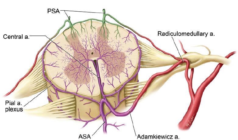
Phase initiale [24h]
• PEC pré-hospitalière
  – Relevage
  – Immobilisation
                              1. Immobilisation
  – Airway                    2. Objectifs hémodynamiques
                              3. Objectifs d’hématose
  – Objectifs tensionnels
• Arrivée à l’hôpital
  – Bilan lésionnel
  – Stratégie opératoire
Traumatisme du rachis : quelle gestion à - saranf
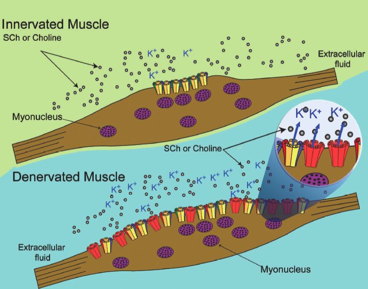
Phase pré-hospitalière
ATLS, Tenth Edition, American College of Surgeons
Stabilisation

Dès la prise en charge
                                    Surtout pour la désincarcération
A retirer dès que la TDM confirme
                                    et la mobilisation
l’absence de lésion
                                    Matelas à dépression
à Confort
                                    Aucune RCT
à Retour veineux jugulaire
sagit
                                                                                                                                                                                                         sure

Cervical Spine Motion During Airway Management: A                                                                                                                                                        NET
                                                                                                                                                                                                         (ILM
                                                                                                                                                                                                         place

Cinefluoroscopic Study of the Posteriorly Destabilized Third                                                                                                                                             FOS-

Cervical Vertebrae in Human Cadavers                                                                                                                                                                     Disc
                                                                                                                                                                                                         Seve
                                                                                                                                                                                                         meth
                                                                                                                                                                                                         C-sp
Joseph Brimacombe, MB, ChB, FRCA, MD*, Christian Keller, MD†, Karl H. Künzel,                                   MD‡,                                                                                    tient
                                                                                                                                                                                                         laryn
Othmar Gaber, MD‡, Michael Boehler, MD†, and Fredrich Pühringer, MD†                                                             Figure 1. Reference lines for determining A, anteroposterior mo-
                                                                                                                                  tion and B, sagittal motion of the third cervical vertebrae. C2, C3,
                                                                                                                                                                                                         tion
                                                                                                                                                                                                         prod
                                                                                                                                  and C4 $ cervical segments.                                            sal in
*University of Queensland, Department of Anesthesia and Intensive Care, Cairns Base Hospital, Australia; and                                                                                             nasa
†Department of Anesthesia and Intensive Care Medicine, and ‡Institute of Anatomy, Leopold-Franzens University,                                                                                           al. (1

Innsbruck, Austria                                                                            Translation max                     Results                                                                a col

        Procédure                                           MILS ?                                                                Rotation max [°]
                                                                                                                                                                                                         and
                                                                                                                                  The mean (range) for age, height, and weight was 79
                                                                                                                                                                                                         obtu

                                                                                                   [mm]
                                                                                                                                  (69 – 87) yr, 168 (152–189) cm, and 66 (42–93) kg, re-
                                                                                                                                                                                                         can c
                                                                                                                                  spectively. The male/female ratio was 7:3. All of the
                                                                                                                                                                                                         tion.
                                                                                                                                  data for the displacement and segmental sagittal mo-
                                                                                                                                                                                                         less m
                                                                                                                                  tion were normally distributed. The image analysis
                                                                                                                                                                                                         colla
                                                                                                                                  software facilitated quantification of displacement and

         Extension max.                                           Non
                                                                   There was a significant increase1,8±1,7                                             0,8
                                                                                                                                                                                                         (6) st
                                                                                                                                  angulation with a resolution of 0.1 mm and 0.05 de-
     We conducted a randomized, controlled, crossover                                               in posterior displace-        grees, respectively. The mean (range) for measure-
                                                                                                                                                                                                         C5-6
     study to determine cervical spine motion for six airway       ment for the FM (1.9 ! 1.2 mm, P " 0.01), OETT (2.6 !                                                                                 batio
                                                                                                                                  ment discrepancies between observers was 0.23 (0 –                     niqu
     management techniques in human cadavers with a pos-              1.6 mm, P " 0.0001), ETC (3.2 ! 1.6 mm, P " 0.0001),        2.68) mm for position and 0.24 (0 –2.5) degrees for                    chin
                                                                                                                                  angulation. Data for the maximum posterior displace-
     teriorly destabilized third cervical (C-3) vertebra. A de-       ILM-OETT (1.7 ! 1.3 mm, P " 0.01), LMA (1.7 !
        Flexion max.                                              Non                                  3,7±1,9                                        -4,5
                                                                                                                                                                                                         the
                                                                                                                                  ment, the maximum segmental sagittal motion of                         study
     stabilized C-3 segment was created in 10 cadavers (6 –           1.3 mm, P " 0.01), FLEX-MAX (3.7 ! 1.9 mm, P "              C2-3, and the phase of maximum motion are given in                     moti
     24 h postmortem). Cervical motion was recorded by                0.0001), EXT-MAX (1.8 ! 1.7, P " 0.01), however, not for    Table 1. Compared with baseline values, the posterior                  and
                                                                                                                                  displacement and segmental sagittal motion after de-                   simil
     continuous lateral fluoroscopy. The following airway             FOS-NETT (0.1 ! 0.7 mm). Posterior displacement was         stabilization was 2.8 (0.8, 1.3– 4.2) mm and 1.2 (3.5,

        Vent. masque                                              Oui                                 1,9±1,2*                                         2,7
                                                                                                                                                                                                         caus
     management techniques were performed in random                   less for the ILM-OETT and LMA than for the ETC (both        !6.0 – 8.0) degrees, respectively. There was a signifi-                tion.
                                                                                                                                  cant increase in posterior displacement for the FM
     order on each cadaver with manual in-line stabilization          P " 0.04). There were no significant increases in seg-      (P " 0.01), OETT (P " 0.0001), ETC (P " 0.0001),
                                                                                                                                                                                                         be ta
                                                                                                                                                                                                         mane
     applied: face mask ventilation (FM), laryngoscope-               mental sagittal motion with any airway manipulation         ILM-OETT (P " 0.01), LMA (P " 0.01), FLEX-MAX                          Lenn
     guided orotracheal intubation (OETT), fiberscope-                other than with FLEX-MAX (#4.5 ! 4.0°, P " 0.01). Pos-      (P " 0.0001), and EXT-MAX (P " 0.01), however, not                     laryn
                                                                                                                                  for FOS-NETT (0.1 # 0.7 mm). Posterior displacement
     guided nasal intubation (FOS-NETT), esophageal                   terior displacement was similar to FLEX-MAX for the         was less for the ILM-OETT and LMA than the ETC
                                                                                                                                                                                                         terio
                                                                                                                                                                                                         They
     tracheal Combitube® (Kendall-Sheridan, Neustadt,                 OETT and ETC; however, it was less for the FM, FOS-         (both P " 0.04). There were no significant increases in                unsta
     Germany) insertion (ETC), intubating laryngeal mask              NETT, ILM-OETT, and LMA (all P " 0.01). Posterior           segmental sagittal motion with any airway manipula-                    after
                                                                                                                                  tion other than with FLEX-MAX (!4.5 # 4.0°, P "                        smal
     insertion with fiberscope-guided tracheal intubation             displacement was similar to EXT-MAX for all airway          0.01). Posterior displacement was similar to FLEX-                     Kiha
     (ILM-OETT), and laryngeal mask airway insertion                  manipulations other than for FOS-NETT (P " 0.001).          MAX for the OETT and ETC; however, it was less for                     patie
     (LMA). Afterward, maximum head-neck flexion                      For cervical motion and the techniques tested, the safest   the FM, FOS-NETT, ILM-OETT, and LMA (all P "                           disea
                                                                                                                                  0.01). Posterior displacement was similar to EXT-MAX                   ILM.
     (FLEX-MAX) and maximum head-neck extension                       method of airway management in a patient with a pos-        for all airway manipulations other than for FOS-NETT                   exten
     (EXT-MAX) without manual in-line stabilization was               teriorly destabilized C-3 segment is FOS-NETT. LMA          (P " 0.001). For all airway manipulations, sagittal                    ment
                                                                                                                                  rotation was less than FLEX-MAX (all P " 0.0001) and                      W
     performed to determine maximum motion. The maxi-                 devices may be preferable to the ETC.                       similar to EXT-MAX. In all cadavers, the point of                      segm
     mum posterior displacement of C-3 and the maximum                                                                            maximum displacement and maximum segmental                             LMA
     segmental sagittal motion of C2-3 were determined.                                       (Anesth Analg 2000;91:1274 –8)

C
      ervical spine (C-spine) injuries occur in 1.5%–3%              to guide the anesthesiologist in selecting the appropri-
      of all major trauma cases and are associated                   ate technique. Donaldson et al. (5) investigated the
      with major morbidity and mortality (1,2). There                effect of airway management on the unstable C1-2 and
                                       Brimacombe
is anecdotal evidence that airway management           J.
                                                     can           Anesth  Analg
                                                                     C5-6 (6)      2000;in91:cadavers
                                                                               segments        1274-8and showed that both
result in catastrophic neurologic injury (3); however,               oro/nasotracheal intubation and chin lift/jaw thrust
« Chin lift & Jaw thrust »

   ATLS, Tenth Edition, American College of Surgeons
sagit
                                                                                                                                                                                                         sure

Cervical Spine Motion During Airway Management: A                                                                                                                                                        NET
                                                                                                                                                                                                         (ILM
                                                                                                                                                                                                         place

Cinefluoroscopic Study of the Posteriorly Destabilized Third                                                                                                                                             FOS-

Cervical Vertebrae in Human Cadavers                                                                                                                                                                     Disc
                                                                                                                                                                                                         Seve
                                                                                                                                                                                                         meth
                                                                                                                                                                                                         C-sp
Joseph Brimacombe, MB, ChB, FRCA, MD*, Christian Keller, MD†, Karl H. Künzel,                                   MD‡,                                                                                    tient
                                                                                                                                                                                                         laryn
Othmar Gaber, MD‡, Michael Boehler, MD†, and Fredrich Pühringer, MD†                                                             Figure 1. Reference lines for determining A, anteroposterior mo-
                                                                                                                                  tion and B, sagittal motion of the third cervical vertebrae. C2, C3,
                                                                                                                                                                                                         tion
                                                                                                                                                                                                         prod
                                                                                                                                  and C4 $ cervical segments.                                            sal in
*University of Queensland, Department of Anesthesia and Intensive Care, Cairns Base Hospital, Australia; and                                                                                             nasa
†Department of Anesthesia and Intensive Care Medicine, and ‡Institute of Anatomy, Leopold-Franzens University,                                                                                           al. (1

Innsbruck, Austria                                                                            Translation max                     Results                                                                a col

        Procédure                                           MILS ?                                                                Rotation max [°]
                                                                                                                                                                                                         and
                                                                                                                                  The mean (range) for age, height, and weight was 79
                                                                                                                                                                                                         obtu

                                                                                                   [mm]
                                                                                                                                  (69 – 87) yr, 168 (152–189) cm, and 66 (42–93) kg, re-
                                                                                                                                                                                                         can c
                                                                                                                                  spectively. The male/female ratio was 7:3. All of the
                                                                                                                                                                                                         tion.
                                                                                                                                  data for the displacement and segmental sagittal mo-
                                                                                                                                                                                                         less m
                                                                                                                                  tion were normally distributed. The image analysis
                                                                                                                                                                                                         colla
                                                                                                                                  software facilitated quantification of displacement and

         Extension max.                                           Non
                                                                   There was a significant increase1,8±1,7                                             0,8
                                                                                                                                                                                                         (6) st
                                                                                                                                  angulation with a resolution of 0.1 mm and 0.05 de-
     We conducted a randomized, controlled, crossover                                               in posterior displace-        grees, respectively. The mean (range) for measure-
                                                                                                                                                                                                         C5-6
     study to determine cervical spine motion for six airway       ment for the FM (1.9 ! 1.2 mm, P " 0.01), OETT (2.6 !                                                                                 batio
                                                                                                                                  ment discrepancies between observers was 0.23 (0 –                     niqu
     management techniques in human cadavers with a pos-              1.6 mm, P " 0.0001), ETC (3.2 ! 1.6 mm, P " 0.0001),        2.68) mm for position and 0.24 (0 –2.5) degrees for                    chin
                                                                                                                                  angulation. Data for the maximum posterior displace-
     teriorly destabilized third cervical (C-3) vertebra. A de-       ILM-OETT (1.7 ! 1.3 mm, P " 0.01), LMA (1.7 !
        Flexion max.                                              Non                                  3,7±1,9                                        -4,5
                                                                                                                                                                                                         the
                                                                                                                                  ment, the maximum segmental sagittal motion of                         study
     stabilized C-3 segment was created in 10 cadavers (6 –           1.3 mm, P " 0.01), FLEX-MAX (3.7 ! 1.9 mm, P "              C2-3, and the phase of maximum motion are given in                     moti
     24 h postmortem). Cervical motion was recorded by                0.0001), EXT-MAX (1.8 ! 1.7, P " 0.01), however, not for    Table 1. Compared with baseline values, the posterior                  and
                                                                                                                                  displacement and segmental sagittal motion after de-                   simil
     continuous lateral fluoroscopy. The following airway             FOS-NETT (0.1 ! 0.7 mm). Posterior displacement was         stabilization was 2.8 (0.8, 1.3– 4.2) mm and 1.2 (3.5,

        Vent. masque                                              Oui                                 1,9±1,2*                                         2,7
                                                                                                                                                                                                         caus
     management techniques were performed in random                   less for the ILM-OETT and LMA than for the ETC (both        !6.0 – 8.0) degrees, respectively. There was a signifi-                tion.
                                                                                                                                  cant increase in posterior displacement for the FM
     order on each cadaver with manual in-line stabilization          P " 0.04). There were no significant increases in seg-      (P " 0.01), OETT (P " 0.0001), ETC (P " 0.0001),
                                                                                                                                                                                                         be ta
                                                                                                                                                                                                         mane
     applied: face mask ventilation (FM), laryngoscope-               mental sagittal motion with any airway manipulation         ILM-OETT (P " 0.01), LMA (P " 0.01), FLEX-MAX                          Lenn
     guided orotracheal intubation (OETT), fiberscope-                other than with FLEX-MAX (#4.5 ! 4.0°, P " 0.01). Pos-      (P " 0.0001), and EXT-MAX (P " 0.01), however, not                     laryn
        IOT
     guided nasal intubation (FOS-NETT), esophageal
     tracheal Combitube® (Kendall-Sheridan, Neustadt,
                                                                  Oui                                  2,6±1,6
                                                                      terior displacement was similar to FLEX-MAX for the
                                                                      OETT and ETC; however, it was less for the FM, FOS-
                                                                                                                                                       2,7
                                                                                                                                  for FOS-NETT (0.1 # 0.7 mm). Posterior displacement
                                                                                                                                  was less for the ILM-OETT and LMA than the ETC
                                                                                                                                  (both P " 0.04). There were no significant increases in
                                                                                                                                                                                                         terio
                                                                                                                                                                                                         They
                                                                                                                                                                                                         unsta
     Germany) insertion (ETC), intubating laryngeal mask              NETT, ILM-OETT, and LMA (all P " 0.01). Posterior           segmental sagittal motion with any airway manipula-                    after
                                                                                                                                  tion other than with FLEX-MAX (!4.5 # 4.0°, P "                        smal
     insertion with fiberscope-guided tracheal intubation             displacement was similar to EXT-MAX for all airway          0.01). Posterior displacement was similar to FLEX-                     Kiha
     (ILM-OETT), and laryngeal mask airway insertion                  manipulations other than for FOS-NETT (P " 0.001).          MAX for the OETT and ETC; however, it was less for                     patie
     (LMA). Afterward, maximum head-neck flexion                      For cervical motion and the techniques tested, the safest   the FM, FOS-NETT, ILM-OETT, and LMA (all P "                           disea
                                                                                                                                  0.01). Posterior displacement was similar to EXT-MAX                   ILM.
     (FLEX-MAX) and maximum head-neck extension                       method of airway management in a patient with a pos-        for all airway manipulations other than for FOS-NETT                   exten
     (EXT-MAX) without manual in-line stabilization was               teriorly destabilized C-3 segment is FOS-NETT. LMA          (P " 0.001). For all airway manipulations, sagittal                    ment
                                                                                                                                  rotation was less than FLEX-MAX (all P " 0.0001) and                      W
     performed to determine maximum motion. The maxi-                 devices may be preferable to the ETC.                       similar to EXT-MAX. In all cadavers, the point of                      segm
     mum posterior displacement of C-3 and the maximum                                                                            maximum displacement and maximum segmental                             LMA
     segmental sagittal motion of C2-3 were determined.                                       (Anesth Analg 2000;91:1274 –8)

C
      ervical spine (C-spine) injuries occur in 1.5%–3%   to guide the anesthesiologist in selecting the appropri-
      of all major trauma cases and are associated        ate technique. Donaldson et al. (5) investigated the
      with major morbidity and mortality (1,2). There     effect of airway management on the unstable C1-2 and
is anecdotal evidence that airway management       Brimacombe
                                                     can        J. Anesth
                                                          C5-6 (6)         Analg
                                                                    segments      2000; 91:
                                                                             in cadavers   and1274-8
                                                                                                showed that both
result in catastrophic neurologic injury (3); however,    oro/nasotracheal intubation and chin lift/jaw thrust
Definitive airway

ATLS, Tenth Edition, American College of Surgeons
v.s

Driver, B.E. & al. JAMA 2018; 319: 2179–2189.
v.s
Driver, B.E. & al. JAMA 2018; 319: 2179–2189.
Success : 96% [93-99%]

                              Success : 82% [76-88%]

                     v.s

Driver, B.E. & al. JAMA 2018; 319: 2179–2189.
Trimmel, H. & al Crit. Care Med. 2016; 44, e470–6.
Manœuvre de Sellick

         •Estomac plein « par définition » :
             • Epistaxis déglutie
             • Alcoolisation
             • Stress, douleur
             • Gastroparésie

         •Morbi-mortalité essentiellement
         respiratoire :
             • précoce
             • tardive
             Durga P. Anesth Analg 2010; 110: 134-40
Sellick ? Jamais !
1. Manœuvre inefficace :                        2. Manœuvre risquée :
Déplacement latéral de l’œsophage p/r au plan   Mobilisation cervicale > LD
vertébral dans 90% des cas.                     Donaldson WF. Spine     1993;
                                                                La Société            18: 2020-3
                                                                           française d’anesthésie et de réanimation

                                                Déplacement postérieur       max de 9mm
                                                                    en collaboration avec
                                                Gabbott DA. Anaesthesia 1997; 52: 586-8
                                                                        L’Association de neuro-anesthésie réanimation de langue française
                                                                                              Le Samu de France

                                                3. Dégradation de la vue laryngoscopique
                                                                        La Société française de chirurgie orthopédique et traumatologique
                                                                                 La Société francophone de médecine d’urgence
                                                                          La Société française de médecine physique et de réadaptation
                                                                                     La Société française de neurochirurgie
                                                                               La Société francophone de neurochirurgie du rachis
                                                                                 La Société de pneumologie de langue française

                                                4. Manœuvre non recommandée
                                                                                Prise en charge d’un blessé adulte
                                                                        présentant un traumatisme vertébro-médullaire

                                                                                              Conférence d’experts

                                                                                                   Texte court
                                                                                                      2003

Smith KJ. Anesthesiology 2003; 99: 60-4
Sellick ? Bien sûr !
 1. Manœuvre efficace :                             3. Manœuvre recommandée
 Compression de l’hypopharynx contre le rachis ou
 les muscles rétropharyngés.

 Rice MJ. Anesth Analg 2009; 109: 1546-52

2. Manœuvre sans risque :
Mobilisation cervicale minime
Translation verticale C5 : 0,5mm[0-1,5]
Helliwell V. Resuscitation 2001; 49: 53-7
                                                      Wilson WC. ASA Newsletter 2005; 69
Birenbaum, A. & al. JAMA Surg. 2018.
Arrivée à l’hôpital
Quel hôpital ?
• Trauma center niveau I

• Possibilité de traitement définitif précoce (
Bilan lésionnel clinique

  ATLS, Tenth Edition, American College of Surgeons
Bilan lésionnel clinique

                             Score ASIA IMSOP
American Spinal Injury Association - International Medical Society of Paraplegia
Bilan lésionnel clinique

                                Polytrauma

   Hoffmann JR & al. N Engl J Med 2000; 343: 94-99
  ATLS, Tenth Edition, American College of Surgeons
Stabilité / Instabilité
  « Une lésion est dite instable si elle génère immédiatement ou
             secondairement un déplacement intervertébral »
• Quid des seuils tolérables ?
   – Angulation < 11°
   – Translation < 3mm
   White AA, Panjabi MM. Clinical Biomechanics of the Spine 1990

• Quid des situations spécifiques ?
   – Anatomiques :
       • Canal cervical étroit
       • Ostéophytose rachidienne
   – Pathologiques :
      • Hypotension artérielle systémique
      • Hypoxémie
Stabilité / Instabilité

   ±Stable      Instable   Instable

     Instable                Instable

    Instable    ±Stable    Stable
Bilan lésionnel radiographique
• Après stabilisation hémodynamique
• TDM multibarette avec reconstruction 3D :
    – Nécessaire et suffisant
    – Permet de lever l’immobilisation
    – Rend inutile la réalisation de clichés dynamiques
Padayachee L. J Trauma 2006; 60: 341-5

•    En l’absence de TDM disponible
    –     Clichés radiographiques standard
    –     AP, latéral et odontoïde bouche ouverte
• SCIWORA à IRM
                                Walters, B.C. & al. Neurosurgery 2013.
Bilan lésionnel radiographique

 Fracture de Jefferson    Hangman’s Fracture

Fracture de l’odontoïde   Fracture de Chance
Vascularisation médullaire

  Martirosyan NL. J Neurosurg Spine 2011; 15:238–251
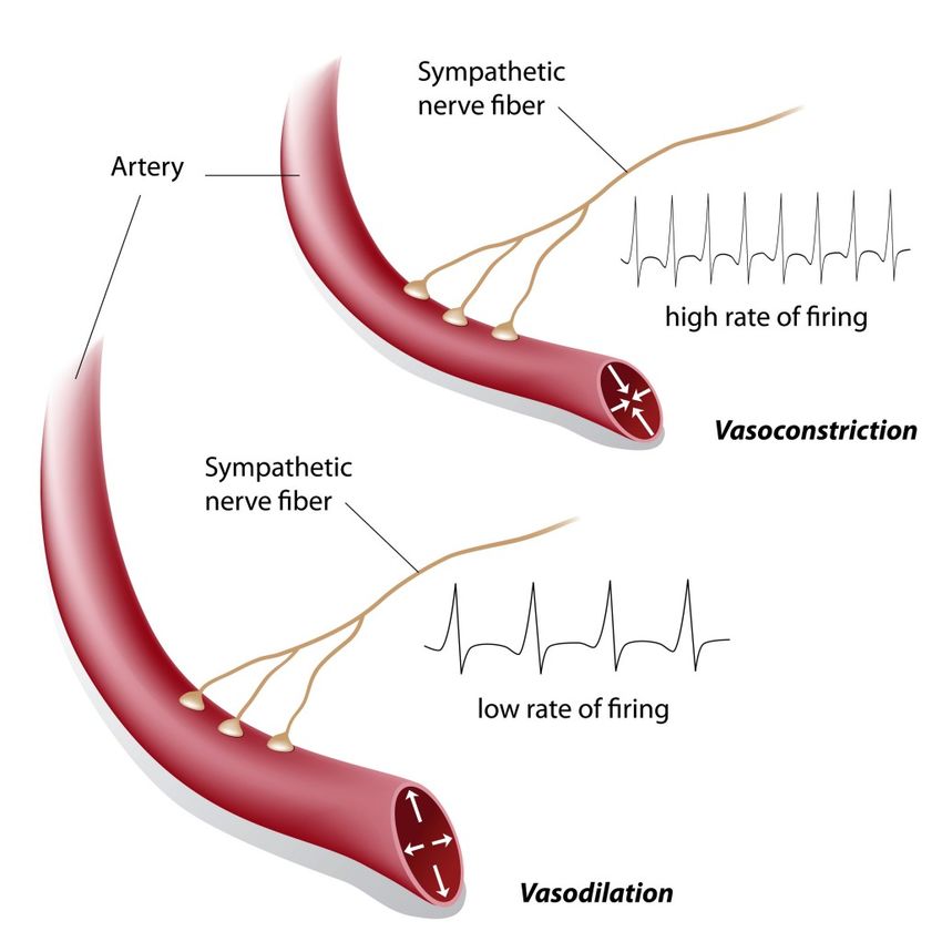
Sympatholyse du choc spinal

   Rq : Face à une hypotension post-traumatique,
   le choc spinal doit rester un diagnostic d’exclusion
Autorégulation médullaire

                                                         Moëlle saine
                      Zone d’autorégulation              Moëlle contuse
                         Vasoco                          Moëlle lésée
ml/100 g/mn

                               nstricti
                                        on
DSM

                                odi l a t a tion
                             Vas

                 50                                150       PPM mmHg
Objectifs hémodynamiques

       70                                 29                         50

PAM [mmHg]                 PIM [mmHg]                            PPM [mmHg]

             Squair, J.W. & al. Neurology 2017; 89, 1660–1667.
En pratique
• Expansion volémique première
  – Surtout guidée par les lésions hémorragiques associées
  – Solutions iso-osmotiques

• Recours précoce aux vasoconstricteurs
  – Noradrénaline diluée
  – PAS > 120mmHg, PAM > 80mmHg, 7 jours
  – 75% des traumatisés médullaires ont au moins un épisode de
    PAS < 90mmHg
Objectifs hémodynamiques
• PAM maintenue 80 mmHg
  – Dès la phase pré-hospitalière
  – Première semaine post-traumatique

  Recommandations américaines
  Walters, B.C. & al. Neurosurgery 2013.

  Recommandations allemandes
  Bouillon, B. & al. Eur J Trauma Emerg Surg 2018; 44, 3–271.

  Recommandations françaises
  Edouard, E. & al Ann Fr Anesth Reanim 2004; 23: 930–945
NASCIS
• NASCIS I
  Bracken MB. JAMA 1984;251:45–52

• NASCIS II
  Bracken MB. N Engl J Med 1990;322:1405– 11.

• NASCIS III
  Bracken MB. JAMA 1997;277;1597–604

• NASCIS n…
  Bracken MB.
  Cochrane Database of Systematic Reviews 2012,
  Issue 1. Art. No: CD001046
Risques
Méthylprednisolone
• Pas d’indication

• Etudes NACSIS II et III non conclusives

• Attente d’autres traitements neuroprotecteurs
sagit
                                                                                                                                                                                                         sure

Cervical Spine Motion During Airway Management: A                                                                                                                                                        NET
                                                                                                                                                                                                         (ILM
                                                                                                                                                                                                         place

Cinefluoroscopic Study of the Posteriorly Destabilized Third                                                                                                                                             FOS-

Cervical Vertebrae in Human Cadavers                                                                                                                                                                     Disc
                                                                                                                                                                                                         Seve
                                                                                                                                                                                                         meth
                                                                                                                                                                                                         C-sp
Joseph Brimacombe, MB, ChB, FRCA, MD*, Christian Keller, MD†, Karl H. Künzel,                                   MD‡,                                                                                    tient
                                                                                                                                                                                                         laryn
Othmar Gaber, MD‡, Michael Boehler, MD†, and Fredrich Pühringer, MD†                                                             Figure 1. Reference lines for determining A, anteroposterior mo-
                                                                                                                                  tion and B, sagittal motion of the third cervical vertebrae. C2, C3,
                                                                                                                                                                                                         tion
                                                                                                                                                                                                         prod
                                                                                                                                  and C4 $ cervical segments.                                            sal in
*University of Queensland, Department of Anesthesia and Intensive Care, Cairns Base Hospital, Australia; and                                                                                             nasa
†Department of Anesthesia and Intensive Care Medicine, and ‡Institute of Anatomy, Leopold-Franzens University,                                                                                           al. (1

Innsbruck, Austria                                                                            Translation max                     Results                                                                a col

        Procédure                                           MILS ?                                                                Rotation max [°]
                                                                                                                                                                                                         and
                                                                                                                                  The mean (range) for age, height, and weight was 79
                                                                                                                                                                                                         obtu

                                                                                                   [mm]
                                                                                                                                  (69 – 87) yr, 168 (152–189) cm, and 66 (42–93) kg, re-
                                                                                                                                                                                                         can c
                                                                                                                                  spectively. The male/female ratio was 7:3. All of the
                                                                                                                                                                                                         tion.
                                                                                                                                  data for the displacement and segmental sagittal mo-
                                                                                                                                                                                                         less m
                                                                                                                                  tion were normally distributed. The image analysis
                                                                                                                                                                                                         colla
                                                                                                                                  software facilitated quantification of displacement and

         Extension max.                                           Non
                                                                   There was a significant increase1,8±1,7                                             0,8
                                                                                                                                                                                                         (6) st
                                                                                                                                  angulation with a resolution of 0.1 mm and 0.05 de-
     We conducted a randomized, controlled, crossover                                               in posterior displace-        grees, respectively. The mean (range) for measure-
                                                                                                                                                                                                         C5-6
     study to determine cervical spine motion for six airway       ment for the FM (1.9 ! 1.2 mm, P " 0.01), OETT (2.6 !                                                                                 batio
                                                                                                                                  ment discrepancies between observers was 0.23 (0 –                     niqu
     management techniques in human cadavers with a pos-              1.6 mm, P " 0.0001), ETC (3.2 ! 1.6 mm, P " 0.0001),        2.68) mm for position and 0.24 (0 –2.5) degrees for                    chin
                                                                                                                                  angulation. Data for the maximum posterior displace-
     teriorly destabilized third cervical (C-3) vertebra. A de-       ILM-OETT (1.7 ! 1.3 mm, P " 0.01), LMA (1.7 !
        Flexion max.                                              Non                                  3,7±1,9                                        -4,5
                                                                                                                                                                                                         the
                                                                                                                                  ment, the maximum segmental sagittal motion of                         study
     stabilized C-3 segment was created in 10 cadavers (6 –           1.3 mm, P " 0.01), FLEX-MAX (3.7 ! 1.9 mm, P "              C2-3, and the phase of maximum motion are given in                     moti
     24 h postmortem). Cervical motion was recorded by                0.0001), EXT-MAX (1.8 ! 1.7, P " 0.01), however, not for    Table 1. Compared with baseline values, the posterior                  and
                                                                                                                                  displacement and segmental sagittal motion after de-                   simil
     continuous lateral fluoroscopy. The following airway             FOS-NETT (0.1 ! 0.7 mm). Posterior displacement was         stabilization was 2.8 (0.8, 1.3– 4.2) mm and 1.2 (3.5,

        Vent. masque                                              Oui                                 1,9±1,2*                                         2,7
                                                                                                                                                                                                         caus
     management techniques were performed in random                   less for the ILM-OETT and LMA than for the ETC (both        !6.0 – 8.0) degrees, respectively. There was a signifi-                tion.
                                                                                                                                  cant increase in posterior displacement for the FM
     order on each cadaver with manual in-line stabilization          P " 0.04). There were no significant increases in seg-      (P " 0.01), OETT (P " 0.0001), ETC (P " 0.0001),
                                                                                                                                                                                                         be ta
                                                                                                                                                                                                         mane
     applied: face mask ventilation (FM), laryngoscope-               mental sagittal motion with any airway manipulation         ILM-OETT (P " 0.01), LMA (P " 0.01), FLEX-MAX                          Lenn
     guided orotracheal intubation (OETT), fiberscope-                other than with FLEX-MAX (#4.5 ! 4.0°, P " 0.01). Pos-      (P " 0.0001), and EXT-MAX (P " 0.01), however, not                     laryn
        IOT
     guided nasal intubation (FOS-NETT), esophageal
     tracheal Combitube® (Kendall-Sheridan, Neustadt,
                                                                  Oui                                  2,6±1,6
                                                                      terior displacement was similar to FLEX-MAX for the
                                                                      OETT and ETC; however, it was less for the FM, FOS-
                                                                                                                                                       2,7
                                                                                                                                  for FOS-NETT (0.1 # 0.7 mm). Posterior displacement
                                                                                                                                  was less for the ILM-OETT and LMA than the ETC
                                                                                                                                  (both P " 0.04). There were no significant increases in
                                                                                                                                                                                                         terio
                                                                                                                                                                                                         They
                                                                                                                                                                                                         unsta
     Germany) insertion (ETC), intubating laryngeal mask              NETT, ILM-OETT, and LMA (all P " 0.01). Posterior           segmental sagittal motion with any airway manipula-                    after
                                                                                                                                  tion other than with FLEX-MAX (!4.5 # 4.0°, P "                        smal

        Combitube                                                 Oui                                  3,2±1,6                                         3,1
     insertion with fiberscope-guided
                             ®             tracheal intubation        displacement was similar to EXT-MAX for all airway          0.01). Posterior displacement was similar to FLEX-                     Kiha
     (ILM-OETT), and laryngeal mask airway insertion                  manipulations other than for FOS-NETT (P " 0.001).          MAX for the OETT and ETC; however, it was less for                     patie
     (LMA). Afterward, maximum head-neck flexion                      For cervical motion and the techniques tested, the safest   the FM, FOS-NETT, ILM-OETT, and LMA (all P "                           disea
                                                                                                                                  0.01). Posterior displacement was similar to EXT-MAX                   ILM.
     (FLEX-MAX) and maximum head-neck extension                       method of airway management in a patient with a pos-        for all airway manipulations other than for FOS-NETT                   exten

        ML
     (EXT-MAX) without manual in-line stabilization was
     performed to determine maximum motion. The maxi-
                                                                  Oui                                 1,7±1,3*
                                                                      teriorly destabilized C-3 segment is FOS-NETT. LMA
                                                                      devices may be preferable to the ETC.
                                                                                                                                                       2,4
                                                                                                                                  (P " 0.001). For all airway manipulations, sagittal
                                                                                                                                  rotation was less than FLEX-MAX (all P " 0.0001) and
                                                                                                                                  similar to EXT-MAX. In all cadavers, the point of
                                                                                                                                                                                                         ment
                                                                                                                                                                                                            W
                                                                                                                                                                                                         segm
     mum posterior displacement of C-3 and the maximum                                                                            maximum displacement and maximum segmental                             LMA
     segmental sagittal motion of C2-3 were determined.                                       (Anesth Analg 2000;91:1274 –8)
        Fastrach                                                  Oui                                 1,7±1,3*                                         1,1

C       Nasofibroscopie                                           Oui
      ervical spine (C-spine) injuries occur in 1.5%–3%            to guide the anesthesiologist0,1±0,7*
                                                                                                 in selecting the appropri-                            0,2
      of all major trauma cases and are associated                   ate technique. Donaldson et al. (5) investigated the
      with major morbidity and mortality (1,2). There                effect of airway management on the unstable C1-2 and
                                       Brimacombe
is anecdotal evidence that airway management           J.
                                                     can           Anesth  Analg
                                                                     C5-6 (6)      2000;in91:cadavers
                                                                               segments        1274-8and showed that both
result in catastrophic neurologic injury (3); however,               oro/nasotracheal intubation and chin lift/jaw thrust
Contraintes appliquées au rachis lors de la
             nasofibroscopie

                     • Reste la technique de référence
                     • Mobilisation cervicale minime
                     • Pratiquée vigile, permet de s’assurer de
                     l’absence de lésions induites par l’intubation
                     Crosby ET. Anesthesiology 2006; 104: 1293-318

                     • Peu réalisable en urgence/extra-hospitalier
                     • Compétences spécifiques…
                     • …non maîtrisées par tous
                     Souvatzis X. Eur J Emerg Med 2008; 15: 344-7
Approche combinée
Induction anesthésique pour intubation

• Intubation naso-trachéale vigile
• Intubation oro-trachéale précoce
   – Hypnotique d’action rapide
      • Maintien de la stabilité hémodynamique car :
          – Dysautonomie constante
          – Pression de perfusion médullaire dépendante de la PAM
      • Etomidate (±) ou kétamine (++)
          – Neuroprotecteurs
          – Pas de différence. Jabre P. Lancet 2009; 374: 293-300
          – Mais des arguments indirects en défaveur de l’étomidate

   – Succinylcholine (lors des premières 24h)
• Quid d’une intubation plus tardive ?
Succinylcholine et hyperkaliémie

      Martyn, JA. Anesthesiology 2006; 104 : 158-169
Succinylcholine et hyperkaliémie

      Martyn, JA. Anesthesiology 2006; 104 : 158-169
Prise en charge opératoire
• Intérêt d’une fixation précoce
  – Après les urgences hémostatiques
  – Surtout dans les atteintes incomplètes
  – De façon à faciliter le nursing en réanimation
  – Doit précéder les ostéosynthèses costales en DV

• Technique chirurgicale
  – Fixation seule, percutanée
  – Laminectomie décompressive
Conclusion
• Immobilisation systématique dès le pré-hospitalier
• Levée après obtention de la TDM avec reconstruction
  3D
• Pas d’improvisation :
  – La technique que l’on connaît le mieux…
  – …est certainement la meilleure
  – Se former aux nouvelles techniques
     • Simulateur
     • Mannequin
Conclusion
• Préhospitalier :
  – Pré-oxygénation sans VM
  – Capnographe systématique
  – MILS sans Sellick…
  – Deux possibilités :
     • Lame métallique avec mandrin souple type Eschmann
       systématique
     • Airtraq®
  – Succinylcholine
Conclusion
• Intra-hospitalier :
     •   Reste une intubation à risque ⎝ 2 opérateurs
     •   Nasofibroscopie
     •   Aitraq® plutôt que Glidescope®
     •   Voire association vidéo-fibroscopie ?
• Attention si délais > 24h et au delà…
     • Pas de succinylcholine
     • Rocuronium+sugammadex
Merci !
julien.pottecher@chru-strasbourg.fr
You can also read