Unmasking the initial stages of HIV Env glycoprotein activation using H/D exchange and X-ray footprinting - Miklos Guttman Kelly Lee's group ...
←
→
Page content transcription
If your browser does not render page correctly, please read the page content below
Unmasking the initial stages of
HIV Env glycoprotein activation
using H/D exchange and X-ray
         footprinting

             Miklos Guttman

             Kelly Lee’s group
    Department of Medicinal Chemistry
     University of Washington, Seattle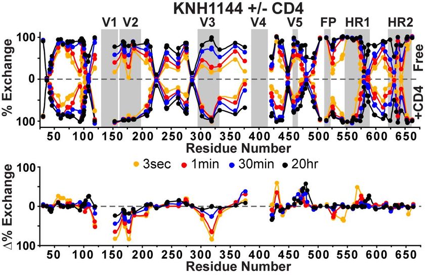
Env is the sole antigenic feature on
              HIV virions
• Mediates viral entry
  through binding host
  receptors
   – Primary: CD4
   – Co-receptor:
     (CCR5/CXCR4)
• Evades immune system
   – High sequence variability
   – Glycan shield
   – Conserved regions only
     exposed upon primary
     receptor (CD4) binding
• Major focus of HIV
  vaccinology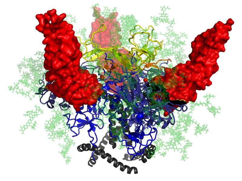
Trimeric structure of Env

       V1/V2          V3      V4   V5          FP     HR1      HR2       TM     CT
                   gp120                                     gp41

• V1/V2 and V3 form
  quaternary interactions at
  the crown of the trimer
   – Hide conserved regions
• CD4 binding opens up the
  trimer to unmask conserved
  regions (V3)
                                                                             gp41

                                   Julien, et al., Science 2013; Lyumkis, et al., Science 2013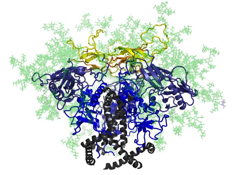
Trimeric structure of Env
                                               s-s

       V1/V2          V3      V4   V5          FP     HR1      HR2       TM     CT
                   gp120                                     gp41

• V1/V2 and V3 form
  quaternary interactions at
  the crown of the trimer
   – Hide conserved regions
• CD4 binding opens up the
  trimer to unmask conserved
  regions (V3)
• Soluble Env trimers                                                        gp41

                                   Julien, et al., Science 2013; Lyumkis, et al., Science 2013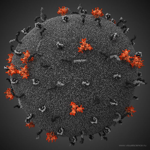
How does CD4 binding alter Env?

• Structures available for many
  ligands with isolated gp120
   – gp120 core +/- CD4 look identical
      • Kwon et al, PNAS 2012
   – No crystal of trimer with CD4
   – EM studies indicate large changes
     in the trimer upon CD4 binding
      • Liu et al, Nature 2008
• Compare unliganded and sCD4
  bound conformations of trimeric
  Env
   – H/D exchange (HDX-MS)
   – X-ray footprinting (XF-MS)

                                         sCD4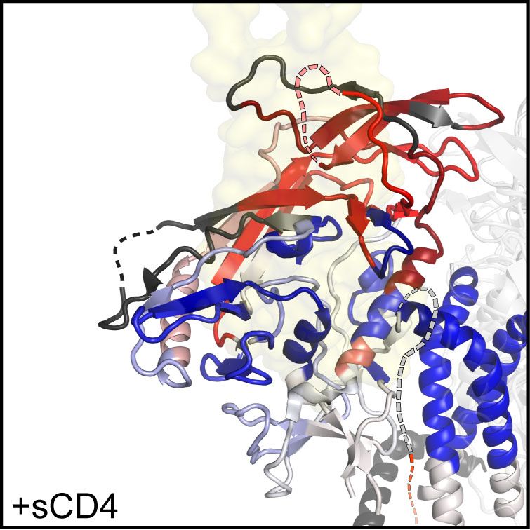
Hydrogen/Deuterium exchange
probes the accessibility of amide
           hydrogens

                  H2O      D2O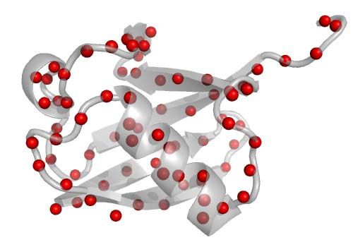
Hydrogen/Deuterium exchange
probes the accessibility of amide
           hydrogens

                  H2O      D2O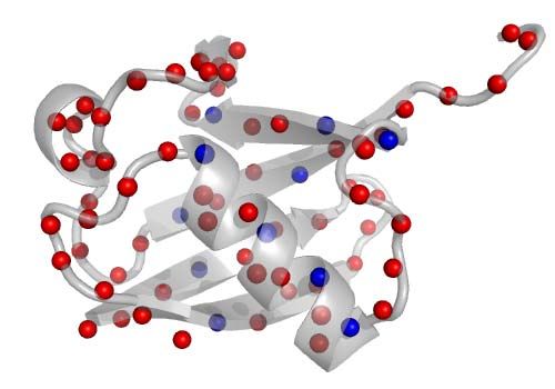
Hydrogen/Deuterium exchange
probes the accessibility of amide
           hydrogens

                  H2O      D2O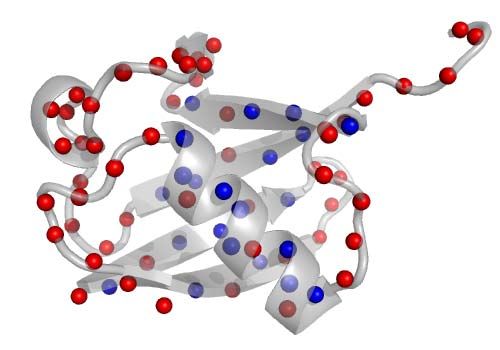
Hydrogen/Deuterium exchange
probes the accessibility of amide
           hydrogens

                  H2O      D2O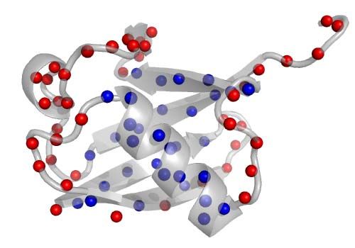
Hydrogen/Deuterium exchange
probes the accessibility of amide
           hydrogens

                  H2O      D2O
Hydrogen/Deuterium exchange
probes the accessibility of amide
hydrogens
H2O D2O
*Labeling doesn’t perturb protein structureHydrogen/Deuterium exchange
probes the accessibility of amide
hydrogens
H2O D2O
Exchange rate (sec-1)
pH *Labeling doesn’t perturb protein structureHDX experimental setup Stock protein (in H2O)
HDX experimental setup

                Dilute into D2O

                     (sec-
Stock protein       hours)
                    pH 7.4
   (in H2O)
HDX experimental setup
Dilute into D2O Quench Pepsin
(sec- pH 2.5 5 min
Stock protein hours) 0°C
pH 7.4 100mM TCEP
(in H2O)
Peptides
Freeze in liquid N2
store at -80°CHDX experimental setup
Dilute into D2O Quench Pepsin
(sec- pH 2.5 5 min
Stock protein hours) 0°C
pH 7.4 100mM TCEP
(in H2O)
Peptides
Freeze in liquid N2
store at -80°C
Thaw &
LC-MS
(at 0°C)HDX experimental setup
Dilute into D2O Quench Pepsin
(sec- pH 2.5 5 min
Stock protein hours) 0°C
pH 7.4 100mM TCEP
(in H2O)
Peptides
Freeze in liquid N2
store at -80°C
Thaw &
LC-MS
(at 0°C)
*Each D increases the
peptide mass by 1 DaHDX experimental setup
Dilute into D2O Quench Pepsin
(sec- pH 2.5 5 min
Stock protein hours) 0°C
pH 7.4 100mM TCEP
(in H2O)
Peptides
Freeze in liquid N2
store at -80°C
Thaw &
LC-MS
(at 0°C)
*Each D increases the
peptide mass by 1 DaHDX experimental setup
Dilute into D2O Quench Pepsin
(sec- pH 2.5 5 min
Stock protein hours) 0°C
pH 7.4 100mM TCEP
(in H2O)
Peptides
Freeze in liquid N2
store at -80°C
Thaw &
+ ligand
LC-MS
(at 0°C)
+ ligand
*Each D increases the
peptide mass by 1 DaVisualizing the H/DX data
+sCD4
-sCD4
• Data for each peptide is plotted on the primary sequence
• Bufferly/mirror plots are useful for comparing HDX for two protein states
(+/- ligand)
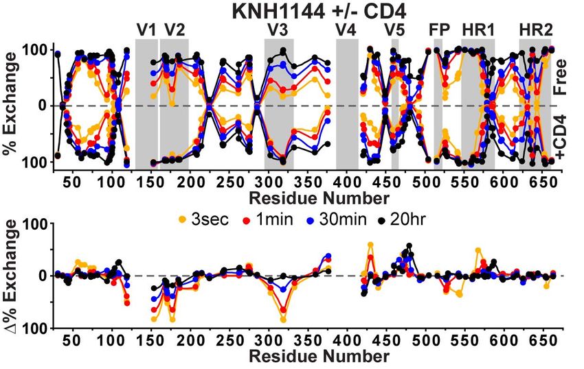
• Difference plots show raw difference at each time pointVisualizing the H/DX data
+sCD4
-sCD4
• Data for each peptide is plotted on the primary sequence
• Butterfly/mirror plots are useful for comparing HDX for two protein states
(+/- ligand)
• Difference plots show raw difference at each time pointVisualizing the H/DX data
+sCD4
-sCD4
+sCD4
• Data for each peptide is plotted on the primary sequence
• Butterfly/mirror plots are useful for comparing HDX for two protein states
(+/- ligand)
• Difference plots show raw difference at each time pointCD4 binding induces elaborate
changes
+sCD4
• CD4 binding surface becomes protectedCD4 binding induces elaborate
changes
V1 V2
V3
+sCD4
• CD4 binding surface becomes protected
• Variable loops V1/V2 & V3 become disorderedCD4 binding induces elaborate
changes
+sCD4
• CD4 binding surface becomes protected
• Variable loops V1/V2 & V3 become disordered
• Crown region of gp120 reorganizedCD4 binding induces elaborate
changes
HR1
+sCD4
FPPR
• CD4 binding surface becomes protected
• Variable loops V1/V2 & V3 become disordered
• Crown region of gp120 reorganized
• In gp41: HR1 is more protected; while FPPR is lessXF-MS at SSRL BL4-2
• Establish dosage
– (Alexa 488 dye)
• Irradiate in quartz
capillary at various flow
rates for different
exposure times
• ~8 to 200 msec
– Eject sample into collection
tubes with methionine
• (20mM final)
– Run wash cycle
• Compare unliganded and
CD4-bound Env trimersXF-MS data processing
Sample processing:
• Denaturation
– GndHCl & DTT
• Cysteine alkylation
– IAA
• Deglycosylation (PNGaseF)
– Dilute sample to 0.5M GndHCl
• Digestion
– Split sample: LysC & GluC
LC-MS:
• 30 minute gradient over C18
column
• ESI-QTOF (Waters Synapt)
• Integrate and measure the
intensity of each unmodified and
oxidized peptide.
• Calculate and plot % modified:
Intensity modified
Intensity of unmodified + modified (all)Internal dosimeters for XF-MS
• Protein concentrations and buffers kept consistent, experiments
performed side by side (in duplicate).
• Internal standard to ensure datasets received identical dosage:
– Leucine enkephalin & Substance P
– Look at the decrease in percent unmodifiedMixed changes near CD4 binding
surface on gp120
M104
M475
M95Increased accessibility at V1/V2
and V3
F316 M161
M150Additional changes within gp41
• Additional changes are seen at M626 and
the C-terminus of gp41
– (not well resolved in the current crystal structures)
gp41H/DX & XF-MS reveal the extensive
changes upon CD4 binding
“closed” “open”
CD4
• Opening of V1/V2 and V3 loops
• Reorganization of the gp120 subunits
• Changes to gp41 may act to “prime” gp41
for subsequent activation (by co-receptor)Acknowledgements
University of Washington Stanford Synchrotron and Radiation
• Kelly K. Lee (PI) Lightsource (SSRL)
• Natalie K. Garcia • Tsutomu Matsui
• Tad M. Davenport
Cornell University
• Al Cupo NIH
• John P. Moore • AIDS reagent program
• F32 award
Scripps Research Institute – F32-GM097805
• Jean-Philippe Julien Bill and Melinda Gates
• Ian A. Wilson Foundation
• CAVD grant OPP1033102
University of Amsterdam
• Rogier SandersYou can also read