Validation of New Gene Variant Classification Methods: a Field-Test in Diagnostic Cardiogenetics
←
→
Page content transcription
If your browser does not render page correctly, please read the page content below
ORIGINAL RESEARCH
published: 01 March 2022
doi: 10.3389/fgene.2022.824510
Validation of New Gene Variant
Classification Methods: a Field-Test in
Diagnostic Cardiogenetics
Mohamed Z. Alimohamed 1,2,3,4*, Helga Westers 1, Yvonne J. Vos 1, K. Joeri Van der Velde 1,
Rolf H. Sijmons 1, Paul A. Van der Zwaag 1, Birgit Sikkema-Raddatz 1† and
Jan D. H. Jongbloed 1*†
1
Department of Genetics, University of Groningen, University Medical Center Groningen, Groningen, Netherlands, 2Department of
Haematology and Blood Transfusion, Muhimbili University of Health and Allied Sciences, Dar-es-Salaam, Tanzania, 3Department
of Research and Training, Shree Hindu Mandal Hospital, Dar-es-salaam, Tanzania, 4Tanzania Human Genetics Organization,
Groningen, Netherlands
Background: In the molecular genetic diagnostics of Mendelian disorders, solutions are
needed for the major challenge of dealing with the large number of variants of uncertain
Edited by:
Pingzhao Hu,
significance (VUSs) identified using next-generation sequencing (NGS). Recently,
University of Manitoba, Canada promising approaches using constraint metrics to calculate case excess scores (CE),
Reviewed by: etiological fractions (EF), and gnomAD-derived constraint scores have been reported that
Rui Chen, estimate the likelihood of rare variants in specific genes or regions that are pathogenic. Our
Baylor College of Medicine,
United States objective is to study the usability of these constraint data into variant interpretation in a
Anna Kostareva, diagnostic setting, using our cardiomyopathy cohort.
Karolinska Institutet (KI), Sweden
*Correspondence: Methods and Results: Patients (N = 2002) referred for clinical genetic diagnostics
Mohamed Z. Alimohamed underwent NGS testing of 55–61 genes associated with cardiomyopathies. Previously
m.z.a.alimohamed@umcg.nl
Jan D. H. Jongbloed
classified likely pathogenic (LP) and pathogenic (P) variants were used to validate the use of
j.d.h.jongbloed@umcg.nl data from CE, EF, and gnomAD constraint analyses for (re)classification of associated
†
These authors have contributed variant types in specific cardiomyopathy subtype-related genes. The classifications
equally to this work corroborated in 94% (354/378) of cases. Next, we reclassified 23 unique VUSs to LP,
increasing the diagnostic yield by 1.2%. In addition, 106 unique VUSs (5.3% of patients)
Specialty section:
This article was submitted to were prioritized for co-segregation or functional analyses.
Statistical Genetics and Methodology,
a section of the journal Conclusions: Our analysis confirms that the use of constraint metrics data can improve
Frontiers in Genetics variant interpretation, and we, therefore, recommend using constraint scores on other
Received: 29 November 2021 cohorts and disorders and its inclusion in variant interpretation protocols.
Accepted: 21 January 2022
Published: 01 March 2022 Keywords: cardiomyopathy, NGS gene panel, variant classification, constraint metrics, cardiogenetics
Citation:
Alimohamed MZ, Westers H, Vos YJ,
Van der Velde KJ, Sijmons RH, INTRODUCTION
Van der Zwaag PA,
Sikkema-Raddatz B and The use of next-generation sequencing in molecular diagnostics of Mendelian disorders has
Jongbloed JDH (2022) Validation of
improved the diagnostic yield significantly. It allows for testing of an increasing number of
New Gene Variant Classification
Methods: a Field-Test in
genes but, unfortunately, also results in the identification of an increasing number of variants of
Diagnostic Cardiogenetics. uncertain significance (VUSs). Moreover, genotype–phenotype associations have not been clearly
Front. Genet. 13:824510. established for all of the genes analyzed in diagnostics, thereby further contributing to unclear clinical
doi: 10.3389/fgene.2022.824510 interpretation of genetic variants (Norton et al., 2013; Duzkale et al., 2013; Das et al., 2014;
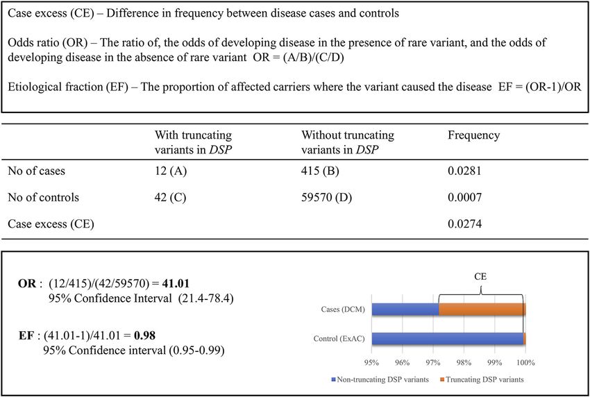
Frontiers in Genetics | www.frontiersin.org 1 March 2022 | Volume 13 | Article 824510Alimohamed et al. Variant Re-Interpretation in Cardiomyopathy FIGURE 1 | Comparing the frequency of rare truncating variants (ExAC MAF
Alimohamed et al.                                                                                         Variant Re-Interpretation in Cardiomyopathy

MATERIALS AND METHODS                                                    in genes with a loss of function (LoF) intolerant (pLI) score >0.90
                                                                         (truncating variants) or a missense (mis_z) score >3 (missense
                                                                         variants) were considered putatively detrimental. Using mis_z
Patients, Genetic Analysis, and Variant                                  and/or pLI scores from the gnomAD database, additional variant
Classification Using Routine Diagnostic                                   types in genes that have an increased risk of being pathogenic
Criteria                                                                 were selected. Notably, our procedure of selecting VUSs for
This study was performed in accordance with UMCG and Dutch               prioritization and reclassification based on constraint metric
national ethical and legal guidelines and complies with the              data is also visualized in Supplementary Figure S1.
regulations stated in the Declaration of Helsinki. Informed
consent was obtained for all patients referred to our clinical           Validation: (L)Ps
genetics laboratory.                                                     We initially validated the potential of using constraint scores for
   In total, a consecutive series of 2,002 patients of mainly            prioritizing and (re)classifying variants by determining the
Caucasian ethnicity from 1,967 families (60.4% male and                  number of variants from our cohort classified as LP or P
39.6% female) were included in our study between 2012 and                using our routine diagnostic criteria (RDC) (Alimohamed
2017. Genes known to be implicated in cardiomyopathies were              et al., 2021) and the number of these variants that would have
selected for analysis. During the patient inclusion period, three        been predicted to have increased risk of pathogenicity using the
versions of the diagnostic panel were used, the second and third         recommendation from the scores from the CE/EF based approach
being updated versions. Variants were classified either as                and therefore may be classified (L)P. Next, the concordance
“benign” (B)—class 1, “likely benign” (LB)–class 2, “variant of          between these groups was calculated.
uncertain significance” (VUS)—class 3, “likely pathogenic”
(LP)—class 4, or “pathogenic” (P)—class 5. Interpretation was            Prioritization and Reclassification: VUSs
largely based on guidelines recommended by the American                  VUSs in genes with significant CE for truncating and/or non-
College of Medical Genetics and Genomics (ACMG) (Richards                truncating variants within the corresponding cardiomyopathy
et al., 2015). Variants detected in our cohort are routinely             subtype, as established by Walsh et al., 2017; Walsh et al., 2019;
submitted to ClinVar (https://www.ncbi.nlm.nih.gov/clinvar).             and Mazzarotto et al., 2020, were selected for prioritization for co-
A comprehensive description of the cohort, genetic analysis,             segregation and/or functional studies and/or reclassification (see
variant interpretation, and classification protocols are                  Supplementary Tables 2A,B). The same was done for VUSs
described in our previous study (Alimohamed et al., 2021).               following the above criterion, but identified in unclassified CM
                                                                         cases. In addition, for genes in our panel to which the results for
Applying Results of Constraint Metric                                    the CE/EF based approach could not be applied, VUSs in genes with a
                                                                         pLI>0.90 (LoF variants) or a mis_z > 3 (missense/in frame del/dup)
Methods for Prioritization and/or (Re)
                                                                         were also selected (Supplementary Table S3). Then, depending on
classification                                                            the respective EF scores of the corresponding variant types in the thus
In recent studies, Walsh and colleagues studied potential CE and         previously analyzed genes, VUSs were either immediately reclassified
EFs of variants identified in cardiomyopathy patients by                  to LP (EF ≥ 0.90) or prioritized for further analyses/studies (EF <
comparing frequency data of rare variants identified in clinical          0.90) (Lek et al., 2016; Karczewski et al., 2020) such as co-segregation
cardiomyopathy cases with the frequency of these variants in             analyses or functional studies, potentially leading to reclassification.
60,706 ExAC reference samples (Walsh et al., 2017; Walsh et al.,         In addition, the latter was also applied to VUSs in genes selected on
2019, Mazzarotto et al., 2020). Initially, CE was determined for         the basis of the gnomAD-based constraint metrics (pLI>0.9 or
truncating (frameshift, nonsense, and RNA consensus splice               mis_z > 3.0).
donor/acceptor) and non-truncating (missense and in frame
insertions and deletions) variants separately for established
cardiomyopathy genes in ACM (N = 8), HCM (N = 20), and
DCM (N = 48) subtypes (Walsh et al., 2017). In addition, the
                                                                         RESULTS
same group reported higher CE and EF scores in additional                Correlation Between Classification via
studies for DCM (Mazzarotto et al., 2020) and HCM (Walsh et al.,
2019). Variant types in genes significantly enriched in patients in
                                                                         Routine Diagnostic Criteria and
these studies are summarized in Supplementary Table S1 and               Classification Using Results of Constraint
were used for variant interpretation for our cardiomyopathy              Metrics-Based Statistical Methods
cohort.                                                                  In our cohort of 2,002 patients, the overall molecular diagnostic yield,
    Additionally, for genes in our panel to which the data of the        defined as patients carrying at least one LP or P variant following RDC
CE/EF-based approach could not be applied, as these genes were           in genes with established associations with the patient’s phenotypes,
not included in the respective analyses, we collected data from an       was 21.5% (430/2002). From the 430 patients carrying these (L)P
alternative, previously reported approach (Lek et al., 2016;             variants, 378 in 369 patients (note that a few patients carry more than
Karczewski et al., 2020) also providing constraint scores. These         one (L)P) were found in genes analyzed within DCM, HCM, or ACM
constraint scores are based on the deviation of observed variant         cohorts (see Table 1 for total and subtype-specific numbers). Of these
counts from variant counts per gene expected by chance. Variants         378 variants, 354 would have been classified as (L)P on the basis of

Frontiers in Genetics | www.frontiersin.org                          3                                      March 2022 | Volume 13 | Article 824510
Alimohamed et al. Variant Re-Interpretation in Cardiomyopathy
TABLE 1 | Impact of retrospective reassessment of variant pathogenicity. (A) Reclassification/prioritisation of variants according to CE/EF scores. (B) Prioritization of variants
in genes with mis_z > 3 or pLI >0.9 according to gnomAD.
(A) Reclassification/prioritisation of variants according to CE/EF scores. (B) Prioritization
of variants in
genes with mis_z
> 3 or pLI >0.9
according to
gnomAD.
CM No. of No. of (i) (ii) Analyses of VUS No. of patients No. of patients
Subtype genes patients with without genetic
No of patients Correlation of No. of No. of patients
with CE LP/P variants diagnosis, but
with LP/p variant patients with without genetic
according to variants (VUS)
variants classification prioritised diagnosis, but
RDC (no. of leading to definitive
according to between RDC variants variants possibly
variants) reclassification
CE/EF scores and CE/EF because leading to
to LP
(no. of scores of CE reclassification
variants)
HCM 14 169 (172) 158 (160) 93% 61 44 19 —
DCM 14 181 (186) 178 (177) 95% 55 46 6 2
ACM 5 19 (20) 17 (17) 85% 1 1 — —
CM — — — — 26 22 — 11
LVNC — — — — — — — 6
TOTAL 33 369 (378) 353 (354) 94% 143 113 25 19
No. of — — — — 113 (6%) 25 (1.2%) 19 (1%)
patients
with
selected
variants
A) 1) Variants classified as (L)P following RDC, CE/EF criteria and their correlations, 2) patients identified, after application of CE/EF scores on VUSs in CM subtype-specific genes, with
reclassified variants and/or variants prioritized for future functional and/or co-segregation studies. B) Additional VUSs prioritized of genes not yet addressed within the CE/EF approach and
having significant missense Z (mis_z) and/or probability of loss-of-function intolerant (pLI) scores adapted from the genome aggregation (gnomAD) database.
case excess (CE) in patient cohorts, showing a 94% (354/378) Tables 2A,B). Of these, 113 patients (46 DCM, 44 HCM, 1
concordance between our RDC- and the CE-based classification. ACM, and 22 CM) did not have a genetic diagnosis yet (the other
Of the remaining 6% of (L)P variants that were not concordant, 4% 30 patients did already carry at least one (L)P), accounting for
were Dutch founder mutations (Alimohamed et al., 2021). In 5.6% of the total cohort (Table 1).
addition, when analyzing genes not included in the Next, in genes not included in our previous approach, variants
aforementioned evaluation, 22 patients carrying 22 (L)Ps in these in genes intolerant to variation, having a pLI>0.9 or mis_z > 3.0
genes were identified. When using the LoF and missense intolerance score, according to gnomAD v2 aggregate data, were selected (Lek
scores as provided by the gnomAD database (Lek et al., 2016; et al., 2016; Karczewski et al., 2020). The resulting variants were
Karczewski et al., 2020) for classification, 12 of these would have prioritized only when identified in genes established to be
been classified as (L)P showing a concordance of 55% (12/22). associated with the patient’s cardiomyopathy subtype (37
different VUSs in 42 patients; Supplementary Table S3). This
resulted in the identification of additional 19 patients (2 DCM, 6
Selection of VUSs for Prioritization/ LVNC, and 11 CM) that did not have a genetic diagnosis yet,
Reclassification accounting for 1% of the total cohort (Table 1).
VUSs were selected based on published constraint metric data
and consisted of three sets: 1) those identified in genes showing Reclassification and Prioritization of VUSs
CE in patients with specific CM subtypes (HCM, DCM, and As described above, 129 unique variants previously classified as
ACM), 2) those identified in the same genes but found in patients VUS were selected for reclassification and/or prioritization for
with unclassified CM where such phenotype matching was not further analyses. The potential to use CE/EF scores for
possible, and 3) those identified in genes not having established prioritization/reclassification was underscored by the fact that
associations with the patient’s subtypes according to the Walsh high scores of pathogenicity prediction programs, often in
and Mazzarotto methods [2017; 2019; 2020] (including those in combination with (very) low population frequencies of these
patients with unclassified CM) and having pLI>0.90 or mis_z > 3. variants, were found for these variants (Supplementary Table
In genes with significant CE for non-truncating and/or truncating S2A). Based on their analyses of CE and EF of HCM-related
variants, a total of 92 unique VUSs in 143 patients were identified genes, Walsh et al. [2019] suggested including EF scores in the
in DCM (N = 55), ACM (N = 1), CM (N = 26), and HCM (N = 61) generally used ACMG guidelines for variant classification. This
to qualify for prioritization/reclassification (Supplementary would result in the adaptation of criterion PM1 (a mutational hot
Frontiers in Genetics | www.frontiersin.org 4 March 2022 | Volume 13 | Article 824510Alimohamed et al. Variant Re-Interpretation in Cardiomyopathy
spot or well-defined functional domain without a benign variant classification, as 94% of variants previously classified as
variation), into PM1_supporting (a non-truncating variant in a (L)P would also have been classified as such using the constraint
gene or protein with 0.8 ≤ EF < 0.9), PM1_moderate (a non- rules. Together, applying these rules in daily practices will lead to
truncating variant in a gene or protein with 0.9 ≤ EF < 0.95), and more diagnoses, as well as guide further analysis of potentially
PM1_strong (non-truncating variants a in gene or protein region causal VUSs.
with EF ≥ 0.95). We decided to implement these adaptations to With current population data, applying the CE/EF criteria
our classification criteria and expanding this also to truncating (Walsh et al., 2019) in our cohort resulted in identifying up to
variants, within the respective subtype for which the EF was 6.6% of additional patients with a potential LP/P reclassification
determined. Also, in addition, these genes/variants were within different CM subtypes. Our results are comparable to
identified in unspecified CM cases. Most of the selected VUSs those reported by Walsh et al. (2019), where 4% of actionable
do also fulfill the (MYH7-adpated—in our opinion these criteria variants in HCM cases were upgraded to LP. The more general
can be generally applied for every established, autosomal use of these CE/EF scores is consistent with the use of recently
dominant-inherited cardiomyopathy gene) ACMG criterion established guidelines for MYH7-associated inherited
PM2 (absent/extremely rare (0.004% scores as one of the criteria for classification is comparable
were not considered for reclassification, such as MYH7 c.2890G > with applying the MYH7-adapted rule PS4. The rule is that
C, p.Val864Leu), and all fulfilled PP3 (multiple lines of the prevalence of the variant in affected individuals is
computational evidence support a deleterious effect on the significantly increased compared with the prevalence in
gene or gene product) and PP4 (patient’s phenotype or family controls—OR—that the variant is identified in ≥15 probands
history is highly specific for a disease with a single genetic with consistent phenotypes, the difference being that we do apply
etiology) (Richards et al., 2015; Kelly et al., 2018). Following the CE/EF scores to all potentially causal VUSs within genes for
the rules for combining criteria to classify sequence variants as which these were established, while the PS4 criterion in Kelly et al.
proposed by the ACMG (Richards et al., 2015), this would mean is applied at the level of individual variants only. Importantly, we
that combining these with the adapted criteria PM1_strong or only reclassify variants in genes with EF ≥ 0.9 and criteria PM2
PM1_moderate would result in 23 unique VUSs in 28 patients (absent/extremely rare (0.9 or mis_z > 3.0 were prioritized for further had a genetic diagnosis now being carrier of two (L)Ps, instead of
analyses such as co-segregation and haplotype-sharing analyses only one, related to their phenotype. Moreover, prioritized VUSs
or functional evaluations. were identified in an additional 23 previously “solved” cases, and
these patients are potentially carrier of multiple (L)Ps related to
their disease. This will have significant effects on the management
DISCUSSION of the respective patients and their family members. In particular,
for family members that were previously shown not to carry the
In this study, we applied data from previously published statistical already known (L)P and for that reason were dismissed of regular
approaches that identified genes and variant types having a higher follow-up.
chance of being disease associated to reduce the amount of VUSs We decided that variant classes with EF scores of 0.9 or mis_z > 3.0 can
Frontiers in Genetics | www.frontiersin.org 5 March 2022 | Volume 13 | Article 824510Alimohamed et al. Variant Re-Interpretation in Cardiomyopathy
help in prioritizing these for follow up like co-segregation or reclassification to LB. Notably, additional research is needed to
functional analyses, in our case 129 unique VUSs in 132 patients further validate whether the use of constraint scores with 0.8 ≤ EF
from our cohort (6.6%). Moreover, when MYH7-adapted co- < 0.9 for variant interpretation is sufficient for definitive
segregation-based criteria (PP1_strong; variant segregates with classification of (L)P variants to P or prioritized VUSs to (L)P.
≥7 meioses, PP1_moderate; variant segregates with ≥5 meioses, To ultimately classify the latter variants as LP or P, further
or PP1_supportive; variant segregates with ≥3 meioses) is more segregation analysis or functional evidence is needed.
generally applied to other cardiomyopathy genes, having a variant Moreover, we have only screened for variants in coding
segregating with disease in only a limited number of family regions and surrounding regions of interest, ±20 bases, in ~60
members could already lead to upgrading a variant in a gene selected cardiomyopathy genes. Deep intronic or regulatory
with CE but EF < 0.9, or a gene with pLI>0.9 or mis_z > 3.0 to an (5′and 3′UTR) variants and variants in novel genes with
LP status. potential functional roles in cardiomyopathies were not
Additional studies need to validate whether the data of included in our analysis. Also, as not all genes in our panels
population-based statistical methods are sufficient for have been analyzed for CE and EF, for those we relied on
definitive reclassification of such variants for scores between gnomAD-derived constraint scores for determining their
0.8 ≤ EF < 0.9. Moreover, to attain EF ≥ 0.90 for all relevant putative causal nature, leading to prioritization for further
genes, if possible at all, more studies are needed to reach higher analyses only. The latter because we felt that the strength of
EFs or identify specific regions within a gene that carry these scores were insufficient to support immediate (L)P
pathogenic variants using larger cohorts for specific classification. This was also underscored by the fact that using
cardiomyopathy subtypes. This underscores the these constraint scores only 55% of RDC derived (L)Ps in the
complementarity of EF with machine-learning (ML)-based respective variant types and genes would have been classified as
variant pathogenicity predictors such as CADD, CAPICE, and (L)P using these scores. Finally, patient phenotypic information
the like (Rentzsch et al., 2019; Li et al., 2020), which present part was obtained from referral forms and not scrutinized according to
of the evidence to classify variants as (L)P in novel regions that definitive phenotypic criteria.
lack CE/EF ≥ 0.90, or help reclassify variants in known CE/EF ≥
0.90 regions, as we have shown 94% of variants classified using
RDC are concordant to the CE/EF method. When sufficient CONCLUSION
‘critical mass’ of pathogenic classifications and population
variants becomes available in a region of interest, a high EF Applying CE scores and EF (i.e., constraint metrics)-based
may be established for a particular variant type (i.e., truncating evaluations confirmed 94% of classified (L)P variants
variants). This criterion may then be used as a rapid and compared to RDC in a cohort of patients with
straightforward variant classifier for unseen variants, perhaps cardiomyopathy, underscoring the fact that such scores can be
supported by an ML predictor as second opinion or safety net. used to complement variant interpretation and classification
When available, we recommend starting the variant classification methods. Most importantly, it led to a 1.2% definitive increase
process for selected genes by first looking at CE/EF values as (VUSs reclassified to LP) and 5.3% relative increase (VUSs
initial criterion. Moreover, as using these values shows to prioritized) in actionable variants in our cardiomyopathy
complement the current variant interpretation framework, we cohort. In addition, using the constraint metrics helped select
therefore propose that including CE/EF classifiers in a variant 37 unique variants in genes with etiological fractions 3 or pLI>0.9 using gnomAD constraint data for future
interpretation and classification approaches. When applied to co-segregation studies and functional assays. Our analysis
other disease genes implicated in Mendelian diseases, this underscores that the use of such constraint metrics scores can
framework offers the potential to generally increase diagnostic improve variant interpretation and we recommend validating this
yield in genetic testing. method in other cohorts and disorders and consider its inclusion
in variant interpretation protocols and implement this for
cardiomyopathies.
LIMITATIONS
For genes with no significant CE and only computational proof DATA AVAILABILITY STATEMENT
for disease association, there is currently insufficient evidence
from the constraint scores methods that a variant will be disease The original contributions presented in the study are included in
causing. Extended CE and/or clustering analyses may establish the article/Supplementary Material, further inquiries can be
their association with disease enabling the use of such constraint directed to the corresponding authors.
scores for variant classification or actually refute disease
association. In this respect, it is important to note that no
variant classes in the cardiomyopathy genes presented ETHICS STATEMENT
significant depletion in cardiomyopathy cases compared to the
gnomAD aggregate database (Walsh et al., 2017), and these The studies involving human participants were reviewed and
variants were therefore not considered for provisional approved by UMCG review board in accordance with the UMCG
Frontiers in Genetics | www.frontiersin.org 6 March 2022 | Volume 13 | Article 824510Alimohamed et al. Variant Re-Interpretation in Cardiomyopathy
and national ethical guidelines and complies with the regulations project (0903-41 JJ), and the Netherlands Heart Foundation
stated in the Declaration of Helsinki. Informed consent was (2010B164 JJ).
obtained for all patients referred to our clinical genetics
laboratory. Written informed consent to participate in this
study was provided by the participants’ legal guardian/next of kin. ACKNOWLEDGMENTS
The authors would like to thank all the clinical geneticists,
AUTHOR CONTRIBUTIONS genetic counselors, and cardiologists for counseling and
referring their patients; technicians and staff members of
MA, HW, RS, BS-R, JJ, and PV contributed to conception and our Genome diagnostics section for excellent technical
design; MA, YV, KV, JJ, and PV acquired, analyzed and interpreted assistance and help in variant interpretation and
data; MA, HW, BS-R, JJ, and PV drafted the manuscript, all classification; and Kate McIntyre for careful editing of this
authors revised it critically for important intellectual content; manuscript.
and all authors gave final approval of the version to be published.
SUPPLEMENTARY MATERIAL
FUNDING
The Supplementary Material for this article can be found online at:
This work was supported by the “Doelmatigheidsfonds” of the https://www.frontiersin.org/articles/10.3389/fgene.2022.824510/
University Medical Center Groningen (JJ), the Fonds NutsOhra full#supplementary-material
Pugh, T. J., Kelly, M. A., Gowrisankar, S., Hynes, E., Seidman, M. A., Baxter, S. M., et al.
REFERENCES (2014). The Landscape of Genetic Variation in Dilated Cardiomyopathy as Surveyed
by Clinical DNA Sequencing. Genet. Med. 16 (8), 601–608. doi:10.1038/gim.2013.204
Alimohamed, M. Z., Johansson, L. F., Posafalvi, A., Boven, L. G., van Dijk, K. K., Rentzsch, P., Witten, D., Cooper, G. M., Shendure, J., and Kircher, M. (2019).
Walters, L., et al. (2021). Diagnostic Yield of Targeted Next Generation CADD: Predicting the Deleteriousness of Variants throughout the Human
Sequencing in 2002 Dutch Cardiomyopathy Patients. Int. J. Cardiol. 332, Genome. Nucl. Acids Res., 47 (D1), D886–D894. doi:10.1093/nar/gky1016
99–104. doi:10.1016/j.ijcard.2021.02.069 Richards, S., Aziz, N., Bale, S., Bick, D., Das, S., Gastier-Foster, J., et al. (2015).
Das K, J., Ingles, J., Bagnall, R. D., and Semsarian, C. (2014). Determining Pathogenicity Standards and Guidelines for the Interpretation of Sequence Variants: A Joint
of Genetic Variants in Hypertrophic Cardiomyopathy: Importance of Periodic Consensus Recommendation of the American College of Medical Genetics and
Reassessment. Genet. Med. 16 (4), 286–293. doi:10.1038/gim.2013.138 Genomics and the Association for Molecular Pathology. Genet. Med. 17 (5),
Duzkale, H., Shen, J., Mclaughlin, H., Alfares, A., Kelly, M., Pugh, T., et al. (2013). A 405–424. doi:10.1038/gim.2015.30
Systematic Approach to Assessing the Clinical Significance of Genetic Variants. Roca, I., Fernández-Marmiesse, A., Gouveia, S., Segovia, M., and Couce, M. (2018).
Clin. Genet. 84 (5), 453–463. doi:10.1111/cge.12257 Prioritization of Variants Detected by Next Generation Sequencing According
Eilbeck, K., Quinlan, A., and Yandell, M. (2017). Settling the Score: Variant to the Mutation Tolerance and Mutational Architecture of the Corresponding
Prioritization and Mendelian Disease. Nat. Rev. Genet. 18 (10), 599–612. Genes. Ijms 19 (6), 1584. doi:10.3390/ijms19061584
doi:10.1038/nrg.2017.52 Walsh, R., Mazzarotto, F., Whiffin, N., Buchan, R., Midwinter, W., Wilk, A., et al.
Karczewski, K. J., Francioli, L. C., Tiao, G., Cummings, B. B., Alföldi, J., Wang, Q., (2019). Quantitative Approaches to Variant Classification Increase the Yield
et al. (2020). The Mutational Constraint Spectrum Quantified from Variation in and Precision of Genetic Testing in Mendelian Diseases: The Case of
141,456 Humans. Nature 581 (7809), 434–443. doi:10.1038/s41586-020-2308-7 Hypertrophic Cardiomyopathy. Genome Med. 11 (1), 5. doi:10.1186/s13073-
Kelly, M. A., Caleshu, C., Morales, A., Buchan, J., Wolf, Z., Harrison, S. M., et al. 019-0616-z
(2018). Adaptation and Validation of the ACMG/AMP Variant Classification Walsh, R., Thomson, K. L., Ware, J. S., Funke, B. H., Woodley, J., McGuire, K. J.,
Framework for MYH7-Associated Inherited Cardiomyopathies: et al. (2017). Reassessment of Mendelian Gene Pathogenicity Using 7,855
Recommendations by ClinGen’s Inherited Cardiomyopathy Expert Panel. Cardiomyopathy Cases and 60,706 Reference Samples. Genet. Med. 19 (2),
Genet. Med. 20 (3), 351–359. doi:10.1038/gim.2017.218 192–203. doi:10.1038/gim.2016.90
Lek, M., Karczewski, K. J., Minikel, E. V., Samocha, K. E., Banks, E., Fennell, T.,
et al. (2016). Analysis of Protein-Coding Genetic Variation in 60,706 Humans. Conflict of Interest: The authors declare that the research was conducted in the
Nature 536 (7616), 285–291. doi:10.1038/nature19057 absence of any commercial or financial relationships that could be construed as a
Li, S., van der Velde, K. J., de Ridder, D., van Dijk, A. D. J., Soudis, D., Zwerwer, L. potential conflict of interest.
R., et al. (2020). CAPICE: a Computational Method for Consequence-Agnostic
Pathogenicity Interpretation of Clinical Exome Variations. Genome Med. 12 Publisher’s Note: All claims expressed in this article are solely those of the authors
(1), 75. doi:10.1186/s13073-020-00775-w and do not necessarily represent those of their affiliated organizations, or those of
MacArthur, D. G., Manolio, T. A., Dimmock, D. P., Rehm, H. L., Shendure, J., Abecasis, the publisher, the editors and the reviewers. Any product that may be evaluated in
G. R., et al. (2014). Guidelines for Investigating Causality of Sequence Variants in this article, or claim that may be made by its manufacturer, is not guaranteed or
Human Disease. Nature 508 (7497), 469–476. doi:10.1038/nature13127 endorsed by the publisher.
Mazzarotto, F., Tayal, U., Buchan, R. J., Midwinter, W., Wilk, A., Whiffin, N., et al.
(2020). Reevaluating the Genetic Contribution of Monogenic Dilated Copyright © 2022 Alimohamed, Westers, Vos, Van der Velde, Sijmons, Van der
Cardiomyopathy. Circulation 141 (5), 387–398. doi:10.1161/ Zwaag, Sikkema-Raddatz and Jongbloed. This is an open-access article distributed
CIRCULATIONAHA.119.037661 under the terms of the Creative Commons Attribution License (CC BY). The use,
Norton, N., Robertson, P. D., Rieder, M. J., Züchner, S., Rampersaud, E., Martin, E., distribution or reproduction in other forums is permitted, provided the original
et al. (2012). Evaluating Pathogenicity of Rare Variants from Dilated author(s) and the copyright owner(s) are credited and that the original publication
Cardiomyopathy in the Exome Era. Circ. Cardiovasc. Genet. 5 (2), 167–174. in this journal is cited, in accordance with accepted academic practice. No use,
doi:10.1161/CIRCGENETICS.111.961805 distribution or reproduction is permitted which does not comply with these terms.
Frontiers in Genetics | www.frontiersin.org 7 March 2022 | Volume 13 | Article 824510You can also read