Viable Second Trimester Cervical Ectopic Pregnancy Managed Successfully with Uterine Preservation: Case Report
←
→
Page content transcription
If your browser does not render page correctly, please read the page content below
Open Journal of Obstetrics and Gynecology, 2021, 11, 1236-1247
                                                                                                 https://www.scirp.org/journal/ojog
                                                                                                           ISSN Online: 2160-8806
                                                                                                             ISSN Print: 2160-8792

Viable Second Trimester Cervical Ectopic
Pregnancy Managed Successfully with Uterine
Preservation: Case Report

Suzan Elsharkawy1*, Abdullah Elrashidy2, Nazem Badran3, Gawed Ekbal3, Shahda Yakob3,
Salamah Elnagar4, Ashraf Elaggan5, Amr Mostafa5, Mohamed Abdelaziz6
1
 Assistant Professor of Obstetrics and Gynecology Alexandria University, ARE and Consultant of Obstetrics and Gynecology,
Hail General Hospital, Hail, KSA
2
 Consultant of Urology, King Khalid Hospital, Hail, KSA
3
 Consultants of Obstetrics and Gynecology, Alkasim Maternity Hospital, Alkasim, KSA
4
 Consultants of Obstetrics and Gynecology, Hail General Hospital, Hail, KSA
5
 Consultants of Radiology, King Salman Hospital, Hail, KSA
6
 Faculty of Medicine Mansoura University, Mansoura, Egypt

How to cite this paper: Elsharkawy, S.,         Abstract
Elrashidy, A., Badran, N., Ekbal, G., Yakob,
S., Elnagar, S., Elaggan, A., Mostafa, A. and   Cervical pregnancy is a rare clinical entity that accounts for less than 1% of all
Abdelaziz, M. (2021) Viable Second Tri-         ectopic pregnancies. It results from implantation of the blastocyst in the cer-
mester Cervical Ectopic Pregnancy Ma-
                                                vical canal below the level of the internal os. Although non-tubal ectopic
naged Successfully with Uterine Preserva-
tion: Case Report. Open Journal of Obste-       pregnancies account for only 5% of ectopics, they contribute to a significant
trics and Gynecology, 11, 1236-1247.            morbidity. The cornerstone in the management of cervical ectopic is early
https://doi.org/10.4236/ojog.2021.119117        diagnosis by high index of suspension and a qualified sonographer. Manage-
                                                ment options for cervical ectopic pregnancies range from conservative drug
Received: August 27, 2021
                                                treatment to radical hysterectomy. Over the last few years, the mortality and
Accepted: September 24, 2021
Published: September 27, 2021                   morbidity rates of ectopic pregnancies have been reduced. This is mainly due
                                                to the early recognition of the condition and the wide availability of mini-
Copyright © 2021 by author(s) and               mally invasive surgical procedures. We present a case of a 33-year-old woman
Scientific Research Publishing Inc.             that was 16 weeks pregnant. She presented initially with recurrent vaginal
This work is licensed under the Creative
Commons Attribution International
                                                bleeding followed by minimal lower abdominal pain. Her early US scans were
License (CC BY 4.0).                            misleading. Several weeks later, a follow up MRI scan suggested cervical ec-
http://creativecommons.org/licenses/by/4.0/     topic. She was managed surgically with uterine preservation.
                Open Access
                                                Keywords
                                                Ectopic Pregnancy, Cervical Ectopic, Uterine Artery Embolization,
                                                Hystroscopic Resection, Conservative Management of Ectopic,
                                                Methotrexate, Cervical Tamponade, Bakri Balloon, Cervical Cerclage

DOI: 10.4236/ojog.2021.119117 Sep. 27, 2021                       1236                   Open Journal of Obstetrics and Gynecology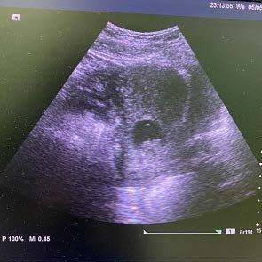
S. Elsharkawy et al.

                                1. Introduction
                                As rare as occurring once in 1000 to 95,000 pregnancies [1], cervical ectopic
                                pregnancy (CEP) is an uncommon clinical situation in gynecology. CEP is the
                                second rarest subtype of ectopic pregnancy following abdominal ectopic [2]. The
                                exact causes are not clear but many theories have been proposed, some suppos-
                                ing an unreceptive endometrium, rapid transport of the fertilized oocyte to the
                                cervical canal before nidation, others questioning the damage to the cervix and
                                the endometrium caused by operative procedures like dilatation & curettage or
                                operative delivery. The risk factors to develop CEP are multiple and include:
                                previous ectopic pregnancy, Asherman’s syndrome, intrauterine device use, his-
                                tory of pelvic inflammatory disease, smoking, anatomic anomalies and in vitro
                                fertilization [3] [4]. Although cervical pregnancies are rare, increased number of
                                cases being reported because of risk factors like high cesarean section rate and
                                increased use of assisted reproductive techniques for management of infertility.
                                According to one review, the incidence of cervical pregnancy is 0.1% among in
                                vitro fertilization pregnancies [5]. Due to its rarity, the absolute risk remains low
                                and the association with the above factors remains small, but CEP is a life
                                threatening condition that must be excluded in any woman presenting with va-
                                ginal bleeding, lower abdominal pain and missed period. Early diagnosis has
                                been the cornerstone for conservative management and reduction of morbidity
                                and mortality, while delay in the diagnosis can cause a bloody compromise that
                                may require hysterectomy by up to 50% of cases [6] [7]. We present a CEP case
                                that had a delay in the diagnosis till it reached seventeen weeks gestation.

                                2. Case Report
                                33-year-old female patient, G4 p3 was presented in emergency ward with a
                                painless attack of extensive vaginal bleeding, her obstetric history has three deli-
                                veries; the first one was preterm vaginal, the second was by caesarian section
                                (CS) and the last one was vaginal birth after CS (VBAC); which preceded this
                                current pregnancy by only 2 months. She also had done a myomectomy surgery
                                for a 7 cm submucous myoma after her first child.
                                  The condition started when she had an attack of moderate vaginal bleeding
                                after five weeks of amenorrhea (7/4/2021), she sought medical advice when she
                                performed a trans-abdominal ultrasound (TA-US) that showed an empty uterus
                                and a human chorionic gonadotropin (HCG) level was 672 mIU/ml, so she was
                                given progesterone IM injection to control the bleeding and asked to follow up
                                after one week to repeat ultrasonic examination.
                                  On the next week (13/4/2021), the BHCG was 8498 mIU/ml, and a small sac
                                with no fetal pole was noticed using the TA-US, nothing abnormal was sus-
                                pected at that time and again the patient was sent back home. On her next ap-
                                pointment (5/5/2021), a cervical gestational sac was obvious on trans-abdominal
                                us, which contained a pulsating fetal pole of 8 weeks gestation; yet her doctor
                                was not able to be sure of the diagnosis of cervical ectopic and the patient was

DOI: 10.4236/ojog.2021.119117                       1237                    Open Journal of Obstetrics and Gynecology
S. Elsharkawy et al.
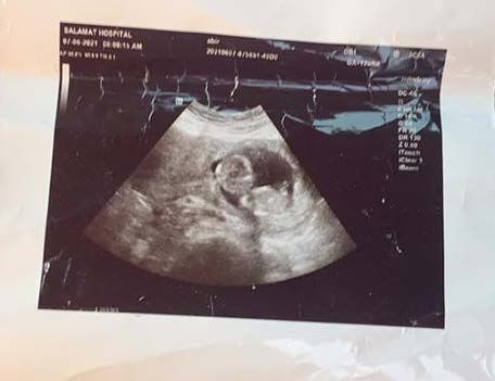
referred for senior consultation (Figure 1).
Unfortunately, the consultant assured her about this pregnancy (from 26/5 to
14/6/2021), alleging it was normal with normal placentation, and said “it is a
boy!!”, which was a desired sex for the patient, so she missed a precious chance
for early intervention and kept following this false assurance till she reached 15
weeks (Figure 2). Then she had a severe attack of vaginal bleeding and admitted
to a primary care hospital emergency ward on the 14th of June 2021.
Figure 1. Early trans-abdominal US showed obvious cervical
ectopic pregnancy with intact 8 weeks fetus. Despite this, the
diagnosis was delayed 8 weeks later.
Figure 2. A follow up trans-abdominal US of the same case
showing the fetus (about 14 week) resting in the ballooned cer-
vix on the right side of the image. On the left side, the uterus is
empty with closed internal os.
DOI: 10.4236/ojog.2021.119117 1238 Open Journal of Obstetrics and GynecologyS. Elsharkawy et al.
On admission, her blood pressure was 119/78 mmHg, pulse 81 b/min, oxygen
saturation 98%, respiratory rate 20 breath/min and temperature 37C. On ex-
amination, moderate vaginal bleeding was found, without any pain per-vaginal
examination (PV) revealed ballooned cervical canal with opened external cervic-
al os by 2 cm. transvaginal us showed ballooned cervical canal occupied with a
second trimester pregnancy, the placenta was lying on the anterior cervical wall
extended to cover the external cervical os with enhanced vascularity and loca-
lized hematoma underneath as shown with color Doppler waves. Trans-abdominal
us showed a 15-week normal looking male fetus surrounded with average
amount of liquor, sitting peacefully in a hugely distended cervical canal!! The
uterus was overriding the pregnancy, empty cavity and closed internal os with a
small interstitial fibroid 3 cm.
She was given Cyclokapron 2 ampules intravenous (500 mg/ampule) and lu-
ton depot 1 ampule intra-muscular (250 mg/ampule) immediately to control the
bleeding, then her investigations were: O +ve blood group, Hb 12.4 g/dl, plate-
lets 208,000/uL, INR 1.02, ALT 33 U/L, AST 16 U/L, serum creatinine 58.5
umol/L. she was generally stable and after 30 minutes the vaginal bleeding
stopped, giving a chance for further evaluation through emergency MRI on the
next day to be sure of the diagnosis. Although the US picture was crystal clear,
but due to the exceptional gestational age of the fetus, more advanced imaging
diagnosis was essential to differentiate between cervical ectopic or low lying pla-
centa.
On 16/6/2021, MRI was done without dye and reported a cervical ectopic
pregnancy 15 weeks gestation totally present in cervical canal, Figure 3. The
placenta was located at the anterior and right lateral aspect of the cervical wall,
with marked stretching, thinning of the underlying myometrium and focal in-
terruptions of the myometrial line together with significant contour bulge (sug-
gestive of placenta accreta spectrum, increta subtype), there were markedly in-
creased regional vascularity and blurring of the urinary bladder wall but no evi-
dence of gross intravesical extension. Prominent vascularity is also noted all over
the cervical and peri-cervical regions, Figure 4. The main uterine cavity proper
showed no sacs and the uterus showed two fibroids, the largest (3 × 3 cm) lo-
cated at the upper aspect of the anterior uterine wall and having sub-mucosal
extension.
When the diagnosis was made sure, the patient was transferred on 18/6/2021
to the tertiary referral hospital of the region 230 km away. On the transfer day,
the patient started leaking liquor till it was drained, which continued for 6
days—the period she stayed under observation as they misdiagnose her condi-
tion for low lying placenta with possibility of accreta, and for consultant com-
mittee to discuss the case and options of management available. The patient re-
ceived methotrexate IM injection on 20/6/2021 (1 mg/kg), she refused uterine
artery embolization done under local anesthesia, asked desperately to keep her
uterus and signed an informed consent for hysterectomy, blood products were
DOI: 10.4236/ojog.2021.119117 1239 Open Journal of Obstetrics and GynecologyS. Elsharkawy et al.
Figure 3. T2 weighted MRI image in sagittal plane, showing the
16 weeks fetus sitting peacefully in a markedly distended cervic-
al canal and placenta implanted on the anterior aspect of it. The
characteristic hour-glass uterus over-rides the pregnancy, with
closed internal os and empty cavity.
Figure 4. T2 weighted MRI image in transverse plane, showing
focal interruptions of the myometrial line together with signifi-
cant contour bulge suggestive of placenta increta, there were
markedly increased regional vascularity and blurring of the uri-
nary bladder wall but no evidence of gross intra-vesical exten-
sion.
DOI: 10.4236/ojog.2021.119117 1240 Open Journal of Obstetrics and GynecologyS. Elsharkawy et al.
prepared and vascular and urology surgeons were informed to attend the
planned major surgery.
On 23/6/2021 at 12:00 PM the surgical team started the operation; surprisingly
aiming to preserve the uterus if they can. The operation took three stressful
hours, during which they did a Pfannenstial incision, ligated the uterine arteries
at the level of isthmus, used diathermy for dissection of the adherent bladder
base from the anterior cervical wall, open the anterior cervical wall transversely,
extract the dead fetus and manually separate the placenta. A considerable
amount of bleeding from the placental bed resulted after removing it. Bakri bal-
lon distended with 150 cc of sterile saline solution was inserted through the ab-
dominal incision into the bleeding cervix, the cervix was sutured with 2-0 vicryl
sutures, and the bladder was checked for any injures and the abdomen was
closed in layers with intra peritoneal drain insertion before closure. Transfusion
of 4 units of blood and 2 units of plasma intra-operative was done and the pa-
tient was transmitted to intensive care unit (ICU) for observation and replace-
ments till the fifth day post-operative. The drain collected 600 cc fresh blood in
the first 6 hours post-operative and around 200 cc of blood lost vaginally, but the
patient’s condition stabilized gradually with the aid of replacements. Bakri bal-
lon and urinary catheter were removed on day two post-operative and the intra-
abdominal drain was removed on the sixth day just before transmission from
ICU to the inpatient ward. After one day inward, she was discharged home on
29th of June, 2021.
Till 18/7/2021, the patient was stable with minimal vaginal spotting, no follow
up was done or special post-operative instructions were given to her. Then she
had a severe attack of painless vaginal bleeding again, transferred to her home
area primary care hospital, vitally she was stable: pulse 107 b/min, BP 105/60, Hb
10 g/dl and her BHCG was 264 mIU/ml. The cervix was ballooned and soft and
vaginal was filled with blood clots by PV examination. TV-US revealed a hete-
rogeneous mass 5 cm in diameter occupied the cervical canal and surrounded
with increased vascularity. Resuscitation with IV fluids, hemostatics, 75 mg me-
thotrexate IM injection, transfusion of one packed RBC unit and trial of vaginal
packing, all were conducted to control bleeding then she was transferred imme-
diately to operative room where the consultant examined the cervix under gen-
eral anesthesia. There were remnants of placental tissue in the anterior cervical
canal, firmly attached to it, manually removed, and then intractable bleeding
occurred from the site of the remnants attachment. To control it, Babri balloon
was inserted in cervix, filled with 150 ml of saline then a tape cerclage was su-
tured at the level of the external os to fix it more. Vaginal packing with two to-
wels was done and one unit of packed RBCs was given intra-operatively.
For the following 3 days, the patient was closely observed for vital signs, va-
ginal bleeding and CBC. Gradual deflation of the balloon was done then re-
moved totally with the vaginal packs on 21/7/2021. The cerclage was left in place
for fear of any trauma to the fragile cervix. Her BHCG was 16 mIU/ml and Hb
10.5 gm/dl. She was stable and discharged on the next day 22/7/2021. On
DOI: 10.4236/ojog.2021.119117 1241 Open Journal of Obstetrics and GynecologyS. Elsharkawy et al.
26/7/2021 her BHCG was 8 mIU/ml.
On 29/7/2021 she did a MRI with contrast examination for follow up. It
showed an irregular soft tissue mass lesion centered upon the right anterolateral
wall of cervix with both intra cervical and exophytic components, measuring
about 4.5 × 4.3 × 4.0 cm in diameters, Figure 5. The mass was presenting com-
plex signal intensity in all pulse sequences and without contrast enhancement.
The exophytic component appears adherent to lower posterior aspect of the uri-
nary bladder without evidence of line of cleavage in between, with markedly
thickened posterior vesical wall measuring about 0.6 cm. No gross intravesical
extension detected. The upper right side of the anterior cervical wall was se-
riously damaged and lacerated, but the left side, the external os and the posterior
wall were perfectly healed and formed again, Figure 6. Her BHCG was 2.8
mIU/ml and she had minimal vaginal spotting, so there was no need for any
farther surgical interventions. The second Post-operative period went smoothly
without any significant events. Twenty days afterwards, the cervix was perfectly
reformed and cerculage was safely removed.
3. Discussion
CEP was first described in 1817, but in 1911 Rubin stated the histo-pathological
criteria required for diagnosis of a cervical pregnancy. These three criteria
Figure 5. T2 weighted MRI image in sagittal plane, showing an
irregular soft tissue mass lesion centered upon the right antero-
lateral wall of cervix with both intra cervical and exophytic
components, measuring about 4.5 × 4.3 × 4.0 cm in diameters.
The exophytic component appears adherent to lower posterior
aspect of the urinary bladder without evidence of line of clea-
vage in between, with markedly thickened posterior vesical wall.
No gross intravesical extension detected.
DOI: 10.4236/ojog.2021.119117 1242 Open Journal of Obstetrics and GynecologyS. Elsharkawy et al.
Figure 6. T1 weighted MRI image in sagittal plane, the right
side of the anterior cervical wall was seriously damaged and
lacerated, but the posterior wall was perfectly healed and
formed again.
require hysterectomy specimens: 1) cervical glands must be opposite the placen-
tal attachment, 2) placental attachment to the cervix must be situated below the
entrance of the uterine vessels or below the reflection of the anterior and post-
erior surface of the uterus and 3) fetal elements must be absent from the corpus
uteri [8]. Because of the efficient early diagnosis of cervical pregnancy, more and
more cases are managed conservatively without hysterectomies. So, the more
practical criteria of Palman and McElin [9] are used now. The five items do not
require hysterectomy for diagnosis and include: a) painless uterine bleeding,
following a period of amenorrhoea, b) a soft enlarged cervix equal to or larger
than the uterine body (an hour-glass shaped uterus), c) products of conception
completely in and firmly attached to the endocervix, d) a closed internal os and
e) a partially opened external os.
In 1997, Ushakov established ultraSonographic diagnostic criteria of CEP
which are very clear and helpful distinguishing it from other similar conditions
and include: 1) empty uterine cavity or thickened endometrium, 2) distended
and/or enlarged cervical canal, 3) gestational sac or placental tissue below the
level of the internal os, 4) negative “sliding organs sign” by transvaginal US, and
5) high peritrophoblastic vascularity on Doppler examination (peak velocity > 20
cm/s, pulsatility index < 1.0) [10].
Differential Diagnosis of CEP include: incomplete abortion, cesarean scar ec-
topic pregnancy, abdominal ectopic pregnancy, cervical tumors, degenerated
cervical leiomyoma, trophoblastic tumor or placenta previa [11] [12] [13]. Ac-
cording to Ushakov et al. [10], the accuracy of cervical pregnancy detection with
DOI: 10.4236/ojog.2021.119117 1243 Open Journal of Obstetrics and GynecologyS. Elsharkawy et al.
a transvaginal scan is 87.5% of cases. Magnetic resonance imaging (MRI) is very
useful in doubtful cases. Jung et al. [14] reported the characteristic MRI findings
in CEP, which include an ill-marginated mass with very heterogenous signal in-
tensity on T2-weighted images, irregular internal high-signal intensities on
T1-weighted images, and a partial or circumferential rim of low-signal intensity.
In the present case, she was misdiagnosed as intrauterine from the early be-
ginning (6 weeks gestation), despite the presence of all clinical and ultrasound
criteria for diagnosis of CEP, The initial ultrasound was also inaccurate, It was
done trans-abdominally and the diagnosis of a cervical ectopic pregnancy was
not thought of at that time, missing a precious chance for early conservative
treatment. Later, she was misdiagnosed again by her consultant as low-lying
placenta and managed expectantly till she reached 16 weeks gestation and had
another attack of painless bleeding. When MRI was done at last, the diagnosis of
cervical ectopic was confirmed, but the opportunity of safe conservative man-
agement was forever lost.
There are no definite guidelines for optimal treatment options for stable pa-
tients with a cervical pregnancy due to the rarity of the clinical entity and lack of
any retrospective studies. The success of conservative treatment depends on the
early diagnosis by ultrasound, which can reduce the chances of a life threatening
hemorrhage necessitating emergency hysterectomy or blood transfusion.
Conservative management includes the use of intramuscular methotrexate
(MTX) and/or an intra-sac injection of potassium chloride. This is effective in
80% - 90% of cases of early cervical pregnancy. Patient’s criteria for methotrex-
ate treatment include a hemo-dynamically stable patient and ectopic size of 3 cm
or less [15]. Relative contraindications for MTX are similar to other types of ec-
topic, like tubal and ovarian pregnancies. Failure rate will be increased in
cases with a gestational age ≥9 weeks, beta hCG ≥10,000 mIU/mL, crown-rump
length >10 mm, or cardiac activity [16] [17]. The multidose regimen is usually
preferred, i.e. 1.0 mg/kg body weight on days 1, 3, 5 and 7 interspaced by leuco-
verin 0.1 mg/kg body weight. According to a retrospective study, methotrexate
chemotherapy in cervical pregnancy did not affect the reproductive performance
of these patients later on [18]. Monteagudo et al. successfully terminated a hete-
rotopic cervical pregnancy with an intra-sac injection of potassium chloride,
leaving the intrauterine pregnancy intact [19].
Uterine artery embolization (UAE) is an alternative option for conservative
management when MTX is not suitable or not tolerated. According to Zakaria et
al., it is to be used for cervical pregnancies when BHCG is more than 34,000
mIU/mL, but may be complicated with uterine infarction, necrosis of the blad-
der or the rectum and sciatic nerve injury [20].
Surgical treatment strategies of CEP rages widely from suction curettage up to
hysterectomy, and the selection of the management plan depends mainly on the
gestational age of the pregnancy, the willing of future fertility and the hemody-
namics of the patient at the moment of diagnosis. For example, an early intact
pregnancy of a patient who desires to keep her uterus, evacuation via suction
DOI: 10.4236/ojog.2021.119117 1244 Open Journal of Obstetrics and GynecologyS. Elsharkawy et al.
curettage may be tried, and control of bleeding can be achieved by tamponade of
the uterine cervix with a Foleys’ catheter, Bakri balloon or vaginal packing [21].
Hysteroscopic resection with simultaneous ablation ofbleeding vessels is a po-
tentially safe and effective option for this patient as it allows direct visualization
and resection of the ectopic gestation.
For decades, the classical treatment for CEP was hysterectomy, and it will re-
main the suitable treatment for advanced gestations and shocked patients. In
1968, Mortimer and Aiken said: “cervical gestations in excess of eight weeks are
best treated by abdominal hysterectomy” [22], and in 2013, Singh observed that
one hundred percent of documented cervical pregnancies beyond 12 weeks’ ges-
tation essentially required hysterectomy [23]. The present case was—by all
means—indicated for hysterectomy, as the pregnancy reached almost seventeen
weeks and there was evidence of adherent placenta accrete by MRI. She suffered
two attaches of severe life threatening bleeding, went to operative room twice,
transfused several units of blood and admitted to the ICU, but the uterus was
preserved at last.
By reviewing the literature, there were few similar cases. A fourteen weeks
gestation was managed conservatively by a combination of Ultrasound-guided
fetal intrathoracic potassium chloride (KCl) injection, Amniocentesis, Ultrasound-
guided D&C and a purse-string suture placed just below the cervico-vaginal ref-
lection for hemostasis of the vascular bed, in a technique similar to a McDonald
cerclage [24]. Another case of a primi-gravida at 24 weeks, complained of an at-
tack of vaginal bleeding with abdominal pain, and was diagnosed as a case of an
inevitable abortion. After termination of the pregnancy, intractable and uncon-
trolled bleeding was noticed, so laparotomy was done and a diagnosis of cervical
ectopic pregnancy was confirmed and necessitated immediate total abdominal
hysterectomy [25].
4. Conclusion
When discovered late, CEP represents a major life threating condition. Conserv-
ative management for those particular cases—although seems possible—but it
really exposes the patients to such a dangerous gamble on their lives. More ef-
forts should be exerted in the direction of early diagnosis of the problem before
it becomes a catastrophe, by keeping CEP in the differential diagnosis of bleed-
ing in early pregnancy, and remembering the golden statement that said: “The
majority of Obstetricians will never see a cervical pregnancy; the minority who
do encounter this complication will wish they had not” [26].
Author Contribution
1) Suzan Elsharkawy, Abdullah Elrashidy: collection of data and manuscript
writing.
2) Ashraf Elaggan, Amr Mostafa: radiology diagnosis.
3) Nazem Badran, Gawed Ekbal, Shahda Yakob: first intervention surgical
DOI: 10.4236/ojog.2021.119117 1245 Open Journal of Obstetrics and GynecologyS. Elsharkawy et al.
team.
4) Salamah Elnagar, Mohamed Abdelaziz: second intervention surgical team.
Conflicts of Interest
The authors declare that they have no conflicts of interest.
References
[1] Celik, C., Baka, A.A., Cezgine, K. and Akyurek, C. (2003) Methotrexate for Cervical
Pregnancy. A Case Report. The Journal of Reproductive Medicine, 48, 130-132.
[2] Hosni, M.M., Herath, R.P. and Rashid, M. (2014) Diagnostic and Therapeutic Di-
lemmas of Cervical Ectopic Pregnancy. Obstetrical & Gynecological Survey, 69,
261-276. https://doi.org/10.1097/OGX.0000000000000062
[3] Taylor, J.E., Yalcinkaya, T.M. and Akar, M.E. (2011) Successful Conservative Man-
agement of Cervical Ectopic Pregnancy: A Case Series. Archives of Gynecology and
Obstetrics, 283, 1215-1217. https://doi.org/10.1007/s00404-010-1529-7
[4] Shinagawa, S. and Nagayama, M. (1969) Cervical Pregnancy as Possible Sequela of
Induced Abortion. Report of 19 Cases. American Journal of Obstetrics & Gynecol-
ogy, 105, 282-284. https://doi.org/10.1016/0002-9378(69)90075-1
[5] Weyerman, P.C., Verhoeven, A.T. and Alberda, A.T. (1989) Cervical Pregnancy af-
ter In-Vitro Fertilization and Embryo Transfer. American Journal of Obstetrics &
Gynecology, 161, 1145-1147. https://doi.org/10.1016/0002-9378(89)90652-2
[6] Tejero, C., Ortega, M., Royo, B., Pilar, A., Lapresta, M., et al. (2010) Gestación
ectópica cervical tratada mediante punción ecoguiada intrasacular de metotrexato.
Revista Chilena de Obstetricia y Ginecología, 75, 325-328.
https://doi.org/10.4067/S0717-75262010000500009
[7] Shavell, V., Ebdallah, M., Zakaria, M., Berman, J., Diamond, M., et al. (2012) Mis-
diagnosis of Cervical Ectopic Pregnancy. Archives of Gynecology and Obstetrics,
285, 423-426. https://doi.org/10.1007/s00404-011-1980-0
[8] Rubin, I.C. (1911) Cervical Pregnancy. Surgery, Gynecology and Obstetrics, 13, 625.
[9] Varghese, U., Fajardo, A. and Gomathinayagam, T. (2008) Cervical Pregnancy.
Oman Medical Journal, 23, 53-54.
[10] Ushakov, F.B., Elchalal, U., Aceman, P.J. and Schenker, J.G. (1997) Cervical Preg-
nancy: Past and Future. Obstetrical & Gynecological Survey, 52, 45-49.
https://doi.org/10.1097/00006254-199701000-00023
[11] Jaswal, T., Sunita, S., Smiti, N., Sagwan, K., Chauhan, M., et al. (2002) Cervicalec-
topic Pregnancy with Placenta Percreta and Bladder Wall Invasion. Acta Obstetricia
et Gynecologica Scandinavica, 81, 991-992.
https://doi.org/10.1034/j.1600-0412.2002.811018.x
[12] Cherng, J., Ma-Lee, K. and Shen, J. (2007) Transvaginal Ultrasound-Guided Treat-
ment of Cervical Pregnancy. Obstetrics & Gynecology, 109, 1076-1082.
https://doi.org/10.1097/01.AOG.0000262052.09350.52
[13] Sherer, D., Gorelick, C., Dalloul, M., Sokolovski, M. and Kheyman, M. (2008)
Three-Dimensional Sonographic Findings of a Cervical Pregnancy. Journal of Ul-
trasound in Medicine, 27, 155-158. https://doi.org/10.7863/jum.2008.27.1.155
[14] Jung, S.E., Byun, J.Y., Lee, J.M., Lee, J.M., Choi, B.G. and Hahn, S.T. (2001) Charac-
teristic MR Findings of Cervical Pregnancy. Journal of Magnetic Resonance Imag-
ing, 13, 918-922. https://doi.org/10.1002/jmri.1131
DOI: 10.4236/ojog.2021.119117 1246 Open Journal of Obstetrics and GynecologyS. Elsharkawy et al.
[15] Slaughter, J.L. and Grimes, D.A. (1995) Methotrexate Therapy—Non Surgical
Management of Ectopic Pregnancy. Western Journal of Medicine, 162, 225-228.
[16] Hung, T.H., Shau, W.Y., Hsieh, T.T., Hsu, J.J., Soong, Y.K. and Jeng, C.J. (1998)
Prognostic Factors for an Unsatisfactory Primary Methotrexate Treatment of Cer-
vical Pregnancy: A Quantitative Review. Human Reproduction, 13, 2636-2642.
https://doi.org/10.1093/humrep/13.9.2636
[17] Cunningham, F.G., Leveno, K.J., Bloom, S.L., et al. (2018) Williams Obstetrics. 25th
Edition, McGraw-Hill Education, London.
[18] Kung, F.T. and Chang, S.Y. (1999) Efficacy of Methotrexate Treatment in Viable
and Nonviable Cervical Pregnancies. American Journal of Obstetrics & Gynecology,
181, 1438-1444. https://doi.org/10.1016/S0002-9378(99)70389-3
[19] Monteagudo, A., Tarricone, N.J., Timor-Tritsch, I.E. and Lerner, J.P. (1996) Suc-
cessful Transvaginal Ultrasound-Guided Puncture and Injection of a Cervical
Pregnancy in a Patient with Simultaneous Intrauterine Pregnancy and a History of a
Previous Cervical Pregnancy. Ultrasound in Obstetrics & Gynecology, 8, 381-386.
https://doi.org/10.1046/j.1469-0705.1997.08060381.x
[20] Zakaria, M.A., Abdallah, M.E., Shavell, V.I., Berman, J.M., Diamond, M.P. and
Kmak, D.C. (2011) Conservative Management of Cervical Ectopic Pregnancy: Utili-
ty of Uterine Artery Embolization. Fertility and Sterility, 95, 872-876.
https://doi.org/10.1016/j.fertnstert.2010.12.024
[21] Sharma, A., Ojha, R., Mondal, S., Cha opadhyay, S. and Sengupta, P. (2013) Cervical
Intramural Pregnancy: Report of a Rare Case. Nigerian Medical Journal, 54, 271-273.
https://doi.org/10.4103/0300-1652.119670
[22] Mortimer, C.W. and Aiken, D.A. (1968) Cervical Pregnancy. Journal of Obstetrics
and Gynaecology of the British Commonwealth, 75, 741-745.
https://doi.org/10.1111/j.1471-0528.1968.tb01554.x
[23] Singh, S. (2013) Diagnosis and Management of Cervical Ectopic Pregnancy. Journal
of Human Reproductive Sciences, 6, 273-276.
https://doi.org/10.4103/0974-1208.126312
[24] Handley, K.F., Bergeron, L.M. and Biggio, J.R. (2020) Conservative Management of
a Second-Trimester Cervical Ectopic Pregnancy. Ochsner Journal, 20, 459-462.
https://doi.org/10.31486/toj.20.0034
[25] Etraim, A., Eljabu, H., Elfortia, I. and Andisha, A. (2021) Delayed Diagnosis and
Management of Second Trimester Cervical Ectopic Pregnancy—Case Report. Health
Science Journals, 15, 785.
[26] Mortimer, C. and Aiken, D. (1968) Cervical Pregnancy. Journal of Obstetrics and
Gynaecology, 75, 741-745. https://doi.org/10.1111/j.1471-0528.1968.tb01554.x
DOI: 10.4236/ojog.2021.119117 1247 Open Journal of Obstetrics and GynecologyYou can also read