Abstract Apathy Antedating and Evolving With Dementia: A Case Report and Insights Into Apathy as a Network Dysfunction - Cureus
←
→
Page content transcription
If your browser does not render page correctly, please read the page content below

Open Access Case
                             Report                                               DOI: 10.7759/cureus.13802

                                             Apathy Antedating and Evolving With Dementia:
                                             A Case Report and Insights Into Apathy as a
                                             Network Dysfunction
                                             Hassan Kesserwani 1

                                             1. Neurology, Flowers Medical Group, Dothan, USA

                                             Corresponding author: Hassan Kesserwani, neuro1815@yahoo.com

                                             Abstract
                                             Apathy is a motivational disorder characterized by a lack of drive or indifference to salient endogenous or
                                             exogenous influences or stimuli. In the classical literature, it was referred to as a lack of mental energy or
                                             vitality and attributed to dysfunction of the ergotropic sympathetic pathway involving the posterior
                                             hypothalamus, thalamus, cingulate cortex, and hippocampal formation. Recent advanced imaging studies
                                             have expanded the ambit of network dysfunction to include the dopaminergic mesolimbic system and the
                                             salience network that gauges the valence or energy of the mesolimbic system. The classic Papez circuit also
                                             has a role in the pathogenesis of apathy. In order to understand the mechanisms of apathy seen with a wide
                                             range of diseases including neurodegenerative disorders, trauma, and strokes, one needs to think of apathy
                                             as a network disorder. In this report, we present a case of apathy antedating and evolving with Alzheimer’s
                                             disease and delve into the network theory of apathy and explore potential pharmacological therapies.

                                             Categories: Neurology, Anatomy
                                             Keywords: alzheimer’s dementia, behavior change

                                             Introduction
                                             Apathy is derived from the Greek word "pathos", which refers to emotion, suffering, or sympathy; apathy,
                                             therefore, means lack of emotion. It is a prominent symptom of mood disorders, psychotic disorders,
                                             neurodegenerative disorders, strokes, and traumatic brain injuries. Its core clinical features include lack of
                                             drive or intention for goal-directed behavior, indifference (neutral interest), and diminished emotional
                                             responsiveness (flat affect or emotional blunting) [1]. Paraphrasing in the vernacular, patients with apathy
                                             seem to be "vacuous" in both drive and emotion. Frontotemporal dementia (FTD) and progressive
                                             supranuclear palsy (PSP) are associated with an exceptionally high prevalence of apathy, with some studies
                                             showing the presence of apathy to be as high as 90% in FTD [2]. In comparison, the prevalence of apathy in
                                             Alzheimer's disease is close to 50% [3]. Apathy being common to both depression and dementia, the critical
                                             distinction between the apathy of depression and the apathy of dementia is that in the former, there is a
                                             frank lack of quantitative and qualitative pleasure in activities (anhedonia or sadness). In the latter, the
Review began 03/01/2021
                                             valence or measure of the emotion is neutral: neither increased nor decreased.
Review ended 03/08/2021
Published 03/10/2021                         The terms abulia, apathy, and akinetic mutism fall on the same clinical trajectory, with apathy synonymous
© Copyright 2021                             with mild abulia and akinetic mutism synonymous with severe abulia. Abulia is derived from the Greek
Kesserwani. This is an open access article   language, meaning "no will", with the term being associated with lack of willpower more than lack of
distributed under the terms of the           emotions. An apt term is "psychic akinesia", referring to abulia as an exclusively mental state and not a
Creative Commons Attribution License
                                             motor phenomenon, with motor akinesia being an occasional secondary manifestation. Apathy is also
CC-BY 4.0., which permits unrestricted
use, distribution, and reproduction in any   referred to as abulia minor, as even though there is diminished will or drive, there is still a limited verbal
medium, provided the original author and     and motor response. On the other end of the spectrum, there is abulia major or akinetic mutism where there
source are credited.                         is virtually no motor or verbal response; this is usually due to severe bilateral involvement of the cingulate
                                             gyri or connecting pathways, as explained in the Discussion section. On the other hand, anhedonia is usually
                                             a core feature of depressive disorders, psychotic disorders, or personality disorders, where there is a frank
                                             inability in experiencing or engaging in pleasurable activities [4].

                                             Apathy can also antedate dementia and, in this context, may be referred to as bradyphrenia or slowness of
                                             thought, a non-specific term that is used to refer to a condition seen in a wide range of disorders including
                                             dementia, schizophrenia, or hypokinetic movement disorders, such as Parkinson's disease (PD). In a meta-
                                             analysis of 16 studies involving 7,365 patients with subjective memory complaints and mild cognitive
                                             impairment, the risk of development of dementia was found to be doubled in those patients with apathy
                                             after a follow-up period that ranged from 1.2 to 5.4 years [5].

                                             Apathy is associated with network dysfunction involving the medial frontal cortex, medial orbitofrontal
                                             cortex, cingulate cortex, and ventral striatum (VS) [6]. The inferior parietal cortex has also been implicated
                                             [7]. In the Discussion section, we will outline the neural network involved in the generation of apathy. At the
                                             outset, it is important to note that a lesion in the mesolimbic system may alter the perception of pleasure or

                              How to cite this article
                              Kesserwani H (March 10, 2021) Apathy Antedating and Evolving With Dementia: A Case Report and Insights Into Apathy as a Network
                              Dysfunction. Cureus 13(3): e13802. DOI 10.7759/cureus.13802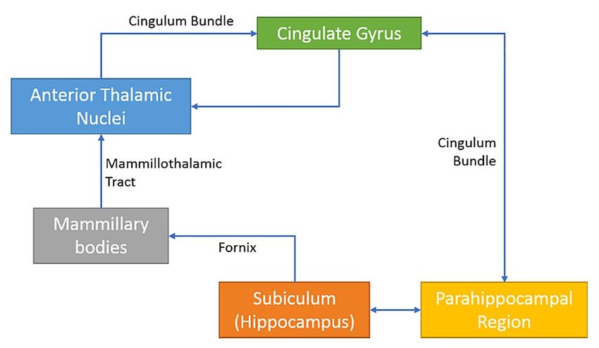
the motivation to act. However, if this pathway is intact, then the value of this signal, its valence, maybe
                                                 under-appreciated by the salience network. This mismatch may lead to apathy. The Papez circuit links the
                                                 hippocampus with the cingulate cortex, and degeneration of the entorhinal cortex by transsynaptic
                                                 degeneration may involve the cingulate cortex, leading to apathy [8].

                                                 Case Presentation
                                                 We present the case of a retired 75-year-old businessman who started experiencing mild memory loss
                                                 pertaining to appointments, conversations, and close family friend’s names. His pre-morbid personality was
                                                 dynamic with a happy, cheerful, and engaging temperament, and he was widely versed in domestic and
                                                 foreign affairs. Over the course of two to three years, he became more introverted and showed no interest in
                                                 calling family, friends, or socializing. He was content sitting at home and watching television. When
                                                 questioned about events in his life, answers tended to be neutral and devoid of any information.
                                                 Conversations were never initiated and he provided simple, binary yes and no answers. Nevertheless, he
                                                 seemed jovial, unconcerned, and there was no report of sadness, hallucinations, delusions, or agitation.
                                                 Sleep and nutrition were adequate with a mild decline in hygiene, such as maintaining his nails and
                                                 bathroom cleanliness after urination. Family gatherings with his grandchildren were not met with the usual
                                                 joy and happiness; instead, he remained quiet and just kept smiling. His wife stated that he did not appear to
                                                 be there and seemed to be disengaged from events around him.

                                                 His past medical history was significant for coronary artery disease treated with atorvastatin 40 mg daily,
                                                 hypertension treated with lisinopril 10 mg daily, and atrial fibrillation treated with rivaroxaban 20 mg daily.

                                                 On examination, his disposition was neutral and calm, but he did not start any conversation or ask any
                                                 questions during the interview. Good eye contact was maintained. There was no obvious aphasia; repetition,
                                                 naming, and reading aloud were normal. Speech prosody and cadence were normal. With immediate
                                                 attention, he registered three out of seven digits. Delayed recall of three objects at two minutes was zero.
                                                 There was visuomotor apraxia with geometric hand pantomime and inability to intersect pentagons. Verbal
                                                 fluency (animal naming) was eight at one minute, with categorical restriction to mammals; no reptiles or
                                                 other vertebrates, insects, or other invertebrates were mentioned. His mini-mental status score was 19 out
                                                 of 30. He had difficulty following multiple-step commands. His knowledge of current affairs was scant, but
                                                 he was able to recollect significant past events such as his wedding and his first business venture.

                                                 No frontal lobe signs were noted. Specifically, a snout reflex, jaw jerk, palmomental reflex, hand-grasp
                                                 reflexes, and paratonia of the arms were all absent. No perseveration of behavior was noted. Limb-kinetic
                                                 praxis tested with coin deftness and ideomotor praxis tested with transitive actions such as unlocking a door
                                                 were both normal. Otherwise, gait, cranial nerve, power, and sensory examination were unrevealing.

                                                 An MRI of the brain showed bilateral parietal and temporal lobe atrophy typical of that seen with
                                                 Alzheimer's disease (Figure 1).

2021 Kesserwani et al. Cureus 13(3): e13802. DOI 10.7759/cureus.13802                                                                                              2 of 6
FIGURE 1: Axial FLAIR MRI of the brain
1A: biparietal lobe atrophy (yellow arrows); 1B: bitemporal lobe atrophy (blue arrows)
FLAIR: fluid-attenuated inversion recovery; MRI: magnetic resonance imaging
A positron emission tomography (PET) scan with an amyloid tag was not deemed necessary, as it would not
have altered the management. A bilateral carotid duplex scan was normal. A thyroid screen and serum
vitamin B12 were within normal limits.
Based on his gradual cognitive decline, coupled with prominent episodic memory loss and findings on the
MRI of the brain, a provisional diagnosis of mild-to-moderate Alzheimer’s disease was made. His lack of
motivation and indifference to emotional signals around him, in the absence of depression, led to
a diagnosis of apathy. The patient was started on a standard dose of donepezil 5 mg daily and titrated to 10
mg daily at one month. Sadly, his condition continued to deteriorate despite the addition of memantine,
and he died of a myocardial infarction five years after the initial diagnosis.
Discussion
Studies utilizing fluorodeoxyglucose positron emission tomography (FDG-PET) have shown that across a
wide spectrum of neurodegenerative disorders including FTD, PD, and PSP, the sine-qua-non of apathy is
hypometabolism of the dorsal anterior cingulate cortex (dACC) and the VS (nucleus accumbens and olfactory
tubercle) in the basal forebrain. Secondary cortical areas of hypometabolism include the dorsolateral
prefrontal cortex (DLPFC) and orbitofrontal cortex (OFC), areas connected with the dACC and VS. These
areas along with the fronto-insular cortex are part of the salience network that weighs the emotional
content of a stimulus or signal, the so-called valence or emotional value of an incoming message. Other sub-
cortical areas with hypometabolism include the thalamus and ventral tegmental area (VTA) (part of the
dopaminergic motivation and pleasure pathway) [6].
The VS consists of large cholinergic cells in the basal forebrain that project to most of the neocortex.
However, it is the gamma-aminobutyric acid (GABA)-containing medium spiny neurons that are heavily
innervated by the dopaminergic mesolimbic system (VTA of the midbrain) via the medial forebrain bundle.
This axis represents the classical reward system, integrating motivation and pleasure [9]. Meanwhile, the
dACC is connected with the DLPFC and the parietal cortex and is involved in attentional mechanisms. Here
reside the von Economo neurons, which are highly specialized large spindle cells with the highest densities
in hominids (human and apes). Bilateral ischemic strokes of the dACC can lead to akinetic mutism, and a loss
of the von Economo neurons is seen in FTD [10].
In summary, the mesolimbic system (VTA and VS) carries the content of a pleasurable feeling or stimulus via
a dopaminergic pathway, which is gauged, weighed, and given value or valence by the dACC, a node of
the salience network. A lesion along this cascade can blunt the emotional content or value of the incoming
signal and suppress the goal-directed motivation or drive [11].
The role of the cingulate cortex makes intuitive sense as it is a key node in the "emotional" Papez circuit. The
cingulum is a major white-matter tract that forms a major limb of the Papez circuit; emerging from the
anterior thalamic nucleus, it arches anteriorly above the genu of the corpus callosum all the way posteriorly
2021 Kesserwani et al. Cureus 13(3): e13802. DOI 10.7759/cureus.13802 3 of 6into the parietal lobe before arching forwards into the parahippocampal gyrus (Figure 2) [12].
FIGURE 2: Diagram illustrating the role of the cingulum bundle and
cingulate cortex in the emotional Papez circuit
Note that the cingulum is part of the conduit from the hippocampus to the cingulate cortex
In 51 patients with probable Alzheimer’s disease who had apathy, diffusion tensor imaging revealed reduced
fractional anisotropy (meaning less dense fiber tracts and hence disconnection) in the left cingulum
independent of depression and the use of psychotropic medications. This further cements the role of the
cingulate cortex in apathy [13].
The temporal lobe and thalamus in Figure 2 are included to emphasize the role of episodic memory in
cognitive function and emotional content. Episodic memory is experiential memory that involves mental
time travel and is context-dependent. The core limbic circuit is heavily interconnected with other cortical
and subcortical regions and may function more as a memory than an emotional network. However, it is a
cohesive network as theta oscillations do resonate through this circuit; these nodes usually degenerate
together in various disease states and the hippocampal-cingulate-diencephalic network all contain head-
direction cells [14]. Alzheimer's disease is associated with the early loss of neural cells in the subiculum
(entorhinal cortex); the latter is connected to the cingulate cortex via the fornix, mammillothalamic tract,
and cingulum, putatively explaining the early onset of apathy in Alzheimer's disease patients [15].
The pathway from the posterior hypothalamus and thalamus to the cingulate cortex via the cingulum to the
hippocampal formation was formerly known as the ergotropic pathway, ergo referring to sympathetic
activation (mental energy). Sectioning of the cingulum in animals led to placidity (calming effect) and to
apathy (indifference of events) [16].
Pharmacologically, since the mesolimbic system is dopaminergically driven, there is merit in the use of
dopaminergic agonists. The dopamine D3 receptor is rich in the basal forebrain (nucleus accumbens) and
pramipexole, which acts through the D2 receptor and may ameliorate motor symptoms and through D3
receptor agonism and may help apathy [17]. Meanwhile, the cholinesterase inhibitor rivastigmine has been
shown to be beneficial in a double-blind placebo-controlled study of 31 patients with PD who had apathy but
were dementia-free [18].
Only two open-label studies out of eight showed improvement in apathy scores, assessed as part of the
Neuropsychiatric Inventory (NPI) or the Clinical Global Impression of Change (CGIC) scales, in patients with
mild-to-moderate Alzheimer's disease treated with the cholinesterase inhibitors donepezil and rivastigmine.
Two small trials (one randomized, the other open-label) showed improvement with the stimulant
methylphenidate at a dose of 10 mg bid with side effects limiting the utility of this drug in patients with
Alzheimer's disease. One large randomized controlled trial involving Ginkgo biloba extract also showed
statistically significant improvement in apathy scores. The addition of modafinil to donepezil in one study
of 23 patients with Alzheimer's disease showed no improvement of apathy [19].
There is a need for randomized placebo-controlled trials about the treatment of apathy, preferably studies
utilizing PET imaging of the brain to determine the network involved in the generation of apathy as this may
help determine the class of drugs one may use. If the mesolimbic system is involved, one may consider a
2021 Kesserwani et al. Cureus 13(3): e13802. DOI 10.7759/cureus.13802 4 of 6dopaminergic agent as opposed to a cholinesterase inhibitor if the basal forebrain and hippocampus are
involved. The white-matter tracts of these networks may be further analyzed with diffuse tensor imaging,
using the connectome library.
Conclusions
Apathy used to be a seemingly nebulous and non-specific concept. However, with the advent of advanced
imaging technology, the understanding of apathy has expanded from a lack of mental energy to a network
dysfunction concept pertaining to a lack of mesolimbic innervation of the basal forebrain and abnormal
calibration of the valence of the mesolimbic pathway by the salience network. The core dysfunction of the
cingulate cortex in a wide variety of diseases associated with apathy concurs with prior clinicopathological
studies and the classical circuit of Papez. In this article, we argue that the involvement of the entorhinal
cortex early in Alzheimer’s disease, by downstream effects via the fornix, mammillothalamic tract, and
cingulum to the cingulate cortex, may explain the presence of apathy in affected patients. Future
connectome studies with diffusion tensor imaging may clarify the picture further. Another area in need of
further research is the pharmacotherapy or neuromodulation of apathy.
Additional Information
Disclosures
Human subjects: Consent was obtained or waived by all participants in this study. Conflicts of interest: In
compliance with the ICMJE uniform disclosure form, all authors declare the following: Payment/services
info: All authors have declared that no financial support was received from any organization for the
submitted work. Financial relationships: All authors have declared that they have no financial
relationships at present or within the previous three years with any organizations that might have an
interest in the submitted work. Other relationships: All authors have declared that there are no other
relationships or activities that could appear to have influenced the submitted work.
References
1. Stanton BR, Carson A: Apathy: a practical guide for neurologists . Pract Neurol. 2016, 16:42-7.
10.1136/practneurol-2015-001232
2. Jenner C, Reali G, Puopolo M, Silveri MC: Can cognitive and behavioural disorders differentiate frontal
variant-frontotemporal dementia from Alzheimer's disease at early stages?. Behav Neurol. 2006, 17:89-95.
10.1155/2006/812760
3. Kuzis G, Sabe L, Tiberti C, Dorrego F, Starkstein SE: Neuropsychological correlates of apathy and depression
in patients with dementia. Neurology. 1999, 52:1403-7. 10.1212/wnl.52.7.1403
4. Marin RS, Wilkosz PA: Disorders of diminished motivation . J Head Trauma Rehabil. 2005, 20:377-88.
10.1097/00001199-200507000-00009
5. van Dalen JW, van Wanrooij LL, Moll van Charante EP, Brayne C, van Gool WA, Richard E: Association of
apathy with risk of incident dementia: a systematic review and meta-analysis. JAMA Psychiatry. 2018,
75:1012-21. 10.1001/jamapsychiatry.2018.1877
6. Le Heron C, Apps MAJ, Husain M: The anatomy of apathy: a neurocognitive framework for amotivated
behaviour. Neuropsychologia. 2018, 118:54-67. 10.1016/j.neuropsychologia.2017.07.003
7. Kos C, van Tol MJ, Marsman JB, Knegtering H, Aleman A: Neural correlates of apathy in patients with
neurodegenerative disorders, acquired brain injury, and psychiatric disorders. Neurosci Biobehav Rev. 2016,
69:381-401. 10.1016/j.neubiorev.2016.08.012
8. Kesserwani H: Delusional infestation secondary to the dopamine agonist ropirinole in a patient with
Parkinson's disease: a case report with an outline of the biology of psychosis. Cureus. 2021, 13:e12880.
10.7759/cureus.12880
9. Ikemoto S: Brain reward circuitry beyond the mesolimbic dopamine system: a neurobiological theory .
Neurosci Biobehav Rev. 2010, 35:129-50. 10.1016/j.neubiorev.2010.02.001
10. Allman JM, Hakeem A, Erwin JM, Nimchinsky E, Hof P: The anterior cingulate cortex. The evolution of an
interface between emotion and cognition. Ann N Y Acad Sci. 2001, 935:107-17.
11. McCutcheon RA, Nour MM, Dahoun T, et al.: Mesolimbic dopamine function is related to salience network
connectivity: an integrative positron emission tomography and magnetic resonance study. Biol Psychiatry.
2019, 85:368-78. 10.1016/j.biopsych.2018.09.010
12. Bubb EJ, Metzler-Baddeley C, Aggleton JP: The cingulum bundle: anatomy, function, and dysfunction .
Neurosci Biobehav Rev. 2018, 92:104-27. 10.1016/j.neubiorev.2018.05.008
13. Kim JW, Lee DY, Choo IH, Seo EH, Kim SG, Park SY, Woo JI: Microstructural alteration of the anterior
cingulum is associated with apathy in Alzheimer disease. Am J Geriatr Psychiatry. 2011, 19:644-53.
10.1097/JGP.0b013e31820dcc73
14. Bubb EJ, Kinnavane L, Aggleton JP: Hippocampal - diencephalic - cingulate networks for memory and
emotion: an anatomical guide. Brain Neurosci Adv. 2017, 1:2398212817723443. 10.1177/2398212817723443
15. Gómez-Isla T, Price JL, McKeel DW Jr, Morris JC, Growdon JH, Hyman BT: Profound loss of layer II
entorhinal cortex neurons occurs in very mild Alzheimer's disease. J Neurosci. 1996, 16:4491-500.
10.1523/JNEUROSCI.16-14-04491.1996
16. Sano K: Sedative neurosurgery with special reference to posteromedial hypothalamotomy . Neurol Med Chir.
1962, 4:112-42.
17. Guttman M, Jaskolka J: The use of pramipexole in Parkinson's disease: are its actions D(3) mediated? .
Parkinsonism Relat Disord. 2001, 7:231-4. 10.1016/s1353-8020(00)00062-6
18. Devos D, Moreau C, Maltête D, et al.: Rivastigmine in apathetic but dementia and depression-free patients
2021 Kesserwani et al. Cureus 13(3): e13802. DOI 10.7759/cureus.13802 5 of 6with Parkinson's disease: a double-blind, placebo-controlled, randomised clinical trial. J Neurol Neurosurg
Psychiatry. 2014, 85:668-74. 10.1136/jnnp-2013-306439
19. Rea R, Carotenuto A, Fasanaro AM, et al.: Apathy in Alzheimer's disease: any effective treatment? .
ScientificWorldJournal. 2014, 2014:421385. 10.1155/2014/421385
2021 Kesserwani et al. Cureus 13(3): e13802. DOI 10.7759/cureus.13802 6 of 6You can also read