Acute Lymphoblastic Leukemia (ALL) in Children and Teens
←
→
Page content transcription
If your browser does not render page correctly, please read the page content below

PROVIDING THE LATEST INFORMATION FOR
                                          PATIENTS & CAREGIVERS

Acute
Lymphoblastic
Leukemia (ALL)
in Children
and Teens

First Edition 2021

Support for this
publication provided by
A six-word narrative about living with blood
   cancer from patients in our LLS Community

Stay strong and keep moving forward. Find the positive in every day.
Be your own best patient advocate. Changed my life for the better.
Accept, learn and focus on present. Learning to live a different life.
Sudden and life changing—be positive. Waiting, worrying, anxiousness/
happy I’m alive! Embrace a new normal each day. 5 years, 41 infusions,
constant fatigue. Patience, positive attitude, hope and faith. Test to test,
I will survive! Treatment, fatigue, treatment, fatigue and survival.
Love life, live better every day. I don’t look back only forward. So far,
so good, live life. Meditation, mindfulness, wellness, faith, nutrition
and optimism. Finding the joy while living with uncertainty. Watch, wait,
treat, regroup, rest, re-energize. Blessed to be doing so well! Eye opening
needed learning and healing. Feel great: uncertain travel plans annoying.
Renewed faith, meditation, diet, mindfulness, gratitude. Watchful waiting
can be watchful worrying. Scary, expensive, grateful, blessings, hope,
faith. Thank god for stem cell transplants! Do not know what to expect.
Extraordinarily grateful, I love my life. Diagnosed; frightened; tested;
treating; waiting; hoping. I’m more generous, impatient less often.
Embrace your treatment day after day. Live today, accept tomorrow, forget
yesterday. Strength you never realized you had. Challenging to our hearts
and minds. Life is what we make it. Live life in a beautiful way.

              Discover what thousands already have at
              www.LLS.org/Community
   Join our online social network for people who are living with or supporting
   someone who has a blood cancer. Members will find
   • T
      housands of patients and caregivers sharing experiences and information,
     with support from knowledgeable staff
   • Accurate and cutting-edge disease updates
   • The opportunity to participate in surveys that will help improve care.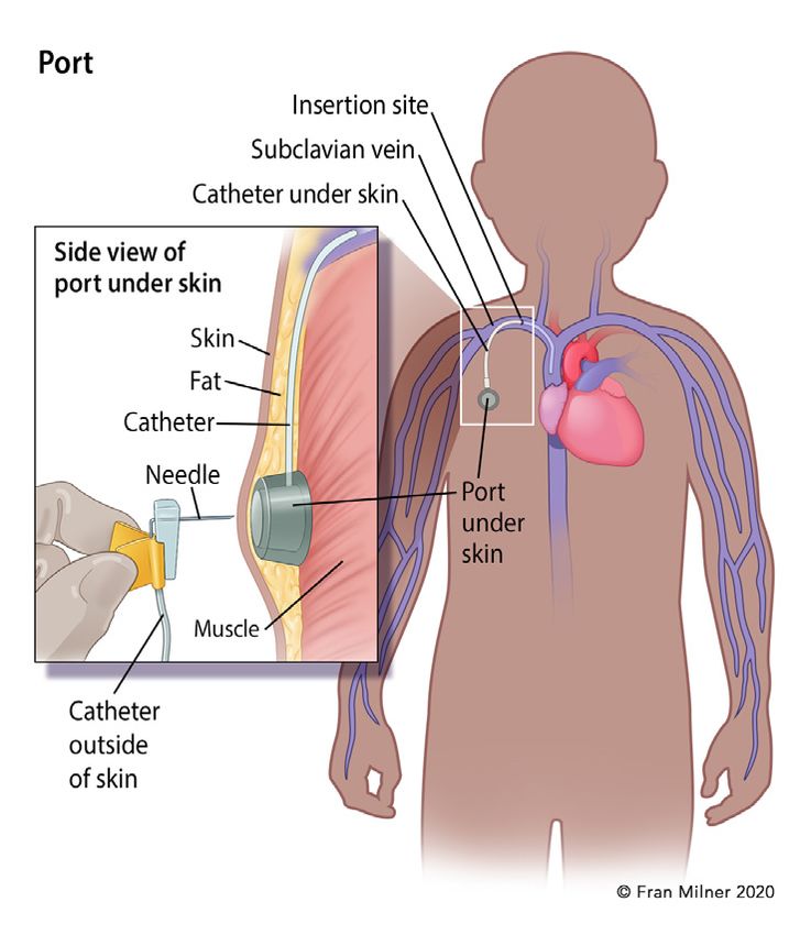
Inside This Booklet
                  2        Introduction
                  2        Leukemia
                  4        Acute Lymphoblastic Leukemia
                  5        Signs and Symptoms
                  6        Diagnostic Testing
                  12       Diagnosis and Cell Classification
                  21       Treatment Planning
                  26       Treatment Options
                  38       Special Treatment Considerations
                  42       Research and Clinical Trials
                  44       Related Disease
                  44       Side Effects and Complications
                  47       Follow-Up Care
                  54       Incidence, Causes and Risk Factors
                  55       Normal Blood and Bone Marrow
                  57       Resources and Information
                  61       Health Terms
                  71       References

Acknowledgement
The Leukemia & Lymphoma Society appreciates the review of this material by

Sarah K. Tasian, MD
Children’s Hospital of Philadelphia
Division of Oncology and Center for Childhood Cancer Research
Associate Professor of Pediatrics
Perelman School of Medicine at the University of Pennsylvania
Philadelphia, Pennsylvania

  New treatments may have been approved since this book was printed.
  Check www.LLS.org/DrugUpdates or call (800) 955-4572.

This publication is designed to provide accurate and authoritative information about the subject matter covered. It is dis-
tributed as a public service by The Leukemia & Lymphoma Society (LLS), with the understanding that LLS is not engaged in
rendering medical or other professional services. LLS carefully reviews content for accuracy and confirms that all diagnostic
and therapeutic options are presented in a fair and balanced manner without particular bias to any one option.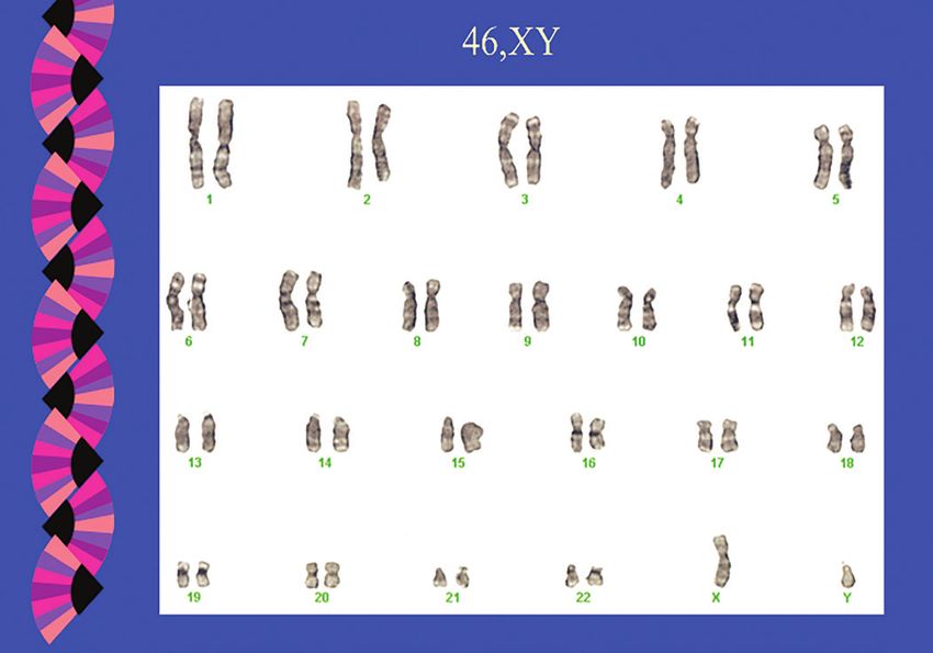
Introduction
This booklet provides information about acute lymphoblastic leukemia (ALL)
in children and also includes information about ALL in young adults. Acute
lymphoblastic leukemia is also known as “acute lymphocytic leukemia” and “acute
lymphoid leukemia.”
People of any age can develop ALL, but most cases are diagnosed in children. It
is the most common childhood cancer in the United States. An average of 2,761
children and young adults younger than 20 years of age were diagnosed with
leukemia each year from 2012 to 2016 in the United States.¹
Over the past several decades, the cure rates and survival outcomes for children
with ALL have improved dramatically. Childhood ALL now has one of the highest
cure rates of all childhood cancers. Today, most young patients diagnosed with
ALL can expect to have full and productive lives after treatment. Many survivors
return to school, attend college, enter the workforce, marry and become parents.
However, more work remains to be done. New therapies are being studied in
clinical trials to find cures for all children who have ALL, including those with
high-risk disease and those who relapse after treatment.
This booklet provides medical information about ALL as well as advice to help
you, your child and your family cope. We trust that this information will provide you
with a good working knowledge of ALL and that it reinforces what you already
know. We hope that you will keep this booklet handy and, should you ever feel
alone when confronting problems, that you will turn to it for information and
guidance to find the support and resources you need.
Visit www.LLS.org/booklets to view, download or order all free
LLS publications mentioned in this booklet.

      New treatments may have been approved since this book was printed.
      Check www.LLS.org/DrugUpdates or call (800) 955-4572.

1
    Source: Facts 2019-2020. The Leukemia & Lymphoma Society. April 2020.

Feedback. Visit www.LLS.org/PublicationFeedback to give suggestions about
this booklet.

Leukemia
Leukemia is a cancer of the blood and bone marrow. Bone marrow is the sponge-
like tissue in the center of most bones, where blood cells form. Leukemia begins
in one of the immature stem cells in the bone marrow. One or more changes

2    I 800.955.4572 I www.LLS.org
(mutations) occur in the DNA of the cell, and it becomes a type of cancer cell,
called a “leukemia cell.”
Leukemia cells do not mature into healthy, functioning blood cells. They grow
more quickly and live longer than normal blood cells. They divide and copy
themselves to make more and more leukemia cells. Over time, the leukemia cells
crowd out and suppress the development of healthy blood cells in the bone
marrow, and they spill out of the bone marrow into the bloodstream.
The four major types of leukemia are:
{   Acute lymphoblastic leukemia (ALL)
{   Chronic lymphocytic leukemia (CLL)
{   Acute myeloid leukemia (AML)
{   Chronic myeloid leukemia (CML)
Disease progression (meaning how quickly the disease gets worse) is one of
the factors that doctors consider when classifying leukemia. Leukemias can be
“acute” or “chronic.” Acute leukemias develop and progress rapidly and typically
get worse quickly if they are not treated. Chronic leukemias usually progress
more slowly. Acute leukemias are much more common in children than chronic
leukemias.
Leukemia is also classified by the type of blood cell that becomes cancerous.
Blood stem cells develop into two primary types: lymphoid and myeloid. As
lymphoid stem cells mature, they become a type of white blood cell called
a “lymphocyte.” The two major types of lymphocytes are B cells and T cells.
Myeloid stem cells eventually become red blood cells, platelets or other types of
white blood cells (other than lymphocytes). Leukemia is called “lymphocytic” or
“lymphoblastic” if the cancerous change begins in a lymphoid cell. Leukemia is
called “myeloid” or “myelogenous” if the cancerous cell change starts in an early
form of a myeloid cell.
This booklet focuses on ALL, but there are other cancers, called “lymphomas,” that
also begin in lymphoid cells. Most lymphomas arise from more mature lymphoid
cells, but in rare instances they can develop from lymphoblasts. The main
difference between lymphoblastic leukemias and lymphoblastic lymphomas is
the location of the cancer cells. Leukemias, such as ALL and CLL, generally affect
the bone marrow and blood. In contrast, lymphomas are mostly located in lymph
nodes or other lymphatic tissues or organs. Patients with lymphoblastic lymphoma
generally benefit from treatment with ALL-like regimens rather than traditional
lymphoma therapy. Therefore, if you have been diagnosed with lymphoblastic
lymphoma, this book may also be helpful for you.
For general information about ALL, visit www.LLS.org/booklets to view the
free LLS booklet The ALL Guide: Information for Patients and Caregivers.

                                     Acute Lymphoblastic Leukemia (ALL) in Children and Teens   I 3
Acute Lymphoblastic Leukemia
How Acute Lymphoblastic Leukemia (ALL) Develops. There are three main
types of blood cells: red blood cells, white blood cells and platelets. Red blood
cells carry oxygen throughout the body. White blood cells help fight infections.
Platelets help stop bleeding by clumping together (clotting) at the site of an injury.
Blood cells begin as hematopoietic stem cells in the bone marrow. Hematopoietic
stem cells are immature (undeveloped) blood cells. In healthy bone marrow, these
blood-forming cells eventually develop into red blood cells, white blood cells and
platelets in a process called “differentiation.”
In people with ALL, a mutation or a series of mutations in the DNA (genetic
material) of the lymphoid stem cell result in the formation of leukemia cells
(lymphoblasts), which are immature cells stuck in the earliest stage of cell
development. These leukemia cells, also referred to as “ALL blasts” or “ALL cells,”
cannot mature into fully functioning lymphocytes which are white blood cells that
help fight infection.
Genetic errors in a mutated cell cause the cell to keep growing and dividing,
whereas a healthy cell would stop dividing and eventually die. Every cell that
arises from the initial leukemia blast cell also has its mutated DNA. As the leukemia
cells multiply uncontrollably and quickly accumulate in the bone marrow, they slow
down or stop the production of normal, healthy red blood cells, white blood cells
and platelets. As a result, there are too many immature leukemic blast cells that
cannot fight infections and too few mature, functional red blood cells, white blood
cells and platelets.
By the time ALL is diagnosed, the number of healthy red blood cells, white blood
cells and platelets is usually lower than normal. Having low levels of blood cells
may result in infections, anemia and excessive bleeding or bruising.
.
Medical term: Description:
Anemia Low red blood cell count
Thrombocytopenia Low platelet count (“thrombocyte”
is another word for platelet)
Neutropenia Low neutrophil count (a neutrophil
is a type of white blood cell)
4 I 800.955.4572 I www.LLS.orgSigns and Symptoms
Signs and symptoms are changes in the body that may indicate the presence of
disease. A sign is a change that the doctor sees during an exam or in a laboratory
test result. A symptom is a change that a patient can see and/or feel.
A person who has signs or symptoms that suggest the possibility of leukemia is
referred to a specialist called a hematologist-oncologist. This is a doctor who has
special training in diagnosing and treating blood disorders and blood cancers
such as leukemia, lymphoma and myeloma. A pediatric hematologist-oncologist
specializes in the care of children with blood disorders and blood cancers.
It is common for someone with ALL to feel a loss of well-being because of the
lack of normal, healthy blood cells. This happens when the leukemia cells in
the bone marrow crowd out the normal blood-making cells. Your child’s blood
counts may show a high number of white blood cells, but if your child has ALL,
these cells are not fully developed and do not fight infection well. In ALL, the
leukemia cells begin to reproduce very quickly and compete with the other
healthy blood cells for nutrients and space. Consequently, children with ALL may
not have enough mature red blood cells, white blood cells and/or platelets, and
often have symptoms related to low blood cell counts.
Symptoms of a low red blood cell count (anemia) include:
{ Fatigue
{ Shortness of breath during normal physical activities
{ Dizziness
{ Pale complexion
Symptoms of a low white blood count (leukopenia) include:
{ Frequent infections
{ Recurrent fevers
Symptoms of a low platelet count (thrombocytopenia) include:
{ Bruising easily
{ Prolonged bleeding from minor cuts
{ The appearance of pinhead-sized red spots on the skin, called “petechiae”
{ Frequent or severe nosebleeds
{ Bleeding gums
{ Heavier or more frequent menstrual periods in females
Acute Lymphoblastic Leukemia (ALL) in Children and Teens I 5Symptoms may also be related to leukemia cells collecting in other parts of the
body. These symptoms may include:
{ Unexplained weight loss or loss of appetite
{ Pain in bones and joints
{ Swollen lymph nodes
{ Enlarged spleen or liver
{ Abdominal pain
{ Wheezing, coughing or painful breathing
The symptoms listed above are common symptoms of ALL, but do not include all
possible symptoms, as children may experience symptoms differently. It is also
important to note that the symptoms of ALL may be similar to those of other blood
disorders or medical conditions. Speak with your doctor if your child has any of
the above symptoms to ensure proper diagnosis and treatment.
Diagnostic Testing
While certain signs and symptoms may indicate that a person has ALL, lab tests
are needed to confirm the diagnosis. It is important to have an accurate diagnosis,
as it helps the doctor to:
{ Estimate how the disease will progress
{ Determine the appropriate treatment
Talk to your doctor about
{ The diagnostic tests that are being done
{ What the results mean
{ Getting copies of the test results
Some of the tests may be repeated both during and after treatment to evaluate if
treatment is working.
Medical History. Your child’s doctor will take a thorough medical history. The
doctor will ask about any health problems or treatments that your child has had.
The history may include information about past illnesses, injuries, other treatments
and medications. Some illnesses run in families, so the doctor may also ask about
the health of your child’s blood relatives.
6 I 800.955.4572 I www.LLS.orgPhysical Examination. The doctor will want to know about your child’s current
symptoms and will conduct a physical examination. During the examination, the
doctor may listen to your child’s lungs and heart and carefully examine the body
for signs of infection and disease. To check the internal organs, the doctor may
also feel different parts of your child’s body. For example, the doctor may feel the
abdomen to see if your child has an enlarged liver or spleen. Because ALL can
cause enlarged lymph nodes, the doctor may check your child’s lymph nodes in
the neck and armpits. In boys, the doctor may also examine the testicles to see if
there are any masses.
Complete Blood Count (CBC) with Differential. This test is used to measure the
number of red blood cells, white blood cells and platelets in a sample of blood. It
also measures the amount of hemoglobin in the red blood cells. The CBC should
include a differential, which measures the numbers of the different types of white
blood cells in the sample.
Children with ALL often have a high number of white blood cells, but most of
these are leukemia cells that do not protect against infection. Meanwhile, they
may not have enough mature white blood cells, red blood cells or platelets.
Even if the CBC findings suggest leukemia, an ALL diagnosis is usually only made
after examination of a sample of bone marrow cells.
Bone Marrow Aspiration and Biopsy. These are two procedures that remove
bone marrow cells and test them for abnormalities. They are generally done at the
same time, either at the doctor’s office or in a hospital. Most children are under
sedation or general anesthesia during these procedures.
The samples are usually taken from the patient’s pelvis (hip bone). Bone marrow
has both a solid and a liquid component. For a bone marrow aspiration, a special
hollow biopsy needle is inserted through the hip bone and into the bone marrow
to remove (aspirate) a liquid sample of bone marrow cells. For a bone marrow
biopsy, a wider needle is used to remove a sample of solid bone that contains
bone marrow. Both samples are sent to the lab where they are examined under a
microscope. See Figure 1 on page 8.
Acute Lymphoblastic Leukemia (ALL) in Children and Teens I 7Figure 1. How Are the Blood and Bone Marrow Tests Done?
Blood Test. A small amount of blood is taken from the patient’s arm with a
needle. The blood is collected in tubes and sent to a lab for testing.
Bone Marrow Aspiration. A sample of fluid with cells is removed from the
bone marrow and sent to a lab for testing.
Bone Marrow Biopsy. A very small amount of bone filled with bone marrow
cells is taken from the body and sent to a lab for testing.
Both bone marrow tests are done with special needles. Adults and older
teens may be given a local anesthetic and be "awake" during this procedure,
but most children are under sedation or given general anesthesia, which
makes them "sleep" briefly during the tests. The sample of cells is usually
taken from the patient’s hip bone.
Blood and bone marrow tests may be done in the doctor’s office or in a
hospital. A bone marrow aspiration and biopsy are almost always done at
the same visit. Blood and bone marrow tests may be done both during and
after treatment. The tests are repeated to see if treatment is working.
Bone Marrow Aspiration and Biopsy
A Bone Marrow Aspiration
samples fluid and cells
A Bone
Common site Marrow
where sample Needles Biopsy
is taken samples
bone and
marrow
Skin
and fat
Patient position Compact
bone
Marrow
Spongy bone
Left: The place on the back of the patient’s pelvic bone where a bone marrow aspiration
or biopsy is done. Right: Where the needle goes inside the bone to collect the liquid
sample for aspiration and the bone sample for biopsy. The needles are different sizes for
each of these tests.
8 I 800.955.4572 I www.LLS.orgCell Assessment. At the lab, a hematopathologist examines the blood and bone
marrow samples. A hematopathologist is a doctor who has special training in
identifying blood diseases by studying cells under a microscope.
The hematopathologist examines the blood and bone marrow cells under a
microscope to determine their size, shape and type, as well as to identify other
cell features. Whether the cells look like normal, mature blood cells or abnormal,
immature blood cells (blast cells) is an important finding. See Figure 2 below.
The percentage of blast cells identified in the samples is another important finding.
Typically, there are no blast cells in the blood, and no more than 5 percent of the cells
in the bone marrow are blast cells. Generally, a diagnosis of ALL in children requires a
finding of 25 percent or more of the cells in the bone marrow to be lymphoblasts.
Figure 2. Acute Lymphoblastic Leukemia (ALL) Cells
Panel A Panel B
Panel A shows a photograph of developing cells in healthy marrow. The variation in the
appearance of the cells is characteristic of normal marrow. Panel B shows a photograph of
bone marrow cells from a patient with acute lymphoblastic leukemia. An unvaried appearance
characterizes the leukemic blast cells.
If leukemia is found, additional tests are done on the blood and bone marrow
samples to gather information about the subtype of ALL.
Flow Cytometry. This laboratory test can detect specific types of cancer cells based
on the antigens or proteins on the surface of the cells. The pattern of the surface
proteins is called the “immunophenotype.” It is used to help diagnose specific types
of leukemia and lymphoma cells.
A bone marrow sample is often used for this test, but it can also be done with a blood
sample. The sample of cells is treated with special antibodies created in a laboratory
that only bind to cells that have a specific antigen on them.
Acute Lymphoblastic Leukemia (ALL) in Children and Teens I 9Depending on the type of leukemia, the leukemia cells can have different antigens on their surfaces. Certain antigens, called “cluster of differentiation (CD) proteins,” are helpful in identifying leukemia cells. Flow cytometry helps to confirm an ALL diagnosis. It is also used to determine the type of lymphocytes (B cells or T cells) in which the disease originated and to assess the maturity of the cells. In addition, flow cytometry is used to check treatment results. Genetic Tests. The following tests are used to examine the chromosomes and genes in a patient’s leukemia cells. Cytogenetic Analysis (Karyotyping). In this test, a hematopathologist or other type of specialist uses a microscope to examine the chromosomes inside cells. In patients with ALL, karyotyping is used to look for abnormal changes in the chromosomes of the leukemia cells. Normal human cells contain 23 pairs of chromosomes, for a total of 46 chromosomes. Each pair of chromosomes is a certain size, shape and structure. In many cases of ALL, the chromosomes of leukemia cells have abnormal changes that can be seen under a microscope. These changes include translocations and/or extra chromosomes. A translocation occurs when a piece of one chromosome breaks off and attaches to another chromosome. Sometimes pieces from two different chromosomes break off and trade places. This results in a “fusion gene,” an abnormal gene formed when two different genes fuse together. Cytogenetic testing can be done with either a bone marrow sample or a blood sample. The leukemia cells in the sample are allowed to grow in the laboratory and then are stained prior to examination. The stained sample is examined under a microscope and photographed to show the arrangement of the chromosomes (called a karyotype). The karyotype shows if there are any abnormal changes in the size, shape, structure or number of chromosomes in the leukemia cells. See Figure 3 on page 11. Cytogenetic analysis provides information for determining a patient’s prognosis and treatment options. This information can predict how the disease will respond to treatment. For example, a translocation between chromosomes 9 and 22 is associated with a diagnosis of Philadelphia chromosome-positive (Ph+) ALL, a subtype of ALL treated differently from other subtypes. See pages 27, 34, 38. Fluorescence in Situ Hybridization (FISH). This lab test is used to identify and examine specific genes or chromosome regions in cells. In cases of ALL, doctors use FISH to detect certain abnormal changes in the genes and chromosomes within leukemia cells, including translocations. Pieces of DNA that contain special fluorescent dyes are made in the laboratory and added to the leukemia cells on a glass slide. When the pieces of DNA bind to specific genes or areas of chromosomes on the slide, they light up when viewed under a fluorescence microscope. Many abnormal changes can be seen with a standard microscope, 10 I 800.955.4572 I www.LLS.org
Figure 3. Normal Male Karyotype

  This figure shows a normal male karyotype. Courtesy of Dr. Dong Chen, hematopathologist,
  Mayo Clinic, Rochester, MN.

but FISH testing can also detect changes that are too small to be seen in more
basic cytogenetic tests.
Molecular Genetic Analysis. Polymerase chain reaction (PCR) is a very sensitive
laboratory technique that is used to detect and measure certain genetic mutations
and chromosomal changes that are too small to be seen with a microscope.
Different types of PCR testing can essentially increase (amplify) small amounts of
specific pieces of either RNA (ribonucleic acid) or DNA (deoxyribonucleic acid) to
make them easier to detect and measure. This test can find a single leukemia cell
among more than 500,000 to 1,000,000 normal cells. PCR testing is one method
used to determine the amount of minimal residual disease (MRD), the small
amount of cancer cells that may remain in the body after treatment. This test can
be done with either a bone marrow sample or a blood sample.
“Next-generation sequencing” is a catch-all term used to describe a number of
different modern genetic sequencing technologies. These technologies allow for
DNA and RNA sequencing and are capable of detecting very small gene fusions
and translocations within leukemia cells that cannot be detected by routine
cytogenetic tests or FISH.
See the free LLS booklets Understanding Genetics and Understanding Lab and
Imaging Tests for more information about these tests.

                                        Acute Lymphoblastic Leukemia (ALL) in Children and Teens   I 11
Diagnosis and Cell Classification
In children, a diagnosis of ALL generally requires a finding that 25 percent or
more of the cells in the bone marrow are leukemic blasts of lymphoid origin
(lymphoblasts). The ALL subtype is determined based on a patient’s lab test results.
Subtypes of ALL. The subtypes of ALL are identified based on certain features
of the leukemia cells. Determining the ALL subtype is an important factor in
treatment planning. The doctor will discuss with you which drug combinations
and “protocols” are indicated based on your child’s ALL subtype. In medicine,
protocols are detailed plans of treatments and procedures. The doctor may also
talk about whether a clinical trial may be an appropriate treatment option.
Immunophenotyping. Leukemia cells can be classified by the antigens, known
as "immunophenotypes," found on their surfaces. The World Health Organization
(WHO) classifies ALL based on the immunophenotype of the leukemia cell in the
following ways (see Table 1 on page 13):
{ B-cell lymphoblastic leukemia or lymphoma. This subtype begins in immature
cells that would normally develop into B cells. In children, if the bone marrow has
25 percent or more lymphoblasts, the disease is called B-cell lymphoblastic
leukemia (B-cell ALL). If the lymphoblasts are restricted to a mass in a lymph
node or other lymph tissue and less than 25 percent of the bone marrow
cells are lymphoblasts, it is called B-cell lymphoblastic lymphoma. Patients
with lymphoblastic lymphoma generally benefit from treatment with ALL-like
regimens rather than traditional lymphoma therapy.
B-cell ALL is the most common ALL subtype, accounting for approximately
80 percent of cases among children with ALL. Within the B-cell lineage, the cell
surface markers (proteins) differ according to the stage of cell maturation.
Before 2008, the WHO classified B-cell lymphoblastic leukemia as “precursor
B-lymphoblastic leukemia.” This older term is sometimes used to distinguish
B-cell ALL from mature B-cell ALL. Mature B-cell ALL is now referred to as
“Burkitt leukemia.” The treatment for Burkitt leukemia is based on therapy for
non-Hodgkin lymphoma and is very different than the treatment used for ALL.
For more information on non-Hodgkin lymphoma, see the free LLS booklet
Non-Hodgkin Lymphoma.
{ T-cell lymphoblastic leukemia or lymphoma. This subtype begins in immature
cells that would normally develop into T cells. If the bone marrow has 25
percent or more lymphoblasts, the disease is called T-cell lymphoblastic
leukemia (T-cell ALL). If the bone marrow has less than 25 percent
lymphoblasts and the lymph nodes are enlarged, it is call T-cell lymphoblastic
lymphoma. This subtype is less common than B-cell ALL and occurs more
often in adults than in children. T-cell ALL accounts for approximately 15 to 20
percent of ALL cases in children.
12 I 800.955.4572 I www.LLS.orgTable 1. World Health Organization Classification of
Acute Lymphoblastic Leukemia (ALL)
B-cell lymphoblastic leukemia/lymphoma
B-cell lymphoblastic leukemia/lymphoma, not otherwise specified (NOS)
B-cell lymphoblastic leukemia/lymphoma with recurrent genetic
abnormalities
B-cell lymphoblastic leukemia/lymphoma with t(9;22)
(q34.1;q11.2); BCR-ABL1
B-cell lymphoblastic leukemia/lymphoma with t(v;11q23.3); KMT2A
rearranged
B-cell lymphoblastic leukemia/lymphoma with t(12;21)(p13.2;q22.1);
ETV6-RUNX1
B-cell lymphoblastic leukemia/lymphoma with hyperdiploidy
B-cell lymphoblastic leukemia/lymphoma with hypodiploidy
B-cell lymphoblastic leukemia/lymphoma with t(5;14)(q31.1;q32.3);
IL3-IGH
B-cell lymphoblastic leukemia/lymphoma with t(1;19)(q23;p13.3);
TCF3-PBX1
Provisional entity: B-cell lymphoblastic leukemia/lymphoma,
BCR-ABL1–like
Provisional entity: B-cell lymphoblastic leukemia/lymphoma with
iAMP21
T-cell lymphoblastic leukemia/lymphoma
Provisional entity: early T-cell precursor lymphoblastic leukemia
Provisional entity: natural killer (NK) cell lymphoblastic leukemia/
lymphoma
Source: Classification of acute lymphoblastic leukemia types created by the World Health Organization
(WHO).
Abbreviations: t, translocation between chromosomes; q, the long arm of a chromosome (the lower
half); p, the short arm of a chromosome (the upper half); v, variable.
Acute Lymphoblastic Leukemia (ALL) in Children and Teens I 13Genetic Changes. In addition to classifying ALL as either B-cell or T-cell, it can
be further classified based on changes to certain chromosomes and genes (see
Tables 2 and 3 on pages 14-16). This identification of specific genetic abnormalities
is critical for disease evaluation, risk stratification and treatment planning.
One type of genetic change that may occur in ALL is the result of numerical
abnormalities. A numerical abnormality is either a gain or loss in the number
of chromosomes from the normal total of 46. A change in the number of
chromosomes can affect growth, development and the functioning of body
systems.
Another type of genetic change associated with ALL is a translocation. In a
translocation, the DNA from one chromosome breaks off and becomes attached
to a different chromosome.
About 75 percent of childhood cases of ALL can be classified into subgroups
based on chromosomal abnormalities and genetic mutations. Not all patients who
have ALL exhibit the same genetic changes. Some changes are more common
than others, and some have a greater effect on the patient’s prognosis.
See the free LLS booklet Understanding Genetics for more information
about genetics and genetic testing.
Table 2. Common Genetic Alterations in Childhood B-Cell ALL
Frequency
Genetic Subtype Common Alterations Prognosis
in ALL
Abnormalities in chromosome number
High hyperdiploidy
25% Favorable
(51-67 chromosomes)
Previously
Low hyperdiploidy
14% unfavorable, now
(47-50 chromosomes)
intermediate
Near-haploidy
Hypodiploidy (24-31 chromosomes),
1-2% Unfavorable
(Table 2 (continued).
Frequency
Genetic Subtype Common Alterations Prognosis
in ALL
Unfavorable
KMT2A (MLL)
5-6% (infants), Interme-
rearrangements
diate (non-infants)
t(1;11)(q21;q23) KMT2A-MLLT11 Less favorable
Particularly
t(4;11)(q21;q23) KMT2A-AFF1 (AF4)
unfavorable
t(9;11)(p22;q23) KMT2A-MLLT3 (AF9)
t(10;11)(p12;q23) KMT2A-MLLT10 (AF10)
t(11;19)(q23;p13·3) KMT2A-MLLT1 (ENL)
Other fusion partners
Unfavorable
prior to TKI
t(9;22)(q34;p11·2) BCR-ABL1 3-5% therapy;
Intermediate with
TKI therapy?
Other
IGH-CRLF2, P2RY8-
Ph-like 7-8% Unfavorable
CRLF2
ABL1, ABL2, CSF1R,
PDGFRB 5-6% Unfavorable
rearrangements
EPOR, JAK2
2% Unfavorable
rearrangements
P2RY8-CRLF2, JAK2 50-60% of
Trisomy 21-associated ALL Intermediate
mutations DS-ALL
Multiple copies of
iAMP21 2% Unfavorable
RUNX1
DUX4 rearrangements IGH-DUX4, ERG-DUX4 3-7% Favorable
MEF2D-BCL9, MEF2D-
MEF2D rearrangements 3-6% Unfavorable
HNRNPUL1
ZNF384 rearrangements EP300-ZNF384 4% Intermediate
Note that percentages may total more than 100% due to co-occurrence of genetic lesions.
Abbreviations: B-ALL, B-cell acute lymphoblastic leukemia; DS-ALL, Down syndrome-associated ALL;
iAMP21, intrachromosomal amplification of chromosome 21; TKI, tyrosine kinase inhibitor; t, translocation
between chromosomes; q, the long arm of a chromosome (the lower half); p, the short arm of a
chromosome (the upper half).
Adapted from Tasian SK, Hunger SP. Genomic characterization of paediatric acute lymphoblastic leukaemia.
British Journal of Haematology. 2017;176:867-882.
Acute Lymphoblastic Leukemia (ALL) in Children and Teens I 15Table 3. Common Genetic Alterations in Childhood T-Cell ALL
Frequ ency
Genetic Subtype Common Alterations Prognosis
of T-cell ALL
Recurrent chromosomal translocations
t(10;14)(q24;q11) TLX1 (HOX11) fusions 5-10% Favorable
t(7;19)(q34;p13) LYL1 fusions 10% Unfavorable
t(1;14)(p32;q11), t(1;7)
TAL1, LMO1, LMO2
(p32;q34), t(11;14)(p15;q11), 50-60% Unfavorable
fusions
t(11;14)(p13;q11)
Unfavorable
(some studies),
t(11;14)(p15;q11), t(5;14) Intermediate
TLX3 (HOX11L2) fusions 20-25%
(q35;q32) (some studies),
Favorable
(some studies)
Probably
t(8;14)(q24;q11) TRA-MYC, TRC-MYC 1%
unfavorable
HOXA10, HOXA9
7p15 translocations 3% Unfavorable
overexpression
KMT2A (11q23) KMT2A-AFF1, Possibly
5%
rearrangements KMT2A-MLLT1 favorable
Unfavorable
PICALM-MLLT10 (some studies),
t(10;11)(p13;q21) 5-10%
(CALM-AF10) Intermediate
(other studies)
Unfavorable
(some studies),
t(9;14)(q34q32) NUP214-ABL1 5-15%
Intermediate
(other studies)
NOTCH1 mutations 50-60% Favorable
Unfavorable
(some studies),
ETP 10-15%
Intermediate
(other studies)
FBXW7 mutation 15%
Other T-ALL 6%
Note that percentages may total more than 100% due to co-occurrence of genetic lesions.
Abbreviations: T-ALL, T cell-acute lymphoblastic leukemia; ETP, early thymic precursor or early
T-cell precursor; t, translocation between chromosomes; q, the long arm of a chromosome (the lower half); p,
the short arm of a chromosome (the upper half).
Adapted from Tasian SK, Hunger SP. Genomic characterization of paediatric acute lymphoblastic leukaemia.
British Journal of Haematology. 2017;176:867-882.
16 I 800.955.4572 I www.LLS.orgLearning About Your Child’s Diagnosis. You are likely to experience a wide
range of emotions from the time your child is diagnosed with cancer as well as
during and after treatment. These emotions may include shock, denial, fear, anger,
guilt and sadness. You may feel that life for your child and family will never be the
same. Allow yourself to feel sad. Understand that you are not to blame for your
child’s diagnosis.
Over time, you and your family will find ways to adapt and gradually develop a
new sense of normalcy. All of these feelings are to be expected, but if you feel
consumed by negative feelings and emotions or are unable to function, seek
professional help. Psychologists, social workers and spiritual advisers may also
help you come to terms with your child’s diagnosis. It is important to work through
your feelings so you can help your child cope and you can continue to manage
other aspects of family life and work.
Talking to Your Child About His or Her Diagnosis. Regardless of age, children
are usually aware when their health causes their parents concern. Your child may
experience a variety of emotions, such as anger, guilt, fear, anxiety and sadness,
possibly all in quick succession.
Sometimes parents wish to shield their child from information about the illness
and its treatment. Keep in mind that children will use their imagination to fill in
perceived gaps of information. Sharing information about the illness and treatment
helps your child build trust in both you and the members of the treatment team, so
that he or she feels comfortable talking about fears and concerns. Encourage your
child to talk about his or her concerns and ask questions.
Introduce your child to treatment team members who can provide psychosocial
support. These include psychologists, social workers, art or play therapists and
child-life specialists. In addition to helping you explain the illness and its treatment
to your child, they can help your child better understand his or her disease
through play or other activities.
Keep the discussion age appropriate when you talk to your child about his or her
diagnosis. Consider the following guidelines, organized by age:
Baby/Toddler (0 to 3 Years)
{ Children this young do not have an understanding of illness or cancer.
However, they are aware of changes to routines and the feelings of
people around them.
{ Children in this age-group may be afraid of the medical staff and medical
procedures.
{ Babies and toddlers may be afraid of abandonment or being left at the
hospital. Offer physical and verbal reassurance.
Acute Lymphoblastic Leukemia (ALL) in Children and Teens I 17Preschool/Kindergarten (4 to 6 Years)
{ Children may have some understanding of an illness, such as a cold, but
may not grasp the implications of a serious illness.
{ Children’s primary focus will be the symptoms they are experiencing in
any specific moment.
{ Children in this age-group may be afraid of pain, so explain tests or
treatments to them in advance.
{ Assure your child that he or she did nothing wrong to cause the cancer.
Elementary/Middle School (7 to 12 Years)
{ Children in this age-group may have a better understanding of serious
illness, but not specifically cancer.
{ They may have heard things about cancer at school, from friends, on TV,
or they may have found information online. Ask your child what he or she
knows and correct any misunderstandings, especially those that cause
distress.
{ Explain tests, treatments, and other medical procedures in advance.
Your child may be afraid of pain and resist some tests or procedures. Be
honest. If a procedure may be painful, work with the healthcare team
and decide how to explain what will be done to lessen pain and why the
procedure is important.
{ Talk to your child in advance about possible changes to his or her
physical appearance.
{ You may need to discuss fertility preservation with your child. Some
cancer treatments can affect fertility. Fertility preservation, such as egg or
sperm banking, may be an option for children who have begun puberty.
Fertility preservation needs to be done before treatment begins. Enlist
members of the healthcare team to help with this sensitive discussion.
{ You may see signs of regression in a child’s behavior, such as thumb
sucking, bed-wetting or tantrums.
{ A child may use play to process the information—play-acting doctor/
patient scenarios, for example.
{ If the cancer treatment will result in any changes to the child’s daily
routine, explain the changes ahead of time so that the child will know
what to expect.
18 I 800.955.4572 I www.LLS.orgHigh Schoolers/Teenagers (13 to 18 Years)
{ Teenagers understand more about cancer and may want to know more.
You may still need to correct any misinformation your teenager has heard
about cancer from school, friends, TV and movies, or has found online.
{ Teenagers may want to participate in decisions about their treatment.
Include them in discussions with members of the healthcare team, as
appropriate.
{ You may need to discuss fertility preservation with teenagers. Some
cancer treatments can affect fertility. Fertility preservation, such as egg
or sperm banking, needs to be done before treatment begins. Enlist
members of the healthcare team to help with this sensitive discussion.
{ High schoolers and teenagers may also be very concerned about
changes to their physical appearance, such as hair loss and losing or
gaining weight, as well as worrying about how their peers will react to
the changes.
{ As teenagers struggle to find independence, a cancer diagnosis may
feel like a setback that can lead to feelings of frustration and anger. They
may try to test their boundaries or engage in risky behaviors, such as
drinking, drug use, or sex.
Ways to Help Your Child Cope. It will help your child cope with his or her
diagnosis if you:
{ Provide structure to increase your child’s sense of control. Children crave
structure in their environment. Make things as consistent as possible. For
example, plan a regular routine that you will follow during your time together in
the hospital or clinic.
{ Acknowledge and praise your child when he or she is doing difficult things.
Intermittent praise is the best way to reinforce the desirable behaviors that you
want to see in your child.
{ Use the same consequences for unacceptable or inappropriate behavior as
you did before your child was diagnosed with cancer. Consistency will maintain
structure and normalcy.
{ Show that you respect your child’s anger, worry, sadness or fear. Give your
child appropriate outlets for expressing these feelings, such as drawing or
keeping a journal.
{ Keep your child busy with activities during treatment to take his or her mind off
difficult and unpleasant experiences.
Acute Lymphoblastic Leukemia (ALL) in Children and Teens I 19{ Help your child stay connected with friends from home and school with phone
calls, emails, or visits, if possible.
{ Ask for professional assistance for your child if he or she is having an
especially difficult time adjusting to the cancer diagnosis and its treatment.
Siblings. When a child is diagnosed with cancer, everyone in his or her family is
affected by the experience, including the child’s brothers and sisters. Siblings can
feel angry, anxious, lonely, sad, guilty, or even resentful of the new attention their
sibling receives. You can help your other children cope with a sibling’s diagnosis
in some of the following ways:
{ Give them the chance to talk about how the experience is affecting them.
{ Be open and willing to answer questions about their brother or sister’s cancer
and treatment.
{ Reassure younger siblings that they cannot “catch” cancer from their brother or
sister. Explain that their brother or sister did not do anything that caused
the cancer.
{ Warn siblings that their brother or sister who has cancer may have less energy
or lose his or her hair.
{ Explain that other concerned family members and friends may ask them about
their sibling’s diagnosis. Talk about appropriate responses.
{ Remember that brothers and sisters still have their own problems, unrelated to
their sibling’s cancer. Their problems are real and require your attention.
{ Provide consistent, fair discipline to all your children, even though it may be
more difficult right now.
{ Let all your children know that you love them and are proud of them.
Siblings need to continue to go to school and participate in their usual activities,
as much as possible. Ask friends, family, other parents and teachers for help.
However, disruptions to routines are inevitable, and siblings may feel lost or
overlooked. Arrange for regular “alone time” with each child.
Make sure the school is aware of the diagnosis. Talk to your other children’s
teachers. Ask your hospital’s social worker or psychologist, or your school
psychologist, whether your community offers any programs for siblings of children
who have cancer. For additional assistance finding programs for siblings, you can
also call an LLS Information Specialist at (800) 955-4572.
SuperSibs, a program of Alex’s Lemonade Stand Foundation, provides
programs and support for the siblings of children with cancer.
Visit www.alexslemonade.org/supersibs to learn more.
For additional support and information, please call an Information Specialist or
visit www.LLS.org/FamilyWorkbook to find information for caregivers.
20 I 800.955.4572 I www.LLS.orgTreatment Planning
Choosing a Hospital and Doctor for Your Child’s Cancer Treatment. Once
you learn that your child has ALL, you need to decide where to go for treatment.
Most children with cancer receive treatment at hospitals that specialize in treating
children with cancer. The doctors and other healthcare providers at these centers
have special training and expertise in giving comprehensive care to children.
These centers are often members of the Children’s Oncology Group (COG). This
is the world’s largest organization devoted to clinical research to improve the care
and treatment of children with cancer.
Going to a specialized children’s cancer hospital helps ensure that your child gets
the best available treatment. You can ask your child’s pediatrician or family doctor
for a referral, or you can call an LLS Information Specialist at (800) 955-4572 to
find hospitals that specialize in treating children with ALL.
Most children with ALL are cared for by a pediatric hematologist-oncologist.
A pediatric doctor (pediatrician) specializes in the treatment of children. A
hematologist is a doctor who has special training in disorders of the blood, and an
oncologist is a doctor who has special training in cancer. A pediatric hematologist-
oncologist specializes in blood cancers in children.
Children who are diagnosed with ALL usually need to start treatment as soon
as possible after diagnosis. Some families may wish to seek a second opinion,
particularly if their child has a high-risk subtype of ALL or if the ALL has come
back (relapsed) after initial treatment. A second opinion may help you feel more
confident about your child’s treatment plan. The second opinion should come
from a pediatric hematologist-oncologist, preferably one who specializes in
childhood ALL. This doctor will usually have the most knowledge and experience
regarding the latest treatment options.
If you are unsure or feel uncomfortable about how to tell your child’s doctor you
are getting a second opinion, call our Information Specialists to discuss a way that
makes you comfortable. You may also want to check with your insurance company
to be sure that a second opinion will be covered.
Pre-Treatment Testing. Before your child starts treatment, the doctor will perform
tests to learn more about your child’s leukemia and overall health, and to find out
if the leukemia has spread to other parts of the body. Doctors use this information
for treatment planning. Some of these tests are summarized below.
Blood Tests. Doctors test blood to help plan treatment. Below are some tests
used for treatment planning.
Acute Lymphoblastic Leukemia (ALL) in Children and Teens I 21Complete Blood Count (CBC) with Differential. This test is used to measure the number of red blood cells, white blood cells and platelets in a sample of blood. It also measures the amount of hemoglobin in the red blood cells. The CBC should include a differential, which measures the numbers of the different types of white blood cells in the sample. Blood Chemistry Profile. This blood test measures the levels of certain substances released into the blood by organs and tissues in the body. These substances include electrolytes (such as sodium, potassium and chloride), fats, proteins, glucose (blood sugar), uric acid and enzymes. Blood chemistry test findings indicate how well a person’s kidneys, liver and other organs are working. Although this test is not used to diagnose leukemia, if the results show that there is an abnormal amount of a particular substance in the blood, it may be a sign of disease or some other health problem. A blood chemistry profile also provides helpful information about any potential organ damage caused by leukemia cells or ALL treatments. Liver Function Tests. The liver is the largest organ inside the body. It is located in the upper right side of the abdomen. It helps the body digest food, store energy and remove toxins from the blood. If leukemia cells are present in the liver, they can affect liver function. Some chemotherapy drugs can also damage the liver and affect liver function. Liver function tests are done to check how well the liver is working. Coagulation Tests. These tests measure the blood’s ability to clot and stop bleeding. Certain proteins, called “coagulation factors,” are needed for clotting. These proteins are made by the liver. In addition to checking how well the blood is able to clot, these tests can determine whether there are deficiencies in some proteins, such as the protein called fibrinogen. Coagulation tests can help assess your child’s risk for excessive bleeding. Tumor Lysis Syndrome (TLS) Panel. Children with ALL may be at high risk for developing a condition called “tumor lysis syndrome (TLS).” This condition can occur when a large number of cancer cells die within a short period of time. As the leukemia cells die, they break apart and release their contents into the bloodstream, which changes the normal balance of chemicals in the blood. This can overwhelm the kidneys because they cannot get rid of the toxic substances all at once. The effects of TLS can be life threatening; they can be severe during the early phases of treatment, especially if white blood cell counts are very high before induction therapy. A TLS panel can help the doctor assess if your child is likely to get or already has TLS. HLA Typing. This consists of a blood test to identify certain proteins, called human leukocyte antigens (HLAs), found on the surface of most cells in the body. These proteins make up a person’s tissue type, which varies from person to person. They also play an important role in the body’s immune response to 22 I 800.955.4572 I www.LLS.org
foreign substances by helping the body distinguish its own cells from foreign
cells. HLA typing is done before allogeneic stem cell transplantation to find
out if there is a tissue match between the donor and the person receiving
the transplant. Although HLA typing is not used to diagnose leukemia, it is
an important test for newly diagnosed ALL patients, if allogeneic stem cell
transplantation is being considered as a treatment option. For more information
on stem cell transplantation, see page 36.
Lumbar Puncture. ALL can spread to the cerebrospinal fluid, the fluid that flows
around the brain and spinal cord. In order to determine whether leukemia cells
have spread to this area, a sample of the cerebrospinal fluid is tested.
The procedure used to collect the cerebrospinal fluid from the spinal column is
called a lumbar puncture or “spinal tap.” After the area over the spine in the lower
part of the back has been numbed with a local anesthetic, a thin needle is inserted
between two bones (vertebrae) and into the cerebrospinal fluid. A sample of the
fluid is withdrawn and examined under a microscope to look for leukemia cells
that may have spread to the brain and spinal cord.
Imaging Tests. These tests create pictures (images) of the inside of the body. A
radiologist is a doctor who specializes in reading these images. Various types of
imaging tests are used to detect where a cancer is located in the body.
Computed Tomography (CT) Scan. In this type of imaging test, a computer linked
to an x-ray machine is used to take a series of detailed pictures of areas inside
the body. In some cases, leukemia may grow outside the bone marrow—most
commonly in the lymph nodes. A CT scan may be used to see whether leukemia
cells are accumulating in lymph nodes in the chest or abdomen, or in organs such
as the spleen and liver.
Positron Emission Tomography (PET) Scan. For this type of imaging test, a
small amount of radioactive glucose (sugar) is injected into a patient’s vein. The
PET scanner detects areas in the body where large amounts of glucose are
being used. In the images, the cancer cells appear brighter than the normal cells
because they use sugar more quickly than normal cells. A PET scan may be done
to see if there are cancer cells in the lymph nodes or organs.
Positron Emission Tomography-Computed Tomography (PET-CT) Scan.
This procedure combines images from a both a PET scan and a CT scan. The
combined scans give a more detailed image of areas inside the body than either
scan can give by itself.

                                  Acute Lymphoblastic Leukemia (ALL) in Children and Teens   I 23
Magnetic Resonance Imaging (MRI) Scan. This imaging test uses magnetic fields
and radio waves to create images of the body’s organs and tissue, as well as the
brain and spinal cord. An MRI scan of the head and/or spinal cord should be done
if a patient has symptoms such as headaches or seizures that suggest that ALL
cells may have spread to the brain and spinal cord.
Ultrasound. This imaging test uses high-energy sound waves to examine tissues
and organs inside the body. For example, it can detect leukemia cells in a boy’s
testicles. If the testicles are not the same size or have any lumps, the doctor may
order an ultrasound to see whether there is a mass in the testicles.
Echocardiogram. A computerized image of the heart is created by bouncing
ultrasound waves off internal tissues or organs in the chest. An echocardiogram
shows the size, shape and position of the heart, as well as its internal structures.
It also shows if the heart is beating and pumping blood normally. Since some
cancer treatments can damage the heart, the doctor may do this test as part of the
treatment planning process to check how well the heart pumps blood.
See the free LLS booklet Understanding Lab and Imaging Tests for more
information about these tests. To view interactive 3D illustrations of some lab
and imaging tests, visit www.LLS.org/3D.
Prognostic Factors. Certain factors can affect a patient’s prognosis—the likely
outcome of a disease or ailment. Doctors use prognostic factors to help predict
how a patient’s disease is likely to respond to treatment. These factors help
doctors plan the most appropriate treatment regimen for each patient.
Prognostic factors for children with B-cell ALL include:
{ Age: The leukemia cells in infants younger than 1 year and children older than
10 years tend to be more resistant to treatment. Stronger treatments may be
needed to kill the leukemia cells.
{ White blood cell count: Children with white blood cell counts of 50x109L or
greater at the time of diagnosis need stronger treatment.
{ Genetic factors: Certain changes in the chromosomes or genes can make
the leukemia cells either easier or harder to treat. See Table 2 on pages 14-15,
and Table 3 on page 16.
{ Central nervous system involvement: Children with ALL who have leukemia
cells in the central nervous system at diagnosis are at a higher risk of disease
relapse.
{ Treatment response: Children who have a better response to the initial
induction therapy have a lower risk of disease relapse.
24 I 800.955.4572 I www.LLS.orgFor children with T-cell ALL, risk stratification is primarily based on their early
treatment response. Children who have a better response to the initial induction
therapy have a lower risk of disease relapse.
Risk Groups for B-cell ALL. Your doctor may describe your child’s ALL in terms
of its risk group. Patients are assigned to a risk group based on age, genetic and
clinical features of the disease and the results of laboratory tests.
Knowing your child’s risk group helps the doctors develop the most effective
treatment plan for your child. Patients with lower-risk ALL are more likely to have a
favorable outcome and need less aggressive treatment. Children in the high-risk
and very high-risk groups usually receive more intense treatment than children in
the lower-risk groups.
Standard (Low) Risk: Children older than age 1 and younger than age 10; low
white blood cell count; favorable response to treatment.
High Risk: Children older than 10 years; high white blood count at the time of
diagnosis; children with unfavorable genetic changes; children with minimal
residual disease, a very small amount of leukemia cells still detectable by sensitive
lab tests after 4 weeks of induction therapy.
Very High Risk: Children younger than age 1; children with certain genetic
changes; children who have a slow response to initial treatment and signs of
leukemia after the first 4 weeks of treatment; children with minimal residual
disease after four weeks of induction therapy.
Fertility. Some cancer treatments can affect your child’s fertility (the ability to have
children in the future). Before your child begins treatment, it is important to talk
with the doctor about whether the treatment could affect your child’s fertility. You
may also want to speak with a fertility specialist. A fertility specialist is a doctor who
diagnoses and treats problems related to infertility. The fertility specialist can talk
to you about possible options for preserving your child’s fertility.
Delaying treatment to address fertility options may not always be recommended.
Many children with ALL need to start treatment right away. Nevertheless, before
treatment begins, it is important to talk with your child’s doctor about the effect
treatment may have on fertility.
For more information about fertility preservation, see the free LLS booklet
Fertility and Cancer.
Acute Lymphoblastic Leukemia (ALL) in Children and Teens I 25Treatment Options
New treatments may have been approved since this book was printed.
Check www.LLS.org/DrugUpdates or call (800) 955-4572.
Many children with leukemia have treatment options including standard treatment
or a clinical trial. It is important to talk to your child’s doctor about the best
treatment option for your child.
Typically, treatment for children with ALL consists of a multi-drug regimen that is
divided into five phases: induction, consolidation, interim maintenance, delayed
intensification and maintenance. High-risk patients may have additional phases of
treatment. Most treatment regimens take 2 to 3 years to complete.
The main treatment for ALL is chemotherapy. Some treatment plans may also
include targeted agents and stem cell transplantation. Treatment regimens for
ALL include central nervous system (CNS) prophylaxis to prevent leukemia cells
from spreading to the area around the brain and spinal cord. CNS prophylaxis
is typically given to children throughout all phases of ALL treatment. For more
information about CNS prophylaxis, see page 27.
Talk to your doctor about
{ Your child’s treatment options and the results you can expect from
treatment
{ The results you can expect from standard treatment
{ The possibility of your child participating in a clinical trial
Induction Therapy. The first phase of chemotherapy is called “induction therapy.”
The goal of induction therapy is to destroy as many cancer cells as possible in
order to achieve (induce) a remission. Remission means that leukemia cells are no
longer found in bone marrow samples and blood counts have returned to normal.
Induction therapy lasts for 4 weeks. The specific drugs, dosages and timing of
administration depend on several factors, including the patient’s age, the specific
features of the leukemia and the overall health of the patient. See Table 4, on
page 30.
Your child may spend some or most of this time in the hospital during this phase
of treatment, depending on your child’s clinical condition. For some children, the
hospital stay is the first time they have been away from home for an extended
period of time. Most hospitals allow a parent to stay at the child’s bedside during
hospitalization.
26 I 800.955.4572 I www.LLS.orgYou can also read