An Analysis of the Neurological and Molecular Alterations Underlying the Pathogenesis of Alzheimer's Disease
←
→
Page content transcription
If your browser does not render page correctly, please read the page content below
cells
Review
An Analysis of the Neurological and Molecular Alterations
Underlying the Pathogenesis of Alzheimer’s Disease
Chantal Vidal and Li Zhang *

                                          Department of Biological Sciences, University of Texas at Dallas, Campbell Road, Mail Stop RL11, 800 W,
                                          Richardson, TX 75080, USA; cxv130930@utdallas.edu
                                          * Correspondence: li.zhang@utdallas.edu; Tel.: +1-972-883-5757

                                          Abstract: Alzheimer’s disease (AD) is a neurodegenerative disorder characterized by amyloid
                                          beta (Aβ) plaques, neurofibrillary tangles, and neuronal loss. Unfortunately, despite decades of
                                          studies being performed on these histological alterations, there is no effective treatment or cure
                                          for AD. Identifying the molecular characteristics of the disease is imperative to understanding the
                                          pathogenesis of AD. Furthermore, uncovering the key causative alterations of AD can be valuable in
                                          developing models for AD treatment. Several alterations have been implicated in driving this disease,
                                          including blood–brain barrier dysfunction, hypoxia, mitochondrial dysfunction, oxidative stress,
                                          glucose hypometabolism, and altered heme homeostasis. Although these alterations have all been
                                          associated with the progression of AD, the root cause of AD has not been identified. Intriguingly,
                                          recent studies have pinpointed dysfunctional heme metabolism as a culprit of the development of AD.
                                          Heme has been shown to be central in neuronal function, mitochondrial respiration, and oxidative
                                          stress. Therefore, dysregulation of heme homeostasis may play a pivotal role in the manifestation of
                                AD and its various alterations. This review will discuss the most common neurological and molecular

                                          alterations associated with AD and point out the critical role heme plays in the development of
Citation: Vidal, C.; Zhang, L. An
                                          this disease.
Analysis of the Neurological and
Molecular Alterations Underlying the
                                          Keywords: Alzheimer’s disease; mitochondria; heme; amyloid beta
Pathogenesis of Alzheimer’s Disease.
Cells 2021, 10, 546. https://doi.org/
10.3390/cells10030546

Academic Editor: Alexander
                                          1. Introduction
E. Kalyuzhny                                    Dementia is a chronic dysfunction of cortical and subcortical function that causes
                                          cognitive decline [1]. It affects about 5% of the elderly population over the age of 65 [1].
Received: 5 January 2021                  Alzheimer’s disease (AD) is the most common and most studied cause of dementia [2].
Accepted: 3 March 2021                    In Europe and North America, AD is more common than vascular dementia [1,3,4]. One
Published: 4 March 2021                   study in Shanghai noted that 65% of all dementias were clinically diagnosed as AD [5].
                                          AD is a progressive neurodegenerative disorder that affects memory and other cognitive
Publisher’s Note: MDPI stays neutral      functions. It is the 6th leading cause of death in the United States, and more than 5 million
with regard to jurisdictional claims in   Americans are currently living with this disease [6]. In the US, the number of people with
published maps and institutional affil-   this disease is projected to double by 2050 [6]. Worldwide, 50 million people are living with
iations.                                  this disease, and by 2050, this community is likely to rise to about 152 million people [7].
                                          Therefore, developing an effective treatment or cure for this disease is essential.
                                                AD can be divided into two subgroups: late and early onset forms of this disease [8].
                                          Early-onset AD, also known as familial AD (FAD), affects individuals under 65 years of
Copyright: © 2021 by the authors.         age and only accounts for 2–10% of the total cases of AD [9]. This form of AD is attributed
Licensee MDPI, Basel, Switzerland.        to mutations in genes such as the amyloid precursor protein (APP), Presenilin 1(PSEN1),
This article is an open access article    and Presenilin 2 (PSEN2) [10–16]. Late-onset AD is considered sporadic (SAD), although
distributed under the terms and           genetic risk factors have been identified, including the apolipoprotein E gene (APOE) [17].
conditions of the Creative Commons        Regardless of the type of AD, there are specific pathologies that are attributed to this
Attribution (CC BY) license (https://
                                          disease, which include the presence of extracellular plaques made of insoluble amyloid
creativecommons.org/licenses/by/
                                          beta peptides (Aβ) and neurofibrillary tangles (NFT) [6,10,18,19]. Recently, mitochon-
4.0/).

Cells 2021, 10, 546. https://doi.org/10.3390/cells10030546                                                      https://www.mdpi.com/journal/cells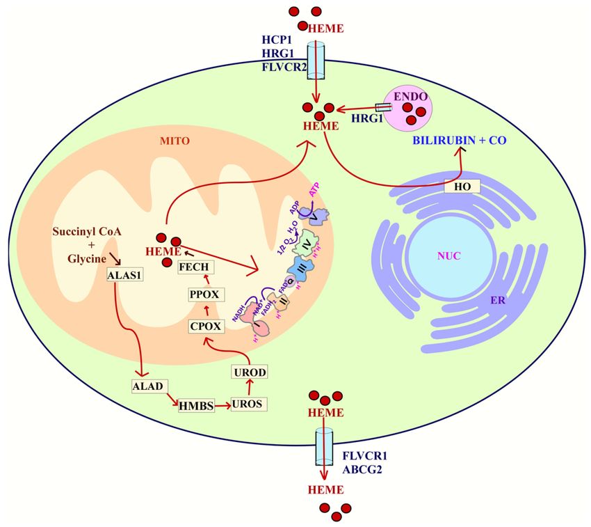
Cells 2021, 10, 546 2 of 27
drial dysfunction, reduced energy metabolism, synaptic loss, altered Wnt signaling, and
inflammation have been implicated in AD [20–22].
The US Food and Drug Administration (FDA) has approved only a few drugs to treat
AD, and they include memantine, donepezil, galantamine, and rivastigmine [23–26]. These
drugs either regulate glutamate activity, a chemical involved in information processing,
or delay the breakdown of acetylcholine, a chemical in the brain essential for memory.
Unfortunately, these drugs only moderately delay cognitive symptoms, and approximately
half of the people who take these drugs do not respond to them [26,27]. To develop effective
therapies that can slow down the cognitive symptoms of AD and halt the disease’s overall
progression, we must understand the molecular alterations that initiate the cascade of
events leading to neuronal dysfunction in AD.
One specific alteration that can play a pivotal role in the development of AD is dys-
functional heme homeostasis. Heme is likely a common factor that links several metabolic
alterations in AD, including dysregulated iron metabolism, decreased mitochondrial com-
plex IV levels, and increased levels of oxidative stress [28–30]. Heme deficiency induced in
two human brain cell lines caused reduced mitochondrial complex IV expression, altered
APP expression, and corrupted iron homeostasis [31]. Furthermore, in a 2019 study, the
presence of anemia was associated with a 41% increased risk for AD [32]. Therefore, this
review will discuss the various alterations seen in AD and point out the critical role heme
plays in AD pathogenesis.
2. Genetic Risk Factors
Family history has shown to increase a person’s chance of developing AD. There are
various genetic risk factors associated with the development of AD. These risk factors
are usually associated with some of the histological alterations previously discussed. The
presence of APP, PSEN1, and PSEN2 mutations as well as other genetic risk factors have
been attributed to an increased risk of developing AD.
2.1. APP, PSEN1, and PSEN2 Mutations
In 1984 Dr. Glenner and Dr. Wong isolated and identified APP, but it was not until
1991 and 1996 that mutations in the APP, PSEN1, and PSEN2 genes were identified as
having a causative role in the production of Aβ peptides and senile plaques [33–35].
Mutations in these genes are commonly associated with Familial Alzheimer’s disease
(FAD).
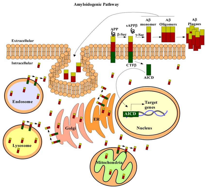
In the amyloidogenic pathway, APP is cleaved by β- and γ-secretases to produce
a 4kDa protein, known as Aβ (Figure 1) [33,36–38]. The first proteolytic cleavage is
produced by the membrane-bound aspartyl protease, β-APP-site cleaving enzyme (BACE).
This protease renders a secreted APP derivative, sAPP, and a membrane-bound protein
fragment of 99 amino acids, β-Secretase-Derived C-Terminal Fragment (CTFβ) [38–41].
CTFβ is further cleaved by γ-secretase containing the four proteins: APH1, PEN2, nicastrin,
and presenilin (PS1 or PS2). Cleavage of CTFβ produces different lengths of Aβ peptides
and an APP intracellular domain (AICD) [36,38]. The 40-residue peptide, Aβ40, makes up
the majority of the total Aβ produced in cells [38]. Less than 5% of the generated Aβ ends
at the residue 42 [38]. Aβ42 has a higher rate of fibrilization and insolubility and therefore
is more prevalent in senile plaques [42,43]. The increased ratio of Aβ42/Aβ40 is one of the
pathogenic hallmarks of AD [42,44–48].
APP gene mutations usually involve those in the β-APP-site cleaving enzyme (BACE)
cleavage site, those at the γ-secretase cleavage site, and those in the mid domain Aβ
region [49]. Mutations in this gene can render either an increase in Aβ42 produced or
an increase in Aβ42/Aβ40 ratios [50,51]. Furthermore, in 2016 a detailed study carried
out by Sun et al. [52] characterized 138 distinct PSEN1 mutations and their effect in Aβ
production. This study revealed that 34 variants increased production of AB42 while
the other 104 caused a reduction in the total production of Aβ40 and Aβ42 [52]. More
importantly, the production of Aβ40 was typically more affected than the production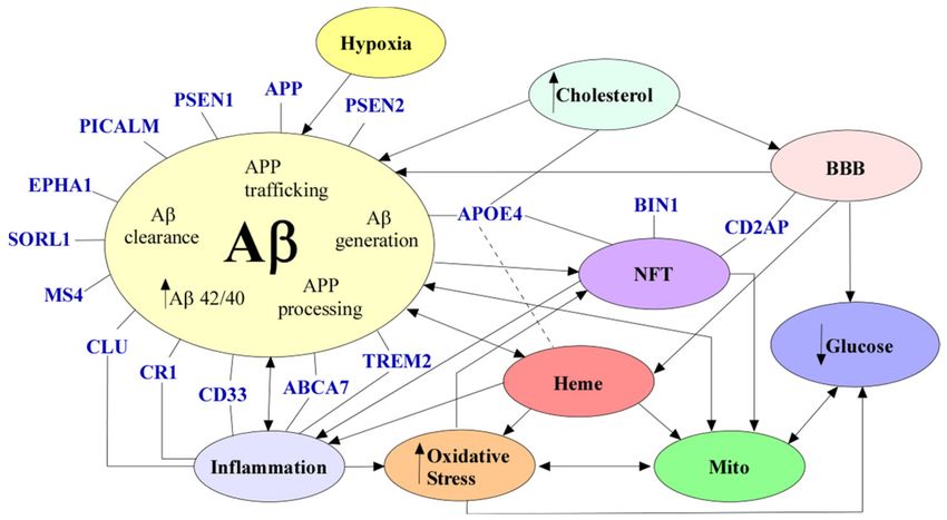
Cells 2021, 10, 546                                                                                                3 of 27

                      of Aβ42, leading to the elevated ratios of Aβ42/Aβ40 [52–54]. Similarly, mutations in
                      PSEN2 increase Aβ42/Aβ40 ratios, suggesting that the shift in the Aβ ratios has a role in
                      developing FAD [55]. An early study analyzing SAD and FAD brains found that the ratio
                      of the long-tail form of Aβ to total Aβ was increased in FAD brains [56]. Aβ42/Aβ40 ratios
                      were also elevated in FAD mutant induced pluripotent stem cells (IPSC) relative to con-
                      trols [57]. APPsw mice expressing apoE4 also exhibited increased Aβ42/Aβ40 ratios [58].
                      Furthermore, Aβ can be generated outside the central nervous system (CNS), contributing
                      to the circulating Aβ pool [59]. Roher et al. [59] found that brains and skeletal muscles
                      from AD patients express significantly more Aβ than non-demented controls.

                      Figure 1. APP processing. In the amyloidogenic pathway APP (in green red and gold) is cleaved by
                      β secretase to produce a soluble form of APP, sAPPβ. Then, γ-secretase cleaves the remaining amino
                      acid protein CTFβ to produce Aβ (red and gold) and AICD (green). Aβ can then form oligomers
                      and plaques which are characteristic of AD. AICD can translocate to the nucleus (beige) and regulate
                      gene expression (DNA is in grey and blue). APP can also be localized to the trans-Golgi network
                      (pink), ER (orange), endosomal (blue), lysosomal (yellow), and mitochondrial membranes (green).
                      Aβ liberation can occur wherever APP and the β- and γ-secretases are localized. Aβ can also be
                      taken up by the cell to form intracellular pools. Abbreviations: amyloid precursor protein (APP),
                      secreted APP derivative (sAPPβ), amyloid beta (Aβ), β-Secretase-Derived C-Terminal Fragment
                      (CTFβ), APP intracellular domain (AICD), β-APP-site cleaving enzyme (β-Sec), γ-Secretase (y-Sec),
                      ER (endoplasmic reticulum).

                      2.2. ApoE4
                           ApoE is a glycoprotein known to regulate the clearance of lipoproteins from the
                      plasma by serving as the ligand that binds to various cell surface receptors [60]. These
                      receptors then internalize apoE-containing lipoprotein particles. ApoE has three iso-
                      forms (Figure 2): apoE2, apoE3, and apoE4. Amino acid sequencing of these isoforms
                      showed that they differ in the residues at positions 112 and 158 (Figure 2) [61,62]. These
                      variations in amino acid residues affect their receptor and lipid-binding affinities. For
                      example, apoE2 has Cys residues at both positions and has a preference for high-density
                      lipoproteins (HDL) [61,62]. ApoE2 also has a low binding affinity to low-density lipoprotein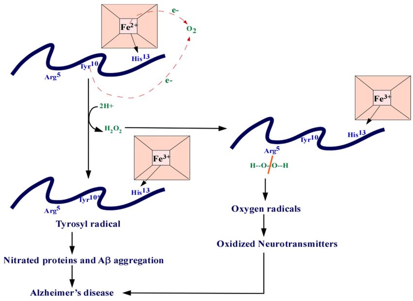
Cells 2021, 10, 546                                                                                             4 of 27

                      (LDL) receptors compared to apoE3 [62,63]. ApoE3 has a Cys residue at position 112 and an
                      Arg residue at 158. Similar to ApoE2, APOE3 can preferentially bind to HDL [62]. APOE4
                      has Arg residues at both positions that allow a higher binding affinity to LDL receptors and
                      larger triglyceride-enriched lipoproteins (TRL) [62,64]. The differences in binding affinities
                      affect their role in lipid metabolism. For example, apoE3 is associated with cholesterol efflux
                      and the formation of APOE-containing HDL, while apoE4 accumulates in the endosomal
                      compartments causing impaired cholesterol efflux, leading to the generation of Aβ [65–69].

                      Figure 2. ApoE contains the N-terminal domain which contains the receptor-binding region and the
                      C-terminal domain containing the lipid-binding region. There are three isoforms of APOE: ApoE2,
                      ApoE3, and ApoE4. ApoE2 contains Cys residues at positions 112 and 158. ApoE3 contains a Cys
                      residue at position 112 and an Arg residue at 158. ApoE4 contains Arg residues at both of these
                      positions. Abbreviations: cysteine (Cys), arginine (Arg), apolipoprotein E(ApoE).

                            ApoE2 can have a protective role against the development of AD [70]. Macrophages
                      expressing apoE2 are more efficient in degrading Aβ than those expressing apoE3 or
                      apoE4 [71]. A meta-analysis carried out in 2015 showed that carriers of the APOE2 allele
                      have a lower rate of brain amyloid presence than APOE3 carriers [72]. ApoE3 is the most
                      common form of apoE and plays a neutral role in AD.
                            ApoE4 exists in approximately 20% of the population and is the most significant
                      genetic risk factor for SAD [62,70]. The association of apoE4 with late-onset Alzheimer’s
                      disease was first discovered in three landmarks studies published in 1993 [73–75]. One of
                      these studies showed that apoE has a high affinity to Aβ and that the APOE4 allele has
                      a higher association with AD [74]. Corder et al. [73] found that the risk for AD increases
                      from 20% to 90% with increasing number of APOE4 alleles in 42 families with late-onset
                      AD. Furthermore, the APOE4 allele was shown to decrease the mean age of onset from
                      84 to 68 years [73].
                            Studies done on human induced pluripotent stem cells showed that APOE upregulates
                      APP expression, and this expression is most prominent for APOE4, followed by APOE3,
                      and finally APOE2 [76,77]. The APOE4 allele is also associated with increased amyloid
                      deposition and NFTs. [78,79]. Mitochondrial dysfunction has also been studied in APOE4
                      carriers. In a study analyzing the neurotoxicity of apoE4 fragments on cultured Neuro-2a
                      cells, apoE4 fragments formed filamentous inclusions in some cells that interacted with
                      mitochondria causing mitochondrial dysfunction [80].

                      2.3. Other Genetic Risk Factors of AD
                           A Genome-wide association study (GWAS) of 74,046 individuals identified 11 genes
                      associated with AD [81]. These genes include APOE, TREM2, CD33, BIN1, CLU, CR1, MS4,
                      CD2AP, ABCA7, PICALM, and EPHA1 [81]. Among these genes, TREM2 variants cause a
                      two-fold increase in the risk for AD [82,83]. TREM2 is primarily expressed in microglia and
                      helps mediate phagocytosis, inhibit inflammatory signals, and promote cell survival [82].
                      TREM2 activation can lead to ligand binding, inducing a signal cascade that results in
                      increased phagocytosis and decreased pro-inflammation [82]. Therefore, a compromised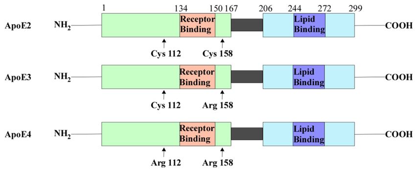
Cells 2021, 10, 546                                                                                           5 of 27

                      function of TREM2 may lead to decreased clearance of cell debris and possibly the removal
                      of Aβ in Alzheimer’s disease [83]. In 2019, Parhizkar et al. [84] found that in the absence of
                      TREM2, amyloid plaque seeding increased and microglia clustering around newly seeded
                      plaques decreased.
                            CD33 is significantly upregulated in AD patients’ brains and can modulate microglial
                      activation and inhibit Aβ clearance [85]. BIN1 has also been linked to AD in early GWAS
                      and is the most important genetic susceptibility locus in AD after APOE [86]. An analysis
                      of 114 AD brain tissues and 167 control brain tissues showed an increased expression of
                      BIN1 in AD brains [87]. Furthermore, loss of the Drosophila BIN1 ortholog AMPH was
                      able to suppress Tau-induced neurotoxicity [87]. This suggests that BIN1 acts as a genetic
                      risk factor for AD by regulating Tau pathology.
                            CLU and CR1 genes, previously identified as having a role in Aβ clearance, were
                      associated with the development of AD in a GWAS of 2032 AD patients and 5328 con-
                      trols [88–90]. MS4 family proteins have also been implicated in the pathogenesis of AD [91].
                      Karch et al. [92] showed that MS4A6A expression correlates with neuropathological mea-
                      sures of AD. Similarly, single nucleotide polymorphisms (SNPs) in the CD2AP gene are
                      associated with the development of SAD [93,94]. The CD2AP protein can modulate Tau-
                      mediated neurotoxicity, regulate Aβ generation, and maintain the blood–brain barrier
                      integrity [93]. For example, Cochran et al. [95] analyzed CD2AP deficient mice and found
                      that these mice exhibit reduced blood–brain barrier integrity, suggesting a cardiovascular
                      role in AD. Loss of function variants in the ABCA7 gene, involved in the Aβ clearance path-
                      way, have also been implicated in the development of AD [94,96,97]. Sakae et al. [98] found
                      that ABCA7 deficiency alters the brain lipid profile and impairs memory. Furthermore,
                      APPPS1 deficient for ABCA7 had an increased amyloid plaque burden.
                            Several GWAS have identified variants within the PICALM gene as risk factors for
                      developing AD [94,99,100]. PICALM, involved in clathrin-mediated endocytosis, likely
                      plays a role in APP endocytosis and thus regulates Aβ generation [101]. A 2011 study
                      analyzing four GWA datasets found EPHA1 variants implicated in AD [94]. Having a
                      minor C allele at SNP (rs11771145) on the EPHA1 gene is associated with a lower chance of
                      being Aβ positive, suggesting its protective role in preventing AD [102]. A meta-analysis of
                      30,000 subjects and a large GWAS associated multiple variants in the SORL1 gene with both
                      late- and early-onset AD [103,104]. Furthermore, SORL1 mutations have been associated
                      with a weakened interaction between the SORL1 protein and the full-length APP, altering
                      the levels of APP trafficking [105]. Overall these newly identified genetic risk factors
                      suggest new genetic and molecular mechanisms underlying AD’s pathogenesis.

                      3. Neurological and Molecular Alterations of AD
                           AD is characterized by various histological and molecular alterations. The most estab-
                      lished hallmarks of this disease include amyloid plaques and neurofibrillary tangles [106].
                      Despite the extensive interest in amyloid plaques and neurofibrillary tangles, several other
                      alterations are associated with this disease [17,28,107–112]. Unfortunately, there is much
                      debate on which of these alterations play a causative role in the development of AD. This
                      section will focus on the neurological and molecular alterations implicated in AD.

                      3.1. Amyloid Beta
                           The pioneering work of Dr. Alios Alzheimer started in 1906 when a patient of the
                      Community Psychiatric Hospital at Frankfurt named Auguste D. died [113]. The patient
                      presented various cognitive impairments, including memory loss and confusion [113]. Dr.
                      Alzheimer analyzed the brain of Auguste D. and discovered the histological alterations
                      that would later be known as plaques and neurofibrillary tangles (NFT) [113,114]. These
                      senile plaques are made of the accumulation of a 39–42 amino acid peptide called amyloid
                      beta (Aβ) [46,115,116]. The characteristic accumulation of this Aβ protein in AD patients
                      has caused many researchers to believe that this histological alteration is the cause of
                      the disease.
Cells 2021, 10, 546 6 of 27
Aβ is well-known to play a pivotal role in AD pathology, but the exact mechanism
has been widely debated. Nuclear magnetic resonance has shown that Aβ42 can form
oligomers that incorporate into the cell membrane and form channels that are highly per-
meable to Ca2+ [117,118]. This causes a disruption in calcium homeostasis, which induces
synaptic degeneration [118,119]. Aβ can also cause neuronal death in vivo through the cas-
pase 3 apoptotic cascade [120]. One study utilizing CK-p25 mice expressing increased Aβ
levels showed differentially expressed genes enriched in cell cycle, immune response, and
synaptic functions compared to controls [21]. It has also been proposed that Aβ42 causes
neuronal apoptosis by activating the caspase pathway, thereby promoting mitochondrial
fission and increasing reactive oxygen species (ROS) [121].
Despite ample evidence of the toxicity of Aβ, there is a poor correlation between the
clinical symptoms in sporadic AD and Aβ plaque deposition [122]. This has caused critics
to suggest that Aβ does not mediate AD [122]. However, in FAD cases, in which disease
pathogenesis is more clearly driven by Aβ, the anatomical correlation between plaques and
neuronal loss is consistent with that of SAD. This implies that Aβ can still drive neuronal
loss without the colocalization of plaques and neurodegeneration [122,123]. Furthermore,
the poor correlation between fibrillar Aβ and neuronal loss in SAD can be attributed
to the different aggregation states of Aβ. There is some evidence that Aβ oligomers
correlate well with AD severity [124]. Aβ oligomers can cause neuron degeneration and
hyperphosphorylation of tau, key characteristics of AD [125].
Accumulation of intracellular Aβ is also associated with AD. Two mechanisms have
been proposed for accumulating intracellular Aβ (Figure 1): intracellular sites of Aβ
production or reuptake of Aβ [126]. APP can be localized to the trans-Golgi network,
ER, endosomal, lysosomal, and mitochondrial membranes [126]. Aβ liberation can occur
wherever APP and the β- and γ-secretases are localized. Therefore, if APP cleavage occurs
within the cell, Aβ can accumulate intracellularly. Extracellular Aβ can also be taken up
by cells to form intracellular Aβ pools [126,127]. In SH-SY5Y, the uptake of Aβ40 and
Aβ42 occurs exclusively via endocytosis [127]. There are also several putative receptors and
transporters associated with the accumulation of intracellular Aβ [128–131]. For example,
binding of Aβ to the scavenger receptor for advanced glycation end products (RAGE)
can cause internalization of Aβ [132]. Similarly, the G protein-coupled formyl peptide
receptor-like 1 (FPRL1) and the NMDA receptors can also uptake Aβ [133,134].
Studies utilizing the Tg2576 mouse model have shown that accumulation of intra-
cellular Aβ can lead to synaptic dystrophy [135]. Intracellular Aβ is also associated with
decreased mitochondrial membrane potential [136]. Furthermore, injections of Aβ in pri-
mary neurons have also been shown to cause significant cell death through the p53-Bax
cell death pathway [137]. These studies suggest that the accumulation of intracellular
and extracellular Aβ and the different physical and aggregated states of Aβ play a role in
neuronal damage, leading to the development of AD.
3.2. Neurofibrillary Tangles
Neurofibrillary tangles (NFTs) are filamentous aggregates of the microtubule-associated
protein tau [138]. Tau is involved in microtubule stability and cytoskeletal trafficking within
mature neurons [139]. Tau has also been seen to copurify with tubulin and plays a major
role in polymerization and hence microtubule assembly [140]. Tau is tightly regulated by
various post-translational modifications, but phosphorylation is the most noted. In the
brain, tau is predominantly expressed in neurons, and its non-phosphorylated form is
restricted to axons [141]. Previous studies on tau revealed that specific modes of phospho-
rylation can cause conformational changes that affect its ability to polymerize tubulin [142].
Phosphorylation at Thr231, Thr214, and Ser235 causes dissociation of tau from micro-
tubules [143,144]. Interestingly, phosphorylation of tau at the C-terminal region causes
self-aggregation [145]. This role of phosphorylated tau can contribute to the formation
of NFTs.Cells 2021, 10, 546 7 of 27
One of the histological characteristics of AD is the presence of NFTs composed of
hyperphosphorylated tau [146–148]. NFTs have been shown to correlate well with disease
progression [149]. It has also been proposed that NFTs can directly cause damage to neurons
and glial cells by displacing cytoplasmic organelles to the periphery, inhibiting proteasome
activity, or disturbing microtubule assembly [147,150,151]. Furthermore, oligomeric tau has
been shown to induce neurodegeneration by decreasing levels of mitochondrial respiratory
complex I activity [152]. NFTs can also prevent mitochondrial transport, causing oxidative
stress and energy deprivation, which in turn leads to neurodegeneration [153,154]. The
role of NFTs and their function in either accelerating or halting neurodegeneration has also
been widely debated [155,156]. A study by Ferrari et al. [157] found that tau was likely to
be aggregated in cells treated with Aβ, suggesting that tau pathology follows Aβ toxicity
in AD.
NFTs are also known to induce oxidative stress [158]. Mitochondria are the main
source of oxidative stress, and the mitochondrial superoxide dismutase 2 (sod2) plays a crit-
ical role in alleviating ROS. To determine if oxidative stress causes NFTs, a study utilizing
sod2 null mice showed that increasing amounts of antioxidants significantly reduced levels
of hyperphosphorylated tau [159]. This suggests that mitochondrial oxidative stress plays
a role in the histological alteration of tau [159]. A Quantitative analysis also showed that
neurons with NFTs have a 40–56% decrease in the levels of 8-hydroxyguanosine (8OHG),
suggesting that NFTs help reduce levels of oxidative stress in neurons [158,160].
3.3. Neuronal Loss/Synaptic Loss
Neuronal loss is a prominent pathological feature of AD. AD is considered a neurode-
generative disease which means the clinical manifestation of AD is correlated with neuronal
loss [111]. There are various mechanisms that might contribute to the loss of neurons seen
in AD. For example, studies have shown that Aβ is attributed to the progression of AD
because of its cytotoxicity [161]. Similarly, mitochondrial dysfunction and oxidative stress
might also play a critical role in neuronal death [107]. Despite the debate on the mechanism,
neuronal death is a key characteristic of AD.
Electron microscopy has also demonstrated a correlation between synapse counts
and scores on the Mini-Mental State Examination. Increased synaptic loss is linked to
lower mental status scores [162,163]. The synaptic markers synaptophysin and syntaxin
and postsynaptic density-95 are known to decrease with age in 5xFAD mice [11]. A meta-
analysis of 57 synaptic markers revealed a consistent synaptic loss across the hippocampus
and frontal cortex. Specifically, the presynaptic markers were seen to be more affected [164].
3.4. Blood–Brain Barrier Dysfunction
The blood–brain barrier (BBB) refers to the microvasculature of the central nervous
system. The BBB serves to separate the CNS from the peripheral tissue. Specifically, the
BBB is known to regulate the neural microenvironment by mediating the entry and exit of
various substances, including metabolites, toxins, and inflammatory mediators [165]. The
endothelial cells that make up the blood vessels of the CNS have tight junctions that limit
vesicle-mediated transcellular transport and transporters [166]. There are two categories of
transporters in CNS endothelial cells, and they include efflux transporters that transport
lipophilic molecules to the blood and nutrient specific transporters that allow uptake of
nutrients to the CNS. The nutrient-specific transporters also help remove waste from the
CNS [167]. Furthermore, these endothelial cells contain high mitochondrial levels that can
drive the ion gradient necessary for transport functions [168]. Another important concept
of the BBB is the presence of collagen, laminin, nidogen, heparin, and other secreted
molecules that provide an additional barrier [167]. Regardless of the tight regulation of the
BBB, various studies have shown that BBB dysfunction is correlated to AD progression.
One such hypothesis of the neurovascular dysfunction in AD is that increased Aβ
in the brain interstitial fluid (ISF) is due to decreased Aβ clearance or increased levels of
Aβ influx receptors [169]. Studies done on the Aβ clearance receptor, lipoprotein receptor-Cells 2021, 10, 546 8 of 27
related protein (LRP), show that Aβ causes proteasome-dependent degradation of LRP,
resulting in the low levels of LRP seen in AD patients. In WT mice, the Aβ influx receptor,
RAGE, decreases cerebral blood flow (CBF) with the addition of Aβ [130]. Moreover,
cerebral blood flow is decreased in some areas of the brain over 50%, leading to reduced
Na/K pup activity and glutamate release [170–172]. Other studies propose that decreased
cerebral blood flow is caused by a decrease in blood vessel diameter, particularly around
senile plaques [173]. A study analyzing vascular smooth muscle cells (VSMC) in AD
revealed that the hypercontractile phenotype of VSMCs could lead to the hypoperfusion
seen in AD [174]. Other studies have shown a breakdown of the BBB with leakage of
blood-borne molecules [169]. All these characteristics have been proposed to induce or
contribute to the cognitive decline seen in AD.
3.5. Inflammation
Neuroinflammation is defined as an immune system response characterized by the
activation of glial cells and the production of inflammatory mediators. [175,176]. Molecular
networks constructed from whole-genome gene-expression data of 1647 postmortem brain
tissues from late-onset AD patients revealed a strong association between activation of
the immune system and AD pathology [177]. Inflammatory cytokines have also been
reported to increase in disease progression or during the conversion of mild cognitive
impairment (MCI) to AD [178,179]. A microarray study of young, aged, and AD cases
showed an upregulation of innate immune system pathways in aging brains and a modest
upregulation of these genes in AD [178]. These results suggest that inflammation is likely
an early event in the preclinical stages of AD [178].
Among the innate immune cells, microglia play an important part in neuroinflamma-
tion [180]. Immunostaining of microglia in postmortem brain sections found an increase
in microglia detection in mid-to-late stage AD [181]. Aβ can bind to various receptors
expressed in microglia and can result in the production of inflammatory cytokines and
chemokines [182]. Activated microglia can also cause neurotoxicity by releasing superoxide
free radicals, NO, and TNFα [183,184]. Furthermore, microglia are known to play a critical
role in the removal of Aβ [182]. However, studies suggest that microglia can lose their
Aβ-clearing capabilities in AD [185,186].
Aβ and tau-containing NFTs can directly activate the classical complement path-
way [109]. The classical complement system consists of a number of proteins and proteases
that are activated in a cascade. Studies in AD patient brains have revealed an increase in
immunoreactivity of C1q, C3b, C4d, C5b-9, and MAC surrounding senile plaques [187,188].
RNA sequencing and histological characterization of brain tissues revealed an upregulation
of C3 in synapses of human AD brains with tau pathology [189]. Deletion of C3 was shown
to rescue plaque-associated synapse loss in PS2APP mice and ameliorate neuronal loss [189].
These results show that the complement system contributes to neurodegeneration, and
blocking C3 might be protective in AD.
3.6. Defective Cholesterol Metabolism
The brain has the highest cholesterol content of all organs, and it plays a critical role
in the development and function of neurons [190]. The supply of neuronal cholesterol in
adult brains is mainly produced by glial cells [190,191]. Apart from the biosynthesis of
cholesterol, astrocytes can generate ApoE to combine with cholesterol and be secreted out
of the cell by the activity of ATP-binding cassette transporters [192,193]. Neurons can then
take up this complex utilizing LDL receptors, and the cholesterol can be stored to meet the
need for neurons [193].
Defects in brain cholesterol have been implicated in AD [194]. One study showed
that plasma cholesterol is 10% higher in AD patients compared to control subjects [195].
Epidemiological evidence has also confirmed that elevated cholesterol is a risk factor
for AD [196]. Cognitive ability can decline faster in AD patients with high levels of
cholesterol [197]. Moreover, a high-cholesterol diet has been shown to induce disruptionCells 2021, 10, 546 9 of 27
of the BBB by reducing the expression of tight junction proteins [198]. High cholesterol
levels can also promote the binding of APPs to lipid rafts and be decomposed into Aβ
through the amyloidogenic pathway [199]. Therefore, the levels of cholesterol can lead to
the generation of Aβ. These studies suggest that cholesterol can have different mechanisms
contributing to the overall pathogenesis of AD.
3.7. Hypoxia
Hypoxia is also associated with the development of dementias like AD [200]. Hy-
poxia leads to the formation of Aβ by modulating APP metabolism. Studies show that
hypoxia induces the expression of BACE1, and this promotes the production of Aβ [201].
Particularly, the promoter of BACE1 contains a hypoxic response element where HIF1α
can bind during hypoxia. Therefore, HIF1α is postulated to be crucial for the induction of
BACE1 and the formation of Aβ [201]. Studies have also shown that hypoxia decreases
Aβ-degrading enzymes, affecting the clearance of Aβ [202,203].
Approximately 30% of AD cases can be attributable to vascular pathologies like
infarct, arteriosclerosis, and amyloid angiopathy [1,3]. Evidence from epidemiologic,
neuroimaging, and neuropathological studies show that vascular risk factors are associated
with an increased risk of AD [204,205]. Several observational studies showed that elevated
blood pressure in middle age was linked to an increased risk of AD [206,207]. However,
studies with a longer follow-up period show that low blood pressure in late life can be
associated to the development of AD [208]. There is also a 90% or higher co-incidence of
cerebral amyloid angiopathy and AD. Stroke and brain infarcts are also associated with an
increased risk of dementia and AD [204]. Studies suggest that cerebrovascular lesions and
neurodegenerative changes in the brain coexist and may promote the clinical expression of
dementia [209].
3.8. Mitochondrial Dysfunction
Neurons are high-energy requiring cells that depend on mitochondria for various
functions, including generating action potentials, neural transmissions, and axonal trans-
port [210]. Mitochondria provide more than 90% of the total ATP produced [211]. Studies
done on induced pluripotent stem cells have shown that these cells shift from glycolysis
to oxidative phosphorylation (OXPHOS) when differentiating into neurons, suggesting
the importance of mitochondria for neuronal development [212]. Mitochondria are known
to be important in axogenesis and neuronal polarity. Depletion of mitochondrial DNA
has been shown to prevent axon formation [213]. One study proposed that mitochondria
help increase the recovery of synaptic transmissions during high synaptic activity by se-
questering Ca2+ [214,215]. Data from a genome-wide transcriptomic study and Western
blot analysis showed that nuclear genes influencing mitochondrial energy metabolism are
under-expressed in AD [22]. Overall, mitochondria play a crucial role in powering various
functions within neuronal cells.
However, ample evidence shows that mitochondrial dysfunction plays a key role in
developing AD [210]. For example, cells treated with Aβ induce mitochondrial-targeted
Aβ accumulation, leading to cellular death [216]. Confocal microscopy has also shown
colocalization of Aβ with complex II of the ETC [210]. Studies have demonstrated that the
APP accumulates in the mitochondrial import channels, causing an increase in H2 O2 [217].
Aβ intracellular accumulation occurs prior to Aβ extracellular deposition implying its
early role in the development of AD [218]. Some studies have also shown that inhibiting
mitochondrial function pushes APP processing to Aβ production [219,220].
Apart from the direct impact of Aβ in mitochondria, studies have also shown that
mitochondrial DNA is defective in elderly and AD patients [221]. One commonly known
theory of AD progression involves the mitochondrial cascade hypothesis, which proposes
that a person’s genes determine their baseline mitochondrial function. Various other factors
can then influence the rate at which mitochondrial function changes, contributing to AD
progression [222]. Early AD specimens have shown a down-regulation of mitochondrialCells 2021, 10, 546 10 of 27
genes in complex I of the electron transport chain [223]. Furthermore, oxidative damage is
associated with damaged mtDNA [224].
3.9. Oxidative Stress
The ETC consists of complexes I, II, III, IV, and V, which work on catalyzing the
phosphorylation of adenosine diphosphate (ADP) to adenosine triphosphate (ATP) [225].
To generate ATP, complex I and II of the ETC must first oxidize NADH and FADH2,
respectively [226]. Electrons are then transferred to ubiquinone (coenzyme Q) and from
there to complex III. From complex III, electrons are further transferred to cytochrome c and
complex IV, where O2 is reduced into H2 0. Finally, ATP is produced by the proton gradient
produced from complexes I, III, and IV via complex V. This reduction of O2 sometimes
leads to a small amount of superoxides [225,226]. These superoxides make up some of the
potent oxidants that are called reactive oxygen species (ROS).
Oxidative stress is an imbalance of pro and antioxidants, leading to an increase in
reactive nitrogen species (RNS) and ROS [227]. Mitochondria are the primary source of
toxic free radicals, which are a product of normal cellular respiration. In normal conditions,
about 1–5% of oxygen is converted to ROS [225]. The major sources of mitochondrial ROS
production can be attributed to two factors: the first is high NADH/NAD ratio in the
matrix and the second is highly reduced coenzyme Q along with high proton gradient and
no ATP synthesis [228]. Importantly, various enzymes can quench ROS, but if the amount
of free radicals exceeds the neuronal capacity, then oxidative stress, mitochondrial damage,
and neuronal damage can occur [229]. MtDNA is one known target of damage from oxida-
tive stress that can continue to exert its effect by downregulating specific mitochondrial
proteins. Furthermore, protein oxidation and nitration are also modifications produced
in response to oxidative stress. These alterations can affect metabolic enzymes within the
ETC [225]. In neurons, these alterations in enzymes might affect their function and lead to
neurodegeneration. In AD, oxidative damage is associated with the accumulation of Aβ
and NFTs [108].
Well-studied targets of oxidative stress are lipids. Studies have shown that cells
treated with Aβ increase lipid peroxidation [230,231]. Lipid peroxidation is initiated by
radicals extracting hydrogen from an unsaturated carbon resulting in a carbon centered
lipid radical. The lipid radical reacts with O2 to form a peroxyl radical (LOO). This peroxyl
radical can react with nearby lipids causing a chain reaction of lipid peroxidation [232].
Lipid peroxidation can lead to 4-hydroxynonenal and other oxidation products that can be
neurotoxic [230,233].
Apart from lipid oxidation, protein oxidation has been studied in AD. Oxidized
proteins can lead to conformational changes, resulting in loss of structural and functional
activity [234]. Particularly, Aβ can increase protein oxidation. A proteomic study revealed
that 14-3-3ζ and glyceraldehyde-3-phosphate dehydrogenase are oxidized in neurons
treated with Aβ [235]. The oxidation of these proteins can lead to some of the commonly
known alterations of AD, such as NFTs and glucose hypometabolism.
3.10. Glucose Hypometabolism
The brain consumes about 25% of the total body glucose in the resting awake state [236].
Carbohydrates are a predominant substrate for oxidative metabolism in the brain [237].
Particularly, glucose is considered a dominant energy substrate for the brain [236]. Glucose
transportation and intracellular oxidative catabolism contribute to the overall cerebral
glucose metabolism [236]. Glucose transportation depends on the BBB and the glucose
transporters. Astrocytes are known to take up glucose from the blood generate lactate
for neuronal energetics [238]. Neurons also have different glucose transporters (GLUTs)
that help uptake glucose from the blood [236,239]. The oxidative catabolism depends on
glycolysis, pentose phosphate pathway, Krebs cycle, and oxidative phosphorylation [240].
Alterations in these processes might affect the overall metabolism of glucose in the brain.Cells 2021, 10, 546 11 of 27
A known feature of Alzheimer’s disease is the reduction of the cerebral metabolic rate
of glucose. FDG-PET studies have shown decreased glucose metabolism, which correlates
with AD’s severity [241]. The reduction in the cerebral metabolic rate of glucose is also
present in pre-symptomatic individuals that carry the autosomal dominant mutations
of familial AD [242,243]. Lee et al. [244] also identified genes that were dysregulated
in both AD and diabetes mellitus, suggesting a common pathophysiology. The cerebral
cortex of AD patients have decreased GLUT1 and GLUT3 levels, potentially resulting in
decreased glucose transport and glucose hypometabolism [245]. Reduced levels of glucose
can contribute to a decline in mitochondrial ATP [236].
3.11. Dysregulated Homeostasis of Metals and Heme
Evidence also suggests that iron (Fe), copper (Cu), and zinc (Zn) play a role in AD
by increasing oxidative stress [227]. The BBB tightly regulates the concentration of Cu,
Zn, and Fe. However, increased amounts of Cu, Zn, and Fe have been reported in AD
brains. Although there is some debate on whether Fe and Cu are significantly upregulated,
several studies have mentioned the dysregulated homeostasis of metals in AD [246,247].
Studies have shown that Aβ plaques contain Cu, Fe, and Zn [247,248]. Furthermore, Aβ
can reduce Fe(III) or Cu(II) to produce H2 O2 , contributing to oxidative stress in AD [249].
Some studies have even suggested that these trace metals can promote Aβ aggregation.
SH-SY5Y cells treated with Fe3+ caused accumulation of APP and B-secretase, leading to
increased Aβ42 [250,251].
Another iron containing molecule that has been associated with AD is heme. Heme,
also known as iron-protoporphyrin IX, is an essential nutrient involved in various physi-
ological and disease processes [252]. A study by Faux et al. [253] found that people with
AD had lower hemoglobin levels, mean cell hemoglobin concentration, and packed cell
volume relative to healthy controls. Participants in this study showed a strong association
between anemia and AD, suggesting that hemoglobin production might be defective in
AD patients [253]. Similarly, in 2013 a study analyzing 2552 older adults found that anemia
is associated with an increased risk of developing dementia [254].
Heme in cells is acquired through two main processes: uptake or synthesis (Figure 3).
Heme is synthesized in an eight-step process that involves both the mitochondria and
cytosol. In the first step, succinyl-CoA and glycine are utilized in the mitochondria to make
δ-aminolevulinic acid (ALA) [255]. This step is initiated by the rate limiting enzyme ALAS1.
Once ALA is made, it is exported to the cytosol. Then, ALA dehydratase (ALAD) catalyzes
the condensation of ALA to form porphobilinogen (PBG). Porphobilinogen deaminase
(PBGD) then condenses four molecules of PBG to form hydroxymethylbilane (HMB).
Uroporphyrinogen III synthase (UROS) then rearranges HMB to form uroporphyrinogen
III. This is converted to coproporphyrinogen III by uroporphyrinogen decarboxylation
(UROD). Coproporphyrinogen III can then go into the mitochondria for the next steps of
heme synthesis. Through decarboxylation and oxidation, coproporphyrinogen oxidase
(CPOX) forms protoporphyrinogen IX [256,257]. Finally, in the last step, ferrochelatase
(FECH) inserts iron into protoporphyrin IX to form heme [256].
The uptake and homeostasis of heme involve several transporters such as HCP1,
HRG1, FLVCR1, FLVCR2, and ABCG2. The import of intracellular heme is mediated by
the heme carrier protein 1 (HCP1), Feline Leukemia Virus subgroup C 2 (FLVCR2), and
the heme-responsive gene 1 (HRG-1) [258]. The export of heme is regulated by the ATP
binding cassette subfamily G member 2 (ABCG2) and the Feline Leukemia Virus Subgroup
C Receptor (FLVCR1) [259]. These transporters help maintain intracellular levels of heme.
Heme is known to be involved in neuronal development. Zebrafish deficient in
HRG-1 have shown impaired neuronal growth and differentiation [258,260]. Inhibition of
FLVCR2 can cause a lack of complexes III and IV of the ETC [258,261,262]. Furthermore,
studies done on PC12 cells showed that inhibiting heme synthesis significantly impairs
neuronal development [263]. Heme deficiency can also cause a decrease in phosphorylation,
expression, and function of the NMDA receptor in neurons [264]. Furthermore, complexesCells 2021, 10, 546 12 of 27
II, III, and IV of the ETC require heme in order to function [265]. Considering the importance
of mitochondria in neurons, heme plays a major role in neuronal function. Consequently,
impaired heme metabolism might play a crucial role in AD.
Figure 3. Heme flux. The uptake of heme is mediated by HCP1, HRG1, and FLVCR2. Heme can
also be synthesized through an 8 enzyme step reaction that is carried out both in the cytoplasm and
mitochondria. Heme acquired either by uptake or synthesis can be utilized for complexes in the
ETC. Heme can also be degraded into bilirubin and carbon monoxide (CO) in a process that involves
HO. Finally, the export of heme is carried out by FLVCR1 or ABCG2. Abbreviations: heme carrier
protein 1 (HCP1), ferrochelatase (FECH), coproporphyrinogen-III oxidase (CPOX), uroporphyrinogen
III decarboxylase (UROD), uroporphyrinogen III synthase (UROS), hydroxymethylbilane synthase
(HMBS), delta-aminolevulinic acid dehydratase (ALAD), Delta-aminolevulinate synthase 1 (ALAS1),
feline leukemia virus subgroup C receptor-related protein 1 (FLVCR1), ATP-binding cassette super-
family G member 2 (ABCG2), nucleus (NUC), endoplasmic reticulum (ER), endosome (ENDO),
Feline leukemia virus subgroup C cellular receptor family, member 2 (FLVCR2), heme oxygenase
(HO), mitochondria (MITO).
Perturbations in heme metabolism can affect the ETC causing loss of complex IV,
dimerization of APP, oxidative stress, and cell death [266]. A study by Sankar et al. [267] also
found that heme can suppress the Aβ42-mediated inflammatory activation of astrocytes,
decreasing Aβ clearance. These are all characteristic alterations seen in AD. Heme can also
bind to Aβ, forming a complex that prevents Aβ aggregation (Figure 4). This complex is
known to have peroxidase activity that oxidizes neurotransmitters, serotonin, and DOPA,
providing a link between heme and the oxidative stress seen in AD [30]. The binding of
heme to Aβ might also lead to a deficiency in heme required for various cellular functions.
For example, inducing heme deficiency in cells can result in APP dimers and loss of
complex IV of the ETC [31,268]. The decrease in complex IV can also cause oxidative
stress [268]. Studies have suggested that the iron accumulation seen in AD could be a result
of heme deficiency [31]. Furthermore, studies have also shown that ALAS1 is significantly
reduced in AD brain [264].Cells 2021, 10, 546 13 of 27
Figure 4. The heme-Aβ complex. Heme can bind to Aβ at the His13 residue. The residues can donate one electron each
which reduces O2 into H202. The Arg5 residues can split the H2 O2 and generate oxygen radicals that are responsible in the
nitration of proteins. The ROS can also cause oxidized neurotransmitters. Abbreviations: amyloid beta (Aβ), arginine (Arg),
tyrosine (Tyr), histidine (Hys), iron (Fe), electron (e-).
Heme degradation has also been shown to be affected in studies of AD. Heme degra-
dation requires the enzyme Heme oxygenase (HO) to produce biliverdin, carbon monox-
ide, and iron. The biliverdin produced from heme degradation can then be reduced by
biliverdin reductase (BVR) to form the powerful antioxidant bilirubin [269]. There are three
known isoforms of HO: HO-1, HO-2, and HO-3. HO-1 is an inducible form of HO induced
by various factors, including hypoxia. HO-2 is a constitutive isoform highly expressed in
the brain. HO-3 does not have enzymatic activity [269]. The role of HO in AD has been
debated, but various studies have implicated its association with AD. For example, cells
containing the APOE4 allele can increase the anti-inflammatory protein HO-1 [270]. Upreg-
ulation of HO-1 in AD can lead to the accumulation of iron seen in AD [271]. However,
studies have also proposed the protective role of HO-1 in reducing ROS by producing
bilirubin [272]. Furthermore, some studies have attributed the oxidative stress seen in
AD to the downregulation of BVR-A, the enzyme involved in producing bilirubin [273].
APP can also interact with HO inhibiting its activity and increasing neurotoxicity [274].
Recent studies have also shown that ALAS1 and HO-2 are selectively decreased in AD
patients and mice. These studies also showed that Aβ reduces the levels of HO-2 and heme
degradation [29]. Regardless of these results, more studies should be done to analyze the
temporal changes of heme flux and how they contribute to the progression of AD.
4. Models of AD
Mouse models are one of the most important tools for analyzing a wide array of
diseases. They provide insight into the mechanisms underlying different diseases and can
help develop treatments. Similarly, cellular models have also been useful in examiningCells 2021, 10, 546 14 of 27
disease progression. Various models of AD have been developed to understand and
characterize the molecular changes that occur in AD. Most importantly, these models can
help elucidate the early and causative factors that are crucial in the development of AD.
4.1. Cell Models
Various cells and cell lines have been established to emulate the phenotypic and
molecular characteristics of neurons. Primary cell cultures are used as a model for neuronal
cells. However, these cells are not homogenous nor immortal. Therefore, working with
these cells is more complicated. Culturing primary neurons requires the separation of
the different cell types [275]. These cells also need to be generated from embryonic or
early postnatal brains [276]. The PC12 cell line has also widely been used as a model for
neuronal differentiation. The PC12 cell line was initially isolated from a tumor in the adrenal
medulla of a rat [277]. These cells can differentiate into sympathetic ganglion neurons when
cultured with nerve growth factor (NGF) [278]. The SH-SY5Y cell line is a neuroblastoma
cell line commonly used to model neurons because it can be differentiated into neuronal
cells. This cell line was generated from the parental neuroblastoma cell line SK-N-SH
and was derived from a bone marrow biopsy with both neuroblast-like and epithelial-like
cells [279]. These cells are human-derived and therefore express human proteins that
are not expressed in rodent primary neurons. The SH-SY5Y cells can be differentiated
using different mechanisms but usually contain retinoic acid (RA) and specific neuronal
growth factors such as brain-derived neurotrophic factors (BDNF) and NGF [280–284].
Differentiation of these cells produces extension of neuritic processes, increased electrical
excitability, and induction of various neuron-specific proteins and enzymes, making them
a suitable model for neurons [284]. For these neuronal cells to serve as a model for AD,
they are usually treated with Aβ [285,286]. Other researchers have utilized these cell lines
and transfected them with mutated or wild type forms of APP [287]. This helps visualize
the effect of Aβ on neurons.
More recently, studies have utilized stem cells as an alternative for culturing primary
neurons. Induced expression of specific genes can reprogram patient-derived somatic cells
into pluripotent stem cells. From this, neural progenitor cells are generated [288]. These
cells can be further differentiated into mature neurons with the addition of various growth
factors [288]. These stem cells can produce electrophysical characteristics and provide an
alternate strategy to create functional neuronal networks [289]. Furthermore, because these
cells are patient-derived, various AD-related mutations can be analyzed. For example,
induced pluripotent stem cells (iPSC) expressing the APOE4 allele or PSEN1 mutations
can provide a good model for analyzing AD. One study carried out in 2013 generated FAD
and SAD iPSC lines and differentiated these cells into neural cells [290]. This model was
useful in understanding whether these oligomers could cause cellular stress and lead to
AD pathogenesis [290]. IPSC lines generated from APOE3/3 and APOE4/4 subjects have
also been useful in illuminating the role of apoE4 in neurons [291]. The conversion of
APOE4/4 to APOE3/3 lead to a decrease in the level of APOE fragmentation and Aβ40 and
Aβ42 secretion into the culture medium [291]. APOE4/4 neurons also generated increased
levels of phosphorylated tau and GABAergic neuron degeneration [291]. Other iPSC
lines have been generated to characterize the pathogenesis of sporadic AD, including
those mentioned in two 2019 studies carried out by Diaz-Guerra et al. [292]. Despite the
importance of iPSC lines for understanding the molecular mechanisms of AD, there are
various limitations in using this model. For example, the reprogramming of iPSC lines can
cause de novo mutations [293]. This model also has an uncontrolled genetic background
and has limited cell–cell interactions [293].
4.2. Mouse Models
Although many cases of AD are sporadic (SAD), there are FAD mutations that can
mimic the clinical and pathological characteristics of SAD. The familial cases offer a genetic
lesion that can be used to model AD in transgenic mice. For example, the first approachCells 2021, 10, 546 15 of 27
to generating these transgenic mice utilized a platelet-derived growth factor-β promoter
to drive a human APP that contained the V717F mutation. This line had an elevated
production of APP protein and Aβ [294]. Other transgenic lines have been developed
utilizing similar approaches of incorporating strong promoters to drive APP expression.
PS1 FAD mutant transgenic lines have also been generated utilizing similar promoters.
However, these lines need to be crossed with APP lines to form a more extensive pro-
duction of Aβ [294]. The APPPS1 mouse model, for example, contains both the APP
KM670/671NL mutation and the PSEN1 L166P mutation, both under the neuron-specific
Thy1 promoter [295]. These mice start showing cerebral amyloidosis at 6–8 weeks and
contain a high Aβ42 to Aβ40 ratio [295].
The 5xFAD line is another commonly used model of AD that expresses the human
APP and PSEN1 transgenes with five AD mutations. This line expresses the Swedish
(K670N/M671L), Florida (I716V), and London (V717I) mutations in APP, and the M146L
and L286V mutations in PSEN1 [11]. They start to accumulate intraneuronal Aβ42 as
young as 1.5 months of age and have an age-dependent decrease in synaptic activity [11].
These are some of the most commonly used mouse models for AD, but several others have
been developed to analyze the different pathologies of AD [294,296]. Utilizing these mouse
models can be useful because they provide a controlled genetic background. However,
they can generate artifacts and unwanted genetic alterations that might affect the overall
interpretation of the results [293].
Another model used for studying AD pathogenesis is the xenograft mouse model, in
which human iPSC-derived cells are transplanted into the mouse brain [293]. This provides
a 3D matrix for human cells and helps reproduce many human features. In a 2017 study,
cortical precursor cells were implanted into newborn mice to understand whether Aβ
generated in an AD mouse model can induce full AD pathology in non-manipulated
human neurons [297]. This xenograft model showed that transplanted neurons show
remarkable signs of neurodegeneration not detected in the mouse host brain [297]. This
suggests that human neurons respond to Aβ pathology differently than mouse cells.
Although these xenograft models can be useful for studying AD, they do have several
limitations. For example, the human-to-mouse cell interactions might affect the overall
results of the experiments. Furthermore, this model requires immune-compromised mice,
which can affect the outcome and interpretation of the findings [293].
5. Conclusion
AD is a neurological disease that affects millions of people throughout the world,
and despite countless studies, there are no effective treatments for this disease. Aβ ac-
cumulation, NFTs, neuronal loss, dysfunctional BBB, inflammation, defective cholesterol
metabolism, hypoxia, mitochondrial dysfunction, oxidative stress, glucose hypometabolism,
and dysregulated heme homeostasis are alterations commonly seen in AD (Figure 5). How-
ever, there is no consensus on which factor is instrumental in causing AD. One specific
factor that seems to link most of the alterations seen in AD is the dysregulation of heme
homeostasis. As previously described, heme is imperative for neuronal function, and
dysfunctional heme metabolism can cause mitochondrial dysfunction, oxidative stress, and
even the accumulation of Aβ seen in AD. Nevertheless, more studies should be conducted
to understand the role of heme and heme metabolism in AD pathology.
The discovery of the genetic risk factors associated with AD has allowed researchers
to design specific models of AD that can help glean the molecular changes that occur in AD
patients. The cell and mouse models previously described can serve as a suitable platform
to analyze the presumptive causative factors of AD. The neuronal cell lines and stem cells
can also provide an insight into the different metabolic pathways essential for neuronal
function. Therefore, utilizing both of these models can be instrumental in understanding
AD pathology.You can also read