Metabolic Hormones Modulate Macrophage Inflammatory Responses
←
→
Page content transcription
If your browser does not render page correctly, please read the page content below
cancers
Review
Metabolic Hormones Modulate Macrophage
Inflammatory Responses
Matthew J. Batty, Gwladys Chabrier, Alanah Sheridan                         and Matthew C. Gage *

                                          Department of Comparative Biomedical Sciences, Royal Veterinary College, 4 Royal College Street,
                                          London NW1 0TU, UK; mb2439@cam.ac.uk (M.J.B.); gchabrier@rvc.ac.uk (G.C.); asheridan20@rvc.ac.uk (A.S.)
                                          * Correspondence: mgage@rvc.ac.uk

                                          Simple Summary: Macrophages are a type of immune cell which play an important role in the
                                          development of cancer. Obesity increases the risk of cancer and obesity also causes disruption to the
                                          normal levels of hormones that are produced to coordinate metabolism. Recent research now shows
                                          that these metabolic hormones also play important roles in macrophage immune responses and so
                                          through macrophages, disrupted metabolic hormone levels may promote cancer. This review article
                                          aims to highlight and summarise these recent findings so that the scientific community may better
                                          understand how important this new area of research is, and how these findings can be capitalised on
                                          for future scientific studies.

                                          Abstract: Macrophages are phagocytotic leukocytes that play an important role in the innate immune
                                response and have established roles in metabolic diseases and cancer progression. Increased adiposity

                                          in obese individuals leads to dysregulation of many hormones including those whose functions are
Citation: Batty, M.J.; Chabrier, G.;      to coordinate metabolism. Recent evidence suggests additional roles of these metabolic hormones
Sheridan, A.; Gage, M.C. Metabolic        in modulating macrophage inflammatory responses. In this review, we highlight key metabolic
Hormones Modulate Macrophage              hormones and summarise their influence on the inflammatory response of macrophages and consider
Inflammatory Responses. Cancers
                                          how, in turn, these hormones may influence the development of different cancer types through the
2021, 13, 4661. https://doi.org/
                                          modulation of macrophage functions.
10.3390/cancers13184661

                                          Keywords: macrophages; hormones; TAMs; obesity; polarisation; metabolism; inflammation; cancer
Academic Editors: Lisardo Bosca,
Antonio Castrillo, Eduardo
Lopez-Collazo and Mary Frances
McMullin
                                          1. Metabolic Hormones Modulate Macrophage Inflammatory Responses
Received: 1 July 2021                     1.1. Introduction
Accepted: 13 September 2021                    Hormones are ubiquitous chemical messengers that mediate physiological communi-
Published: 17 September 2021              cation. Classically, they are defined as being produced by specialised cells within endocrine
                                          glands and released into the bloodstream in which they are carried until they reach their
Publisher’s Note: MDPI stays neutral      target cells. A tightly controlled spatiotemporal network of hormone signals mediates
with regard to jurisdictional claims in   crosstalk within and between different organ systems to maintain healthy homeostasis.
published maps and institutional affil-
                                          Disturbances to this network as a result of diet, lifestyle, or environmental factors can
iations.
                                          lead to obesity and diseases such as cancer [1]. Increased adiposity in obese individuals
                                          leads to dysregulation of many hormones [2] including those whose functions are to coor-
                                          dinate metabolism (which we refer to as ‘metabolic hormones’) (Table 1 and Figure 1A).
                                          Macrophages are phagocytotic leukocytes that play an important role in inflammation
Copyright: © 2021 by the authors.         and have established roles in metabolic diseases [3] and cancer progression [4,5]. Recent
Licensee MDPI, Basel, Switzerland.        evidence suggests many metabolic hormones play additional roles in inflammation, which
This article is an open access article    includes modulating macrophage inflammatory responses.
distributed under the terms and
conditions of the Creative Commons
Attribution (CC BY) license (https://
creativecommons.org/licenses/by/
4.0/).

Cancers 2021, 13, 4661. https://doi.org/10.3390/cancers13184661                                           https://www.mdpi.com/journal/cancers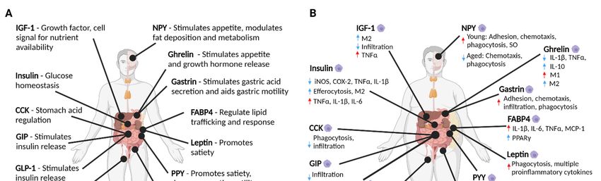
Cancers 2021, 13, 4661 2 of 28
Table 1. Summary of the metabolic hormones reviewed.
Primary Metabolic
Hormone Trigger Origins Metabolic Target Receptor
Functions
Stimulates release of digestive
CCK Fatty acids, proteins Small intestine I-cells Pancreas CCK1R & CCK2R
enzymes and insulin
Adipocytes Adipocytes Absorption of fatty acids
FABP4 Lipolysis PPARγ
Macrophages Macrophages M2 macrophage polarisation
Food intake Stomach G-cells
Gastrin Gastrin releasing Duodenum Stomach CCK1R & CCK2R Stomach acid regulation
peptide Pancreas
Stomach
Regulates food intake, energy
Intestine Brain
Ghrelin Food intake GHSR expenditure, glucose
Brain Adipose tissue
homeostasis, adiposity
Macrophages
K-cells in the
GIP Glucose, fatty acids duodenum and Pancreatic β-cells GIPR Stimulates insulin release
jejunum
L-cells of the small Pancreatic β-cells Stimulates insulin release
GLP-1 Hexose, fats GLP1R
intestine Brain Induces satiety
Muscle
INSR Glucose uptake
Insulin Hyperglycaemia Pancreatic β-cells Liver
IGF1R Inhibition of gluconeogenesis
Adipose
Liver Bones
Growth Hormone IGF1R Stimulates bone and tissue
IGF-1 Macrophages Smooth muscle
(GH) INSR growth
Adipocytes Neurons
Leptin Food intake Adipocytes Brain OBR Regulation of food intake
Food intake Central and
NPY Receptors
NPY (High levels of Central nervous system peripheral nervous Regulation of food intake
(GPCRs)
dietary fat and sugar) systems
Amino acids L-cells of the ileum and Central and
PYY Receptors Gastric emptying
PYY Short-chain fatty colon peripheral nervous
(GPCRs) Gut motility
acids systems
Gonads, adipose tissue, ERα Primary female sex hormone
Luteinizing hormone
Estrogen bone, skin, liver & Systemic Erβ Fat distribution
(LH)
brain GPER Metabolism
Primary male sex hormone
Luteinizing hormone Leydig cells of the testis
Testosterone Cancers 2021, 13, x FOR PEER REVIEW Systemic AR Fat 3distribution
of 30
(LH) & adrenal glands
Muscle mass
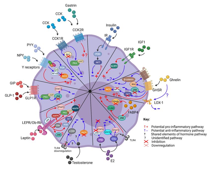
Figure 1. Schematic
Figure 1. summarising (A) the(A)
Schematic summarising classical functions
the classical of metabolic
functions of metabolichormones
hormones inina ahealthy
healthy individual,
individual, and
and (B) (B) how these
how
these hormones can modulate macrophage inflammatory responses when potentially dysregulated in
hormones can modulate macrophage inflammatory responses when potentially dysregulated in an obese state. Red arrows an obese state. Red
arrows indicate proinflammatory actions, blue arrows indicate anti-inflammatory actions. This figure was created with
indicate proinflammatory
Biorender.com. actions, blue arrows indicate anti-inflammatory actions. This figure was created with Biorender.com.
This review highlights key classical metabolic hormones (Table 1 and Figure 1A) that
become dysregulated in obesity and cancer and discusses their emerging roles in
macrophage inflammatory responses (Figure 1B)—which have the potential to influence
cancer progression (Figure 2). In this review, we have also included the sex hormones
estrogen and testosterone due to their important direct and indirect roles in metabolism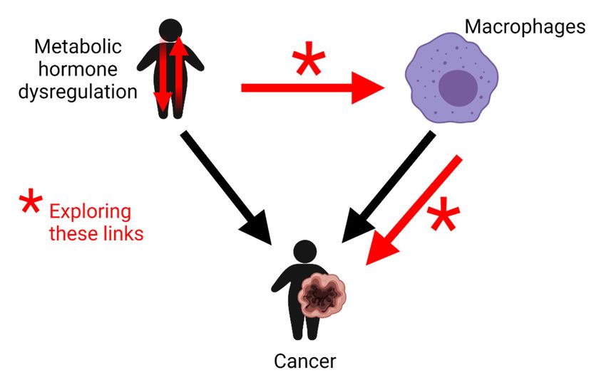
Figure 1. Schematic summarising (A) the classical functions of metabolic hormones in a healthy individual, and (B) how
    these hormones can modulate macrophage inflammatory responses when potentially dysregulated in an obese state. Red
    arrows indicate proinflammatory actions, blue arrows indicate anti-inflammatory actions. This figure was created with 3 of 28
Cancers 2021, 13, 4661
    Biorender.com.

                                    This review highlights key classical metabolic hormones (Table 1 and Figure 1A) that
                               become   dysregulated
                                     This                  in obesity
                                           review highlights              and metabolic
                                                                key classical   cancer and    discusses
                                                                                          hormones        their
                                                                                                      (Table     emerging
                                                                                                             1 and  Figure 1A)roles
                                                                                                                                 that in
                                                                                                                                      be-
                               macrophage     inflammatory
                                come dysregulated               responses
                                                       in obesity            (Figure
                                                                    and cancer       1B)—which
                                                                                 and discusses     have
                                                                                                their     the potential
                                                                                                      emerging    roles into influence
                                                                                                                           macrophage
                               cancer progression
                                inflammatory          (Figure(Figure
                                                 responses       2). In this  review, have
                                                                         1B)—which    we have    also included
                                                                                             the potential         the sex cancer
                                                                                                             to influence   hormonespro-
                               estrogen
                                gressionand    testosterone
                                          (Figure   2). In thisdue   to their
                                                                 review,  we important    direct and
                                                                               have also included   theindirect roles in estrogen
                                                                                                         sex hormones     metabolismand
                               through  their due
                                testosterone   influence
                                                    to theironimportant
                                                               body fat distribution    and effect
                                                                           direct and indirect       onincancer
                                                                                                 roles           sexual through
                                                                                                           metabolism    dimorphismtheir
                               (Table 1 and
                                influence   onFigure
                                                body 1A).
                                                       fat distribution and effect on cancer sexual dimorphism (Table 1 and
                                Figure 1A).

                                 Figure 2. Schematic illustrating the links this review will explore (red arrows) between potential
    Figure 2. Schematic illustrating the links this review will explore (red arrows) between potential metabolic hormone
    dysregulation, macrophagemetabolic      hormone
                                   inflammatory       dysregulation,
                                                   responses         macrophage
                                                              and cancer           inflammatory
                                                                             development.    Thisresponses  and cancer
                                                                                                   figure was    createddevelopment.
                                                                                                                          with
    Biorender.com.               This figure was created with Biorender.com.

                                1.2. The Role of Macrophages in Cancer Development
                                      Macrophages are phagocytic cells of the hematopoietic lineage that play a central role
                                in the innate immune response and also have established roles in metabolic diseases [3]
                                and cancer initiation, malignancy, and metastasis [4,5]. Macrophages pervade almost
                                every organ system and can exhibit a wide range of phenotypes depending on their
                                particular microenvironment. During the last two decades, the conceptual framework for
                                macrophage activation has evolved. Initially, macrophages were polarized into classically
                                (M1) or alternatively (M2) activated cells [6] representing two polar inflammatory or
                                anti-inflammatory extremes, respectively. The M1 vs. M2 model has been useful in
                                describing immune responses during acute infections, allergies, asthma, and obesity [7].
                                However, observations from macrophages involved in chronic inflammation such as type
                                2 diabetes and cancer strongly suggest a much broader, context-dependent transcriptional
                                repertoire in which macrophages adopt a spectrum of phenotypes that go beyond the rigid
                                M1/M2 nomenclature [6,8]. Recent transcriptomics studies have now made considerable
                                contributions to a better understanding of immune cell function and regulation; there are
                                now at least nine distinct macrophage activation programs recognised [8], and within these
                                programs, there are multiple unique gene expression signatures that enable macrophages
                                to exist in a spectrum of activation states [8]. Nevertheless, to better compare the findings
                                of the literature referenced within this review and in the absence of a framework to more
                                accurately reflect the new macrophage phenotype spectrum, we will continue to use the
                                M1/M2 convention as appropriate.
                                      Tissue-resident macrophages may originate from yolk sac-derived erythromyeloid
                                progenitors or circulating monocytes from bone marrow resident haematopoietic stem
                                cells [9]. Tissue-resident macrophages display specific characteristics local to the tissues
                                they reside in [10], which influence their function and hence their effect on their surround-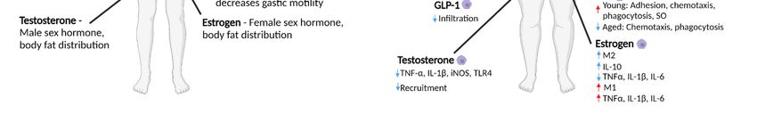
Cancers 2021, 13, 4661                                                                                           4 of 28

                         ing tissues. Macrophages can be highly influential on tumour development through the
                         induction of inflammation, stimulation of neoangiogenesis, immune suppression or in-
                         duction of metastasis. Macrophages that populate a tumour’s surrounding environment
                         (the tumour microenvironment (TME)) are referred to as tumour-associated macrophages
                         (TAMs); we direct the reader to the very recent review by Cendrowicz et al. [11] for a
                         detailed description of the contribution of TAMs to the formation and development of
                         tumours. Both clinical and experimental evidence has found that a high density of TAMs
                         within a TME is strongly correlated with poor prognosis and reduced survival in a number
                         of cancer types [12]. While TAMs have been found to display a wide spectrum of pheno-
                         types, the majority are reported to have M2-like immunosuppressive properties due to the
                         higher expression of IL-10 and TGF-β in TMEs, which is thought to help tumours evade
                         cancer cell elimination by the immune system [12]. In contrast, pro-inflammatory M1-like
                         TAMs are thought to establish a tumour-inhibiting phenotype by allowing tumoricidal
                         activity to resume through the reversal of immunosuppressive mechanisms. Macrophages
                         express hormone receptors [13] and because of the systemic nature of these metabolic
                         hormones and the significant role that macrophages play in tumour development, the
                         potential of dysregulated hormone levels to modulate tumour microenvironments and
                         hence macrophage inflammatory responses may be significant.

                         2. Metabolic Hormones Play Roles in Modulating Macrophage Inflammatory
                         Responses
                         2.1. Cholecystokinin (CCK)
                         2.1.1. Origin and Function
                              The peptide hormone Cholecystokinin (CCK) is well-established as being a metabolic
                         hormone secreted from I cells of the small intestine when high levels of dietary fatty
                         acids or proteins are detected [14]. CCK regulates digestion by stimulating the release of
                         digestive enzymes and insulin from the pancreas and mediates satiety by binding to CCK
                         receptors in the vagal afferent neurons of the gut–brain axis [15]. However, CCK is also
                         a neurotransmitter, growth factor and anti-inflammatory cytokine expressed as multiple
                         different bioactive peptides by neurons, endocrine, and epithelial cells (recently reviewed
                         in [16]). The effects of CCK are mediated through two types of receptors; CCK1R and
                         CCK2R [17,18]. CCK1R is mainly located in peripheral tissues and shows higher selectivity
                         for CCK than CCK2R [19].

                         2.1.2. CCK and Cancer Association
                              Diets rich in long-chain saturated fatty acids lead to the overexpression of CCK which
                         alongside obesity, is a significant risk factor for pancreatic cancer [20,21] and elevated CCK
                         levels are also associated with the development of pancreatic metastases in mice [22]. CCK
                         receptors can be over-expressed in a range of human cancers including stomach, pancreas,
                         colon, rectum, oesophagus, lung, and liver [23,24].

                         2.1.3. CCK Modulates Macrophage Inflammatory Responses
                              Studies to date indicate an anti-inflammatory role of CCK in several diseases and
                         animal models of disease [25–32] demonstrated by ablation of CCK or CCKR, or treat-
                         ment with CCKR antagonists which exert pro-inflammatory effects [27,30,33]. Both CCK
                         receptors are expressed in macrophages [34,35] although CCK-1R is the predominant me-
                         diator of CCK’s immunomodulatory effects [36]. The CCK-8 isoform negatively modulates
                         macrophage functions such as phagocytosis and tissue infiltration [26,27,37] and inhibits
                         inflammatory response through downregulation of CD68, ICAM-1, TGF-β, and TNFα gene
                         expression and inhibition of NF-κB activity [30]. In peritoneal and pulmonary interstitial
                         macrophages, CCK-8 treatment blocks LPS-induced IL-1β production, reduces nitric oxide
                         production and attenuates iNOS and TNFα mRNA expression [29,36,38]. These studies
                         indicate the mechanism through which CCK exerts anti-inflammatory effects is through
                         modulation of p38 and NF-κB activity via inhibition of PKC and activation of the cAMP-
Cancers 2021, 13, 4661 5 of 28
PKA pathway [29,30,36–38]. It is therefore possible that the pancreatic tumour growth
associated with CCK expression can be attributed to the capacity of CCK to promote
macrophages to adopt a pro-tumour, M2 phenotype.
2.2. FABP4
2.2.1. Origin and Function
Fatty acid-binding proteins (FABP) are at least a nine-member family of 14–15 kDa pro-
teins that facilitate the absorption and utilisation of water-insoluble dietary long-chain fatty
acids [39]. The different family members are uniquely expressed in distinct tissues involved
in active lipid metabolism, including adipocyte FABP (known as FABP4). FABP4 is highly
expressed by mature adipocytes [40,41] and macrophages [42,43] and the major regulator
of FABP4 signalling is peroxisome proliferator-activated receptor (PPAR) γ [43,44].
2.2.2. FABP4 and Cancer Association
Evidence suggests that FABP4 levels impact diseases ranging from metabolic syn-
drome, type two diabetes, atherosclerosis [45–48] and various forms of cancer including
breast, liver, colon, and ovarian [49–52]. FABP4 has been shown to promote tumour pro-
gression via enhancement of new blood vessel formation and tumour growth mediated
by its effects on adipocytes and tumour cells [53,54]. However, in contrast to these find-
ings, decreased levels of FABP4 have also been associated with hepatocellular carcinoma
tumours and FABP4 was shown to suppress proliferation and invasion of hepatocellular
carcinoma cells [50], suggesting that the influence of FABP4 on cancer development and
progression may depend on the cancer type and microenvironment situation.
2.2.3. FABP4 Modulates Macrophage Inflammatory Responses
FAPB4 is expressed in macrophages [42,43], with substantial crosstalk between macro-
phages and adipocytes occurring upon inflammatory activation [55]. The regulation of
FABP4 signalling by PPARγ was demonstrated by Garin-Shkolnik et al. through FABP4 trig-
gering proteasomal degradation of PPARγ which inhibited PPARγ-related functions [56]
including its role in inhibiting the expression of inflammatory cytokines and directing the
differentiation of immune cells towards anti-inflammatory phenotypes [57,58]. As PPARγ
interferes with NF-κB, AP-1 and STAT transcriptional activity [58,59], it inhibits the upregu-
lation of pro-inflammatory genes, such as IL-1β, IL-6, and TNFα. The repression of PPARγ
is therefore associated with the initiation of inflammatory pathways and impaired alterna-
tive M2 macrophage activation [60]. Indeed, FABP4-deficient macrophages are seen to have
reduced basal and stimulated expression of pro-inflammatory cytokines including TNFα,
IL-1β, MCP-1, and IL-6 due to decreased/NF-κB activity and deficient activation-induced
expression of iNOS [42,61,62]. Additionally, FABP4 is shown to exacerbate LPS-induced
inflammation by forming a positive feedback loop with the JNK signalling cascade [63],
and subsequently influences the production of inflammatory cytokines. However, in
contrast, some studies have suggested that FABP4 may enhance the activities of PPARγ
during the differentiation of macrophages, providing a positive feedback loop between
the two proteins [64]. These-conflicting findings have been suggested to arise from FABP4
exerting a concentration dependent effect on PPARγ regulation. Other regulators that have
been seen to induce FABP4 expression in macrophages include rapamycin [65], which can
increase the expression of genes involved in cholesterol transport and triglyceride synthe-
sis. Notably, intracellular FABP4 has been observed to enhance pro-tumour macrophage
function. FABP4 is highly expressed in a small subset of TAMs of the CD11b + F4/80 +
MHCII − Ly6C − CD11c − phenotype [49]. FABP4-positive TAMs accumulate in late-stage
mammary tumours, promoting their growth through the enhancing effect FABP4 has on
NF-κB expression, thereby increasing the secretion of pro-tumour IL-6 signalling. Indeed,
genetic ablation or chemical inhibition of FABP4 in TAMs has been shown to suppress
mammary tumour growth [49].Cancers 2021, 13, 4661 6 of 28
2.3. Gastrin
2.3.1. Origin and Function
Gastrin is a stomach acid secretion-regulating peptide hormone produced by en-
docrine G-/gastrin cells in the pyloric antrum of the stomach, duodenum, and pancreas.
The gastrin gene in humans encodes a 101 amino-acid precursor peptide, which is sub-
sequently cleaved to generate progastrin before being cleaved once again to form gastrin
itself—the dominant forms of which in human plasma are gastrin-34 and gastrin-17 [66].
Once synthesised, gastrin peptides are stored in the basal part of the G-cells until they
are released either in response to food intake or induced by the neurotransmitter gastrin-
releasing peptide (GRP) acting on basolateral receptors in the G cells. Once released, gastrin
modulates its effects through the CCK receptors; CCK1R and CCK2R, the latter of which
has a higher affinity for gastrin.
2.3.2. Gastrin and Cancer Association
Gastrin has been observed to directly induce the expression of pro-inflammatory
molecules such as IL-8, CINC-1 and the enzyme COX-2 in gastric epithelial cells [67,68].
COX enzymes are known to catalyse the synthesis of prostaglandins, a pathway shown
to play an important role in cancers. Furthermore, COX-2 inhibition has been shown to
suppress cell proliferation and induce apoptosis in various gastrointestinal cancer cell lines
in vitro [69]. The pro-inflammatory effect of gastrin has also been hypothesised to play a
role in cancer initiation through its association with H. pylori which is known to induce
gastric cancer development and progression [70]. Gastrin-releasing peptide (GRP) and
its receptor (GRPR) have also been linked to cancerous malignancies [71] and GRPR has
been shown to induce the release of IL-8 and vascular growth factor in the case in human
prostate cancer cell lines [72].
2.3.3. Gastrin Modulates Macrophage Inflammatory Responses
Both CCK1R and CCK2R have been identified in an array of human leukocyte
cell types, including lamina propria macrophages [73], peripheral blood mononucleo-
cytes [34,74], circulating polymorphonuclear leukocytes (PMNs) [75] and also PMNs found
within human malignant colorectal tumours [76]. One of the first links between gastrin
and macrophages was provided by Okahata et al. in 1985 when they demonstrated that
gastrin treatment resulted in increased immunoreactivity in pure populations of human
PMNs [77] and further in vitro studies have shown that murine macrophages and human
PMNs treated with gastrin induces chemotaxis and increases adherence and phagocyto-
sis [78,79]. Alvarez et al. also noted that gastrin treatment increased leukocyte rolling
and adhesion, decreased rolling velocity and increased leukocyte infiltration into the in-
terstitium and as CCK2R, but not CCK1R antagonists abrogated these effects it is thought
that CCK2R mediates gastrin’s inflammatory effects [80]. The gastrin receptor CCK2R has
been further implicated in the pro-inflammatory response due to its promoter containing
an IFN-γ regulatory site [81]. In addition to these direct effects on macrophages, gastrin
may modulate their function by acting on the surrounding tissue. Gastrin, mediated by
CCK2R, is reported to induce the release of IL-8 from human endothelial cells and increase
the synthesis of the adhesion molecules VCAM-1 and P-selectin, resulting in increased
adhesivity for human mononuclear leukocytes [82,83].
2.4. Ghrelin
2.4.1. Origin and Function
Ghrelin is an orexigenic hormone primarily produced by enteroendocrine cells in the
stomach. Although its highest levels are found in the stomach and intestine [84], it is also
expressed in the brain [85] and by macrophages [86]. Ghrelin has been demonstrated to
regulate food intake, energy expenditure, glucose homeostasis, adiposity, body weight,
inflammation, and growth hormone (GH) secretion [87]. There are two isoforms of ghre-
lin as a result of post-translational modifications; desacyl-ghrelin and acyl-ghrelin. TheCancers 2021, 13, 4661 7 of 28
desacetylated form of ghrelin, desacyl-ghrelin, can undergo octanoylation performed by
ghrelin O-acyltransferase (GOAT) to become acyl-ghrelin, which is also referred to as the
active although least abundant form of the hormone. Ghrelin’s function is mediated by
the Growth Hormone Secretagogue Receptors (GHSR). The acyl-ghrelin form can bind to
GHSR1α and GHSR1β forms [88–90]. These receptors are highly expressed in the brain,
poorly expressed in adipose tissue, and are localized within several immune cell types
including monocytes and macrophages [86,90–92]. Ghrelin is down-regulated in obese
patients and up-regulated under conditions of negative energy balance in humans and
mice [93,94].
2.4.2. Ghrelin and Cancer Association
Ghrelin regulates several processes related to cancer progression recently reviewed
in [95] including cellular proliferation, inflammation, and energy homeostasis. However,
the precise relationship between the ghrelin axis and cancer development remains unclear
and controversial due to conflicting results observed between in vitro [96] studies involving
a range of tumour cells lines and clinical studies [97]. Several recent attempts have been
made recently to standardise these findings [95,98] leading to the hypothesis that the
systemic nature of ghrelin signalling may contribute to confounding local factors that make
delineating ghrelin’s direct role in cancer progression complex. Furthermore, experimental
variations such as different cell lines and dosages used, add additional difficulties to
interpreting the effects of ghrelin from these in vitro studies. In vivo animal and human
studies have also revealed that ghrelin expression is often downregulated in cancer tissues
and blood plasma while having no clear correlation with tumour development [99].
2.4.3. Ghrelin Modulates Macrophage Inflammatory Responses
In macrophages the effects of ghrelin are complex; exogenous ghrelin treatment in
the RAW264.7 macrophage cell line has been shown to inhibit LPS-induced production of
pro-inflammatory cytokines IL-1β, TNFα and promote the release of the anti-inflammatory
marker IL-10 [86]. However, in contrast, acyl ghrelin treatment in RAW264.7 macrophages
promoted macrophage polarization to M1 under an inflammatory state by enhancing the
effect of LPS and weakening the effect of IL-4 [100]. Although both studies used the same
in vitro model, the doses of LPS used and the incubation time differed between the two
studies (1000 ng/mL vs. 10 ng/mL) which may explain the contradictory results. In vivo
work [101] has demonstrated that GHSR knockout, in aged mice, promotes macrophage
phenotypic shift towards an anti-inflammatory M2 state. Yet, in contradiction to these
results, a separate study showed GHSR knockout in mice fed a high-fat diet, to have a
beneficial effect on adipose tissue [100]. The mice had reduced adipose tissue inflam-
mation, decreased macrophage infiltration, and improved insulin sensitivity. Moreover,
macrophages were polarized into an M2 type. Several additional studies show ghrelin to be
anti-inflammatory [102–105], thought to be the result of JNK inhibition and activation of the
Wnt/β-catenin pathway [92,106,107]. Recently [108,109], the effects of ghrelin, and ghrelin
analogue hexarelin, on macrophages induced with oxidised-LDL (ox-LDL) and observed
inhibition of LOX1 gene expression as a result of reduced ox-LDL uptake and decreased
TNFα and IL-6 release. Similar protective effects of ghrelin have been demonstrated in
alveolar macrophages induced with LPS where ghrelin treatment was able to reduce the
level of IL-1β, TNFα, and IL-6, as well as decreasing iNOS and Akt activity. LOX-1-NF-κB
and NF-κB/iNOS pathways or Akt signalling are the pathways suggested to mediate these
anti-inflammatory effects [108–110].
2.5. Incretins
2.5.1. Origin and Function
The incretin hormones; glucose-dependent insulinotropic polypeptide (GIP, also
known as gastric inhibitory polypeptide) and glucagon-like polypeptide-1 (GLP-1) are
secreted by the small bowel in response to nutrients and stimulate the release of insulinCancers 2021, 13, 4661 8 of 28
from pancreatic β cells. Both GIP and GLP-1 are rapidly degraded by dipeptidyl peptidase
4. GIP is a 42-amino acid peptide secreted by enteroendocrine K cells of the small intestine.
Its action is mediated by gastric inhibitory peptide receptor (GIPR), which is expressed
mainly in the pancreas but can also be found in other tissues and immune cells [111,112].
GLP-1 is expressed in L cells of the small and large intestines [113] and is derived from
the proglucagon gene [114]. GLP-1 action is mediated by the glucagon-like polypeptide-1
receptor GLP1R [115].
2.5.2. Incretins and Cancer Association
GLP1R is reportedly overexpressed in many tumour cell types [116]. However, the
role of GLP1R overexpression in neoplasia remains unknown Incretin mimetic drugs such
as exenatide and liraglutide are powerful and widely used treatments for type II dia-
betes [117]. Like GLP-1 and GIP, they bind to GLP1R on the pancreatic β cells to stimulate
insulin secretion. Exenatide has been shown to promote apoptosis in SKOV-3 and CAOV-3
human ovarian cancer cell lines and reduce the production of the pro-metastatic adhesion
molecules ICAM-1 and VCAM-1 by TNFα stimulated vascular endothelial cells [118].
Similar observations have been made in studies using breast [119], colon [120], and ovar-
ian [121] cancer cell lines, likely due to the inhibition of the PI3K/Akt signaling pathway
by exenatide. However, whilst concerns have been raised over the safety of incretin-based
treatments for those living with both cancer and diabetes meta-analysis of clinical data has
not revealed any association between the development of lung [122], gastrointestinal [123]
or pancreatic [124] cancer and the prescriptions of the incretin analogues.
2.5.3. Incretins Modulate Macrophage Inflammatory Responses
Incretins have demonstrated overall metabolic benefits and anti-inflammatory effects
in macrophages in the context of inflammation-mediated obesity [125]. Treatment with
GLP1, or GLP1 agonist, improves insulin sensitivity, glucose, and insulin tolerance, nor-
malises glycemia, and reduces fat mass, adipocyte size and macrophage number in adipose
tissue as a result of macrophage infiltration inhibition [126–128]. In vitro studies on several
different models of macrophage cells (peritoneal macrophages, LPS treated RAW264.7 cells
and adipose tissue macrophages (ATM)) have consistently shown the anti-inflammatory
effect of GLP1 and its agonists. These studies have shown a decrease in pro-inflammatory
markers (iNOS, IL-1β, IL-6, TNFα and MCP1, MMP2, MMP9, ROS) [129–131]) while in-
creasing anti-inflammatory markers (IL-10, mannose receptor-1 (MRC-1), macrophage
galectin-1 (MGL-1), arginine-1 (ARG-1)) [128–130,132], PGE2 and COX2 mRNA and COX2
protein level [129]. Studies have demonstrated these effects to be mediated through JNK
inhibition, STAT3 activation [132,133], and also the cAMP/PKA/NF-κB signalling path-
way [126,128–130,132]. In vitro, GLP1 has been shown to decrease cell migration through
its reduction of MCP-1 and MMP-9 activity which may be mediated by the inhibition of
Nf-κB, JNK, ERK, and p38 in LPS activated macrophages [134,135]. GLP1 and GIP inhibitor
DPP4 (also known as CD26) enhances the expression of TNFα and IL-6 mRNA and protein
in THP-1 cells [136] while GIP prevents proinflammatory macrophage activation leading
to reduced LPS-induced IL-6 secretion [134].
2.6. Insulin
2.6.1. Origin and Function
Insulin is an anabolic hormone secreted by the β cells of the pancreas. Insulin reg-
ulates glucose homeostasis, lipid metabolism [137] and cell growth [138]. The effects of
insulin are mediated through the insulin receptor (INSR) [139,140] and the insulin-like
growth factor 1 receptor (IGF-1R) [141]. Upon binding to its receptor, insulin activates two
major downstream pathways, the PI3K pathway which mediates its metabolic effects [142]
including the translocation of GLUT4 in metabolic tissues such as muscle and adipose, and
the MAPK kinase pathway [143] which regulates mitogenesis and growth. The insulin
receptor has also been shown to act directly as a transcription factor [144], which mayCancers 2021, 13, 4661 9 of 28
illuminate previously unrecognised mechanisms of the long-term effects of insulin normal
physiology and disease.
2.6.2. Insulin and Cancer Association
Metabolic diseases such as obesity and type 2 diabetes that are characterised by hy-
perinsulinemia are associated with the development of several types of cancer including
colon, breast, endometrium, oesophagus, kidney, liver, and pancreas [145–148]. Insulin
can promote cancer through its mitogenic actions [149] and the INSR is often overex-
pressed in tumour cells [150–152]. More recently, a new mechanism has been demonstrated
showing that hyperinsulinemia promotes epithelial tumourigenesis by abrogating cell
competition [153].
2.6.3. Insulin Modulates Macrophage Inflammatory Responses
Macrophages express insulin receptors [154] and intracellular signalling machinery.
Existing reports demonstrate diverse effects that include promoting pro-inflammatory
responses through increasing phagocytosis [155], and TNFα production [156]. However,
other studies report an anti-inflammatory response involving decreased apoptosis [157]
and decreased pro-inflammatory cytokine production [158,159]. Discrepancies may be due
to inconsistencies in the macrophage cells used (mouse [160] or human cell lines [156,157];
mouse tissue macrophages [161] or human peripheral cells [162]), and lack of consistency of
insulin concentration and duration used. We would direct the reader to the very recent re-
view [163] detailing insulin’s inflammatory and anti-inflammatory effects on macrophages.
2.7. Insulin-Like Growth Factor-1
2.7.1. Origin and Function
Insulin-Like growth factor-1 (IGF-1) is a polypeptide hormone mainly produced in
the liver where growth hormone (GH) binds to its receptor to drive most of the IGF-
1 synthesis [164]. IGF1R is also expressed in adipocytes [165] endothelial cells and
macrophages [166,167]. Its actions can be endocrine, paracrine, or autocrine and are
mediated by the tyrosine kinase receptor IGF-1R. IGF-1 is involved in growth, regulation
of metabolism, and inflammation [168]. Structural similarities with insulin enable IGF-1 to
bind to both IGF-1 and insulin receptors [169]. IGF-1 signal transduction requires insulin
receptor substrates (IRS) [170] and involves the downstream phosphoinositide-3 kinase
(PI3-K) and MAPK activation pathways [171].
2.7.2. IGF-1 and Cancer Association
Above normal levels of circulating IGF-1 are associated with an elevated risk of
developing several primary cancer types including colorectal [172] and breast [173]. IGF-1
has been demonstrated to enable tumour growth by preventing apoptosis through the
induction of the PI3K and MAPK signalling cascades [174]. Furthermore, IGF-1 has been
shown to increase the migratory and proliferative capabilities of IGF-1R-overexpressing
HCT116 colon cancer cells, leading to metastases in mice transfected with these cells [175].
Finally, IGF-1R is known to be overexpressed by cancer cells contributing further to the
malignant phenotype [176].
2.7.3. IGF-1 Modulates Macrophage Inflammatory Responses
Macrophages polarized into an anti-inflammatory M2 subtype express high levels
of IGF-1 [166,177,178]. A recent study assessing IGF-1 implication in the development of
obesity [166] using mice with myeloid cell-specific ablation of IGF-1R and challenged with a
high-fat diet showed increased macrophage infiltration in adipose tissue leading to insulin
resistance and also suggested an anti-inflammatory role for IGF-1 on macrophages. In
contrast, however, other studies have reported IGF-1 to have a pro-inflammatory effect on
macrophages; IGF-1 treatment on murine macrophages has been shown to increase produc-
tion and expression of TNFα mediated by IGF-1R and tyrosine kinase factor activation [179],Cancers 2021, 13, 4661 10 of 28
and to stimulate LDL uptake and cholesterol esterification [180]. Ablation of IGF-1R in
macrophages was also seen to significantly decrease NLRP3 inflammasome-dependent
caspase-1 and IL-1β activation when induced by ageing-relevant damage-associated molec-
ular patterns (DAMPs) [181].
2.8. Leptin
2.8.1. Origin and Function
Leptin is a pleiotropic peptide hormone encoded by the ob gene and secreted by
adipose tissue in proportion to its abundance [182]. It was first reported to control food
intake and body weight through anorexigenic effects in the brain [183,184]. Leptin receptors
(known as LEPR or OBR) are class I cytokine receptors expressed in many cell types,
including macrophages [185] where Ob-Rb is the most well-characterized isoform [186,187].
2.8.2. Leptin and Cancer Association
The role of the leptin-Ob-Rb signalling axis in cancer development is well documented
with both leptin and OBR becoming overexpressed in several cancer types including head
and neck, pancreatic, and breast cancer [188]. Leptin signalling has been implicated as
a driver of angiogenesis in colorectal cancer [189] and glioblastoma [190], resulting in
metastasis and tumour growth. Leptin increases the expression of inflammatory cytokines
such as TNFα and IL-1β which contributes to tumour-associated inflammation and subse-
quently, immunosuppression of tumouricidal CD8+ cytotoxic T cells in breast cancer [191].
Furthermore, leptin is known to have direct effects on ovarian cancer cell proliferation
and growth by activating the PI3K/Akt and MEK/ERK1/2 signalling pathways [192].
Obesity is also associated with elevated serum leptin levels, with many obese individuals
experiencing ‘leptin resistance’ in which leptin is no longer able to effectively regulate food
intake. The exact reason for the upregulation of leptin during obesity is unknown, but it
has been speculated that leptin may be a critical link between obesity and cancer [193,194].
2.8.3. Leptin Modulates Macrophage Inflammatory Responses
Leptin is an adipokine, a member of the superfamily of cytokines [195] and is impli-
cated in inflammation, infection, and immune responses [182]. Genetic abnormalities in
leptin or leptin receptors are reported to impair macrophage phagocytosis and promote pro-
inflammatory cytokines production, while leptin treatment increases both [196]. Similarly,
in vitro studies on the murine macrophage J774A.1 cell line, human monocyte-enriched
mononuclear cells, LPS stimulated Kupffer cells and human adipose macrophages have
shown that leptin can stimulate the phagocytotic activity of macrophages [197] and the
proliferation and activation of monocytes [198]. In addition, leptin may stimulate produc-
tion of proinflammatory cytokines TNFα, resistin, IL-6, and IL-1β, IL-1Ra, IL-10, MCP-1,
and MIP-1α and enhance CC-chemokine ligand expression [199–201]. These effects might
be mediated through activation of a JAK2-STAT3 pathway [199] and may also activate
ERK1/2, P8 MAPK, JNK, AMPK, PKC and PI3K/Akt pathways [185,202–205].
2.9. Neuropeptide Y (NPY) and Peptide YY (PYY)
2.9.1. Origin and Function
Both NPY and PYY belong to a family of neuropeptides bearing a close resemblance to
each other, consisting of 36-amino acids with a unique hairpin turn called the PP-fold [206].
NPY is highly abundant and is found in all levels of the gut-brain axis as well as being
highly expressed in the central nervous system where it is widely known for its activity as
a regulator of food intake and energy balance [207]. PYY is almost exclusively associated
with the digestive system and is predominantly expressed in L cells in the ileal and
colonic mucosa and released into the bloodstream post-prandially in proportion to calorie
intake [207–210]. NPY and PYY peptides can also be truncated, yielding the fragments
NPY(3-36) and PYY(3-36) [211]. In humans, NPY and PYY’s functions are mediated by
diverse G-protein coupled Y receptor subtypes, of which seven have been noted, but onlyCancers 2021, 13, 4661 11 of 28
four are widely functional (Y1, Y2, Y3 and Y4). NPY(1-36) and PYY(1-36) are thought to
bind to all the receptors with an equal affinity, whilst NPY(3-36) and PYY(3-36) exhibit the
highest affinity for Y2 [212–214].
2.9.2. NPY and PYY and Cancer Association
Investigation of PYY and NPY, have collectively revealed that they are implicated in a
variety of inflammatory disorders, such as autoimmune diseases, asthma, atherosclerosis,
and cancer [215–217]. Y receptors have recently attracted attention due to their overex-
pression in various human cancers, including breast carcinomas and neuroblastomas,
creating interest in their use as a possible target for cancer imaging and therapy [218].
The Y receptors mediate tumour development through their direct effect on cancer and
endothelial cells promoting tumour cell proliferation, survival, and migration, as well as
angiogenesis) [219].
2.9.3. NPY and PYY Modulate Macrophage Inflammatory Responses
In macrophages, neuropeptides have been found to exert varying effects depending
on the age of the subject. In one of the first studies examining their role in macrophage
function, both NPY and PYY were found to increase adhesion, chemotaxis, and phagocyto-
sis in murine peritoneal macrophages, as well as increasing the production of superoxide
anions in young adult mice [220]. The authors noted that this effect was produced through
the stimulation of PKC due to a significant increase in its activation following NPY and
PYY treatment. However, in more aged mice, this effect was potentiated, with chemotaxis
and phagocytosis being decreased. These changes have been hypothesised to be dependent
on the activity of dipeptidyl peptidase 4, an enzyme that terminates the activity of neu-
ropeptides on the Y1 receptor subtype and whose activity is seen to change with age [221].
This age-dependent impact in modulating the immune response was also found to be true
concerning PYY and NPY acting via Y1 receptors to potentiate nitric oxide production in
rat peritoneal macrophages, with production being suppressed in older rat cells [222]. Y
receptors are known to be widely expressed in immune cells, particularly Y1, which has
been found in almost every type of immune cell [223].
The expression of Y receptors is also significantly upregulated after antigen or inflam-
matory stimulation [224–226]. Studies have also demonstrated the ability of neuropeptides
to modulate macrophage cytokine secretion. However, contradictory results have been
found. Y1 ablation in macrophages has been seen to lead to an increased pro-inflammatory
phenotype displaying increased inflammatory response and exacerbated secretion of MCP-
1 and TNFα, and a similar response was seen in macrophages isolated from double NPY and
PYY knockout mice, suggesting an anti-inflammatory role [227]. Additionally, NPY was
shown to decrease the production of TNFα and IL-1β following LPS treatment [228,229]
and increase that of TGFβ1 in RAW264.7 cells [230]. In contrast, other studies have reported
NPY to increase the production of pro-inflammatory mediators, with NPY being found
to significantly increase the expression of TNFα, C-reactive protein, MCP-1 and reactive
oxygen species in RAW264.7 macrophages mediated by the Y1 receptor [231]. NPY has also
been shown to stimulate IL-1β secretion in aged animal macrophages [232]. Furthermore,
in whole blood cells from healthy subjects, NPY upregulated IL-6, IL-1β and TNFα pro-
duction [233]. Some suggestions for the observed duality have been differences in species,
cell type and cell environment. Additionally, the activation of different Y receptor types is
seen to mediate different effects, and there is evidence that along with Y1, Y4 and 5 may
also play a role in cytokine modulation [227]. A relatively recent study by Cheng et al.
found sympathetic stimulation of prostate cancer cells in vitro led to the release of NPY,
which in turn was seen to promote myeloid cell trafficking and increased IL6 synthesis in
TAMs, promoting tumorigenesis [234]. However, the connections between neuropeptides,
immune regulation and cancerous disease have not yet been fully explored, and indeed
it may be found that neuropeptides have divergent effects on immune cells in cancer
development as observed in their general effects on macrophage function.Cancers 2021, 13, 4661 12 of 28
2.10. Estrogen
2.10.1. Origin and Function
Estrogens are a class of steroid hormones and are the main female sex hormones, but
also play an essential role in both male and female reproductive and non-reproductive pro-
cesses [235,236]. Estrogen is predominantly synthesised in the gonads in pre-menopausal
women; however, non-gonadal sites such as adipose tissue, bone, skin, and the liver and
brain can also produce a small but significant amount of estrogen. In humans, estradiol
(E2 or 17β-estradiol) is the most prevalent and active form of estrogen. Estrogens play an
important role in body weight, fat distribution, energy expenditure and metabolism [237].
The effects of estrogen are mediated by two intracellular estrogen receptors (ERs), ERα
and ERβ [238] and by a plasma membrane protein, G protein-coupled estrogen receptor
(GPER) [239]. ERα is predominantly expressed in the uterus, ovaries, and breasts, while
expression of ERβ is mainly found in the nervous system, ovaries, cardiovascular system,
and the male reproductive system [238]; however, all of these receptors are expressed in
both rodent and human macrophages [240–244].
2.10.2. Estrogen and Cancer Association
Obesity is often associated with elevated estrogen levels [245] and estrogens are
thought to be involved in the sex differences observed to cancer susceptibility [246] and
survival rates, with men having higher risk and mortality than women across various cancer
types, excluding notable exceptions such as breast cancer [247,248]. Various experimental
studies have demonstrated ER activity to exert anti-cancer effects recently reviewed in [249].
One example of this is ERβ activation shown to suppress the viability and migration
of PC-3 and DU145 prostate cancer cell lines by suppressing the inflammatory NF-κB
signalling pathway [250]. Another study reported that high levels of ERα expression in
cancer-associated fibroblasts suppressed prostate cancer invasion by reducing macrophage
migration via its suppression of the chemokine CCL5 [251]. In contrast, other experimental
studies implicate estrogen as a potential mediator of tumour immune evasion through its
association with the accumulation and increased activity of myeloid-derived suppressor
cells, a set of immune cells associated with tumours and treatment resistance [252]. This
relationship is thought to arise through ERα mediated activation of the STAT3 pathway,
which has separately been linked to cancer cell survival and the expansion of myeloid-
derived suppressor cells in cancerous growths [253].
2.10.3. Estrogen Modulates Macrophage Inflammatory Responses
Estrogen has been shown to alter macrophage function via its receptors in a variety of
ways including their proliferation [254], polarisation and cytokine production [246]. How-
ever, it should be noted that RNA transcription levels in resting macrophages indicate that
ERα and GPER are mainly responsible for mediating estrogen action under physiological
conditions [255]. The exact splice variants remain controversial, with different variants
being reported as the major receptor present in macrophages across various reproductive
and non-reproductive tissues and between the sexes [240,244].
During the past decade, the ability of macrophages to proliferate locally has been
demonstrated in adipose tissue during obesity [256] and to be an important driver of
atherosclerosis development in advanced atherosclerotic plaques [257–260]. A compre-
hensive description of the genomic responses induced in peritoneal macrophages by a
mimicked “estrogen surge” found that estrogen regulates several genes associated with
proliferation [254]. However, the exact mechanism by which estrogen is exerting its effect
on macrophage proliferation remains unclear. Although estrogen response elements ap-
pear to be present in the promoter region of several cell-cycle genes, including Chafa1a,
CcnB2 and Wee1, suggesting estrogen may have a direct effect, in vitro assays studying
estrogen’s effect on peritoneal cells were unable to support macrophage proliferation. It
therefore cannot be ruled out that indirect mechanisms involving other peritoneal cells
were responsible for the observed increase in proliferative genes observed.Cancers 2021, 13, 4661 13 of 28
The study by Pepe et al. [254], also showed that macrophages were initially found to
adopt an M2-resembling subtype upon exposure to the mimicked “estrogen surge” with
conversion to the pro-resolving phenotype as shown by the induction of the key immuno-
suppressive cytokine IL-10. Similarly, Campbell et al. [261], demonstrated that treatment
with E2 or an ERβ agonist significantly dampened the 6 h post-stimulation increase in
Nos2 expression usually associated with pro-inflammatory activation of BMDMs upon
LPS and IFN-γ stimulation [261], suggesting that estrogen induces an inhibitory effect
on pro-inflammatory polarisation. In another study, pre-treatment with ERα agonist was
seen to strongly induce Arg1 expression (a later marker of alternatively activated, M2-like
macrophages) after only 6 h of stimulation with IL-4, suggesting a direct ERα-mediated
transcriptional effect. However, in LPS-induced inflammatory conditions, E2 was once
again found to influence macrophage polarisation resolution upon inflammatory insult
by accelerating the progression of the inflammatory process towards the IL-10 dependent
“acquired deactivation” phenotype via SOC3 and STAT3 signalling pathways [262]. In addi-
tion, opposing reports regarding estrogen’s role in polarisation have also been noted. Yang
et al. [263] found estradiol to repress alternative activation in TAMs through the inhibition
of the JAK1-STAT6 pathway via ERβ, thereby inhibiting hepatocellular carcinoma tumour
growth [263]. In ovariectomised rats, estradiol was found to promote M1-like macrophages
through cadherin-11 after the induction of temporomandibular joint inflammation [264].
This discrepancy regarding estrogen’s effect on macrophage polarisation state may arise
from differences in the specific microenvironment and subsequent ER activation pathways,
as a ‘yin-yang’ relationship has been demonstrated with respect to estrogens’ effect on
tissues mediated by the activation state of ERα and ERβ. For example, in hormone-related
cancers, ERα has been shown to promote proliferative effects, whereas ERβ is found to
inhibit cancer cell proliferation [265].
Seemingly contradictory data also exists regarding estrogen-mediated effects on
macrophage cytokine production. One of the ways estrogen’s apparent heterogeneous
nature appears most evident is in its enhancement or suppression of TNFα, IL-6 and IL-1β
gene expression [266–273]. However, this discordance has not been as readily explained
by discrepancies in species, estrogen concentration or other culture conditions, leading to
the hypothesis that there may be multiple distinct pathways by which estrogen influences
cytokine expression. The enhancement or suppression of various cytokines may depend
on the specific activation of these distinct pathways in the individual cell context. Alterna-
tively, the increase or decrease in cytokine production seen may be due to certain intrinsic
or extrinsic coregulators of estrogen action that are able to change the response, based
again on the particular experimental conditions. This discrepancy, particularly with re-
spect to in vivo studies, may also arise in part due to pharmacological differences between
hormonal replacement and endogenous estrogen secretion. One example of this is a study
in which estrogen’s effect on IL-6 concentration in healthy fertile rats did not correspond
with that of ovariectomised rats given exogenous E2 [267]. Moreover, there is evidence
that estrogen aids tumoral M2-like TAM invasion and promotes macrophage secretion of
tumour growth factors, such as VEGF [274]. However, Yang et al. noted opposite findings
of estrogen’s effect on TAM polarisation [263]. Further studies are therefore needed to fully
understand the complex interaction of estrogen and TAMs in context-specific situations.
2.11. Testosterone
2.11.1. Origin and Function
Testosterone is one of four androgen hormones in humans, the others being dihy-
drotestosterone (DHT) (a metabolite of testosterone), androstenedione and dehydroepiandros-
terone [275]. Although DHT is the most potent of these androgens, testosterone is the principal
sexual steroid hormone in men, with the highest concentration in adult male serum [276]. In
males, testosterone is primarily synthesized in the testes’ Leydig cells and has a characteristic
four ring C18 steroid structure [275]. Testosterone is known to exert genomic effects through
binding to intracellular androgen receptors (ARs), which are ligand-inducible nuclear tran-Cancers 2021, 13, 4661 14 of 28
scription factors [277]. However, rapid physiological responses to testosterone have also been
reported. As these occur too quickly to be explained by the classical AR genomic pathway,
it is now generally accepted that androgens must also exert non-genomic effects, which are
assumed to be mediated through unconventional receptors in the plasma membrane [278].
2.11.2. Testosterone and Cancer Association
Obesity is frequently associated with low androgen levels in men [279], whilst women
with central obesity have higher total and free testosterone levels than normal-weight
women [280]. Cancer sex-disparity in incidence, aggressiveness and prognosis has long
been observed and testosterone’s modulating effect on the immune system has been in-
vestigated in relation to cancer development and progression. In one study of induced
thyroid cancer in male mice, gonadectomy led to an upregulation of tumour-suppressor
genes, Glipr1 and Sfrp1 [281] suggesting that testosterone promotes thyroid cancer pro-
gression through the suppressed expression of these genes. This suppression of Glipr1 is
also thought to impact the immune response through its modulation of Ccl5 secretion, a
chemokine that plays important roles in chemotaxis and activation of immune cells. The
reduced Ccl5 secretion associated with Glipr1 knockdown led to reduced tumour infiltra-
tion by inflammatory macrophage and CD8+ T cytotoxic T cells, thereby aiding tumour
immunity, as infiltration by these immune cells is usually associated with reduced tumour
growth. Androgen deprivation therapy is also associated with increased infiltration of
macrophages and T lymphocytes in prostate cancer patients [282]. Thus, testosterone’s
observed cancer-promoting effects may be the result of its immunosuppressive ability;
however, further investigation is warranted.
2.11.3. Testosterone Modulates Macrophage Inflammatory Responses
Testosterone and other androgens, such as DHT, are generally regarded as immuno-
suppressors [283]. In line with this, testosterone deficiency has been associated with
several disease states involving inflammation, such as cardiovascular disease [284], and
various metabolic disorders such as type 2 diabetes mellitus [285]. Furthermore, testos-
terone replacement therapy has been reported to reduce circulating inflammatory cytokines
in hypogonadal men, whilst promoting the secretion of the anti-inflammatory cytokine
IL-10 [286].
Specifically, regarding macrophages, several studies suggest that testosterone can
modulate macrophage cytokine production and macrophage activity/function. In vitro
investigations have shown that androgen treatment diminishes the production of the
pro-inflammatory cytokines TNFα and IL-1β in both rodent and human macrophage cell
lines [287,288], and in a rat model of experimental autoimmune orchitis, testosterone re-
placement was found to down-regulate TNFα, IL-6 and MCP-1 mRNA expression in the
testis, whilst inhibiting macrophage recruitment (simultaneously increasing the number
of immunosuppressive regulatory T-cells) [289]. There is also evidence of testosterone
exerting anti-inflammatory effects through regulating macrophage production of reactive
oxygen intermediates [290] and nitrites via the inhibition of iNOS [291]. Alongside this,
another mechanism by which androgens are thought to regulate macrophage action and
exert immunosuppressive effects is through the downregulation of Toll-like receptor 4
(TLR4) [292]. Rettew et al. demonstrated that in vitro testosterone treatment of RAW
264.7 murine macrophage-like cells significantly decreased TLR4 expression, and further
evidence of this was seen when castrated animals expressed elevated prostate TLR4 expres-
sion compared to intact [292,293]. The activation of TLR4 triggers downstream intracellular
signalling cascades, including extracellular signal-regulated kinase (ERK), which medi-
ate the secretion of inflammatory cytokines [294]. Therefore, it has been suggested that
the TLR4 pathway may represent a key aspect of the increased inflammation seen with
testosterone deficiency, and emerging studies have supported this, showing that removal
of endogenous testosterone results in elevated ERK activity [295].You can also read