Application of SERS quantitative analysis method in food safety detection
←
→
Page content transcription
If your browser does not render page correctly, please read the page content below
Reviews in Analytical Chemistry 2021; 40: 173–186

Review Article                                                                                                            Open Access

Hualan Zhou*, Xiaodi Li, Lehui Wang, Yingfang Liang, Aikedan Jialading, Zishuo Wang,
and Jianguo Zhang*

Application of SERS quantitative analysis method
in food safety detection
https://doi.org/10.1515/revac-2021-0132                                   residues, biotoxins, antibiotics, and pathogens in food
received December 21, 2020; accepted March 01, 2021                       due to its high sensitivity and rapid, trace detection
                                                                          features. SERS is a highly sensitive elastic scattering
Abstract: Food safety and quality have gained much
                                                                          method that uses ordinary Raman scattering to detect
attention and the capability to evaluate food quality and
                                                                          the surface of a sample adsorbed with coarse nanopar-
safety in a sensitive, rapid, and reliable manner is of
                                                                          ticles (such as gold, silver, or copper), resulting in a
great importance in the food industry. Surface-enhanced
                                                                          stronger Raman signal for the sample to be measured,
Raman scattering (SERS) with the advantages of excel-
                                                                          specifically by a factor of 103-106 [1]. SERS technology is
lent sensitivity, high selectivity, non-destructive nature,
                                                                          widely used in environmental monitoring [2], explosives
and significant enhancement to identify the target has
                                                                          detection [3], biomedical [4], food safety [5], and pesti-
demonstrated a great potential for quick detection of
                                                                          cide detection [6] due to its high sensitivity, trace level,
the food sample. The enhancement of Raman signals
                                                                          fast response, and details about the characterization of
for SERS is not only related to the interactions between
                                                                          the “fingerprint”. For example, Huang et al. success-
substrates and samples but also the functionalization
                                                                          fully prepared snowflake-like gold nanoparticles used
of substrates to gain SERS active substrates. In the
                                                                          as SERS substrates, and the results showed that the
present review, this paper summarized the progress
                                                                          snowflake-like Au NPs substrates have excellent SERS
of SERS quantitative analysis and application in food
                                                                          activity with a detection limit of about 3×10 -9 mol·L-1 in
safety detection. The future trends and perspectives
                                                                          aqueous solution of rhodamine 6G and 1×10 -8 mol·L-1 for
were also given.
                                                                          organophosphorus pesticides in solution [7]. Ilhan et al.
                                                                          developed a paper-based SERS platform and an immu-
Keywords: surface enhanced Raman scattering, food
                                                                          nomagnetic enrichment assay for the specific detec-
safety, application
                                                                          tion of Escherichia coli in milk, and the SERS results
                                                                          obtained were consistent with conventional plate colony
                                                                          counting methods [8]. SERS can even reach the level of
1 Introduction                                                            single-molecule detection under the near-infrared laser,
                                                                          so it has a great prospect of application in food quality
In recent years, vibrational spectroscopy such as Raman                   and safety evaluation. The purpose of this review is to
spectroscopy has been widely used to detect pesticide                     introduce the theoretical basis of SERS, SERS active sub-
                                                                          strates, SERS detection techniques, and the application
                                                                          of SERS in food safety evaluation, and to analyze the
* Corresponding author: Hualan Zhou, School of Medical Instrument
                                                                          current challenges and prospects of SERS technology in
and Food Engineering, Center for Food Rapid Detection, University
of Shanghai for Science and Technology, 200093, Shanghai, China,
                                                                          the field of food safety detection. Based on the previous
e-mail:hlzhou@usst.edu.cn                                                 summary, this review mainly focuses on the application
* Corresponding author: Jianguo Zhang, School of Medical                  of SERS technology in recent years and summarizes
Instrument and Food Engineering, Center for Food Rapid Detection,         the SERS detection technology. Finally, it summarizes
University of Shanghai for Science and Technology, 200093,                the current challenges and potential future trends of
Shanghai, China, e-mail: jgzhang@usst.edu.cn
                                                                          SERS technology, hoping to provide readers with a good
Xiaodi Li, Lehui Wang, Yingfang Liang, Aikedan Jialading,
Zishuo Wang: School of Medical Instrument and Food Engineering,           understanding of the latest applications of SERS tech-
Center for Food Rapid Detection, University of Shanghai for Science       nology and encourage more applications of SERS for
and Technology, 200093, Shanghai, China                                   food safety evaluation.

   Open Access. © 2021 Hualan Zhou et al., published by De Gruyter.   This work is licensed under the Creative Commons
Attribution 4.0 International License.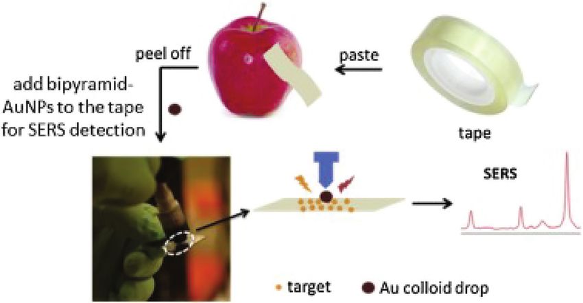
174       Hualan Zhou et al.

2 Theoretical bases of SERS                                     transfer between the chemical bonding, surface com-
                                                                plexes, light-induced and the substrate, which is gene-
Raman spectroscopy is an inelastic scattering of an inci-       rally in the range of 10-103, and the enhancement effect
dent photon with a molecule, discovered by C.V. Raman           is much lower than the physical enhancement [13]. Due
in 1928, which allows the analysis of scattering spectra        to the differences between the molecules and their inter-
at frequencies different from the incident light to obtain      actions with metal nanoparticles, the chemical enhan-
information about the vibration and rotation of mole-           cement mechanism is not universal compared to the
cules and is applied to the study of molecular structure.       physical enhancement mechanism. Although the mecha-
Raman scattering involves only small energy changes,            nism of SERS enhancement is not conclusive, SERS is
and typical Raman scattering cross sections are small,          widely used due to its high sensitivity, fast response, and
with values of about 10-29 cm2/sr [9], 10 orders of magni-      “fingerprint” recognition features. In addition to these
tude lower than those of infrared or fluorescence spec-         features, the good compatibility with water systems and
troscopy, making typical Raman spectroscopy too weak to         the linear relationship between spectral intensity and
be used for trace detection in routine analysis, which also     analytical concentration determines that SERS techno-
makes Raman spectroscopy a great challenge for trace            logy can be used for qualitative [15], semi-quantitative
detection [10]. Due to the general weakness of Raman            [16], and quantitative analysis [17].
scattering (typically one millionth of the incident light
frequency) [11], only solid samples or biological samples
with high concentrations of aqueous solutions show
good Raman scattering, but when nanometer roughness             3 SERS-active substrates
metals (such as gold and silver) are adsorbed on the
sample surface, the Raman signal of the sample is sig-          As can be seen above, the SERS phenomenon is relatively
nificantly enhanced, allowing the detection of samples          complex, and there is no uniform substrate to detect all
at very low levels (picomolar to femtomolar concentra-          target analytes, so it is necessary to develop different
tions), also known as SERS [12].                                high-sensitivity SERS substrates to cope with a variety
     Although SERS technology has undergone more than           of analytes, making SERS technology a powerful tool to
40 years of development, there is still no definite con-        detect specific target molecules at trace levels. In recent
clusion on the enhancement mechanism, and it is now             years, with the continuous development of nanotechnol-
generally accepted in academic circles that the main            ogy, different shapes, sizes, compositions, and spatial
mechanisms of SERS are physical enhancement mecha-              distances of SERS substrates have been developed,
nism (EM) and chemical enhancement mechanism (CT),              because the shape, size, and composition of the sub-
and the result of the joint action of the two classical         strates can affect the sensitivity of SERS detection, and
mechanisms [13]. EM mechanism is the mechanism of EM            they can generate different enhancement factors (EFS) to
field enhancement. The reason for the enhancement is            enhance SERS to different degrees [18].The enhancement
that when the EM wave interacts with the metal surface,         factor is often used to reflect the activity of the substrate
if the metal surface is rough, then the EM wave will excite     and the sensitivity of SERS is greatly influenced by the
a local surface plasma on the surface, which leads to the       “hot spots”, which are the largest electric field enhance-
amplification of the electric field near the surface. Surface   ment sites on metal nanoparticles, mainly in the tiny gaps
plasma is the collective oscillation of free electrons in a     between the metal nanoparticles [19]. Due to the occur-
metal under a photoelectric field, and the local electro-       rence of localized surface plasmon resonance (LSPR) of
magnetic field enhancement caused by surface plasmon            the metal nanoparticles, the electric field of the metal
resonance (SPR) is also considered to be the main core          surface part is significantly enhanced, showing a good
of SERS enhancement [14]. The electromagnetic enhan-            Raman signal. Therefore, the enhanced Raman signal is
cement mechanism is a purely physical description of            not only related to the interaction between the substrate
the local electromagnetic field enhancement caused by           and analyte, but also to the functionalization of the sub-
plasma resonance on the surface of metallic nanopartic-         strate to obtain a better substrate activity [20]. For food
les, which has been confirmed by many scholars at home          quality evaluation, it is particularly important to select
and abroad. Unlike the physical enhancement mecha-              functionalized substrates for more accurate detection of
nism, the chemical enhancement mechanism mainly                 target analytes. The common active substrates for SERS
considers the charge transfer interactions between the          in recent years are divided into metallic nanomaterials
metal nanoparticles and the sample, that is the charge          (noble metals and transition metals) and non-metallic
Application of SERS in food detection 175
nanomaterials (graphene, semiconductors, etc.) [21]. For rapid reaction of the reduction method, it is not easy to
example, Ekmen et al. synthesized surface molecularly control the nucleation rate of nanoparticles, which leads
imprinted magnetic nanoparticles (MIP@Fe3O4NPs) for to irregular and inhomogeneous size and shape of nano-
the sensitive and selective quantification of malachite particles, and ultimately leads to lower stability and repro-
green in tap water and carp samples through a recently ducibility of chemical reduction [28]. Therefore, in the
developed active mechanism called reversible chain following long time, many scholars in China and abroad
transfer catalytic polymerization (RTCP) [22]. Wang et al. have tried to improve the SERS strength by improving the
constructed a highly sensitive SERS substrate based on shape and size of metal nanoparticles. Bastús et al. used
Ag-nanoplates decorated graphene-sheets for ultrasen- growth kinetics to control the growth rate of metal nano-
sitive SERS detection of organic pesticides (including particles and prepared gold and silver nanoparticles with
thiram and methyl parathion and their mixtures). The high uniformity and adjustable particle size by a step-
Ag-nanoplates@graphene hybrids (Ag-NP@GH) substrate wise growth method [29]. As mentioned above, the size
showed low detection limits of 40 and 600 nM for thiram and shape of metal nanoparticles affect the intensity of
and methyl parathion, respectively, and more importantly, SERS through surface plasmon resonance, so the devel-
Ag-NP@GH substrate showed good SERS signal reproduc- opment of nanosubstrates of different sizes and shapes is
ibility with relative signal deviation as low as 5.6% [23]. of great significance for high sensitivity detection. D’Elia
Guselnikova et al. proposed functionalized surface equi- et al. fabricated gold nanorods for ultra-trace detection of
partition oscillator supported gold grating surfaces with cocaine in unprepared oral fluid samples, and the results
metal-organic backbone (MOF-5) for sensitive, selective, confirmed the increased sensitivity and high stability and
and reproducible SERS detection of organophosphorus reproducibility of the detection with gold nanorods [30].
pesticides, in addition to selective detection and identi- Wang et al. developed a novel solvothermal method to
fication of several relevant organic contaminants such as synthesize Ag nanocubes with controllable size. The PVP
azo dyes and mycotoxins [24]. Gold nanoparticles (AuNPs) was used as the capping agent, ethylene glycol as reduc-
and silver nanoparticles (AgNPs) were used more often in ing agent and glycerol as the viscosity regulator in the
these studies. There are also nuclear-shell nanoparticles growth of Ag nanocubes. The results show that Ag nano-
developed on this basis. To enhance the SERS signal, these cubes are highly SERS-active substrates and the detecting
noble metal nanoparticles are transformed into various limit of thiram can reach as low as 10-9 M [31].
shapes, such as nanorods, nanodendrites, nanoflowers, Numerous experiments have confirmed that gold
nanosheets, nanomembranes, and nanostars [25,26]. nanoparticles have better stability and significantly
In general, SERS active nanosubstrates can be divided more controllability than silver nanoparticles, and silver
into colloidal nanosubstrates and solid nanosubstra- nanoparticles have higher SERS activity than gold nano-
tes. Colloidal substrates are easier and more sensitive to particles. Therefore, many scholars often use the two in
make. However, since it is a solution system, it is not easy combination to obtain more powerful SERS substrates. Pu
to control the assembly of metal nanoparticles and evalu- et al. developed two-dimensional (2D) stable Au-Ag core-
ate the exact position of the target analyte, so the develo- shell nanorod (Au@AgNRs) nanoarray substrates with
ped solid nanosubstrates can solve this problem, and the high-performance SERS activity based on an interfacial
solid substrates have good stability and durability. self-assembly strategy and successfully applied them for
the detection of thiram in apple samples. A broad linea-
rity range of 0.01-10 mg/L and a low limit of detection of
3.1 Colloidal substrates 0.018 mg/L were achieved for thiram solution. The subst-
rates were stable and showed satisfactory sensitivity with
Gold and silver nanoparticles with diameters of 10-200 93-116% recovery after 4 weeks of storage at ambient tem-
nm are typically used to make colloidal substrates, perature [32].
which are readily available and inexpensive. Due to its On the other hand, electrostatic repulsion exists
high sensitivity, ease of fabrication, and stability, it is between the metal nanoparticles in the colloidal matrix
widely used for the detection of pesticide residues, bio- system, and the stability of the system is also maintained
toxins, and antibiotics, etc. [27]. In addition to chemical by electrostatic repulsion, so when testing liquid food, the
reduction methods, also known as wet synthesis tech- charge present in the sample will have a great impact on
niques, radiation reduction and laser ablation are used the adsorption ability between the metal nanoparticles
to prepare metalloid substrates, but the most widely used and the target, thus leading to a reduction in the repro-
method is chemical reduction. However, due to the more ducibility of the test results. Besides, the pH of the sample176 Hualan Zhou et al.
matrix can affect the metal colloidal solution. For a more popularity of SERS for trace energy level detection, differ-
commercial development of colloidal matrices, improve- ent commercial solid surface SERS substrates have also
ments in their stability and reproducibility are needed. been developed. These include Q-SERS (USA), P-SERS
(USA), Enspectr SERS substrates (Russia), and inkjet
printed SERS substrates. These solid substrates are more
3.2 Solid substrates expensive, but the assay process is simpler and less
time-consuming and has been used by many research-
In order to overcome the poor stability and reproduc- ers. For example, Liu et al. used Q-SERS solid substrates
ibility of the colloidal matrix, many immobilization with gold-coated SERS-active nanosubstrates for SERS
techniques have been developed to adsorb metal nan- measurements. Three pesticides (carbaryl, phosmet,
oparticles onto solid substrates, the most commonly and azinphos-methyl), which are widely used in apples
used techniques are nanolithography, microsphere and tomatoes, were selected for the study quantitative
lithography, and self-assembly techniques. In previous and qualitative models. The study showed that SERS
studies, self-assembly techniques were found to be more could detect 4.51 ppm carbaryl, 6.51 ppm phosmet, and
effective and easier to prepare than nanolithography 6.66 ppm azinphos-methyl in apples; 5.35 ppm carbaryl,
[33]. Self-assembly technology mainly uses the morpho- 2.91 ppm phosmet, and 2.94 ppm azinphos-methyl in
logical control of surface functional groups and nanos- tomatoes at 99.86% confidence interval [37].
tructures to form ordered structures, which can prepare In addition, flexible solid SERS substrates have
highly dense and orderly and controlled nanoarrays [34]. been developed for “in-situ” and real-time inspection of
Zhang et al. developed a simple self-assembly method to irregular surfaces. Therefore, many scholars have made
fabricate ordered Ag nanofilms, and the results showed a lot of efforts to develop flexible substrate materials,
excellent SERS enhancement for the detection of methyl such as cotton, tape, polymers, and paper [38]. Wu et al.
parathion with detection limits up to 10-7 M [35]. Mean- developed a simple and effective SERS tape sensing
while, different homemade solid surface substrates such strategy based on double pyramidal gold nanoparticles
as graphene-gold film-gold nanorods, graphene-gold (BP-AuNPs), using the tape to collect methyl parathion
nanorods, and gold film-gold nanorods have been devel- from the surfaces of cucumbers, apples, and tomatoes by
oped for the detection of hazardous substances in food. a simple “stick/peel” procedure (Figure 1) [39]. It is clear
Sivashanmugan et al. arrayed gold nanodots (AuNDs) on from practice that the development of soft, adhesive SERS
thin graphene (GR) layer, which was mainly prepared by substrates is of great significance for “in-situ” detection
chemical vapor deposition, by focused ion beam tech- and has a very promising application.
nique and used rhodamine 6G as a molecular probe. The Compared with colloidal matrices, the stability and
results showed that the detection limit of rhodamine reproducibility of solid matrices have been significantly
6G was as low as 10-12 M and the Raman enhancement improved, but the preparation process is relatively more
factor was increased to 108 fold [36]. Due to the increasing complicated.
Figure 1: Schematic diagram of BP-AuNPs-based SERS tape sensor for trace sensing on peel surfaces Reprinted with permission from
Ref. [39].Application of SERS in food detection 177
4 SERS detection technology multivariate analysis [42]. Du et al. synthesized novel
multifunctional Fe3O4@TiO2@Ag particles for the immu-
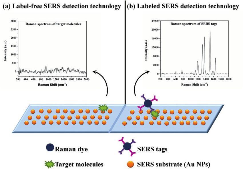
At present, the detection techniques for SERS include noassay of prostate-specific antigen (PSA) with a low
label-free SERS detection and labeled SERS detection detection limit of 16.25 pg/mL using a label-free SERS
(Figure 2). assay [43]. The use of label-free SERS assays for highly
sensitive and quantitative determination in food samples
remains a challenge. Therefore, the development of a
4.1 label-free SERS detection technology more sensitive and selective SERS detection technique is
the focus of many scholars.
Label-free SERS detection is considered to be the most
direct and reliable way to obtain SERS spectra without
the need for secondary Raman dyes and synthetic SERS 4.2 Labeled SERS detection technology
labels (also known as reporter molecules) to obtain a
unique “fingerprint” of the target analyte, relying primar- The labeled SERS method is considered as an indirect
ily on the interaction between the target analyte and the approach. The method of functionalizing a SERS active
SERS substrate [40]. In label-free assays, molecular inter- substrate with a label and then detecting the SERS signal
actions are directly converted into multiple (mechanical, of that molecule can significantly improve the specific-
electrical, or optical) signals and can be detected without ity of a given substance [44]. Labeled SERS detection
any labeling. Therefore, label-free SERS assays offer the use external signal tags for analyte measurements. Com-
advantages of simplicity, flexibility, high specificity, and monly used tags include electrochemically active mole-
real-time tracking [41]. Hernández et al. developed a low- cules, isotopic elements, organic dyes, and nanoparticles
cost, selective, and reliable gold nanoprism-based label- [45]. SERS tag-based detection methods have been shown
free nanoadaptor sensor for the detection of trichostatin to be very promising for a wide range of detection appli-
A (OTA) by SERS. Label-free SERS provides the molecular cations, overcoming the drawbacks mentioned above in
markers of the aptamer-OTA complex used and can dif- Label-free SERS detection. Lu et al. developed a method
ferentiate the OTA solution from 10 to 250 ppb through combining SERS and sandwich-type immunolabel for
Figure 2: Schematic diagram of (a) label-free SERS detection and (b) labeled SERS detection Reprinted with permission from Ref. [12].178 Hualan Zhou et al.
sensitive detection of TYR, in which 4-mercaptoben- 5 Application of SERS in food safety
zyl cyanide is embedded between Au core and Au shell
(Au4MB@Au). Core-shell structure is used as SERS label,
detection
which is expected to achieve sensitive and quantitative
detection of TYR in complex body fluids [46]. Zhang et al. 5.1 Heavy metal detection
developed a multiple SERS-based lateral flow immu-
nosensor for the identification of six major fungal toxins The accumulation and concentration of heavy metals,
in maize. Using two characteristic Raman reporter mole- especially harmful heavy metals, in the human body can
cules 5,5-dithiobis-2-nitrobenzoic acid (DTNB) and 4-mer- cause acute and chronic poisoning. Researchers have
captobenzoic acid labeled synthetic Au@Ag core-shell done a lot of work on highly sensitive detection of heavy
nanoparticles, the detection limits for zearalenone were metals in food, as is shown in Table 1.
6.2 pg/mL and 0.26 ng/mL for fumonisin B, which are Song et al. proposed a novel colorimetric/SERS dual-
below instrumental analysis and most other biosensors, mode detection of the Hg2+ method by using SERS-active
the limits of detection for zearalenone and fumonisin B peroxidase-like Au@AgPt NPs. The dual-mode probe
were 6.2 pg/mL and 0.26 ng/mL, which are lower than integrated the advantages of facile detection by colori-
those of instrumental analysis and most other biosen- metric analysis and high-sensitive trace assay by SERS.
sors, and well below the tolerance limits set by the EU, The limit of detections (LODs) of colorimetric analysis
US, and China [47]. and SERS assay were 0.52 and 0.28 nM, respectively [48].
Table 1: The application of SERS in food safety detection
Classification Target analyte SERS substrate LOD Reference
Heavy metal pollution Hg(II) Au@AgPt NP 0.28 nM [48,51-53]
B40/PSS PAH @AuNP 0.5 ppb
4,4’-bipyridine (Dpy) to modify silver 0.1 ppb
nanoparticles (AgNPs)
AuNP@Thiosemicarbazone 79×10-8 mol·L -1
Cd(II) B40/PSS PAH@AuNP 0.5 ppb [51]
Pb(II) DNAzymes and enzyme-free CHA 0.42 pM [49]
amplification system
Cr (VI) Ag nanoparticles decorated Ag@ZrO2 0.5 μM [50]
As Au@Ag core-shell nanoparticles 0.1 ppb [54]
foodborne pathogen Staphylococcus aureus VAN-Au NPs 1.0×10³ cfu·mL-1 [55,58,59,62]
Fe-MIL-88 was fabricated as artificial 1.95 cfu·mL-1
enzyme to catalyze leuco malachite
green
AuNPs-PDMS 13 cfu·mL-1
aptamer-Fe3O4@Au MNPs 3 cell·mL-1
S. typhimurium functionalized polymeric magnetic 10 cells·mL−1 [56]
nanoparticles (FPMNPs)
Shigella sonnei Au@Ag NPs or SiO2@Au NPs 10 cfu·mL-1 [57]
Staphylococcus aureus, Listeria black phosphorus-Au (BP-Au) filter 10-106 cfu·mL-1 [60]
monocytogenes and Escherichia coli paper
E. coli O157:H7 aptamer-4-ATP-GNPs 102 cfu·mL-1 [61]
Illegal additives melamine Au@HC3N4 2.7×10⁻⁹ M [63]
AgNPs/AgNWs 10-10 mol·L-1 [66]
rhodamine B AuNPs/AgNWs 10 -15 mol·L-1 [67]
clenbuterol hydrochloride (CLB) Fe3O4@Au@AgNP 0.003 ng mL-1 [64]
acid orange II and brilliant blue Fe3O4@Au core–shell 1 μg·mL-1,0.5 [68]
μg·mL-1
formaldehyde the self-assembled substrates of 0.86 nM [65]
GNRs were prepared by ethanol
modulation
(continued)Application of SERS in food detection 179
Table 1: (continued)
Classification Target analyte SERS substrate LOD Reference
biotoxin ochratoxin A (OTA) Ag@AuCSNPs 0.83 fg·mL-1 [69]
domoicacid (DA) animo-AgNPs 0.033 ppb [72]
patulin MIP-SERS 8.5×10⁻¹¹ M [76]
aflatoxin B1 aptamer-SERS sensing chip 0.4 fg·mL-1 [73]
Yersinia pestis, Francisella SERS-based LFA 43.4 cfu·mL-1, [74]
tularensis, and Bacillus anthracis 45.8 cfu·mL-1,
357 cfu·mL-1
tetrodotoxin (TTX) Fe3O4/SiO2/Au–Ab 0.01 μg·mL-1 [71]
tropane alkaloids (TAs): Ag NPs 10.0 µg·L-1, [70]
scopolamine hydrobromide 1.0 µg·L-1,
(SH),methscopolamine bromide (MB) 1.0 µg·L-1
and scopolamine butylbromide (SB)
pesticide residues 2,4-dichlorophenoxyacetic acid hollow Au@Ag bimetallic nanoflowers 0.11 ng·mL-1 [76]
(2,4-D)
4-aminothiophenol, methomyl silver nanoparticle-bacterial 3.6×10⁻⁷ M [77]
nanocellulose paper
thiabendazole Silver colloids synthesized by 4 ppm [78]
reduction of AgNO3 by trisodium
citrate
crystalviolet (CV), thiram Ag NPs/PDMS 1×10⁻⁷M,1×10-5M [79]
thiram (TMTD), methyl parathion gecko-inspired nanotentacle surface- 0.0012 mg·kg-1, [80]
(MPT), malachite green (MG) enhanced Raman spectroscopy 0.02 mg·kg-1,
(G-SERS) 0.0003 mg·kg-1
thiabendazole standard solution, gold nanoparticles were physically 0.01μg·mL-1, [81]
thiabendazole immobilized on the UF 0.125 μg·mL-1
thiabendazole (TBZ) CNF-AgNP 5 ppm [82]
Residues of veterinary thiabendazole, ferbam Au@Ag-TGANPs 0.12 ppm, [83]
drug detection 0.003 ppm
marbofloxacin β-cyclodextrin-modified silver 1.7 nmol·L-1 [84]
nanoparticles (β-CD-AgNPs)
thiram, thiabendazole, malachite Ag/Nanocellulose fibers 0.05 ppm, [85]
green, enrofloxacin 0.09 ppm,
0.0014 ppm,
0.069 ppm
amantadine the flower-like gold nanoparticles 0.005 ng·mL-1 [86]
(AuNFs)
fipronil SiO2@Au nanoparticles 10−7 M [87]
kanamycin 2-mercaptobenzothiazole (MBT) 2 pg·mL-1 [88]
labeled Au@Ag core–shell
nanoparticles
malachite green (MG) glass fiber paper modified with AgNPs 5×10⁻¹⁰ mol·L-1 [89]
Researchers have also developed a novel “signal-on” SERS substrates to sense Cr (VI) in the water at different
SERS sensing platform for Pb2+, which was developed concentrations, and the linear correlation between SERS
based on CHA amplification and DNAzymes. The biosen- intensity and Cr (VI) concentration was R2 = 0.97 with a
sor is based on the excellent performance of Pb2+-specific detection limit of 0.5 μM [50].
DNAzymes and enzyme-free CHA amplification system. In another study, Mukherji et al. developed the detec-
The linear detection range of Pb2+ was 1 pM to 100 nM, and tion of metal ions (Hg2+, Cd2+) added to deionized and tap
the detection limit was 0.42 pM [49]. Moreover, Li et al. water using E. coli B40 as bioreceptor with a detection
designed an Ag nanoparticle decorated Ag@ZrO2 for SERS limit of 0.5 ppb [51]. In addition, Dou et al. prepared a
to detect Cr (VI). Ag@ZrO2@Ag nanospheres were used as SERS-based capillary sensor for the detection of mercury180 Hualan Zhou et al.
ions (Hg2+) in water using 4,4’-bipyridine (Dpy) functiona- 5.3 Illegal additives detection
lized silver nanoparticles with LOD of 0.1 ppb [52]. Also,
de Souza et al. developed AuNP@thiosemicarbazone as Frequently exposed food safety incidents seriously threat-
a substrate for the selective detection of Hg2+ with LOD ened residents’ health and affected public trust in food
of 79×10-8 mol·L-1[53]. Moreover, Zhou et al. proposed a safety. Tang et al. synthesized holey g-C3N4 embedded
novel type of sensor for detecting As, combining arsenic with Au nanoparticles, and this holey structure of high-
aptamer with Au@Ag NPs, and measuring the actual con- density Au nanoparticles also inhibited their own aggre-
centration of arsenic (III) in the lake water [54]. gation. The detection limit of crystal violet is 2.7×10⁻⁹ M,
the enhancement factor (EF) is 6.8×10⁵. The milk samples
tested by this method showed a good linear range of
5.2 Foodborne pathogens detection 1×10⁻⁴-5×10⁻⁷ M (R2 = 0.986) and 1×10⁻³-5×10⁻⁶ M (R2 = 0.971)
[63]. In another study, Duan et al. synthesized Fe3O4@Au@
Pathogenic bacteria directly or indirectly contaminate Ag nanoparticles with strong SERS enhancement were as
food and water sources, and oral infection in humans can active substrates. The aptamer against clenbuterol hydro-
lead to intestinal infections, food poisoning, and epidem- chloride (CLB) was immobilized on the surface of the sub-
ics of infectious diseases in livestock and poultry. strate to form a capture probe. The detection limit was
Liu et al. modified the design of Fe-MIL-88 enzyme 0.003 ng·mL-1 [64]. Yan et al. synthesized gold nanorods
to catalyze colorless malachite green to malachite green. (GNRs) through a seed growth method to prepare self-
Milk and chicken meat were selected as actual samples assembled substrates by ethanol regulation. The detec-
to evaluate the performance of the Staphylococcus aureus tion limit of HCHO was 0.86 nM, and the EF was 3.9×10⁷.
(ATCC 29213) detection system by detecting the MG signal The relative standard deviation (RSD) in the detection of
and quantifying S. aureus in the range of 101-106 CFU·mL-1 rice noodles, steamed bread and liquor with methanol
with a detection limit of 1.95 CFU·mL-1 [55]. Compared addition was less than 6.74%, and the recovery was 89.45-
with the traditional enzyme-linked immunosorbent 103.2% [65]. Xu et al. also determined melamine in milk by
assay, the 5,5′-dithiobis (succinimidyl-2-nitrobenzoate) preparing dendritic AgNPs/AgNWs with the limit of detec-
(DSNB)-based SERS immunosensor developed by this tion of 10-10 mol·L-1 [66]. Moreover, Xu et al. have prepared
method is very sensitive, even in the presence of cross- centimeter-scale AuNPs/AgNWs composite materials with
contamination[56]. In addition, Wu et al. constructed an high nanometer-level roughness. The limiting concentra-
aptamer-based sensor by synthesizing metal complex- tion of Rhodamine B for this SERS substrate is 10-15 mol·L-1
linked gold nanoparticle dimer, which AuNP dimer has [67]. Researchers also synthesized Fe3O4@Au core-shell
the dual function of active substrate and Raman repor- nanomaterials, which can be used to detect acid orange II
ter molecule and modified the aptamer against Shigella and brilliant blue in food samples [68].
sonnei onto the surface of this bifunctional material. The
method achieved recoveries of 92.6-103.8% with LOD of
10 CFU·mL-1 [57]. 5.4 Biotoxin detection
In another study, Zhao et al. constructed a new sand-
wich LFA format to use VAN and pig IgG for the detec- Natural toxins are a general term for a large class of bio-
tion of Staphylococcus aureus with a detection limit of logically active substances, including animal toxins, phy-
1.0×10³ CFU·mL-1 [58]. Zhu et al. synthesized hollow Au@ totoxins, and microbial toxins. Different from synthetic
Ag NFs SERS aptamer sensors to detect S. aureus with LOD toxic compounds, they can also be divided into marine
of 13 CFU·mL-1 [59]. Besides, Huang et al. invented a 3D toxins and agricultural toxins. Huang et al. reported a
SERS substrate based on BP-Au filter paper that can spe- dual amplification strategy for the ultra-sensitive detec-
cifically identify and differentiate Staphylococcus aureus, tion of biotoxins with the detection limit of 0.83 fg·mL-1.
Listeria monocytogenes, and E. coli [60]. Some researchers This method was based on the specific target recognition
also used aptamer-4-ATP-GNPs to detect E. coli O157:H7 of DNA aptamers, and the developed satellite core com-
sensitively and highly specifically, with LOD value of ponents generate many “hot spots” that can effectively
102 CFU·mL-1, and a recovery rate of 99-113% [61]. Also, Pang enhance SERS signals [69]. Using the electrostatic inter-
et al. designed a dual identification SERS biosensor for action between halide ion, target, and Ag NPs, Tian et al.
pathogen detection. The recovery rate of Staphylococcus found that the coadsorption with the specific adsorbed
aureus for actual samples is 95.0-106.4%, and the relative I- during the formation of hotspots could significantly
standard deviation (RSD) is less than 5.3% [62]. improve the detection sensitivity. With the example ofApplication of SERS in food detection 181
the trace analysis of three tropane alkaloids (TAs) includ- polymers (MIPs) with SERS to detect thiabendazole in
ing scopolamine hydrobromide (SH), methscopolamine orange juice, the LOD was 4 ppm [78]. Moreover, resear-
bromide (MB), and scopolamine butylbromide (SB), it chers assembled a transparent AgNPs/PDMS composites
was performed at a minimum detection concentration of on flexible PDMS surfaces, and measured crystal violet (CV)
1 g·L-1 for these three substances under optimized condi- and thiram concentrations as low as 1×10⁻⁷ and 1×10⁻⁵ M in
tions [70]. In addition, Sun et al. developed Fe3O4/SiO2/ contaminated fish skin and orange peel, respectively [79].
Au/magnetic nanoparticles conjugated with tetrodotoxin In addition, Han et al. designed a gecko- inspired nano-
(TTX) antibodies (Ab) and used as a Raman active sub- tentacle SERS (G-SERS) substrate to detect TMTD, MPT
strate (Fe3O4/SiO2/Au-Ab), the concentration of TTX with a and MG in cucumbers, grapes, and apples. The LOD
limit of detection of 0.01 μg·mL-1 and a detection linearity were 0.0012, 0.02, and 0.0003 mg·kg-1, respectively [80].
range of 0.01-0.5 μg·mL-1 [71]. Also, Hong et al. developed a highly homogeneous plas-
In another study, Müller et al. used amino- monic SERS substrate by immobilizing gold nanopar-
functionalized Ag nanoparticles as SERS probes to ticles on ultrafiltration (UF) membranes using a suction
detect domoic acid (DA) biotoxins causing amnesic technique. The LOD of thiabendazole standard solution
shellfish poisoning (ASP) in seawater with a detection and thiabendazole in orange extract were 0.01 μg·mL-1 and
limit of 0.033 ppb [72]. Besides, Han et al. proposed an 0.125 μg·g-1 [81]. In another study, a cellulose nanofiber
exonuclease-assisted SERS sensing strategy and suc- (CNF) composite material coated with silver nanopartic-
cessfully constructed an aptamer-SERS for the determi- les (AgNPs) was also developed to detect thiabendazole
nation of aflatoxin B1 (AFB1) in peanuts with a limit of (TBZ) in apples with a detection limit of 5 ppm [82].
detection (LOD) of 0.4 fg·mL-1 [73]. A novel SERS-based
lateral flow (LF) assay has been reported by another
researcher, which could detect Yersinia pestis, Fran- 5.6 Residues of veterinary drug detection
cisella tularensis, and Bacillus anthracis with LODs at
43.4, 45.8, and 357 cfu·mL-1, respectively [74]. Moreover, Veterinary drug residues have become the most common
Han et al. combined the sensitivity of SERS technology contamination problem. The development of SERS-based
to prepare a new type of MIP-SERS substrate for the technology is particularly important for the detection of
detection of patulin. The linear range of the method was veterinary drug residues. For example, Sun et al. devel-
5×10 -10-10 -6 M with a limit of detection of 8.5×10 -11 M [75]. oped a surface-enhanced Raman spectrometer based on
thioglycolic acid (TGA)-functionalized silver-coated gold
nanoparticles (Au@Ag-TGANPs) for rapid screening of thi-
5.5 Pesticide residues detection amethoxam (TBZ) and ferbam in liquid milk with detec-
tion limits of 0.12 and 0.003 ppm [83]. Moreover, Zhao
In order to protect the health of consumers, many studies et al. developed a novel type of SERS of marbofloxacin
have reported the application of SERS for pesticide res- established by using the interaction between marboflox-
idues detection. For example, Chen et al. synthesized acin and β-cyclodextrin-silver nanoparticles. The method
hollow Au@Ag-nanoflower-SERS sensor for competi- was used to determine the content of Marbofloxacin in
tive detection of 2,4-dichlorophenoxyacetic acid (2,4-D), chicken and duck mea t[84]. Huang et al. also devel-
and they developed antibody-functionalized magnetite oped an Ag/nanocellulose fibers for in-situ detection of
nanoparticles (antibody-MNPs) as enrichment probe. foodstuffs, used rhodamine 6G as a probe to rapidly and
The LOD was 0.11 ng·mL-1, and its linear range was 0.001- accurately detect harmful residues on fish-fosfomycin,
100 ng·mL-1 with recoveries of 89.73-100.27% and RSDs thiabendazole, malachite green, and enrofloxacin with
of 2.56-4.97% [76]. In addition, Ekgasit et al. designed a the detection limits of 0.05, 0.09, 0.0014, and 0.069 ppm,
simple and effective “paste-and-read” SERS method and respectively [85].
prepared a biodegradable plasma silver nanoparticle- In addition, Wang et al. proposed a novel ultrasensi-
bacterial nanocellulose paper (AgNP-BNCP). A 3D SERS tive SERS immunosensor based on the flower-shaped gold
hot spot was formed on the substrate to directly detect nanoparticles and magnetic beads to detect amantadine
4-aminothiophenol and methomyl pesticides on oranges in chicken meat with a detection limit of 0.005 ng·mL-1
and apples, and the detection limit for methomyl was [86]. Also, Huang et al. fabricated uniform SiO2@Au nano-
3.6×10⁻⁷ M [77]. particles with excellent SERS activity, which can detect
In another study, Lu et al. designed a novel MISPE- fipronil in 0.1 ppm [87]. In addition, another study used
SERS chemosensor by combining molecularly imprinted anti-kanamycin functionalized hybrid magnetic (Fe3O4)182 Hualan Zhou et al.
nanoparticles (MNPs) and 2-mercaptobenzothiazole-labe- Acknowledgments: Financial support from the National
led Au core@Ag shell nanoparticles as substrates. This Natural Science Foundation of China (Grant No. 31870045)
sandwich assay measured the limit of detection (LOD) of and partial support from the Shanghai Foundation for In-
kanamycin in milk to be 2 pg·mL-1 [88]. Moreover, Deng ternational Science and Technology Cooperation (Grant
et al. prepared a SERS substrate using glass fiber paper No. 19230742900) is greatly appreciated.
to detect malachite green residues in fish. The detection
limit was 5×10⁻¹⁰ mol·L-1 [89]. Funding information: This work was supported by the
National Natural Science Foundation of China (Grant No.
31870045). This work was also partially supported by the
International Science and Technology Cooperation Foun-
6 Future trends and perspectives dation of Shanghai (Grant No. 19230742900).
As a kind of quick and sensitive detection technology, Author contributions: Hualan Zhou: supervision, writing –
SERS played a very important role in detecting heavy review and editing, funding acquisition; Xiaodi Li: data
metal pollution, agricultural, and veterinary drug curation, writing – original draft, investigation; Lehui
residue, misuse of food additives, foodborne pathogenic Wang: data curation, writing – original draft; Yingfang
micro-organisms and so on. However, the SERS technol- Liang: investigation; Aikedan Jialading: Investigation;
ogy is still in its initial stage in the practical application Zishuo Wang: investigation; Jianguo Zhang: writing –
of food safety detection, and it also faces correspond- review and editing, supervision.
ing challenges in theoretical analysis and quantitative
Conflict of interest: Authors state no conflict of interest.
analysis. First, SERS mechanism has not yet reached a
clear point, and some SERS phenomena cannot be fully
explained. Electromagnetic field enhancement mech-
anism and chemical enhancement mechanism are two References
main theoretical supports at present, but they have not
[1] Li D, Yao D, Li C, Luo Y, Liang A, Wen G, et al. Nanosol
been unanimously agreed. Therefore, the theoretical
SERS quantitative analytical method: A review. TrAC-Trend
research on SERS still needs to be carried out continu- Anal Chem. 2020; https://doi.org/127.10.1016/j.trac.
ously. Second, although there were various kinds of SERS 2020.115885.
substrate, problems such as instability, poor reproduci- [2] Jiang GH, Wang ZY, Zong SF, Yang K, Zhu K, et al. Peroxidase-
bility, high cost and uneven structure were still faced. like recyclable SERS probe for the detection and elimination
of cationic dyes in pond water. J Hazard Mater. 2020:124426.
Complex two-dimensional or three-dimensional nano-
https://doi.org/10.1016/j.jhazmat.2020.124426.
structures can generate many “hot spots”, but they are
[3] López-López M, García-Ruiz C. Infrared and Raman spectros-
rarely used in food safety analysis, so further research in copy techniques applied to identification of explosives. TrAC-
this area is not possible. Thirdly, since the signal strength Trend Anal Chem. 2014;54:36-44. https://doi.org/10.1016/j.
of the same sample to be tested will be different under trac.2013.10.011.
different test conditions, it is still a challenge to apply [4] Joseph MM, Narayanan N, Nair JB, Karunakaran V, Ramya
AN, Sujai PT, et al. Exploring the margins of SERS in practical
SERS to quantitative analysis. The quantitative accuracy
domain: An emerging diagnostic modality for modern
still needs to be further studied. Fourth, the rapid deter- biomedical applications. Biomater. 2018;181:140-81. https://
mination of different samples requires the establishment doi.org/10.1016/j.biomaterials.2018.07.045.
of a unified Raman spectrum library. However, since the [5] Shen Z, Fan Q, Yu Q, Wang R, Wang H, Kong X. Facile detec-
strength of SERS signal was affected by detection envi- tion of carbendazim in food using TLC-SERS on diatomite thin
layer chromatography. Spectrochim Acta, Part A: Mol Biomol
ronment, the establishment of this gallery still needed
Spectrosc. 2021;247:119037. https://doi.org/10.1016/j.
to be done.
saa.2020.119037.
SERS technique showed great potential in the field [6] Pang S, Yang T, He L. Review of surface enhanced Raman
of food safety. In the future, SERS technology can be spectroscopic (SERS) detection of synthetic chemical
combined with other techniques to improve the sensiti- pesticides. TrAC-Trend Anal Chem. 2016;85:73-82. https://doi.
vity, such as chemical separation technology, biological org/10.1016/j.trac.2016.06.017.
[7] Huang D, Zhao J, Wang M, Zhu S. Snowflake-like gold
capture technology, etc. In order to obtain the results
nanoparticles as SERS substrates for the sensitive
quickly and conveniently, the portable Raman spectrome- detection of organophosphorus pesticide residues. Food
ter can be developed which has a broad prospect in the Control. 2020;108:106835. https://doi.org/10.1016/j.
field of rapid detection. foodcont.2019.106835.Application of SERS in food detection 183
[8] Ilhan H, Guven B, Dogan U, Torul H, Evran S, Çetin D, et al. [22] Ekmen E, Bilici M, Turan E, Tamer U, Zengin A. Surface
The coupling of immunomagnetic enrichment of bacteria with molecularly-imprinted magnetic nanoparticles coupled with
paper-based platform. Talanta. 2019;201:245-52. https://doi. SERS sensing platform for selective detection of malachite
org/10.1016/j.talanta.2019.04.017. green. Sensor Actuat B-Chem. 2020;325:128787.
[9] Blackie EJ, Le Ru EC, Etchegoin PG. Single-Molecule Surface- https://doi.org/10.1016/j.snb.2020.128787.
Enhanced Raman Spectroscopy of Nonresonant Molecules. [23] Wang X, Zhu C, Hu X, Xu Q, Zhao H, Meng G, et al. Highly
J Am Chem Soc. 2009;131(40):14466-72. https://doi. sensitive surface-enhanced Raman scattering detection
org/10.1021/ja905319w. of organic pesticides based on Ag-nanoplate decorated
[10] Lu X, Al-Qadiri HM, Lin M, Rasco BA. Application of Mid- graphene-sheets. Appl Surf Sci. 2019;486:405-10.
infrared and Raman Spectroscopy to the Study of Bacteria. https://doi.org/10.1016/j.apsusc.2019.05.008.
Food Biopro Technol. 2011;4(6):919-35. https://doi. [24] Guselnikova O, Postnikov P, Elashnikov R, Miliutina E, Svorcik
org/10.1007/s11947-011-0516-8. V, Lyutakov O. Metal-organic framework (MOF-5) coated SERS
[11] Huang C-C, Cheng C-Y, Lai Y-S. Paper-based flexible surface active gold gratings: A platform for the selective detection of
enhanced Raman scattering platforms and their applications organic contaminants in soil. Anal Chim Acta. 2019;1068:70-9.
to food safety. Trends Food Sci Tech 2020;100:349-58. https://doi.org/10.1016/j.aca.2019.03.058.
https://doi.org/10.1016/j.tifs.2020.04.019. [25] Waiwijit U, Chananonnawathorn C, Eimchai P, Bora T, Hornyak
[12] Neng J, Zhang Q, Sun P. Application of surface-enhanced GL, Nuntawong N. Fabrication of Au-Ag nanorod SERS
Raman spectroscopy in fast detection of toxic and harmful substrates by co-sputtering technique and dealloying with
substances in food. Biosens Bioelectron. 2020;167. selective chemical etching. Appl Surf Sci. 2020;530:147171.
https://doi.org/10.1016/j.bios.2020.112480. https://doi.org/10.1016/j.apsusc.2020.147171.
[13] Tong Q, Wang W, Fan Y, Dong L. Recent progressive prepara- [26] Wang Q, Xu Z, Zhao Y, Zhangsun H, Bu T, Zhang C, et al.
tions and applications of silver-based SERS substrates. Bio-inspired self-cleaning carbon cloth based on flower-like
TrAC-Trend Anal Chem. 2018;106:246-58. https://doi. Ag nanoparticles and leaf-like MOF: A high-performance and
org/10.1016/j.trac.2018.06.018. reusable substrate for SERS detection of azo dyes in soft
[14] Liu Gk, Zheng H, Lu Jl. Recent progress and perspective drinks. Sensor Actuat B-Chem. 2020:129080. https://doi.
of trace antibiotics detection in aquatic environment org/10.1016/j.snb.2020.129080.
by surface-enhanced Raman spectroscopy. Trends [27] Chamuah N, Bhuyan N, Das PP, Ojah N, Choudhary AJ,
Environ Anal. 2017;16:16-23. https://doi.org/10.1016/j. Medhi T, et al. Gold-coated electrospun PVA nanofibers
teac.2017.10.002. as SERS substrate for detection of pesticides. Sensor
[15] Mosier-Boss PA. Review of SERS Substrates for Chemical Actuat B-Chem. 2018;273:710-7. https://doi.org/10.1016/j.
Sensing. Nanommaterials-Basel. 2017;7(6):142. https://doi. snb.2018.06.079.
org/10.3390/nano7060142. [28] Bastús NG, Comenge J, Puntes V. Kinetically Controlled
[16] Kearns H, Goodacre R, Jamieson LE, Graham D, Faulds K. SERS Seeded Growth Synthesis of Citrate-Stabilized Gold
Detection of Multiple Antimicrobial-Resistant Pathogens Nanoparticles of up to 200 nm: Size Focusing versus Ostwald
Using Nanosensors. Anal Chem. 2017;89(23):12666-73. Ripening. Langmuir. 2011;27(17):11098-105. https://doi.
https://doi.org/10.1021/acs.analchem.7b02653. org/10.1021/la201938u.
[17] Li C, Ouyang H, Tang X, Wen G, Liang A, Jiang Z. A surface [29] Bastús NG, Merkoçi F, Piella J, Puntes V. Synthesis of Highly
enhanced Raman scattering quantitative analytical platform Monodisperse Citrate-Stabilized Silver Nanoparticles of
for detection of trace Cu coupled the catalytic reaction and up to 200 nm: Kinetic Control and Catalytic Properties.
gold nanoparticle aggregation with label-free Victoria blue Chem Mater. 2014;26(9):2836-46. https://doi.org/10.1021/
B molecular probe. Biosens Bioelectron. 2017;87:888-93. cm500316k.
https://doi.org/10.1016/j.bios.2016.09.053. [30] D’Elia V, Rubio-Retama J, Ortega-Ojeda FE, García-Ruiz
[18] Yaseen T, Pu H, Sun D-W. Functionalization techniques for C, Montalvo G. Gold nanorods as SERS substrate for the
improving SERS substrates and their applications in food ultratrace detection of cocaine in non-pretreated oral fluid
safety evaluation: A review of recent research trends. Trends samples. Colloid Surface A. 2018;557:43-50. https://doi.
Food Sci Tech. 2018;72:162-74. https://doi.org/10.1016/j. org/10.1016/j.colsurfa.2018.05.068.
tifs.2017.12.012. [31] Wang H, Li KB, Xu C, Xu SC, Li GH. Large-scale solvothermal
[19] Xu H, Aizpurua J, Käll M, Apell P. Electromagnetic contribu- synthesis of Ag nanocubes with high SERS activity. J
tions to single-molecule sensitivity in surface-enhanced Alloy Compd. 2019;772:150-6. https://doi.org/10.1016/j.
Raman scattering. Phys. Rev. E. 2000;62(3):4318-24. jallcom.2018.09.043.
https://doi.org/10.1103/PhysRevE.62.4318. [32] Pu H, Huang Z, Xu F, Sun D-W. Two-dimensional self-
[20] Willets KA, Duyne RPV. Localized Surface Plasmon assembled Au-Ag core-shell nanorods nanoarray for sensitive
Resonance Spectroscopy and Sensing. Annu Rev Phys detection of thiram in apple using surface-enhanced Raman
Chem. 2007;58(1):267-97. https://doi.org/10.1146/annurev. spectroscopy. Food Chem. 2021;343:128548. https://doi.
physchem.58.032806.104607. org/10.1016/j.foodchem.2020.128548.
[21] Fu X, Wang Y, Liu Y, Liu H, Fu L, Wen J, et al. A graphene [33] He XY, Ge C, Zheng XQ, Tang B, Chen L, Li SB, et al. Rapid
oxide/gold nanoparticle-based amplification method identification of alpha-fetoprotein in serum by a micro
for SERS immunoassay of cardiac troponin I. Analyst. fluidic SERS chip integrated with Ag/Au Nanocomposites.
2019;144(5):1582-9. https://doi.org/10.1039/ Trends Food Sci Tech. 2018; https://doi.org/10.1016/j.
C8AN02022A. tifs.2018.02.020.184 Hualan Zhou et al.
[34] Toderas F, Baia M, Baia L, Astilean S. Controlling gold separation. Anal Chim Acta. 2020;1138:150-7. https://doi.
nanoparticle assemblies for efficient surface-enhanced org/10.1016/j.aca.2020.09.042.
Raman scattering and localized surface plasmon resonance [47] Zhang W, Tang S, Jin Y, Yang C, He L, Wang J, et al. Multiplex
sensors. Nanotechnology. 2007;18(25):255702. https://doi. SERS-based lateral flow immunosensor for the detection of
org/10.1088/0957-4484/18/25/255702. major mycotoxins in maize utilizing dual Raman labels and
[35] Zhang L. Self-assembly Ag nanoparticle monolayer triple test lines. J Hazard Mater. 2020;393:122348. https://
film as SERS Substrate for pesticide detection. Appl doi.org/10.1016/j.jhazmat.2020.122348.
Surf Sci. 2013;270:292-4. https://doi.org/10.1016/j. [48] Song C, Li J, Sun Y, Jiang X, Zhang J, Dong C, et al.
apsusc.2013.01.014. Colorimetric/SERS dual-mode detection of mercury ion via
[36] Sivashanmugan, K., Nguyen, V.-H. & Nguyen, B.-S. Tailoring SERS-Active peroxidase-like Au@AgPt NPs. Sensor Actuat
a novel Au nanodot arrays on graphene substrate for a B-Chem. 2020;310:127849. https://doi.org/10.1016/j.
highly active Surface-Enhanced Raman Scattering (SERS). snb.2020.127849.
Mater Lett. 2020;271:127807. https://doi.org/10.1016/j. [49] Wu Y, Fu C, Xiang J, Cao Y, Deng Y, Xu R, et al. “Signal-on”
matlet.2020.127807. SERS sensing platform for highly sensitive and selective
[37] Liu, B. et al. Detection of Pesticides in Fruits by Surface- Pb2+ detection based on catalytic hairpin assembly. Anal
Enhanced Raman Spectroscopy Coupled with Gold Chim Acta. 2020;1127:106-13. https://doi.org/10.1016/j.
Nanostructures. Food Bioprocess Tech. 2012;6: 710-718, aca.2020.06.038.
https://doi.org/10.1007/s11947-011-0774-5. [50] Zhou L, Yang J, Wang X, Song G, Lu F, You L, et al. Ag nanopar-
[38] Cottat M, Lidgi-Guigui N, Tijunelyte I, Barbillon G, ticles decorated Ag@ZrO2 composite nanospheres as highly
Hamouda F, Gogol P, et al. Soft UV nanoimprint active SERS substrates for quantitative detection of hexava-
lithography-designed highly sensitive substrates for SERS lent chromium in waste water. J Mol Liq. 2020;319:114158.
detection. Anoscale Res Lett. 2014;9(1):623. https://doi. https://doi.org/10.1016/j.molliq.2020.114158.
org/10.1186/1556-276X-9-623. [51] Halkare P, Punjabi N, Wangchuk J, Nair A, Kondabagil K,
[39] Wu H, Luo Y, Hou C, Huo D, Zhou Y, Zou S, et al. Flexible Mukherji S. Bacteria functionalized gold nanoparticle matrix
bipyramid-AuNPs based SERS tape sensing strategy for based fiber-optic sensor for monitoring heavy metal pollution
detecting methyl parathion on vegetable and fruit surface. in water. Sensor Actuat B-Chem. 2019;281:643-51. https://
Sensor Actuat B-Chem. 2019;285:123-8. https://doi. doi.org/10.1016/j.snb.2018.10.119.
org/10.1016/j.snb.2019.01.038. [52] Zhao Y, Yamaguchi Y, Ni Y, Li M, Dou X. A SERS-based
[40] Han XX, Ozaki Y, Zhao B. Label-free detection in biological capillary sensor for the detection of mercury ions in envi-
applications of surface-enhanced Raman scattering. TrAC- ronmental water. Spectroc Acta Pt A-Molec Biomolec Spectr.
Trend Anal Chem. 2012;38:67-78. https://doi.org/10.1016/j. 2020;233:118193. https://doi.org/10.1016/j.saa.2020.118193.
trac.2012.05.006. [53] Franciscato DS, Matias TA, Shinohara J, Gonçalves JM, Coelho
[41] Liu Y, Zhou H, Hu Z, Yu G, Yang D, Zhao J. Label and NP, Fernandes CS, et al. Thiosemicarbazone@Gold nanopar
label-free based surface-enhanced Raman scattering ticle hybrid as selective SERS substrate for Hg2+ ions.
for pathogen bacteria detection: A review. Biosens Spectroc Acta Pt A-Molec Biomolec Spectr. 2018;204:174-9.
Bioelectron. 2017;94:131-40. https://doi.org/10.1016/j. https://doi.org/10.1016/j.saa.2018.06.038.
bios.2017.02.032. [54] Song L, Mao K, Zhou X, Hu J. A novel biosensor based on Au@
[42] Hernández Y, Lagos LK, Galarreta BC. Development of a label- Ag core–shell nanoparticles for SERS detection of arsenic
free-SERS gold nanoaptasensor for the accessible determina (III). Talanta. 2016;146:285-90. https://doi.org/10.1016/j.
tion of ochratoxin A. Sens Bio-Sens Res. 2020;28:100331. talanta.2015.08.052.
https://doi.org/10.1016/j.sbsr.2020.100331. [55] Liu S, Li H, Hassan MM, Ali S, Chen Q. SERS based artificial
[43] Du Y, Liu H, Chen Y, Tian Y, Zhang X, Gu C, et al. Recyclable peroxidase enzyme regulated multiple signal amplified
label-free SERS-based immunoassay of PSA in human system for quantitative detection of foodborne pathogens.
serum mediated by enhanced photocatalysis arising Food Control. 2020:107733. https://doi.org/10.1016/j.
from Ag nanoparticles and external magnetic field. Appl foodcont.2020.107733.
Surf Sci. 2020;528:146953. https://doi.org/10.1016/j. [56] Chattopadhyay S, Sabharwal PK, Jain S, Kaur A, Singh H.
apsusc.2020.146953. Functionalized polymeric magnetic nanoparticle assisted
[44] Zheng X-S, Jahn IJ, Weber K, Cialla-May D, Popp J. Label-free SERS immunosensor for the sensitive detection of S.
SERS in biological and biomedical applications: Recent typhimurium. Anal Chim Acta. 2019;1067:98-106. https://doi.
progress, current challenges and opportunities. Spectrochim org/10.1016/j.aca.2019.03.050.
Acta, Part A: Mol Biomol Spectrosc. 2018;197:56-77. [57] Wu S, Duan N, He C, Yu Q, Dai S, Wang Z. Surface-enhanced
https://doi.org/10.1016/j.saa.2018.01.063. Raman spectroscopic–based aptasensor for Shigella sonnei
[45] Li H, Arroyo-Currás N, Kang D, Ricci F, Plaxco KW. Dual- using a dual-functional metal complex-ligated gold nanopar-
Reporter Drift Correction To Enhance the Performance of ticles dimer. Colloid Surf B-Biointerfaces. 2020;190:110940.
Electrochemical Aptamer-Based Sensors in Whole Blood. https://doi.org/10.1016/j.colsurfb.2020.110940.
J Am Chem Soc. 2016;138(49):15809-12. https://doi. [58] Zhao M, Yao X, Liu S, Zhang H, Wang L, Yin X, et al.
org/10.1021/jacs.6b08671. Antibiotic and mammal IgG based lateral flow assay for
[46] Lu D, Lin X, Chen C, Lu Y, Feng S, Huang Z, et al. Interference- simple and sensitive detection of Staphylococcus aureus.
free SERS tags for ultrasensitive quantitative detection Food Chem. 2021;339:127955. https://doi.org/10.1016/j.
of tyrosinase in human serum based on magnetic bead foodchem.2020.127955.Application of SERS in food detection 185
[59] Zhu A, Ali S, Xu Y, Ouyang Q, Chen Q. A SERS aptasensor [71] Neng J, Wang X, Jia K, Sun P. Rapid Detection of Tetrodotoxin
based on AuNPs functionalized PDMS film for selective Using Surface-Enhanced Raman Spectroscopy and
and sensitive detection of Staphylococcus aureus. Biosens Fe3O4/SiO2/Au Gold/Magnetic Nanoparticles. J Appl
Bioelectron. 2021;172:112806. https://doi.org/10.1016/j. Spectrosc. 2018;85(1):160-5. https://doi.org/10.1007/
bios.2020.112806. s10812-018-0627-3.
[60] Huang D, Zhuang Z, Wang Z, Li S, Zhong H, Liu Z, et al. [72] Müller C, Glamuzina B, Pozniak I, Weber K, Cialla D,
Black phosphorus-Au filter paper-based three-dimensional Popp J, et al. Amnesic shellfish poisoning biotoxin detec-
SERS substrate for rapid detection of foodborne bacteria. tion in seawater using pure or amino-functionalized Ag
Appl Surf Sci. 2019;497:143825. https://doi.org/10.1016/j. nanoparticles and SERS. Talanta. 2014;130:108-15. https://
apsusc.2019.143825. doi.org/10.1016/j.talanta.2014.06.059.
[61] Díaz-Amaya S, Lin L-K, Deering AJ, Stanciu LA. Aptamer-based [73] Li Q, Lu Z, Tan X, Xiao X, Wang P, Wu L, et al. Ultrasensitive
SERS biosensor for whole cell analytical detection of E. coli detection of aflatoxin B1 by SERS aptasensor based on
O157:H7. Anal Chim Acta. 2019;1081:146-56. https://doi. exonuclease-assisted recycling amplification. Biosens
org/10.1016/j.aca.2019.07.028. Bioelectron. 2017;97:59-64. https://doi.org/10.1016/j.
[62] Pang Y, Wan N, Shi L, Wang C, Sun Z, Xiao R, et al. Dual- bios.2017.05.031.
recognition surface-enhanced Raman scattering(SERS) [74] Wang R, Kim K, Choi N, Wang X, Lee J, Jeon JH, et al. Highly
biosensor for pathogenic bacteria detection by using sensitive detection of high-risk bacterial pathogens
vancomycin-SERS tags and aptamer-Fe3O4@Au. Anal using SERS-based lateral flow assay strips. Sensor Actuat
Chim Acta. 2019;1077:288-96. https://doi.org/10.1016/j. B-Chem. 2018;270:72-9. https://doi.org/10.1016/j.
aca.2019.05.059. snb.2018.04.162.
[63] Tang W, An Y, Ho Row K. Fabrication of Au nanoparticles [75] Zhu Y, Wu L, Yan H, Lu Z, Yin W, Han H. Enzyme induced molecu-
embedded holey g-C3N4 as SERS substrates for sensitive and larly imprinted polymer on SERS substrate for ultrasensitive
reliable detection. Chem Eng J. 2020;402:126194. https://doi. detection of patulin. Anal Chim Acta. 2020;1101:111-9.
org/10.1016/j.cej.2020.126194. https://doi.org/10.1016/j.aca.2019.12.030.
[64] Duan N, Qi S, Guo Y, Xu W, Wu S, Wang Z. Fe3O4@Au@Ag [76] Xu Y, Kutsanedzie FYH, Hassan MM, Zhu J, Li H, Chen
nanoparticles as surface-enhanced Raman spectroscopy sub- Q. Functionalized hollow Au@Ag nanoflower SERS
strates for sensitive detection of clenbuterol hydrochloride in matrix for pesticide sensing in food. Sensor Actuat
pork with the use of aptamer binding. LWT. 2020;134:110017. B-Chem. 2020;324:128718. https://doi.org/10.1016/j.
https://doi.org/10.1016/j.lwt.2020.110017. snb.2020.128718.
[65] Zhao YX, Zhu WW, Wu YY, Chen YY, Du FK, Yan J, et al. Sensitive [77] Parnsubsakul A, Ngoensawat U, Wutikhun T, Sukmanee T,
surface-enhanced Raman scattering for the quantitative Sapcharoenkun C, Pienpinijtham P, et al. Silver nanoparticle/
detection of formaldehyde in foods using gold nanorod bacterial nanocellulose paper composites for paste-and-read
substrate. Microchem J. 2021;160:105727. https://doi. SERS detection of pesticides on fruit surfaces. Carbohydr
org/10.1016/j.microc.2020.105727. Polym. 2020;235:115956. https://doi.org/10.1016/j.
[66] Xu D, Jiang H, Zhang S, Yang W, Guo Q, Zhang Y, et al. carbpol.2020.115956.
Centimeter-scale high nanoscale roughness silver nanopar- [78] Feng J, Hu Y, Grant E, Lu X. Determination of thiabendazole
ticles decorated silver nanowires: A highly sensitive material in orange juice using an MISPE-SERS chemosensor. Food
towards melamine in milk. Opt Mater. 2020;109:110471. Chem. 2018;239:816-22. https://doi.org/10.1016/j.
https://doi.org/10.1016/j.optmat.2020.110471. foodchem.2017.07.014.
[67] Xu D, Jiang H, Zhang S, Yang W, Zhang Y, Wang Z, et al. High [79] Alyami A, Quinn AJ, Iacopino D. Flexible and transparent
roughness gold nanoparticles/silver nanowires composites: Surface Enhanced Raman Scattering (SERS)-Active Ag NPs/
Fabrication, characterization and ultrasensitive SERS detec- PDMS composites for in-situ detection of food contaminants.
tion towards Rhodamine B. Microchem J. 2020;158:105136. Talanta. 2019;201:58-64. https://doi.org/10.1016/j.
https://doi.org/10.1016/j.microc.2020.105136. talanta.2019.03.115.
[68] Xie Y, Chen T, Guo Y, Cheng Y, Qian H, Yao W. Rapid [80] Wang P, Wu L, Lu Z, Li Q, Yin W, Ding F, et al. Gecko-Inspired
SERS detection of acid orange II and brilliant blue in Nanotentacle Surface-Enhanced Raman Spectroscopy
food by using Fe3O4@Au core–shell substrate. Food Substrate for Sampling and Reliable Detection of
Chem. 2019;270:173-80. https://doi.org/10.1016/j. Pesticide Residues in Fruits and Vegetables. Anal Chem.
foodchem.2018.07.065. 2017;89(4):2424-31. https://doi.org/10.1021/acs.
[69] Huang D, Chen J, Ding L, Guo L, Kannan P, Luo F, et al. analchem.6b04324.
Core-satellite assemblies and exonuclease assisted double [81] Hong J, Kawashima A, Hamada N. A simple fabrication of plas-
amplification strategy for ultrasensitive SERS detection of monic surface-enhanced Raman scattering (SERS) substrate for
biotoxin. Anal Chim Acta. 2020;1110:56-63. https://doi. pesticide analysis via the immobilization of gold nanoparticles
org/10.1016/j.aca.2020.02.058. on UF membrane. Appl Surf Sci. 2017;407:440-6.
[70] Lu J, Cai Z, Zou Y, Wu D, Wang A, Chang J, et al. Silver https://doi.org/10.1016/j.apsusc.2017.02.232.
Nanoparticle-Based Surface-Enhanced Raman [82] Liou P, Nayigiziki FX, Kong F, Mustapha A, Lin M. Cellulose
Spectroscopy for the Rapid and Selective Detection nanofibers coated with silver nanoparticles as a SERS
of Trace Tropane Alkaloids in Food. ACS Applied Nano platform for detection of pesticides in apples. Carbohydr
Materials. 2019;2(10):6592-601. https://doi.org/10.1021/ Polym. 2017;157:643-50. https://doi.org/10.1016/j.
acsanm.9b01493. carbpol.2016.10.031.You can also read