Biomechanical evaluation of modified track shoes - LSU ...
←
→
Page content transcription
If your browser does not render page correctly, please read the page content below
Louisiana State University
LSU Digital Commons
LSU Master's Theses                                                                                                              Graduate School

2010

Biomechanical evaluation of modified track shoes
Marlon Alberetos Greensword
Louisiana State University and Agricultural and Mechanical College, mgree15@tigers.lsu.edu

Follow this and additional works at: https://digitalcommons.lsu.edu/gradschool_theses
     Part of the Construction Engineering and Management Commons

Recommended Citation
Greensword, Marlon Alberetos, "Biomechanical evaluation of modified track shoes" (2010). LSU Master's Theses. 2524.
https://digitalcommons.lsu.edu/gradschool_theses/2524

This Thesis is brought to you for free and open access by the Graduate School at LSU Digital Commons. It has been accepted for inclusion in LSU
Master's Theses by an authorized graduate school editor of LSU Digital Commons. For more information, please contact gradetd@lsu.edu.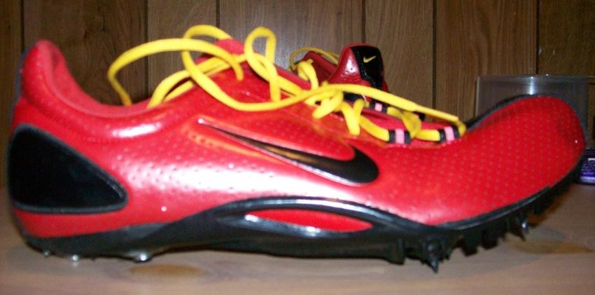
BIOMECHANICAL EVALUATION OF MODIFIED TRACK SHOES

                              A Thesis

              Submitted to the Graduate Faculty of the
                  Louisiana State University and
                 Agricultural Mechanical College
                     in partial fulfillment of the
                  requirements for the Degree of
             Master of Science in Industrial Engineering

                                 in

The Department of Construction Management and Industrial Engineering

                                By
                   Marlon Alberetos Greensword
                        B.S., L.S.U., 2007
                        B.A., L.S.U., 2005
                            May 2010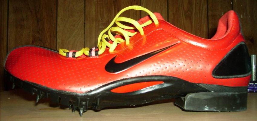
ACKNOWLEDGMENTS

       I must first give thanks to God the Father, Jesus the Son and the Holy Spirit for their

guidance, wisdom, and understanding through my journey in graduate school and in my life.

       I would like to thank my advisor and thesis director, Dr. Fereydoun Aghazadeh, for his

continuous guidance, encouragement, and support throughout my graduate school experience. I

insist on expressing my sincere appreciation as he has been one of my most influential advisors,

guides, or mentors. I would also like to thank committee members Dr. Ikuma and Dr. Nahmens,

who helped me perform to the best of my ability. Your insight, patience, and recommendations

are greatly appreciated.

       I wish to bestow my deep gratitude to my father Anthony Greensword and mother Joan

Bowen. I also want to express special thanks to my brother Mark Greensword and my Uncle

Simon Bowen for their support.

       Dr. Ashish Nimbarte, I am grateful for your advice and guidance during my research.

I am as well indebted to all my subject volunteers for their time and the physical participation in

the lengthy series of experiments.

       Coach Mark Elliott, thank you for helping me identify the needs of a track athlete. I

would not be where I am if it were not for your continuous encouragement and guidance in my

personal, scholarly, and athletic experience. I would also like to show appreciation to my friend

Mario Kelly and sister-in-law Angelique Ngandu for their support.

       Finally, I dedicate this thesis to my wife Sylviane Kalenga Greensword; without her love

and support, this project would not have been a reality.

                                                 ii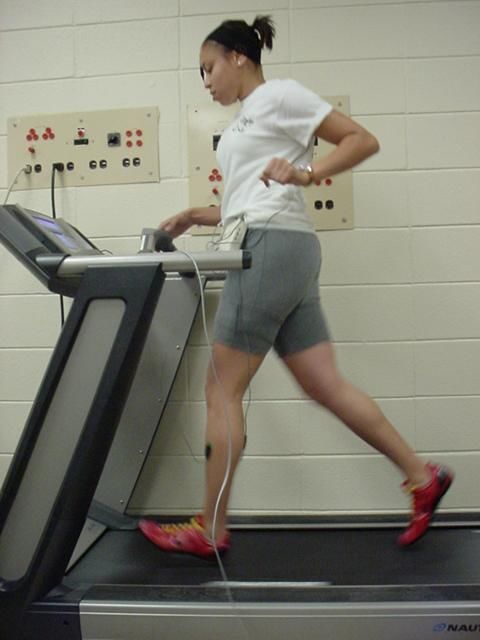
TABLE OF CONTENTS

ABSTRACT ...................................................................................Error! Bookmark not defined.

ACKNOWLEDGMENTS .............................................................................................................. ii

LIST OF TABLES .......................................................................................................................... v

LIST OF FIGURES ....................................................................................................................... vi

ABSTRACT ................................................................................................................................. viii

1.      INTRODUCTION AND BACKGROUND ........................................................................... 1

2.      LITERARY REVIEW ............................................................................................................ 4
     2.1. Common Running Injuries ............................................................................................... 4
     2.2. Running and Walking Techniques and the Common Track Spike Shoe .......................... 6
        2.2.1.    Running and Walking Biomechanics ...................................................................... 6
        2.2.2.    Muscles Involved: The Extrinsic Foot Muscles .................................................... 11
            2.2.2.1.   The Tibialis Anterior Muscle ....................................................................... 13
            2.2.2.2.   The Gastrocnemius Muscle.......................................................................... 14
     2.3. Transition between Running and Walking ..................................................................... 16
     2.4. Consequences of Inappropriate Equipment .................................................................... 18
     2.5. Electromyography (EMG) .............................................................................................. 19
        2.5.1.    Definition and Explanation ................................................................................... 19
        2.5.2.    EMG Applications in Biomechanics, Work Physiology, and Kinesiology .......... 21
     2.6. Previous Use of Removable Heels ................................................................................. 22
            2.6.1. Orthopedics ....................................................................................................... 22
        2.6.2.    Bowling ................................................................................................................. 23
        2.6.3.    Basketball .............................................................................................................. 25
        2.6.4.    Ladies‟ High Heel Shoes with Removable Heels ................................................. 27
     2.7 Summary ......................................................................................................................... 32

3.      RATIONALE ........................................................................................................................ 34

4.      OBJECTIVES ....................................................................................................................... 35

5.      METHODOLOGY AND PROCEDURES ........................................................................... 36
     5.1. Participants ..................................................................................................................... 36
     5.2. Equipment / Apparatus ................................................................................................... 37
     5.3. Task Design .................................................................................................................... 37
        5.3.1.  Installation of the Heel .......................................................................................... 38
        5.3.2.  EMG Experiment .................................................................................................. 39
        5.3.3.  Body Maps ............................................................................................................ 44
     5.4. Nature of the Data ........................................................................................................... 46
     5.5. Hypothesis: Statement and Parameters ........................................................................... 47
        5.5.1.  Hypothesis Testing ................................................................................................ 47
                                                                      iii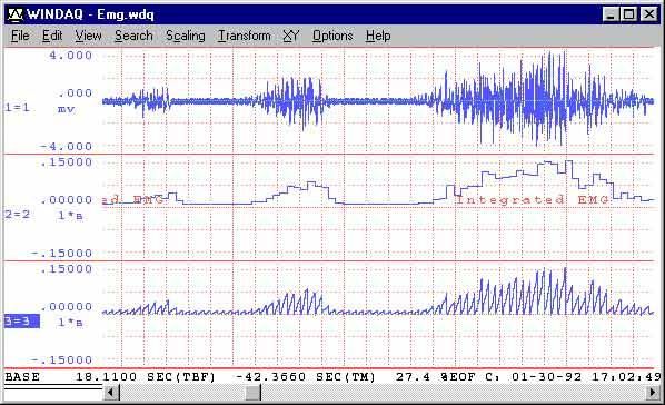
5.5.2.        Statistical Analysis ................................................................................................ 48
        5.5.3.        Steps for Data Processing ...................................................................................... 49

6.      RESULTS ............................................................................................................................. 50
     6.1. EMG ............................................................................................................................... 50
        6.1.1.    Muscles Evaluated ................................................................................................. 53
        6.1.2.    Impact of Speed on Muscle Activity ..................................................................... 53
        6.1.3.    Impact of the Participant‟s Running Background ................................................. 53
        6.1.4.    Statistical Results .................................................................................................. 57
            6.1.4.1.    Hypothesis 1................................................................................................. 58
            6.1.4.2.    Hypothesis 2................................................................................................. 59
     6.2. Body Maps ...................................................................................................................... 59
        6.2.1.    Statistical Results: Hypothesis 3 ........................................................................... 61
        6.2.2.    Relevance of the Participants‟ Athletic Background ............................................ 62
            6.2.3. Remaining Discomfort ..................................................................................... 64

7.      IMPLICATIONS .................................................................................................................. 65
     7.1. Shortcomings .................................................................................................................. 65
        7.1.1.  Limited Number of Participants ............................................................................ 65
        7.1.2.  Limited Prototype Quantity ................................................................................... 65
     7.2. Further Developments .................................................................................................... 66
        7.2.1.  Running and Monitoring Conditions..................................................................... 66
        7.2.2.  Participants Selection and Shoe Characteristics .................................................... 66
        7.2.3.  Further Assessment ............................................................................................... 66
     7.3. Final Remarks ................................................................................................................. 67

REFERENCES ............................................................................................................................. 68

APPENDIX A: CHANGES IN MUSCLE ACTIVITY................................................................ 72

APPENDIX B: BODY MAPS ...................................................................................................... 75

VITA ............................................................................................................................................. 80

                                                                         iv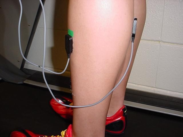
LIST OF TABLES

Table 5.1: Participant running profile ........................................................................................... 36

Table 6.1: Average EMG values ................................................................................................... 50

Table 6.2: Percent average of muscle activity decrease from walking without heels to walking
with heels ...................................................................................................................................... 54

Table 6.3: Analysis of variance for gastrocnemius and tibialis anterior activity.......................... 58

Table 6.4: Averaged discomfort values before and after heel installation ................................... 60

Table 6.5: ANOVA formulas by area ........................................................................................... 61

Table 6.6: ANOVA of body map results ...................................................................................... 61

Table A.0.1: Detailed fatigue change for each subject from EMG results ................................... 72

Table B.0.1: Discomfort values before and after heel installation ............................................... 75

Table B.0.2: Histogram of EMG Results of Tibialis Anterior Activity for Each Participant ....... 76

Table B.0.3: SAS results for discomfort before and after heel installation .................................. 77

                                                                         v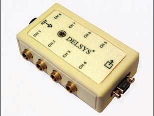
LIST OF FIGURES
Figure 1.1: Example of a typical track spike shoe used by runners ................................................ 2
Figure 2.1: Common overuse running injuries of the lower limbs ................................................. 5
Figure 2.2: Biomechanics of a normal human ankle during level-ground walking........................ 7
Figure 2.3: Ankle torque versus angle during level-ground walking ............................................. 7
Figure 2.4: Pendulum apparatus used to test impact forces .......................................................... 11
Figure 2.5: Anterior and lateral views of extrinsic foot muscles .................................................. 13
Figure 2.6: and muscle activity during normal walking gait cycle ............................................... 15
Figure 2.7: Electromyogram - Example 1..................................................................................... 20
Figure 2.8: Bowling Shoe with Famolare's Separate Heel and Sole Pad ..................................... 23
Figure 2.9: Bowling Shoe with Attached Sole Pad and Heel ....................................................... 24
Figure 2.10: View of Lombardino's Shoe Sole with Original Heel Removed ............................. 25
Figure 2.11: Profile View of Lombardino's Shoe with No Heel Attachment ............................... 26
Figure 2.12: View of Lombardino's Shoe with New Heel Attachment ........................................ 26
Figure 2.13: Shoe construction with self-seating removable heel ................................................ 27
Figure 2.14: Shoe construction with self-seating removable heel_2 ............................................ 28
Figure 2.15: Side view of shoe with high heel and low heel ........................................................ 30
Figure 2.16: The high heel removal process ................................................................................. 30
Figure 2.17: Side and top views of the adjustable shank .............................................................. 31
Figure 2.18: View of shoe with low and high heel when adjusted with the adjustable shank ..... 31
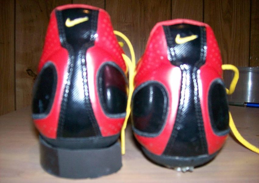
Figure 5.1: Spike shoe with removable heel - side view .............................................................. 38
Figure 5.2: Spike shoes: heel v. no heel - rear view ..................................................................... 39
Figure 5.3: Myomonitor Main Unit .............................................................................................. 40
Figure 5.4: DE-2.3 Single Differential Surface EMG Sensor ...................................................... 41
vi
Figure 5.5: DE Sensor Geometry .................................................................................................. 41

Figure 5.6: Input Module .............................................................................................................. 42

Figure 5.7: EMG alignment .......................................................................................................... 43

Figure 5.8: Subject performing during treadmill experiment ....................................................... 43

Figure 5.9: EMG Sensor orientation with respect to the muscle fibers ........................................ 43

Figure 5.10: Body map sections.................................................................................................... 45

Figure 5.11: Borg's scale for fatigue measurement ....................................................................... 46

Figure 6.1: EMG results at 2 mph ................................................................................................. 51

Figure 6.2: EMG results at 3 mph ................................................................................................. 51

Figure 6.3: Effect of heels on EMG for different speeds .............................................................. 52

Figure 6.4: Percent decrease in EMG when the heel is added ...................................................... 53

Figure 6.5: Percent decrease in tibialis activity for each participant group .................................. 55

Figure 6.6: Plot diagram comparing decrease in tibialis activity for each group according to
speed ............................................................................................................................................. 56

Figure 6.7: Percent decrease in gastrocnemius activity for each participant group...................... 56

Figure 6.8: Diagram comparing decrease in gastrocnemius activity for each group according to
speed ............................................................................................................................................. 57

Figure 6.9: Average discomfort when walking without heels and walking with heels ................ 60

Figure 6.10: Graphed ANOVA results by area ............................................................................. 62

Figure 6.11: Average discomfort difference by participant athletic category .............................. 63

Figure A.1: Histogram of EMG Results of Gastrocnemius Activity for Each Participant ........... 73

Figure A.2: Histogram of EMG Results of Tibialis Anterior Activity for Each Participant ........ 73

Figure B.1: Graph of Body Map Reports for Each Participant..................................................... 76

                                                                         vii
ABSTRACT
Track and field runners, especially sprinters and mid-distance runners, face many
problems due to walking in spike shoes. Due to the fact that track and field spike shoes are
designed specifically for running, the runner‟s feet remain in an uncomfortable, flexed position
when walking between workouts and races. Problems caused by the dangerous foot-positioning
include, but are not limited to, the following: back pain, shin splints, bone spurs, blisters, and
overall decreased level of running performance. Over time, runners wearing improper footwear
for walking may face chronic injuries such as plantar fasciitis, shin splints, Achilles tendinitis,
chondromalacia, and iliotibial band syndrome. To address this problem, a modified spike shoe
was tested. The modification consists of adding a removable heel to the shoe. The removable
heels were attached to the sole after exercise or between races to shoe angle of flexion, so that
the foot can be leveled. The modified shoes were tested in terms of health and comfort through
the use of two experimental protocols. Nine healthy, resistance-trained participants volunteered
to perform walking drills on a treadmill. They walked with regular spikes at 2 mph and 3 mph.
Then, they repeated the drill with the redesigned spike shoes. EMG measurements were used to
evaluate the participant‟s muscle activity, fatigue, and stress during the exercise. The analyzed
muscles were the tibialis anterior and the medial gastrocnemius. The statistical tool used for the
mathematical interpretation of the data was ANOVA, the hypotheses being tested with the
softwares Statistix 9.0. and SAS 9.1 English version. Complementarily, participants were
individually asked to rate their discomfort on a scale of 1 to 10, using a body map as a further
evaluation of the effects of the removable heel. Results showed a 22 % average decrease in EMG
muscle activity from walking without heels to walking with heels in the tibialis anterior and a
24.25% average decrease in the gastrocnemius. Results were consistent for all participants.
viiiSimilarly, when rating discomfort from walking without heels to walking with heels, the body
map survey results indicate that participants noticed an average superior comfort of 2.7 points in
the knees, 2.6 points in the calves, 3.9 points in the ankles, and 4.2 points in the feet on an
ergonomic scale of 10 discomfort points. Thus, results showed that the removable heel helps
reduce muscle fatigue and stress and therefore its related musculoskeletal problems.
ix1. INTRODUCTION AND BACKGROUND
Track-and-field athletes wear spike shoes, which are designed exclusively for running
during training and competition (track meets). Based on observations of the LSU track team
during regular track season, athletes wore their spike shoes for a daily average of one hour and
25 minutes, of which 45 minutes are spent walking. In addition, further observations indicated
that, between workouts, athletes walk in their spikes on an average of 5 hours per week, which
indicated an excessive time spent in footwear not meant for walking. Over a span of time,
depending on the spike shoe design, some sprinters and mid-distance athletes (800 m runners)
suffered several injuries, especially while walking in their spike shoes, which are made only for
running. Such injuries include plantar fasciitis, shin splints, Achilles tendinitis, chondromalacia,
and iliotibial band syndrome (McGrath and Finch, 1996).
Spike shoes' shapes differ depending on the athlete‟s area of competition (mid-distance,
sprint/short distance (50 to 400 m), long distance (1 mile to marathon), jumps, etc.). These
lightweight shoes are named after the small metal spikes that range in size, depending on the
track surface on which an athlete competes. The metallic pieces are attached to the bottom of the
shoes. Thus, they are removable, and the athlete may replace them with longer or shorter spikes
according to his or her needs. Spikes also differ in aerodynamics, depending on the event.
Runners use this specific type of shoe, because the shoe enables their feet to stick to the mondo
or rubber surface, thereby minimizing shock and sliding.
This project is designed to test the feasibility of an innovative track-and-field spike shoe.
In the first approach, a description of the ideal shoe that track-and-field athletes need provides
data such as shape and materials needed.
1Figure 1.1: Example of a typical track spike shoe used by runners
The second part exposes the main problems currently encountered with some of the
classic spikes. This section is introduced with pictures showing different dimensions of the spike
and is followed by an analysis of the shoe‟s potential harm to the athlete‟s feet. In effect, the
shoes were created to fit runners, and therefore, wearing them for a different activity – in this
instance, walking – may cause back pain, shin splits, bones pure, and blisters. It also tends to
decrease running performance, since injured runners cannot perform to the best of their abilities.
Thirdly, the solution proposed and tested in this project is a shoe with a removable heel,
that is, the opportunity for every track-and field athlete to get their own custom-built spikes
specified to his or her activity – running and walking. The efficiency of this feature is tested on
the classic Nike spikes, in terms of health protection and athletic performance, focusing on
muscle fatigue.
Finally, this thesis concludes by elaborating on the implication of the study, including
notes for further research.
Regarding the commerciality of the product, one must note that some of the features
hereby proposed have already been presented but have never been applied to spike shoes. If Nike
2were to adopt such a proposition, they would gain a wider clientele on the track-and-field
market, more satisfied customers, as well as promotional benefits in terms of their interest in
preserving the athlete‟s health.
32. LITERARY REVIEW
2.1. Common Running Injuries
McGrath and Finch‟s (1996) report features a cause-and-effect analysis of common
injuries among track runners. The document provides detailed descriptions of what causes the
problems faced when runners wear track spikes. This research showed the following related
injuries that can occur when the spike shoes do not fit the athlete:
Plantar fasciitis: an inflammation of the thick band of tissue from the heel to the base of
the toes in the bottom of the foot. When placed under stress, the plantar fascia stretches
and tears, leading to inflammation.
Shin splint, also called tibial stress syndrome: the inflammation of the tendons on the
inside of the front of the lower leg, i.e. the shins.
Achilles tendinitis: occurs when the Achilles tendon, a large tendon connecting the two
major calf muscles (gastronemius and soleus), is placed under too much stress causing
inflammation. If the inflamed Achilles continues to be stressed, it can tear or rupture.
Chondromalacia: a cracking or wearing away of the cartilage under the kneecap,
resulting in pain and inflammation.
Iliotibial band syndrome: inflammation and pain on the outside of the thigh, where the
iliotibial band rubs against the femur.
Such findings are comparable to the previously mentioned observations of the Louisiana State
University track-and-field team. They also coincide with statements from the track-and-field
team staff.
In a personal interview (2007), Assistant Coach Mark Elliott, who coaches distance and
mid-distance at Louisiana State University, stated that track athletes wear spikes on a minimum
basis of four days per week and three hours per day. Elliott added that from two months to one
4year, the amount of time spent wearing spikes accumulates, and athletes tend to develop
problems in their legs, lower back, tibia, metatarsals, and patella, as illustrated in Figure 2.1.
Figure 2.1: Common overuse running injuries of the lower limbs
Source: McGrath and Finch (1996, 37-38)
5The research of Myburgh et al. (1988) indicated that 84% of injuries among runners
occur on the tibia (shin) area, and 13% are linked to the Achilles tendon. This project thus
focuses on these two areas and the related muscles.
2.2. Running and Walking Techniques and the Common Track Spike Shoe
2.2.1. Running and Walking Biomechanics
Au et al. (2006) provided a model for the ankle-foot walking process, in which the study
described each phase and its corresponding foot angle. The walking process is divided into two
major stages: stance and swing. Stance, which constitutes the majority of the walking process
(about 60%), is made up of three phases or moments:
Controlled plantarflexion: begins at heel-strike and ends at foot-flat. During this
phase, the heel and forefoot make initial ground contact.
Controlled dorsiflexion: begins at foot-flat and continues until the ankle reaches a
state of maximum dorsiflexion. The main function of the human ankle during
controlled dorsiflexion is to store the elastic energy needed to propel the body
upwards and forwards during the next phase
Powered plantarflexion: begins at maximum dorsiflexion and ends at toe-off.
During this phase, additional energy is supplied, along with the spring energy
stored during the previous phase to achieve the high plantarflexion power during
late stance.
Swing is thus the phase that separates each stance, starting at toe-off and ending at heel-
strike. During swing, the foot is lifted, proceeding to actual geographic locomotion. The
following figure illustrates the biomechanics of walking and its distinct phases:
6Figure 2.2: Biomechanics of a normal human ankle during level-ground walking
Source: Au et al. (2006)
Figure 2.3: Ankle torque versus angle during level-ground walking
Source: Au et al. (2006)
Figure 2.3 graphs the ankle angle change in combination with the torque, that is, the
rotary moment during walking. Segments 1-2, 2-3, and 3-4 represent the ankle torque-angle
behaviors during Controlled Plantarflexion, Controlled Dorsiflexion, and Powered Plantarflexion
phases of gait, respectively. One must note, however, that the foot does not touch the ground
during the swing phase and therefore little pressure is exerted upon the heel, the Achilles tendon,
7or the tibialis anterior. The research concerning this project is more centered on the stance phase.
Furthermore, one may notice on Figures 2.2 and 2.3 that the swing phase of one leg corresponds
to the stance phase of the other.
Given the fact that each athlete has a unique running technique (gait), a shoe should be
adjustable to facilitate and support his/her motion. Raptopoulos et al. (2006) conducted a gait
analysis of gait modes among men and women. Their results showed stronger hip movements
among women. However, women‟s walking and running pattern indicate use of extrinsic foot
muscles similar to that of men. When combined with observations from the Louisiana State
University track team and testimonials from the team staff regarding female injuries,
Raptopoulos et al.‟s (2006) gait analysis indicate that female runners could benefit from the
removable heel as much as men could.
In Principles of Human Anatomy, Tortora (2002) defines the movements that characterize
different walking and running techniques:
Inversion (to turn inward) is a movement of the soles medially at the intertarsal
joints (between the tarsals).
Eversion (to turn outward) is a movement of the soles laterally at the intertarsal
joints.
Dorsiflexion refers to bending of the foot at the ankle or talocrural joint
(between the tibia, fibula, and talus) in the direction of the dorsum (superior
surface). Dorsiflexion occurs when one stands on one‟s heels.
Plantarflexion involves bending of the foot at the ankle joint in the direction of
the plantar or inferior surface, as when standing on one‟s toes. Dorsiflexion is
true flexion, whereas plantar flexion is true extension.
8The classic spikes are designed for plantarflexion, that is, for short and mid-distance
runners. Yet, these athletes walk in dorsiflexion mode (flat-footed), while common spikes are
generally designed for athletes to run on the toes. Thus, those who walk on their heels after
extreme physical activity have minimal stability and cushion and therefore, can easily hurt their
Achilles‟ tendons, as much pressure is exerted onto their tibia and gastrocnemius.
In addition, Donley and Leyes (2001) wrote that extreme or repetitive dorsiflexion can
lead to direct trauma, along with anterior bony ankle impingement, that is, the formation of
osteophytes (bone spurs) on the anterior edge of the distal tibia. This is, according to the article, a
common problem among runners. Injuries related to extreme dorsiflexion are limiting the athlete
not only in terms of painful symptoms, but also because they eventually prevent the runners from
performing to the best of their abilities: “patients will complain of painful limitation of
dorsiflexion, catching, and swelling of the ankle. These symptoms can be debilitating and
considerably limit their athletic performance.” Such injuries would increase in intensity and
gravity were dorsiflexion to be practiced in shoes designed for plantar flexion. For instance, the
non-operative treatments Donley and Leyes (2001) proposed in a successfully experiment
included rest, rubber-sole wedge shoes, and, interestingly, an internal or external heel lift.
Saunders et al. (1953) established a list of gait determinants that differentiate normal and
pathological walking. According to their research, an inefficient, pathological gait pattern is
characterized by numerous lateral and vertical excursions in the body‟s center of gravity. Thus,
using the argument that “locomotion is the translation of the center of gravity through space
along a pathway requiring the least expenditure of energy supplies” (Saunders et al., 1953),
minimizing these excursions improves the quality of the gait. The article states that gait
assessment is accomplished upon observation of these six major factors, also called major
determinants:
9pelvic rotation,
pelvic tilt,
knee flexion,
hip flexion,
knee and ankle interaction, and
lateral pelvic displacement
The determinant of interest in this thesis is the knee and ankle interaction. In effect, Thompson‟s
(2002) study of Saunders et al. (1953) designated this determinant as one of the limitations to the
troughs (or low points) in the sinusoidal pathway that occur during the gait cycle. An analysis of
the biomechanics of walking (see Figure 2.3) finds that walking in spike shoes without a heel
challenges the ankle in maintaining balance in the body, creating a vertical excursion in the
center of gravity as pressure is exerted on the tibialis anterior and the Achilles tendon. Thus,
when compared to the normal human gait pattern, walking in spike shoes with no heels qualifies
as a pathological gait, and therefore requires correction. This project presents a removable heel
as a potential correction to such a pathological gait.
Wakeling et al. (2001) conducted a study of the muscle activity as a response to ground
reaction forces. Their project began from the starting point in which the human body reacts to the
impact forces that occur at heel strike. Their study thus tests the level of muscle activity in the
lower extremity muscles (among which are the gastrocnemius and the tibialis anterior) as they
respond to the rate of impact forces, using a pendulum to deliver impacts to the heel repetitively,
using various materials in the subjects‟ shoes, as seen in Figure 2.4. The pendulum apparatus, as
set in the illustration, was pulled back to a reference stop. It swung for the subject to impact the
wall with his right heel.
10Figure 2.4: Pendulum apparatus used to test impact forces
Source: Wakeling et al. (2001)
The results of this study showed that there is a ratio of 96% in the tibialis anterior and 48% in the
gastrocnemius between the pre-activation intensity and the muscle activity intensity. Therefore,
conditions in which the impact to the ground is increased, such as when walking in heelless spike
shoes, implicate a more intense muscle activity in the gastrocnemius and tibialis anterior,
eventually or occasionally resulting in stress or fatigue. Inversely, a feature that would absorb
some of the intensity of the impact, such as a removable heel, should reduce the intensity of
muscle activity and thereby reduce risks of injuries related to stress and fatigue in the lower
extremities.
2.2.2. Muscles Involved: The Extrinsic Foot Muscles
Extrinsic foot muscles can be defined as the “muscles that insert on the foot but originate
proximal to the foot” (O‟Connor et al., 2004). This group is constituted by the following muscles
(Smith et al., 1996):
Triceps surae
o gastrocnemius
o soleus
11tibialis posterior
flexor digitorum longus
flexor hallucis longus
peroneus longus and brevis
tibialis anterior
extensor hallucis longus
extensor digitorum longus
Because of their impact on foot motion, this specific group of muscles is related to
numerous running injuries among track-and-field athletes. O‟Connor et al. (2004, 2006) studied
the role of extrinsic foot muscles during running using mfMRI technology and, more
extensively, electromyography. According to the authors, these muscles act as “invertor muscles
of the foot and are attributed the primary role in resisting foot pronation during the first stance”
(O‟Connor et al., 2006). Pronation being the act of turning one‟s feet downward – as opposed to
supination – extrinsic foot muscles are responsible for maintaining balance and channeling
energy toward the purpose of geographic motion (walking or running). Figure 2.5 provides a
labeled visual representation of the extrinsic foot muscles.
Due to the importance of their role during the walking and running stance and to the
preponderance of tibial stress syndrome and Achilles tendinitis injuries among track-and-field
runners, two extrinsic foot muscles, the tibialis anterior and the gastrocnemius, were selected to
provide insight into the quality of walking after running, thus evaluating biomechanically
modified track-and-field spike shoes. These two muscles were targeted in the study as their
muscular reaction to the removable heel provides valuable information as to any reduction in the
risk of exercise-induced injury.
12Figure 2.5: Anterior and lateral views of extrinsic foot muscles
Source: www.chionline.com/anatomy
2.2.2.1. The Tibialis Anterior Muscle
This muscle is an invertor of the foot. Reber et al. (1993) and Hunt et al.‟s (2001)
measurements of extrinsic foot muscles‟ impact indicate some degree of tibialis “overuse”
among track-and-field athletes during running, but a failure to relieve and rest this muscle after
exercise, as walking demands effort from this muscle as well if the foot does not have the
support needed such as that provided by a heel. In effect, “tibialis anterior fires above the fatigue
threshold for 85% of the time. This may account for the high number of fatigue-related injuries
to the tibialis anterior muscle seen in runners” (Reber et al., 1993). This statement is particularly
relevant in light of the tibialis anterior‟s controlling role on heel stress. Indeed, during walking,
this muscle “restrains rearfoot plantarflexion from heel contact to 10% stance (see Figure 2.3)
13and eversion between 10% stance and footflat” (Hunt et al., 2001). Thus, literature suggests that
if extreme stress is inflicted to the tibialis anterior during running, an athlete must have extra heel
support to make up for a fatigued foot-invertor muscle.
2.2.2.2. The Gastrocnemius Muscle
This muscle is responsible for controlling foot motion, exerting much force during
walking and running for plantar flexion and foot pronation resistance (O‟Connor et al., 2004,
2006). Thompson (2002) provides a visual of the role of this muscle during a normal walking
gait, as depicted in Figure 2.6 that illustrates the gastrocnemius activity during stance, illustrating
the intensity of the pressure exerted on the ankle and the gastrocnemius during walking.
Regarding Achilles tendinitis, the second most common injury among track athletes, it is
important to understand to impact of the gastrocnemius muscle activity. Effectively, this injury is
an inflammation of the Achilles tendon, which is constituted of tendons of the gastrocnemius and
soleus muscles (see Figure 2.5). Roy (1988), who studied and experienced running injuries,
explains that running shoes are supposed to possess a heel wedge to reduce Achilles tendon
stretch. Similarly, Reilly‟s (2009) historic of the ergonomics of running shoes explains that
shock absorption properties such as outer and midsole with a wedge in between at the shoe‟s
back, air bubbles, or heel counters aids in stabilizing the rearfoot and decreasing the risk of
Achilles tendinitis. Nevertheless, such features apply to shoes designed for long distance runners,
since their stance is longer than that of sprinters and mid-distance runners; this stance increases
the need for heel support during running. Yet, heelless sprint and mid-distance spike shoes are
elevated at the toes to aid in speeding up the stance during running. In addition, they provide no
heel wedge during the longer walking stance, which means that the Achilles tendon of a walking
athlete is stretched more intensely – due to the shape of the shoe – and for a longer amount of
time –walking taking longer than running – if the athlete does so in his / her spike shoes.
14Figure 2.6: and muscle activity during normal walking gait cycle
Source: Thompson (2002)
15In regard to another common injury among track-and-field runners that involves the
gastrocnemius, namely plantar fasciitis, the main cause is an inflammation of the plantar fascia
due to abnormal pronation (Roy, 1988), that is, a failure of the gastrocnemius to properly resist
pronation.
2.3. Transition between Running and Walking
The biomechanical modification hereby tested enables runners to rest their heels as well
as reduce gastrocnemius and tibialis fatigue after or before exercise or competition. Indeed, the
study of O‟Connor et al. (2006), examining the role of extrinsic muscles through mfMRI and
EMG measurements, indicates that these muscles control rearfoot motion to alter the activation
in order to maintain a preferred movement pattern. Thus, transition from one gait to another
(from running to walking), is a naturally challenging phenomenon for the runner‟s extrinsic foot
muscles. This suggests that a runner‟s extrinsic foot muscles could benefit from assistance or
intervention in altering foot motion, especially after an intensive race.
Measurements of muscle fatigue during transition from running to walking and vice-
versa have been performed and reported in a published study from the Department of Movement
and Sport Sciences at Ghent University (Segers et al., 2006). The study mostly consisted of EMG
measurements on a group of subjects‟ tibialis anterior. According to Segers et al. (2006), tibialis
anterior fatigue is attributable to more than just the metabolic change or cost, which is to say, the
“change to another type of locomotion reduces oxygen consumption.” In effect, differences of
foot angle when switching from plantarflexion (running) to walking tends to affect the intensity
of tibialis anterior activity. EMG of the tibialis anterior during walking and running has a typical
pattern with a burst during the eccentric foot plantar flexion movement following heel contact.
Such eccentric activity tends to increase exertion, which might serve as protective mechanism to
prevent further damage. Walking and running, when performed at speeds in proximity of the
16transition-speed, differed in the fact that the touch-down angle of the foot is smaller during
running. Another possible explanation could be the greater instability of the foot after TA
fatigue. During the heel strike stage of walking stance (see Figure 2.3), the eversion load is
maximized. If the TA fails to sufficiently counteract foot eversion, with secondary function
preventing eversion, this would cause a medial shift of the center of pressure and a lateral shift of
the center of mass.
This report thus indicates not only that the foot tends to be more instable when walking after
running, but also that a reduction in heel pressure would certainly reduce fatigue of the tibialis
anterior as well.
Similarly, Nigg et al. (2003) examined the influence of shoe soles on muscle activation
and energy through EMG measurements. Their research indicated a change in oxygen
consumption and a change in lower extremities muscle activation (including tibialis anterior and
medial gastrocnemius), as the subjects ran in shoes with different heel materials. During running,
track athletes experience forces of impact that range between 1.0 and 2.5 times their body
weight. Running thus constitutes a great effort on muscles in the lower extremities, which could
benefit from after-running support to absorb and provide relief them from some of the shock due
to heel impact when walking away from the track after competition or exercise. In addition, the
authors use the expression “muscle tuning” to refer to the muscles‟ tendency to adopt an
activation pattern. This complements the conclusions of O‟Connor et al. (2006). Nigg et al.
explained that muscles become conditioned to acting a certain way during a specific activity,
thereby making a transition to another activity increase muscle effort and sometimes trigger
fatigue. In the case of the transition from running to walking, Nigg et al. suggested that “a
reduction of muscle activity before heel strike is associated with fatigue.” Since walking implies
a reduction of muscle activity from running, it can be deducted that a decrease in locomotion
17speed (transitioning from running to walking) can also trigger fatigue due to the effort of pulling
away from the previous “muscle tuning” experienced during running.
Thus, in light of Segers et al. (2006), it may be concluded that the heel material directly
influences muscle activation and the subsequent oxygen consumption. One can therefore expect
a significant change in muscle activity during the experiment performed in this thesis. However,
one major difference from the studies of Nigg et al. (2003) and Segers et al. (2006) is that the
task assigned to the subjects was to run in various heel and sole conditions, whereas in this
thesis, participants were asked to walk at different speeds and in different heel conditions.
Nevertheless, Nigg et al. stated that the phenomena studied and reported should provide insight
into understanding “many aspects of human locomotion, including work, performance, fatigue,
and possible injuries,” thus making their research and the corresponding results relevant for
studying the locomotion mode of walking in various heel conditions.
2.4. Consequences of Inappropriate Equipment
Research regarding the ergonomics of sports shoes is a relatively new phenomenon. Reilly
(2009) wrote a historical analysis of the running shoes market, whereupon sports shoes
companies became attentive to safety issues in the 1970s-1980s explosion in the popularity of
track-and-field events. Before that, runners carried lightweight spike shoes for the track, as well
as heavier shoes to exercise outside the track. Manufacturers then introduced new materials
based on “ergonomics criteria that prioritized comfort, safety, and performance” (Reilly, 2009),
designed by Dr. Peter Cavanagh.
A personal interview with Kaitlin Smith (2007), an athletic trainer at Louisiana State
University, was conducted on relative subject matters. Smith stated that among LSU track and
field athletes, the most common injuries that resulted from wearing cleats, under normal
circumstances (properly worn and fitted, and used only during the event), were turf toe, a sprain
18of the ligaments in the big toe, and plantar fasciitis, the inflammation of the interface between the
fascia and the first layer of intrinsic muscles. After returning from a period that excluded
training in their spikes, Smith notes that the athletes complained a lot about overall soreness.
Some examples were blisters, metatarsal bruising, and a tight Achilles or calf muscle. She further
commented that the worst danger in over-use of cleats would be chronic Achilles tightness,
shortening of the calf muscles, tight hamstring muscles, low back pain, associated foot soreness,
and an altered running stride.
A level spike shoe to reduce some, if not all, of these risks was suggested. Smith replied:
“A leveling of the heel increases heel striking, therefore making the runners slower. However,
during times where the athletes do not need to be in spikes, the level cleat would benefit them. In
conjunction with the level cleat, communication with the coaching staff of any problems is key,
especially with the dangers of over-use of spikes.” According to Smith, the athletes would be
interested in the use of a level cleat with a removable heel. This would not only cause an easy
switch from walking footwear to competition footwear, but this would also reduce the number of
various types of footwear that the athletes carry.
2.5. Electromyography (EMG)
2.5.1. Definition and Explanation
EMG is the use of various electronic devices that use volts to measure muscle activity in
the body. It aims at measuring a specific muscle‟s activity by interpreting electrophysiological
signals. EMG is used in various industries, such as a) medicine, b) sports medicine and
kinesiology, c) work physiology, and d) biomechanics, to provide “objective evaluation of the
musculoskeletal stress” (Lee et al., 1986). Further, EMG monitors physiological parameters for
the generation of ergonomic or medical solutions to problems related to muscle pain, fatigue, and
abnormal / inappropriate activity (Solomonow et al., 2003).
19There are several types of EMG systems, depending on the user‟s needs in regard to area
/ discipline. One of he most commonly used in biomechanics is surface EMG. Surface EMG is
characterized by the use of adhesive electrodes (called non-invasive) that are placed on the skin
that covers the muscle of interest. Other types of equipment include wire and needle electrodes
that are inserted through the skin to the muscle of interest (Nimbarte, 2009).
For this study, surface EMG was employed (see Figure 5.7). A wireless, battery-operated
device was used to provide the flexibility and freedom of movement needed for the participants
to complete the walking drills (lightweight and transmission up to 250 m). Participants were
tested with a Myomonitor IV Wireless Transmission and Datalogging system by Delsys.
Results of EMG testing are displayed in a graph called an electromyogram, sometimes
called an EMG graph. Figure 2.7 provide examples of typical electromyograms:
Figure 2.7: Electromyogram - Example 1
Source: Weimer, 2009
Using a software such as Excel, an electromyogram‟s appearance can be modified by
adding comments, legends, or titles. To compile the data, to average it, or to compare different
20subjects‟ information, EMG data must be normalized. Sommerich et al. (2000) provide an
analytical review of the use of surface EMG. They present normalization as the process used “to
address variation introduced in the measurement process by differences in electrode spacing,
natomical factors, and variation in electrode placement in order to facilitate comparisons
between differenct muscles and individual subjects.” Failure to proceed to normalization can
affect the reliability of the results because the amplitude‟s percentage will include exertion
movement that are irrelevant to the study.
Weimer (2009) explained EMG waveform interpretation, stating that a solitary graph
does not mean much. However, when compared to another one, a standard case or a different
condition undergone by the same subject / patient / participant, an electromyogram reveals
“which case represents the greatest amount of work done by the muscle”.
2.5.2. EMG Applications in Biomechanics, Work Physiology, and Kinesiology
Nigg et al. (2003) provided an example of EMG application for ergonomic purposes. The
study used electromyography to identify muscle activity for selected muscles, including medial
gastrocnemius and tibialis anterior, with 20 participants performing drills in two differently-
heeled running shoes. Similar to the experiment conducted in this thesis, the data collected “were
compared for the different conditions using an ANOVA (α = 0.05).” The same testing procedure
was used in this study, within a similar controlled environment, with the exception of the
following major elements:
Rather than changing shoes, the participants kept the same shoes, yet exchanged
the removable heel for one of the drills.
Body maps were also used to complement the objective scientific EMG data with
the subjective ratings, to indicate how the subject experienced the heel/without
heel change in terms of human pain and discomfort.
212.6. Previous Use of Removable Heels
The concept of a shoe with a removable heel is not a new invention per se. Several
inventors have examined this option and even submitted patents in the past, as early as the 1880s.
The earliest patents promoted removable heels, because heels historically represent the fastest
worn part of a shoe. Replacing a heel is thus less expensive than buying a whole new pair of
shoes. Later on, the literature indicates a certain diversity in the design and use of removable
heels, and the concept has been adapted and adopted in various areas, including the arena of
sports.
2.6.1. Orthopedics
Orthopedic heel elevations are common practice. They usually consist in adding a
prosthesis inside the shoe. People with pathologically asymmetrical leg length use the prosthesis
to even leg length and restore balance in their hips and back. The device is usually small enough
to fit inside the shoe in a virtually invisible manner.
Orchard et al. (1996) used a similar device with long-distance track athletes who suffer
from iliotibial band friction syndrome (ITBFS) (see Figure 2.1). This type of injury is common
among distance runners (McGrath and Finch, 1996), because of their recurrent knee flexion to
the angular zone in which friction occurs, that is, 30˚ (whereas not only do sprinters flex their
knees beyond the impingement zone, but the flexion moment spent in the friction zone is much
shorter, since they run faster than distance runners do). The study consisted in a cadaveric
anatomical examination of 11 normal knees and a video analysis of 9 distance runners suffering
from ITBFS who ran on a treadmill for 2 minutes twice. For this dynamic section of the study,
the subjects ran once with normal running shoes, and then ran for a second time with a 50 mm
heel raise.
22Despite the methodological resemblance between the biomechanical model proposed in
Orchard et al.‟s (1996) study and the removable heel proposed in this thesis, the purposes differ
in that the 50 mm heel elevation device was designed for etiological evaluation and assessment
of ITBFS. Indeed, the model aimed at identifying angular patterns in the gait of distance runners
with ITBFS, not at correcting these patterns, whereas the removable heel evaluated in this thesis
is proposed as a solution to prevent running injuries among sprint runners.
2.6.2. Bowling
Famolare (1994) designed a bowling shoe, in which the removable heel functions to
“vary the friction of the bowling shoe sole on the bowling surface.” This shoe is characterized by
a hook and pile fastener that makes a sliding pad.
Figure 2.8: Bowling Shoe with Famolare's Separate Heel and Sole Pad
Source: Famolare (1994)
23Figure 2.9: Bowling Shoe with Attached Sole Pad and Heel
Source: Famolare (1994)
As illustrated in Figures 2.8 and 2.9, the attachment proposed by Famolare is different
from the removable heel evaluated in this study, in that it has a sliding mechanism, rather than
being screwed to the sole. Moreover, the bowling shoe is modified by increasing the height of
the sole together with that of the heel. In contrast, the track-and-field spike shoes considered in
this thesis were modified exclusively at the heel area, since the purpose is to establish a balance
of the foot, which is lost in the use of the original curved, spiked, track shoe.
The sliding mechanism is commendable in that it requires less effort – and less time – to
install than the time required to screw a removable heel underneath a track spike shoe. However,
this action requires a perfect match in dimensions, whereas the screw-on removable heel
evaluated here can adapt to different models of spike shoes.
242.6.3. Basketball
In 1996, Lombardino proposed a removable heel for athletic shoes. Even though this
invention represents the closest model to the modified spike shoes evaluated in this thesis, one
major difference is that Lombardino designed a shoe with two heel options. In other words, the
athlete can choose between two shapes of heels to fit the activity, whether it is plyometric
training or general use. Another difference is that these shoes are better suited for basketball
practice, as shown in Figures 2.10 through 2.12, whereas this thesis evaluates a shoe specifically
designed for track.
Figure 2.10: View of Lombardino's Shoe Sole with Original Heel Removed
Source: Lombardino (1996)
25Figure 2.11: Profile View of Lombardino's Shoe with No Heel Attachment
Source: Lombardino (1996)
Figure 2.12: View of Lombardino's Shoe with New Heel Attachment
Source: Lombardino (1996)
The above figures display an attachment that slides in and locks at the sole. Again, this
marks another difference with the removable heel of the track spike shoe.
26You can also read