Biomedical and Tissue Engineering Strategies to Control Foreign Body Reaction to Invasive Neural Electrodes
←
→
Page content transcription
If your browser does not render page correctly, please read the page content below
REVIEW
                                                                                                                                            published: 25 May 2021
                                                                                                                                   doi: 10.3389/fbioe.2021.659033

                                            Biomedical and Tissue Engineering
                                            Strategies to Control Foreign Body
                                            Reaction to Invasive Neural
                                            Electrodes
                                            Manuele Gori 1,2* , Gianluca Vadalà 1 , Sara Maria Giannitelli 3 , Vincenzo Denaro 1 and
                                            Giovanni Di Pino 4
                                            1
                                              Laboratory for Regenerative Orthopaedics, Department of Orthopaedic Surgery and Traumatology, Università Campus
                                            Bio-Medico di Roma, Rome, Italy, 2 Institute of Biochemistry and Cell Biology (IBBC) - National Research Council (CNR),
                                            Rome, Italy, 3 Laboratory of Tissue Engineering, Department of Engineering, Università Campus Bio-Medico di Roma, Rome,
                                            Italy, 4 NeXT: Neurophysiology and Neuroengineering of Human-Technology Interaction Research Unit, Università Campus
                          Edited by:        Bio-Medico di Roma, Rome, Italy
                Sandro Mussa-Ivaldi,
              Northwestern University,
                       United States
                                            Neural-interfaced prostheses aim to restore sensorimotor limb functions in amputees.
                        Reviewed by:
                                            They rely on bidirectional neural interfaces, which represent the communication bridge
                                Xin Liu,    between nervous system and neuroprosthetic device by controlling its movements
   University of California, San Diego,
                                            and evoking sensory feedback. Compared to extraneural electrodes (i.e., epineural
                          United States
        Senentxu Lanceros-Mendez,           and perineural implants), intraneural electrodes, implanted within peripheral nerves,
        Basque Center for Materials,        have higher selectivity and specificity of neural signal recording and nerve stimulation.
    Applications and Nanostructures,
                                 Spain
                                            However, being implanted in the nerve, their main limitation is represented by the
                 *Correspondence:
                                            significant inflammatory response that the body mounts around the probe, known
                        Manuele Gori        as Foreign Body Reaction (FBR), which may hinder their rapid clinical translation.
                 m.gori@unicampus.it
                                            Furthermore, the mechanical mismatch between the consistency of the device and the
                    Specialty section:
                                            surrounding neural tissue may contribute to exacerbate the inflammatory state. The
         This article was submitted to      FBR is a non-specific reaction of the host immune system to a foreign material. It is
             Bionics and Biomimetics,
                                            characterized by an early inflammatory phase eventually leading to the formation of a
                a section of the journal
       Frontiers in Bioengineering and      fibrotic capsule around intraneural interfaces, which increases the electrical impedance
                        Biotechnology       over time and reduces the chronic interface biocompatibility and functionality. Thus, the
         Received: 26 January 2021          future in the reduction and control of the FBR relies on innovative biomedical strategies
           Accepted: 27 April 2021
           Published: 25 May 2021
                                            for the fabrication of next-generation neural interfaces, such as the development
                             Citation:
                                            of more suitable designs of the device with smaller size, appropriate stiffness and
     Gori M, Vadalà G, Giannitelli SM,      novel conductive and biomimetic coatings for improving their long-term stability and
       Denaro V and Di Pino G (2021)
                                            performance. Here, we present and critically discuss the latest biomedical approaches
   Biomedical and Tissue Engineering
   Strategies to Control Foreign Body       from material chemistry and tissue engineering for controlling and mitigating the FBR in
           Reaction to Invasive Neural      chronic neural implants.
                           Electrodes.
 Front. Bioeng. Biotechnol. 9:659033.       Keywords: neural electrodes, foreign body reaction, coatings, biomaterials, hydrogel, tissue engineering,
     doi: 10.3389/fbioe.2021.659033         microfluidics, nanofabrication techniques

Frontiers in Bioengineering and Biotechnology | www.frontiersin.org              1                                             May 2021 | Volume 9 | Article 659033
Gori et al. Neuroengineering and Foreign Body Reaction
INTRODUCTION in long-term implants (Anderson and Weir, 2019). Although
some of the current peripheral nerve interfaces can shorten
Since scientists started to invasively study the function of the latency and provide single axon specificity, their performances
central nervous system (CNS) and peripheral nervous system tend to degrade with time due to the biological response of the
(PNS), single electrodes, and later on electrode arrays, have been organism to the electrode, which is triggered by the damage
implanted to record neuronal activity and to stimulate single or provoked by the implant procedure itself (Anderson and Weir,
groups of neurons to artificially induce their activation, in light of 2019). The body tends to insulate and exclude the foreign material
decoding their functions. from the surrounding microenvironment, leading to scar tissue
Once study protocols moved from acute tests to chronic growth around the device that is made of a fibrous capsule.
implantations and the safety of implants performed in primates In the conductive surface, the dielectric constant, dissipation
suggested the possibility to move to studies in humans, a further factor and dielectric loss factor rise with the increase of the
possible application of invasive neural electrodes, beside that capsule thickness. The increase of the electrical impedance
to investigate neuronal functions, became concrete. Electrodes is proportional to the development of the fibrotic tissue,
started to be employed to decode subject motor intention which determines difficulties to distinguish the signal from
and, bypassing neural or osteo-muscular lesions, to artificially background noise (Szostak et al., 2017) and, eventually, the drop
interface the nervous system to the external environment. of stimulation and registration capacities (Guadarrama-Santana
When this happened, neural interfaces -often named brain and Garcia-Valenzuela, 2007; Jayamani et al., 2014).
to computer or to machine interfaces- and the field of The immune-mediated response responsible for the capsule
neuroprosthetics were born. Depending on the site and the growth is known as Foreign Body Reaction (FBR). FBR reduction
subject receiving the implant, electrodes can also be interfaced over time is probably the main challenge for future neural
with sensory area and fibers and, by relaying afferent streams of electrode applications in neuroprosthetics to extend the reliability
information, convey artificial sensory feedback. of the interface (Lotti et al., 2017).
Insofar, some applications for stimulating neural electrodes, The aim of this review is to analyze the latest tissue engineering
particularly deep brain stimulation (DBS) and cochlear implants, strategies and biomedical approaches for controlling and evading
have gained the maturity to be commonly applied in clinical FBR around implantable interfaces.
practice. Other applications targeting a more spatially-selective Although the FBR process can occur in any living tissue
information exchange, such as cortical or peripheral nerve implanted with foreign material, such as molecularly engineered
implants, are very-promising, yet still in a developmental phase. surfaces and medical devices (Anderson et al., 1999; Luttikhuizen
Their not-complete maturity is mostly due to the lack of long- et al., 2006), we will restrict our field of investigation and focus
lasting stability of their performance over time, mainly because the review toward intraneural electrode applications to interface
of the reaction that the body mounts around them. This factor robotic prosthetic limbs.
hampers to a less extent cochlear electrodes, because they do not We analyze factors supposed to be the main causes of
penetrate the neural structures, and DBS, because these electrodes acute and chronic neural tissue reactions, such as scarce
do not need to achieve the level of stimulation selectivity biocompatibility, excessive size, poor flexibility, reduced
needed by information exchange. The long-term functionality electrical properties, low compliance, mechanical mismatch
and longevity of cochlear implants and deep brain stimulators and micromotion.
have already been widely demonstrated (Deuschl et al., 2006; Finally, we examine the shortcomings of current electrode-
Woeppel et al., 2017). producing technologies and discuss possible cutting-edge
Contrarily, the use of invasive multichannel electrodes, solutions for the development of promising alternatives to
implanted within stump peripheral nerves to control cybernetic the present intraneural interfaces. Strategies and technologies
hand prostheses, is an application field of neural interfaces where analyzed in light of the specific application we pursue could be
electrodes should achieve an intimate contact with neural fibers potentially tailored to any electrode inserted in the CNS or PNS,
required to reach a reliable information transmission, and where and interfaced with different artificial devices.
implantable solutions seem to favor exchange selectivity.
Since peripheral nerves contain both motor and sensory fibers,
peripheral nerve electrodes can achieve proper bidirectional MOLECULAR MECHANISMS AND
communication through the use of a single device by stimulating CELLULAR COMPONENTS OF THE FBR
afferent axons (Xavier and Jaume, 2014).
Regained sensory feedback from hand prosthesis has the In a living tissue or a nerve, any implantation of foreign
potential to improve motor control (Valle et al., 2018; Zollo material, including advanced biomaterials that surround an
et al., 2019), discrimination abilities (Raspopovic et al., 2014), and invasive electrode, triggers an unbalanced biological reaction (i.e.,
to reverse aberrant brain plasticity triggered by the amputation characterized by scarce wound healing and chronic inflammatory
(Rossini et al., 2010; Di Pino et al., 2012, 2014; Ferreri et al., 2014; state) of the non-specific immune system, known as FBR, which is
Serino et al., 2017). the natural protection mechanism of the host to the foreign body
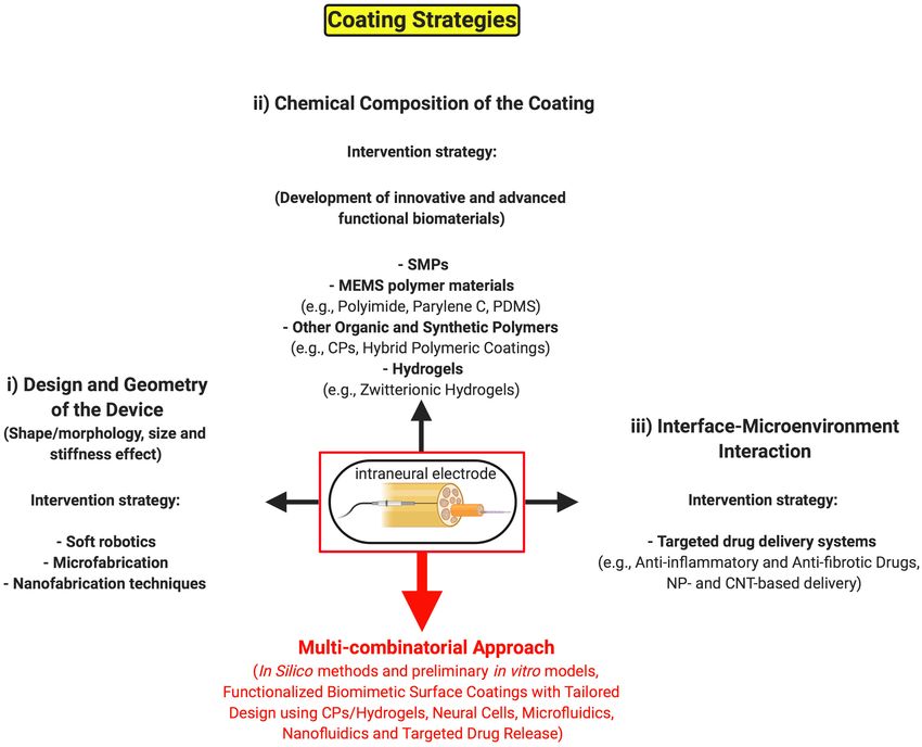
Unfortunately, the standard control systems of prosthetic (Anderson et al., 1999; Luttikhuizen et al., 2006). This complex
limbs rely on surface electromyogram, which is mainly limited by host reaction (Figure 1) consists in a sequential and orderly
problems of high latency, as well as low specificity and robustness cascade of molecular events that involves adhesive blood and
Frontiers in Bioengineering and Biotechnology | www.frontiersin.org 2 May 2021 | Volume 9 | Article 659033Gori et al. Neuroengineering and Foreign Body Reaction FIGURE 1 | Onset, progression and resolution of the Foreign Body Reaction. Sequence of cellular events of the non-specific immune response elicited by the biomaterial surrounding the invasive electrode implanted into the nervous tissue, which is perceived as a nerve injury: (a) onset, similarly to the wound healing, the adsorption of blood and plasma proteins [in particular, fibrinogen and antibodies (IgG), which will be recognized by the white blood cells of the immune system, and the complement system providing specific binding sites and chemoattractants for circulating leukocytes and monocytes] to the surface of the implant leads to the second step of the process, (b) the progression of the FBR, during which leukocyte and monocyte extravasation that is due to the influence of various chemokines, such as TGF-β, promotes their attraction and adhesion to the electrode surface. Recruited monocytes differentiate into activated M1 macrophages that fuse together into multinucleated FBGCs, which carry out multiple functions including: the increase of the inflammatory response both through a positive feedback mechanism (mainly via additional TGF-β production) and through the recruitment of further monocytes and macrophages, the digestion of the electrode surface while promoting the recruitment of the fibroblasts and their activation to myofibroblasts in the last step of the process, (c) the resolution of the FBR, during which the myofibroblasts secrete the different ECM components around the implant that are responsible for the formation of the fibrotic capsule, which ultimately isolates the electrode from the surrounding tissue. IgG, immunoglobulin G; CCLs, CC chemokines; TGF-β, transforming growth factor β; FBGCs, foreign body giant cells. Created with BioRender.com. plasma proteins, tissue and infiltrated inflammatory cells, and leukocyte extravasation, attraction and adhesion to the surface, inflammatory cytokines. along with the activation of the coagulation cascade (Richardson The first step (onset), which is similar to wound healing, upon et al., 1976; Smiley et al., 2001; Szaba and Smiley, 2002). foreign body implantation is the adsorption of blood and plasma Fibrinogen is hydrolyzed to fibrin that creates a sort of matrix proteins, such as fibrinogen, fibronectin, albumin and antibodies able to attract circulating leukocytes and local macrophages to the implant surface (Andrade and Hlady, 1987; Jenney and around the implanted surface under the chemoattractive Anderson, 2000). The type of the proteins adsorbed and the influence of different chemokines (Tang et al., 1998; Smiley progression of the FBR depend on the surface shape, chemistry et al., 2001; Tao and Kobzik, 2002; Lishko et al., 2004). At composition and charge (Tang and Eaton, 1993; Hunt et al., the onset of the FBR and during its progression, circulating 1996; Thull, 2002). In the second step (progression), the adsorbed platelets first and macrophages then secrete transforming growth protein layer and its composition in turn promote monocyte and factor β (TGF-β). This pivotal cytokine serves as chemoattractant Frontiers in Bioengineering and Biotechnology | www.frontiersin.org 3 May 2021 | Volume 9 | Article 659033
Gori et al.                                                                                               Neuroengineering and Foreign Body Reaction

and activator of monocytes, besides being responsible for the               peripheral nerve are cuff electrodes (Navarro et al., 2001; Ortiz-
continuum of the inflammation and its exacerbation until                    Catalan et al., 2013) and Flat Interface Nerve Electrodes (FINEs)
fibrosis (DiPietro et al., 1998; Crowe et al., 2000). Leukocytes            (Tyler and Durand, 2002; Freeberg et al., 2020).
express and secrete a series of other inflammatory cytokines,                   Intraneural electrodes should offer a better signal-to-noise
such as CCL2, CCL3, CCL5, which are the principal players                   ratio during neural recording and the reduced current intensity
involved in the recruitment of blood-borne monocytes and local              necessary to reach the appropriate nerve stimulation (Navarro
macrophages in the foreign body microenvironment (DiPietro                  et al., 2005). Nonetheless, being implanted within the nerve, these
et al., 1998; Hancock et al., 2000; Ono et al., 2003; Armstrong             interfaces are traumatic for the surrounding tissue triggering
et al., 2004). Afterward, extravasated monocytes differentiate              an early inflammatory response caused by the injury of the
to macrophages that, once activated under the stimulation                   vascularized connective tissue. Indeed, as the electrode proximity
of activated T cells, fuse together to form multinucleated                  to the nerve increases, a higher selectivity of neural recording
foreign body giant cells (FBGCs). FBGCs start releasing                     of the signal and stimulation can be obtained. However, the
further inflammatory cytokines, thus boosting the inflammatory              formation of the fibrotic capsule around the interface reduces
response through a mechanism of positive feedback, giving                   recording and stimulation long-term stability compared to
rise to a chronic inflammation (Anderson, 2000; Kyriakides                  extraneural electrodes (Rossini et al., 2010; Badia et al., 2011,
et al., 2004). This cell recruitment from the bloodstream is                2016; Lotti et al., 2017).
allowed by vasodilatation and increase of vessels permeability,                 The chronically implanted devices stimulate the
which is induced by the platelet release of the angiogenic                  aforementioned multistep cascade of foreign body response,
cytokine vascular endothelial growth factor (VEGF) (Banks                   ending in scar tissue formation and electrode encapsulation, and
et al., 1998; Ferrara et al., 2003). The biological activity                thus in the need of increased currents (i.e., power consumption)
of the FBGCs represents a hallmark of the FBR, as it is                     to maintain appropriate nerve stimulation due to a progressive
aimed to protect implanted tissue against the foreign body,                 increase of the electrical impedance. The most frequently used
mediating its surface damage and digestion through the release              metals for the fabrication of neural electrodes are gold, tungsten,
of various proteases and acids (Kyriakides et al., 2004). In                platinum (Pt) and Platinum-Iridium (Pt-Ir) alloy, with Pt being
the last step of the process (resolution), macrophages play a               considered the preferred choice for long-term neuroprosthetic
key role via the production of TGF-β. This multifunctional                  applications due to its electrochemical stability, safety, resistance
cytokine has a paramount importance as it will stimulate                    to corrosion and limited reactivity within a tissue environment
the fibroblast-mediated extracellular matrix (ECM) production,              (Brummer et al., 1983; Geddes and Roeder, 2003; Merrill et al.,
while reducing at the same time inflammation (Bellingan, 1996;              2005; Polikov et al., 2005). However, the stiffness of Pt has a
Ashcroft, 1999). Thus, the role of the recruited macrophages                traumatic impact on the surrounding soft neural tissue (Green
is to promote further monocyte and macrophage recruitment                   et al., 2012), causing a shear stress that over time induces an
and to stimulate the growth and differentiation of quiescent                inflammatory reaction, which can be further stimulated by the
fibroblasts to myofibroblasts. Myofibroblasts are eventually                tissue movements and electrode micromotion (Rousche et al.,
responsible for the massive production and secretion of ECM                 2001; Leach et al., 2010). In addition, another weakness of Pt
components, including collagen I, collagen III, fibronectin                 and other metallic electrodes is due to their fabrication, which
and proteoglycans that give rise to the dense fibrotic capsule              is usually performed with smooth surfaces that do not allow
around the implanted electrode (Luttikhuizen et al., 2006;                  complete nervous tissue adhesion and integration. As a result,
Anderson et al., 2008; Ward, 2008). In the very final stage                 immune cells may invade the remaining space between device
of the process, the capsule becomes impermeable to the non-                 surface and target nerve in the implanted area, fostering the
specific immune system and to many chemicals, including                     FBR (Aregueta-Robles et al., 2014). Therefore, the strength of
some therapeutic inhibitors of inflammation, and responsible                the implant-tissue integration is influenced by the presence of
for the augmentation of the electric impedance and progressive              FBGCs and monocytes/macrophages (Fink et al., 2008). On
isolation of the implanted device, impairing its long-term                  the other hand, manufacturing excessively rough surfaces may
functionality (Anderson et al., 1999, 2008; Luttikhuizen et al.,            risk increasing the local strain and producing friction forces,
2006).                                                                      thereby causing tissue damage. It is also known that rougher
                                                                            surfaces are able to alter cell adhesion, growth, activation and
                                                                            behavior (Fink et al., 2008; Gamboa et al., 2013; Hulander
INTRANEURAL VS. EXTRANEURAL                                                 et al., 2013) including macrophage fusion (Chen et al., 2010),
ELECTRODES IN FBR                                                           although these effects depend on the different cell types as
                                                                            well as on the materials used and their fabrication methods.
To interface with a peripheral nerve invasive intraneural and               Consequently, the right compromise should be sought between
extraneural electrodes can be employed. Among intraneural                   the optimal flatness, smoothness and suitable roughness that
electrodes, the most used are Multielectrode arrays (MEAs),                 meet the texture of the nerve tissue, thus avoiding local insults
Longitudinal Intra-Fascicular Electrodes (LIFEs) and Transverse             and hazardous damages that could trigger inflammation and
Intrafascicular Multichannel Electrode (TIME) (Yoshida and                  a deranged wound healing process. Because of these intrinsic
Stein, 1999; Branner et al., 2004; Badia et al., 2011; Yildiz et al.,       limitations in metallic electrode efficiency, the continuous search
2020). The extraneural electrodes developed to interface with               for valid alternatives and chemical modifications to material

Frontiers in Bioengineering and Biotechnology | www.frontiersin.org     4                                        May 2021 | Volume 9 | Article 659033
Gori et al. Neuroengineering and Foreign Body Reaction
composition is encouraged. For example, electrodes can be materials”) (Wichterle and Lím, 1960; Rao et al., 2011; Gutowski
coated with conductive and soft polymers, like a core of flexible et al., 2015; Heo et al., 2016), although some limitations that
and insulating polyimide with metallic tracks of Pt or Pt-Ir, as were somehow addressed combining PEG with other polymers.
detailed below. Such a strategy can be adopted for mitigating However, PEG shows high susceptibility to oxidative damage
the stiffness disparity between device and host tissue and for in vivo, it may activate severe immune response, and its
relieving the biological rejection of the nerve tissue (Geddes and functionalization is usually troublesome, thereby limiting its
Roeder, 2003; Merrill et al., 2005; Polikov et al., 2005). application for neural interfaces that require long-term stability
So far, diverse strategies are being pursued (Figure 2) to (Ostuni et al., 2001; Ward et al., 2002; Knop et al., 2010).
create minimally invasive neural implants that may address the Likewise, poly(2-hydroxyethyl methacrylate) (PHEMA), which
FBR issue and guarantee their long-term use, which can be is, together with PEG, the most widely used coating material
summarized as follows: (i) working on the design and geometry for implantable devices (Campioni et al., 1998; Ratner, 2002) is
of the device (such as surface roughness, electrode shape, size, susceptible to non-specific protein adsorption, and eventually to
and flexibility); (ii) working on the chemical composition of the fibrotic encapsulation (Zhang et al., 2017), thereby raising the
coating material to develop novel organic and synthetic polymer same problems faced with PEG for the extended stability over
substrates that can be tolerated much better by the host tissue. time and for restraining an immune reaction. Instead, hydrogels
Finally, another important approach consists in (iii) working made of zwitterionic polymers, such as poly(carboxybetaines),
on the interaction between interface and microenvironment poly(sulfobetaines), and poly(phosphobetaines) (Chen et al.,
for controlling the local delivery of therapeutic molecules 2005; Jiang and Cao, 2010; Sin et al., 2014a,b) are biocompatible
(e.g., anti-inflammatory and anti-fibrotic drugs) making use of and highly hydrated materials, showing anti-inflammatory
functionalized biomimetic and biodegradable coatings. and ultralow-fouling characteristics in vivo, which hold great
potential to reduce FBR a way better than PEG hydrogels (Jiang
and Cao, 2010; Zhang et al., 2013; Wu et al., 2018), as further
Working on the Design and Geometry of discussed below in the hydrogel section.
the Electrode In the last decades, many endeavors have been made
The shape and topography of medical-grade polymers implanted in different biomedical and clinical frameworks, merging
in animal models profoundly influences the FBR at the microengineering and material chemistry skills with molecular
implant surface, with the broadly accepted experimental outcome biology knowledge, to modify the physicochemical features of
that circular and smooth surfaces, in intramuscular and implanted interfaces and tuning their structural and surface
percutaneous implants, minimally affect the aggressive behavior features with the aim to control the FBR and increase their
of macrophages (Matlaga et al., 1976; Salthouse, 1984). The use neurocompatibility. For example, it was initially proposed the use
of flexible implants of multifunctional polymeric fibers (Canales of the focused ion beam technology as high precision machining
et al., 2015), and microfabrication of the electrode shape with technique to create and modify the surface morphology of the
a new flexible sinusoidal design and a 3D spheroid tip that interface material, up to nanometric scale, by controlling the
reduce local strain and tissue damage caused by micromotion ion milling of the substrate or its coating in three dimensions,
(Sohal et al., 2014) may represent alternative strategies to gain and thereby modulating in vitro the neural cell adhesion
some mechanical benefits, without remarkably modifying the (Raffa et al., 2007). Afterward, other promising solutions
size of neural implants, and improve their in vivo longevity developed for patterning the design and morphology of the
and recording performances. The importance to focus on the surface are briefly summarized as follows: the generation of
coating stiffness and geometric configuration (i.e., size effect), combinatorial libraries of cationic polymer coatings in mice
to reduce the mechanical mismatch between chronic implanted (Ma et al., 2011); the intramuscular implantation, in rat
electrodes and neural tissue, has been highlighted by a recent spinotrapezius muscles, of biodegradable poly(l-lactide-co-d/l-
work of Spencer et al. (2017). They investigated the ability of lactide) (PLA), as membranes and uncoated electro-spun fiber
soft polyethylene glycol dimethacrylate (PEG-DMA) hydrogel meshes with a positively charged plasma-polymer coating, to
coatings, compared to hard implants of identical diameter, to alter material morphology (Lucke et al., 2018). Finally, the
reduce chronic glial scar formation on the surface of neural development of a method to control surface porosity of poly(2-
probes in rodent brains, by lowering the local strain and hydroxyethyl methacrylate-co-methacrylic acid) (pHEMA-co-
diameter (from 400 to 150 µm) of the coating. The authors MAA) hydrogels, consisting in the fabrication of parallel channels
suggest that a similar technique could be adapted to coat interconnected to a micrometer-sized spherical pore network
more complex geometries through a dip coating, or spray (Madden et al., 2010). These surface-modified scaffolds were
coating method, including electrodes made of various materials, able to increase neovascularization and reduce the inflammation
such as metal, silicone and polymer implants, by slightly and tissue scarring. This last work represents another smart
changing the chemistry. approach to control channel size and spacing of a functionalizable
The strategy of coating neural electrodes with hydrogels surface, which can be achieved by varying the dimensions of the
of PEG and PEG-based copolymers, leveraging their high microsphere templates. With regard to changing the geometry
versatility, low-fouling and bioinert properties, has long been of the electrode material, the anionic polysaccharide, alginate,
used with moderate success in many studies (as reported with is a naturally-derived polymer able to form biocompatible
various examples in the subsection “other advanced biomedical hydrogels, with the addition of divalent cations, to encapsulate
Frontiers in Bioengineering and Biotechnology | www.frontiersin.org 5 May 2021 | Volume 9 | Article 659033Gori et al. Neuroengineering and Foreign Body Reaction FIGURE 2 | Possible coating strategies of invasive neural implants to minimize the long-term FBR. Schematic representation of the main tissue engineering strategies for coating intraneural electrodes against the FBR: (i) the long-term stability and performance of invasive interfaces can be enhanced through the manipulation of the electrode shape, size, geometry, flexibility and surface roughness to create minimally invasive neural implants by leveraging on micro- and nano-manufacturing methods. (ii) An alternative intervention strategy consists in the development of novel chemical coatings, making use of advanced functional biomaterials, as biocompatible surfaces. (iii) Lastly, the functionalization of the coating with therapeutic drugs and, accordingly, innovative drug-delivery systems may help better integrate and tolerate the invasive device by the host nervous tissue. All of the above intervention strategies could be hopefully integrated into a unique multi-combinatorial approach (red arrow) in the next future. SMPs, shape memory polymers; MEMS, micro-electro-mechanical systems; PDMS, poly(dimethylsiloxane); CPs, conductive polymers; NP, nanoparticle; CNT, carbon nanotube. Created with BioRender.com. cells and materials for biomedical applications (Lee and Mooney, to smaller spheres. In conclusion, they demonstrated that size 2012; Veiseh et al., 2015; Vegas et al., 2016; Bochenek et al., (1.5 mm in diameter or greater) and spherical shape, rather 2018). Semi-permeable alginate spheres have been developed than stiffness, of alginate hydrogels as well as ceramic, metal since long time as a common tissue engineering strategy to and plastic surfaces represent critical features for obtaining isolate implanted biological material from the effect of the prolonged biocompatibility and for resisting to fibrosis rejection. local immune cells, thus reducing the FBR in vivo (Chang, So, this biomaterial design strategy is potentially applicable to 1964; Lim and Sun, 1980; Veiseh et al., 2015). Significantly, intraneural interfaces although, at present, such dimensions are in one of these works, the authors showed for the first time not always achievable for all the intraneural electrodes. Instead, the importance of the size and spherical geometry not only the strategy proposed by Rubehn and Stieglitz (2010) consists for the SLG20 alginate hydrogel encapsulation of pancreatic in a novel 3D design of a spiked ultraflexible neural (SUN) islets, but also for stainless steel, glass and polystyrene spheres interface that integrates spiked structures for intrafascicular on the fibrotic response in immunocompetent and fibrosis- nerve recording from the PNS with an ultraflexible substrate, prone rodent and non-human primate models (Veiseh et al., thereby enabling a unique conformal interface to the target nerve. 2015). They tested different sizes and time windows, including The advantage is represented by the features of the material a chronic time-point (i.e., 6 months), for transplanted grafts used, which is an insulating polyimide substrate that does not encapsulated with the SLG20 alginate capsules and found the cause excessive inflammation. Hitherto, this new sensor model 1.5 mm-sized spheres as the ideal geometry to protect grafted cells has been used only in acute animal experiments, whereas for and surfaces from macrophage activation and fibrosis compared chronic implantations important challenges still remain to be Frontiers in Bioengineering and Biotechnology | www.frontiersin.org 6 May 2021 | Volume 9 | Article 659033
Gori et al.                                                                                             Neuroengineering and Foreign Body Reaction

faced such as, among others, the FBR with fibrotic scar tissue that       matrix that can be part of an intraneural electrode. In the
could displace the electrode from its original position and thus          past decades, flexible polymer-based microelectrodes have been
jeopardize the quality of the neural signal (Rubehn and Stieglitz,        developed also for neural prosthetic devices (e.g., testing different
2010; Wang J. et al., 2018).                                              device size, shape, surface smoothness and structural stiffness)
    Overall, it seems very important to modify the electrode              taking advantage of microfluidic and micromachining techniques
surface with more smooth and circular shape, without major                (Szarowski et al., 2003; Lee et al., 2004; Polikov et al., 2005).
changes in size, to reduce both the local strain of the                   Despite these microelectrodes provide multiple and high-quality
material and the mechanical mismatch between the device                   stimulation and recording sites, the lack of long-term stability
and the host tissue. The consequences of altering the surface             has been reported due to the neural tissue reaction and scar
topography, in particular the effect of the roughness, are                formation following extended microelectrodes implantation (Lee
still debated and quite complex to understand (Fink et al.,               et al., 2004). To overcome this limitation, scientists sought
2008). In fact, topography-induced changes seem to affect                 to integrate microfluidic channels into flexible microelectrodes
macrophage behavior (e.g., cell adhesion, fusion and cytokine             combining different techniques for achieving controlled delivery
secretion in vitro) in the FBR to diverse polymer surfaces                of anti-inflammatory and anti-fibrotic drugs through the
(Chen et al., 2010). Furthermore, the continuous search for               microchannels, as further reviewed in Section “Interface-
novel 3D surface design of the device, with coatings at high              Microenvironment Interaction.” However, micromachining of
flexibility, which can be able to adapt to the microenvironment,          the electrode polymer through a lamination technique (Metz
shaping themselves to the nervous tissue would represent a                et al., 2004), micromolding and thermal bonding of the polymer
plus for improving more and more the implant integration.                 (Ziegler et al., 2006), combined electrochemical deposition of
To this aim, 3D bioprinting of hydrogels and thin-film                    conductive polymer and drugs on the electrode (Wadhwa
deposition technologies of biocompatible and soft polymers will           et al., 2006), turned out to be complex and expensive for a
facilitate the task.                                                      large-scale use. Hence, novel microelectrodes, combining thin-
                                                                          film fabrication with poly(dimethylsiloxane) (PDMS) molding
                                                                          and a more rapid, easy, and cost-effective bonding technique,
MODIFICATIONS OF THE INTRANEURAL                                          enabled long-term drug release for a more stable recording
                                                                          performance (Gao et al., 2013). A new hybrid cuff electrode
ELECTRODES BY INTEGRATING SOFT                                            that integrates microelectrodes, for recording and stimulation,
ROBOTICS, MICROFABRICATION OF                                             embedded within microfluidic channels for drug delivery is
MICROFLUIDIC SYSTEMS AND CARBON                                           an example of flexible thin-film polymer device fabricated via
NANOTUBES                                                                 surface micromachining techniques on a temporary silicon wafer
                                                                          carrier (Elyahoodayan et al., 2020). The electrode was designed
In the research field of neural electrodes and probes continuous          and developed to improve fascicular selectivity and sensitivity
efforts are being made in search of smaller and more flexible             in rat sciatic nerves following minimal handling during surgical
devices to reduce the trauma caused by their insertion and, in            implantation. Its main advantage is represented by the combined
turn, the biological tissue response (chronic inflammation and            possibility to acutely stimulate, record and deliver lysing drugs, to
fibrosis), leveraging on micro- and nano-fabrication techniques.          remove connective tissue (i.e., epineurium layer) that separates
Recently, an innovative soft robotics approach has been devised           electrodes from nerve fibers, and neurotrophic factors that
to mitigate the FBR by controlling fluid flow and shear stress            promote axonal sprouting from the exposed fibers. Nevertheless,
perceived by the host cells (Dolan, 2019). In a rat model,                the authors stated that future studies will be necessary for
the authors implanted subcutaneously a milliscale dynamic soft            functional testing in prolonged implant conditions to check
reservoir (DSR), surrounded by an actuatable polyurethane                 for chronic electrophysiological recording as well as nerve
membrane, and modulated the biomechanics of the biotic-abiotic            health and interface stability after collagenase delivery to verify
interface via tunable pressure. After 14 days, an important               possible levels of axonal inflammation and fibrosis. Regarding
reduction in the number of αSMA+ myofibroblasts and in fibrotic           novel and advanced production methods of microelectrodes,
encapsulation of the implantable device was observed through              a great deal of interest has recently emerged in the additive
histological and immunohistochemical analysis. Furthermore,               manufacturing techniques, a versatile and powerful tool to
as an example of a proof-of-concept study using a porous                  overcome various shortcomings of conventional lithography
and permeable actuating membrane, they were also able to                  techniques. Additive manufacturing of microelectrode arrays
regulate therapeutic delivery of epinephrine, used as a model             or microneedle arrays provides a novel, quick and low-cost
pharmacological agent, to test its functional effect in the               method to fabricate custom-shaped electrochemical devices, by
adjacent tissue. Hence, the presented DSR may have the                    rapid prototyping, for a wide range of applications (Yang et al.,
potential to be integrated into intraneural electrodes for an             2016; Morrison et al., 2019; Soltanzadeh et al., 2020). For
extended period to modulate the inflammatory and fibrotic                 example, the manufacturing method performed by an aerosol
response, making it a promising tool also for future neural               jet technology, for the fabrication of the microelectrode arrays
applications. In fact, the design of the platform can be easily           used in a biosensor platform for electrochemical measurements,
modified and tailored to be integrated into diverse types                 was based on the use of a silver nanoparticle (NP) ink and
of implantable devices through its incorporation into a thin              a UV-curable polymer (Yang et al., 2016). Instead, in another

Frontiers in Bioengineering and Biotechnology | www.frontiersin.org   7                                        May 2021 | Volume 9 | Article 659033
Gori et al. Neuroengineering and Foreign Body Reaction work, compared to microfabricated microneedle arrays, 3D- (MWCNTs) and poly(3-octylthiophene-2,5-diyl) (POT), with the printed arrays, made of an amorphous polymer of acrylonitrile- immobilization of the polymer on the carbon nanostructures, butadiene-styrene, showed almost identical geometric properties preventing its spontaneous and unwanted partition to the and equivalent performance with high frequency biosignals (such membrane phase. The obtained sensors were characterized with as in electromyogram recordings), whereas for recording low good performance, high conductivity as well as high stability frequency signals they turned out to be not suitable (Soltanzadeh of potential readings over time. Nevertheless, although the et al., 2020). However, in these works, only preliminary and remarkable electrical and physical properties of CNTs that can be short-term tests were run to measure their functionality (e.g., exploited for enhancing the functionality of metallic electrodes electrical stimulation in mouse brain, signal recording ability and (Aregueta-Robles et al., 2014), the main concern for their long- impedance characteristics) either in human subjects in a non- term use in vivo remains related to their cytotoxicity and invasive manner (Soltanzadeh et al., 2020), or in mice (Morrison to the risk of causing intracellular damages. Indeed, because et al., 2019) and as electrochemical laboratory biosensors of their elevated stiffness and reduced size (Krishnan et al., (Yang et al., 2016), thus requiring further and deeper in vivo 1998), CNTs can easily penetrate cellular membranes (Kagan investigation to establish the real advantages and drawbacks et al., 2006; Gilmour et al., 2013) and damage nuclei and of 3D-printed microelectrodes and the biocompatibility of the cytoplasmic organelles. Additionally, they are known to be materials used before their clinical application. cytotoxic at high concentrations in different cell types (Bottini To date, microelectrode technologies present important et al., 2006; Tian et al., 2006). In spite of such significant risks, limitations mainly due to the stiffness mismatch between metals which need to be carefully evaluated before clinical applications, or micromachined silicon, used for electrode microfabrication, nanoscale features of CNTs enable their escape from the immune and surrounding tissue, particularly soft brain tissue (Winslow system surveillance, thereby providing an undoubtedly appealing and Tresco, 2010). Thus, the mismatch results in fibrotic resource for the future development of innovative intraneural encapsulation of the microelectrode in chronic implants (Polikov electrodes. A summary of the intervention strategies based on et al., 2005). Furthermore, the problem of controlling possible the design and geometry of the electrode with representative micromotion of the interface that can change its position in examples is reported in Table 1. the tissue may also gradually increase the inflammatory reaction (Gilletti and Muthuswamy, 2006). Similar issues can occur with Developing Innovative and Advanced chronic implants of microfabricated peripheral nerve devices. Functional Biomaterials Thus, another group developed a novel fluidic microdrive Recently, other research groups worked on the development of technology to implant and microactuate ultraflexible electrodes, more suitable materials that can be tolerated by the neural tissue, with a parylene-coated core of carbon nanotube (CNT) fibers, leveraging on material chemistry, micro- and nano-fabrication in animal models that could find useful applications also in techniques (Fekete and Pongrácz, 2017). Many different polymers peripheral nerves (Vitale et al., 2018). Indeed, following fluidic turned out to be possible substrates of neural interfaces due implantation into the nervous tissue, the authors were able to to their proper flexibility, stability, insulation properties and perform electrophysiological recordings, enhancing the stability biocompatibility (Svennersten et al., 2011; Ordonez, 2012; of the device without the need of increasing the stiffness and Ware et al., 2013; Nguyen et al., 2014; Arreaga-Salas et al., thickness of the microdevices, and thus preventing also the onset 2015; Boddupalli et al., 2016). Noteworthy, among these of inflammatory responses. Fluidic microdrives were fabricated are: shape memory polymers (SMPs) [such as polyurethanes, in PDMS by conventional replica molding technique and the polylactides, polystyrenes, poly(cyclooctene), thiol-enes and microelectrodes insertion was obtained via viscous drag force due poly(vinyl acetate)]; the widely used micro-electro-mechanical to the finely controlled liquid flow in the microfluidic channel, systems (MEMS) polymer materials, namely polyimide, parylene limiting tissue damage at a negligible extent. Such brilliant C, PDMS and SU-8 (an epoxy-based photoresist suitable for strategy could be further implemented for peripheral nerve microelectronic applications). In the soft neural tissue, the electrodes, envisioning exciting opportunities for their chronic use of new smart SMPs is gradually overcoming the one of implants. Wireless and flexible film-based ion-selective electrodes more stiff materials, as the former seem to drastically reduce (ISEs) have also been recently developed as miniaturized systems the inflammatory response in the surrounding tissue becoming for performing highly sensitive and non-invasive measurements compliant after implantation (Ware et al., 2013; Nguyen et al., (Lim et al., 2020). These sensor systems, made of carbon–polymer 2014; Minev et al., 2015). Likewise, in the PNS the use of flexible composite transducers integrated onto a flexible circuit, enable polymer materials seems to eliminate the mechanical mismatch ions detection in body fluids with high accuracy and selectivity of compliance between the implanted electrode and the biological and for prolonged lifetime, showing great potential for their tissue (Blakney et al., 2012; Nguyen et al., 2014). application also in health studies and clinical systems. Another recent approach to drastically reduce the risk of alteration of the performance of the transducer material used for sensors MEMS POLYMER MATERIALS and electrodes, was the development and characterization of solid contact ion-selective electrodes using novel composite Polyimide material (Kałuża et al., 2019). The formulation of the present It is a highly resistant and biocompatible polymer, made by nanocomposite was based on multi-walled carbon nanotubes imide monomers, among the most widely used substrates Frontiers in Bioengineering and Biotechnology | www.frontiersin.org 8 May 2021 | Volume 9 | Article 659033
Gori et al.                                                                                                                   Neuroengineering and Foreign Body Reaction

TABLE 1 | Intervention strategies based on the design and geometry of the electrode.

                                                                         (i) Design and geometry

Features                      Examples                                                                             References

Size effect                   PEG-DMA hydrogel coatings and deep and spray coating method                          Spencer et al., 2017
                              PEG-based coatings                                                                   Reviewed in Knop et al. (2010)
                                                                                                                   Wichterle and Lím, 1960; Rao et al., 2011; Gutowski
                                                                                                                   et al., 2015; Heo et al., 2016; Lee et al., 2017
                              PHEMA-based coatings                                                                 Reviewed in Ratner (2002)
                                                                                                                   Campioni et al., 1998; Jhaveri et al., 2009; Zhang et al.,
                                                                                                                   2017
Surface morphology            FIB technology as machining technique to modify surface morphology                   Raffa et al., 2007
Shape                         Flexible implants of multifunctional polymeric fibers                                Canales et al., 2015
Design and topography         Physical properties, surface micro-/nano-topography and surface chemistry            Reviewed in Ware et al. (2013)
                              modifications                                                                        Anderson et al., 1999; Thull, 2002; Fink et al., 2008;
                                                                                                                   Chen et al., 2010; Gamboa et al., 2013; Hulander et al.,
                                                                                                                   2013
                              3D design of spiked ultraflexible substrates                                         Rubehn and Stieglitz, 2010; Wang M. et al., 2018
                              Neural probe with sinusoidal design and a 3D spheroid tip                            Sohal et al., 2014
                              Microgeometry and implant thickness effect                                           Ward et al., 2002
Material morphology           Cationic polymer coatings and PLA and electro-spun fiber meshes with                 Ma et al., 2011; Lucke et al., 2018
                              plasma-polymer coating
Surface porosity              Channel size control through (pHEMA-co-MAA) hydrogels                                Madden et al., 2010
                              PU-based porous implants                                                             Ward et al., 2002
Size and spherical geometry Alginate spheres/capsules                                                              Veiseh et al., 2015
Intervention strategy
Soft robotics                 Control over fluid flow and shear stress through milliscale dynamic soft reservoir   Dolan, 2019
                              with actuatable membrane
Microfabrication              Micro-machined neural prosthetic devices: flexible polymer-based                     Reviewed in Szarowski et al. (2003); Lee et al. (2004),
                              microelectrodes with different shape, size and geometry                              Metz et al. (2004); Polikov et al. (2005), Spataro et al.
                                                                                                                   (2005); Ziegler et al. (2006), Winslow and Tresco (2010);
                                                                                                                   Blau et al. (2011), Gerwig et al. (2012); Gao et al.
                                                                                                                   (2013), Minev et al. (2015); Qi et al. (2017), Vitale et al.
                                                                                                                   (2018); Kozai (2018), Fallahi et al. (2019), and Kumar
                                                                                                                   et al. (2020)
                                                                                                                   Elyahoodayan et al., 2020
                              Encapsulation technologies of flexible microelectrodes                               Reviewed in Ahn et al. (2019)
                              Electrically-responsive flexible microfibers                                         Chen et al., 2017
                              Microfabrication of a neural probe with sinusoidal design and a 3D spheroid tip      Sohal et al., 2014
                              Wireless, flexible, film-based carbon-polymer composite microelectrode system        Lim et al., 2020
                              Additive manufacturing of microelectrode arrays and microneedle arrays               Yang et al., 2016; Morrison et al., 2019; Soltanzadeh
                                                                                                                   et al., 2020
Nanofabrication               CNTs                                                                                 Reviewed in Aregueta-Robles et al. (2014)
                                                                                                                   Castagnola et al., 2016
                              Parylene-coated flexible CNTf microelectrodes                                        Vitale et al., 2018
                              Conducting-polymer carbon nanotubes                                                  Abidian et al., 2010; Gerwig et al., 2012; Alba et al.,
                                                                                                                   2015; Mandal et al., 2015; Samba et al., 2015; Du
                                                                                                                   et al., 2018; Altun et al., 2019; Kałuża et al., 2019;
                                                                                                                   Zheng et al., 2019
                              PPy nanowires                                                                        Reviewed in Qi et al. (2017)
                              PPy nanoparticles                                                                    Hosseini-Nassab et al., 2017
                              SWCNT-PPy/PEGDA composite hydrogels                                                  Xiao et al., 2012
                              PPy/CNT films                                                                        Luo et al., 2011
                              Graphene oxide nanocomposite films of PPy                                            Weaver et al., 2014
                              PLGA nanoparticles embedded in alginate hydrogels                                    Kim and Martin, 2006
                              Nanoparticle-coated nanoelectrodes                                                   Bazard et al., 2017
                              Nanoscale biomimetic surfaces                                                        Reviewed in Von Der Mark et al. (2010)

PEG, polyethylene glycol; DMA, dimethacrylate; PHEMA, poly(2-hydroxyethyl methacrylate); FIB, focused ion beam; PLA, poly(l-lactide-co-d/l-lactide); pHEMA-co-MAA,
poly(2-hydroxyethyl methacrylate-co-methacrylic acid); PU, polyurethane; CNTs, carbon nanotubes; CNTf, carbon nanotube fiber; PPy, polypyrrole; SWCNT-PPy/PEGDA,
single-walled carbon nanotubes-polypyrrole/poly(ethylene glycol) diacrylate; PLGA, poly(lactic-co-glycolic acid). References: except were specifically indicated as
‘Reviewed in,’ all others are research articles.

Frontiers in Bioengineering and Biotechnology | www.frontiersin.org                   9                                                  May 2021 | Volume 9 | Article 659033
Gori et al. Neuroengineering and Foreign Body Reaction for the fabrication of the core of novel neural electrodes Parylene C with metallic tracks, such as Pt and gold, often coated with Parylene C is a variety of high flexible and chemically inert different biomaterials for counteracting and delaying the onset poly(p-xylylene) polymer commonly used as biocompatible of the FBR (Oddo et al., 2016; Delgado-Martínez et al., coating and substrate material of electrodes for soft neural 2017; Wurth et al., 2017; de la Oliva et al., 2018c). Indeed, implants (Fekete and Pongrácz, 2017). In a recent work, the among the possible neuroprosthetic applications of this polymer, authors tested parylene C as a substrate material for peripheral the group of Navarro X. developed a novel double-aisle nerve interfaces both in vitro and in vivo (de la Oliva et al., electrode to regenerate separated nerve fascicles, made of a 2018a). In this study, longitudinal devices made of parylene double-side thin-film of polyimide (Delgado-Martínez et al., C and polyimide were implanted in the rat sciatic nerve for 2017). Although such interface allowed regeneration of nerve up to 8 months and the induced FBRs were compared one branches, it caused FBR in chronic implants. The reaction another. In spite of the advantage to produce parylene C-based was indeed similar to that obtained previously with other thinner substrates than polyimide ones, with no harmful effect chronically implanted polyimide intrafascicular electrodes and on nerve function, long-term stability of such electrodes could non-obstructive regenerative electrodes (Lago et al., 2007; Garde be affected by a thicker tissue capsule than polyimide devices. et al., 2009), thus affecting the quality of neural signal over time. Indeed, the authors observed much more fibroblasts surrounding This common limitation when using polyimide electrodes might the former device, thus making parylene C not suitable for be overcome through the functionalization of the polyimide core chronic implantations (Lecomte et al., 2017; Mueller et al., 2017; with advanced biomimicry ultra-low fouling organic or synthetic de la Oliva et al., 2018a). However, the diverse pattern of FBR coatings that can be much more tolerated by the implanted tissue. around parylene C vs. polyimide, due to their different chemical Toward this direction, diverse efforts have been made to reduce structures, deserves further investigation before parylene C drops the inflammatory response and electrode encapsulation through out of other possible invasive neural applications. For example, in new biomimetic solutions. One of these involved the coating another study the authors microfabricated and tested in vivo up with bioresorbable layers of molten saccharose for intracortical to 24 months, even though in the rabbit brain, a sinusoidal probe insertion in rat models (Hassler et al., 2016). Another option electrode made of a tungsten titanium alloy (WTi) core encased was a superhydrophobic coating from a natural Xanthosoma in flexible layers of parylene C with novel design features (Sohal sagittifolium leaf nanocasted on an electroactive polyimide et al., 2014). Interestingly, over the chronic experimental period surface (Chang et al., 2013). A different nanotechnological of the study the electrode performances and neuronal integration approach was attempted using hybrid conductive material: were better than other conventional electrodes used for recording an indium tin oxide substrate associated to a nanostructured of neuronal activity in humans, showing low levels of gliosis. polyimide film deposited on a glass surface, using a new and Another interesting attempt to improve the long-term stability simple nanopatterning technique (Rombaut et al., 2019). Very in vivo of an intrafascicular neural interface (i.e., a flexible recently, a flexible and transparent polyimide-based electrode microelectrode array with a recording system), was made through was fabricated with a trilayer-stacked geometry that exploits a mechanically enhanced flexible interconnection cable using a the properties of a high-quality ultrathin film of graphene. combination of parylene C and polyimide (Kang et al., 2019). This solution showed enhanced power and current efficiencies, The former provided chemical and electrochemical stability while with properties comparable to indium tin oxide-based diodes, the latter improved the mechanical strength and handling, with increased flexibility and long-term stability in different devices no damage reported, during the implantation procedure of the (Lee et al., 2019). Finally, another strategy to increase the long- whole neural interfacing device in canine sciatic nerves. However, term reliability, while maintaining high flexibility, of a polyimide- before clinical translation, these promising results need more based neural interface in free-moving rats, was the one adopted investigation to test their reproducibility in chronic implants of by a research group from China, through a MEMS fabrication peripheral nerves in larger animal models. Despite the many approach (Ji et al., 2018). This group developed an innovative benefits of parylene C as conformational coating, such as its optogenetics tool consisting in a polyimide-based hybrid (opto- chemical inertness, there are also significant disadvantages that electric) flexible device that integrates 16 micro-LEDs and can limit its wider application compared to the liquid epoxy 16 IrOx-modified microelectrode arrays. Such device allowed or silicon coatings. Notably, a better performance and a more simultaneous, high-resolution optical stimulation and electrical controlled deposition process of the latter that are, moreover, recording of cortical areas. Using this tool, they observed little much more cost-effective in their production-run make them reduction in the electrical or optical performance for 3 months. a preferable choice for researchers. Furthermore, the chemical Although the fabrication process was quite complex, the device vapor deposition process required to apply parylene C onto a revealed itself to be a promising neural interface for further surface, especially a conductive-metal one, is not only time- neuroscience applications, expandable also to larger animals consuming but also costly in the attempt to increase its metal (e.g., non-human primates) and possibly to human patients. adhesion through different methods. However, in order to evade the issue of non-specific protein and cell absorption on the polyimide surface, several groups tried to devise valid alternatives to polyimide substrates, using Poly(Dimethylsiloxane) (PDMS) either diverse MEMS polymers or newly emerged biomedical This silicon-based organic polymer is the elective material materials, as shown below. for microfabrication of microfluidic devices including Frontiers in Bioengineering and Biotechnology | www.frontiersin.org 10 May 2021 | Volume 9 | Article 659033
Gori et al.                                                                                                Neuroengineering and Foreign Body Reaction

microelectrodes, with tissue-like elastic modulus, easily                    drugs, such as dexamethasone (Alba et al., 2015; Boehler et al.,
compliant to neural tissue. These flexible electrodes are usually            2017; Kleber et al., 2019). It can also be conjugated with other
realized through the process of replica molding, from a master               biocompatible and bioinert materials, such as PDMS thin films,
obtained by soft photolithography with a SU-8 photoresist (Qin               CNTs, tetrafluoroborate (TFB), poly(styrenesulfonate), alginate
et al., 2010). Alternatively, they can be fabricated via simple              and nafion to guarantee electrochemical stability both in vivo
and cost-effective photolithography-free methods, such as laser              and in vitro (Blau et al., 2011; Alba et al., 2015; Charkhkar et al.,
micromachining and master molding of PDMS. Such versatile                    2016; Ferlauto et al., 2018; Carli et al., 2019). To date, PEDOT
processes give rise to planar metal electrodes with microfluidic             functionality has already been demonstrated in vitro in terms
channel geometries (Chatzimichail et al., 2018), and stable neural           of improvement of neurite outgrowth bioactivity, and stability
interfaces (Gao et al., 2013; Minev et al., 2015).                           of neural micro-stimulation (Green et al., 2009; Mandal et al.,
   Poly(dimethylsiloxane) micromachining is not only cheap,                  2015). Nonetheless, the long-term performance and integrity
and easy to realize with high parallelization, but also suitable             in vivo of such coatings for chronic recordings have yet to be
for the fabrication of long-term neural implants that are able               verified, despite some interesting data collected from short-term
to produce lower inflammatory response than polyimide-based                  epicortical and epidural recordings (Blau et al., 2011). However,
electrodes (Minev et al., 2015). Flexibility and elasticity of               these aspects start to be evaluated with promising long-term
PDMS are clearly advantageous features for the fabrication of                results, such as for the chronic intracortical neural recordings
neural electrodes, as well as in promoting neuronal maturation               with high stability and activity in rat motor cortex and mice visual
(Teixeira et al., 2009; Yang and Suo, 2018). Notwithstanding,                cortex, which deserve further investigation (Charkhkar et al.,
because of PDMS hydrophobicity, achieving its stable adhesion                2016; Ferlauto et al., 2018; Carli et al., 2019). Another important
to hydrated surfaces and materials, such as hydrogels, can be                example was provided by a research team that developed a
problematic (Yang and Suo, 2018). Furthermore, the proper                    metal-free electrode array of polypyrrole/polycaprolactone-
stability and adhesion between different layers of elastic polymers          block-polytetrahydrofuran-block-polycaprolactone             (PCTC)
in implantable electronic devices, such as stretchable electrodes,           sandwiched in between films of PDMS. This group compared
is difficult to achieve. Actually, under the pressure of muscle              the in vivo performance of such all-polymer interface with a
contraction and of the strain imposed by the micromotion                     Pt electrode of the same area in a rat (Guo et al., 2014). They
between nerve tissue and the implant, the electrode can crack.               demonstrated a lower impedance of the metal-free device,
This issue can eventually jeopardize the device functionality.               along with excellent electrical stimulation performances in a
Therefore, alternative solutions have been pursued using all-                stimulated rat hind-limb muscle following squeezing of the
polymer and metal-free microelectrode arrays with a mixture of               sciatic nerve and higher charge injection capacity compared
various stretchable polymers and via replica molding with PDMS               to the Pt electrode, as well as to other PEDOT-coated metal
(Blau et al., 2011; Guo et al., 2014; Qi et al., 2017), although with        electrodes. Future work from the same group will be necessary
mixed fortunes, as described in the next section.                            to improve and characterize the device physical integrity and
                                                                             mechanical performance in long-term in vivo assays also in
                                                                             peripheral nerves.
OTHER ADVANCED BIOMEDICAL                                                        Two of the most widely used synthetic polymers for
MATERIALS                                                                    coating electrodes are poly (ethylene glycol) PEG (Drury and
                                                                             Mooney, 2003; Gutowski et al., 2015) and PHEMA (Jhaveri
From the close collaboration between the bioengineering field                et al., 2009; Mario Cheong et al., 2014), as they can form
and the biomedical research area in the development of novel                 hydrogels with low- or non-fouling characteristics in vivo,
biomaterials for chronic neural applications, diverse strategies             thus enhancing tissue response around implanted electrodes.
are being pursued to decrease the FBR in the next-generation                 However, their long-term use is limited by oxidative mechanisms
neural interfaces. Some of them are based on the use of                      that partially compromised non-specific protein absorption and
organic and synthetic polymeric coatings, including conductive               device performance. Therefore, recent hybrid solutions have
polymers (CPs). Among organic coatings, CPs have been recently               been proposed to overcome some of the issues related to their
investigated with the aim to improve the long-term performance               prolonged stability and sensitivity in vivo, such as hybrid thin film
of neural electrodes as they can increase their effective surface,           photopatternable polymers, combining the properties of PEDOT
thereby decreasing the impedance, and enhance the electrical                 with the long-term (over 10 days) moisture stability of PEG (Zhu
properties of neural interfaces, thus seeming the most promising             et al., 2017). Another successful test was the integration between
materials (Wilks et al., 2011; Charkhkar et al., 2016). In                   PEDOT-poly(styrene sulfonate) (PSS)-CNT nanocomposites
particular, Poly (3,4-ethylenedioxythiophene) PEDOT, and some                and biocompatible PHEMA hydrogels (Castagnola et al., 2016),
of its modified and hybrid versions, have been shown to be safe              for potential acute and chronic flexible and high sensitivity
and reliable candidates in neuroprosthetic applications, being               electronic applications in rat brains. Thus, the PHEMA hydrogel
stable and able to improve neural adhesion, electrochemical                  was able to guarantee the electrochemical performance of
impedance and dramatically reduce electrical noise and host                  the device and improve the quality of intracortical recording
tissue response (Abidian et al., 2010; Green et al., 2013; Ferlauto          until 28 days after the implant, along with the advantage of
et al., 2018; Ganji et al., 2018). Moreover, PEDOT can be                    reducing the mechanical mismatch between neural tissue and
easily doped and bio-functionalized with anti-inflammatory                   device preventing the nanomaterial detachment. Instead, other

Frontiers in Bioengineering and Biotechnology | www.frontiersin.org     11                                        May 2021 | Volume 9 | Article 659033
You can also read