Bispecific repurposed medicines targeting the viral and immunological arms of COVID 19 - Nature
←
→
Page content transcription
If your browser does not render page correctly, please read the page content below
www.nature.com/scientificreports
OPEN Bispecific repurposed
medicines targeting the viral
and immunological arms
of COVID‑19
Martin A. Redhead1*, C. David Owen2,3, Lennart Brewitz6, Amelia H. Collette1,
Petra Lukacik2,3, Claire Strain‑Damerell2,3, Sean W. Robinson1, Patrick M. Collins1,
Philipp Schäfer1, Mark Swindells1, Chris J. Radoux1, Iva Navratilova Hopkins1,
Daren Fearon2,3, Alice Douangamath2,3, Frank von Delft2,3,9,10, Tika R. Malla6, Laura Vangeel7,
Thomas Vercruysse7, Jan Thibaut7, Pieter Leyssen7, Tu‑Trinh Nguyen8, Mitchell Hull8,
Anthony Tumber1, David J. Hallett1, Christopher J. Schofield6, David I. Stuart2,3,4,5,
Andrew L. Hopkins1 & Martin A. Walsh2,3*
Effective agents to treat coronavirus infection are urgently required, not only to treat COVID-19,
but to prepare for future outbreaks. Repurposed anti-virals such as remdesivir and human anti-
inflammatories such as barcitinib have received emergency approval but their overall benefits remain
unclear. Vaccines are the most promising prospect for COVID-19, but will need to be redeveloped
for any future coronavirus outbreak. Protecting against future outbreaks requires the identification
of targets that are conserved between coronavirus strains and amenable to drug discovery. Two
such targets are the main protease (Mpro) and the papain-like protease (PLpro) which are essential
for the coronavirus replication cycle. We describe the discovery of two non-antiviral therapeutic
agents, the caspase-1 inhibitor SDZ 224015 and Tarloxotinib that target Mpro and PLpro, respectively.
These were identified through extensive experimental screens of the drug repurposing ReFRAME
library of 12,000 therapeutic agents. The caspase-1 inhibitor SDZ 224015, was found to be a potent
irreversible inhibitor of Mpro (IC50 30 nM) while Tarloxotinib, a clinical stage epidermal growth factor
receptor inhibitor, is a sub micromolar inhibitor of PLpro (IC50 300 nM, Ki 200 nM) and is the first
reported PLpro inhibitor with drug-like properties. SDZ 224015 and Tarloxotinib have both undergone
safety evaluation in humans and hence are candidates for COVID-19 clinical evaluation.
The Coronavirus disease 2019 (COVID-19) pandemic caused by Severe Acute Respiratory Syndrome coronavirus
2 (SARS-CoV-2) is the largest global health emergency to emerge this century1. Severely affected patients can dis-
play sepsis, through inappropriate recruitment and expansion of the innate immune response and even patients
who have cleared the virus may continue to suffer, in part due to fibrotic lesions2. To date there are limited direct
antiviral medicines approved for treatment of COVID19, thus we aimed to determine whether any molecules
previously approved for clinical study could be repurposed for the treatment of COVID19.
Vaccines against COVID-19 are reducing COVID19 outbreaks and mortality3, although viral mutations may
compromise the longevity of current vaccines4 and may not protect against future coronaviral disease. In response
to this there are calls to develop a ‘universal vaccine’5. Outside of vaccination small molecule inhibitors may
1
Exscientia, The Schrödinger Building, Oxford Science Park, Oxford OX4 4GE, UK. 2Diamond Light Source Ltd.,
Harwell Science and Innovation Campus, Didcot OX11 0DE, UK. 3Research Complex at Harwell, Harwell Science
and Innovation Campus, Didcot OX11 0FA, UK. 4Division of Structural Biology, Wellcome Centre for Human
Genetics, University of Oxford, Oxford OX3 7BN, UK. 5Instruct-ERIC, Oxford House, Parkway Court, John Smith
Drive, Oxford OX4 2JY, UK. 6Department of Chemistry, Chemistry Research Laboratory,, The Ineos Oxford Institute
for Antimicrobial Research, 12 Mansfield Road, Oxford OX1 3TA, UK. 7KU Leuven Department of Microbiology,
Immunology and Transplantation, Rega Institute, 3000 Leuven, Belgium. 8Calibr, Scripps Research, 11119 N Torrey
Pines Road, La Jolla, CA 92037, USA. 9Structural Genomics Consortium, University of Oxford, Old Road Campus,
Roosevelt Drive, Headington OX3 7DQ, UK. 10Department of Biochemistry, University of Johannesburg, Auckland
Park 2006, South Africa. *email: mredhead@exscientia.co.uk; martin.walsh@diamond.ac.uk
Scientific Reports | (2021) 11:13208 | https://doi.org/10.1038/s41598-021-92416-4 1
Vol.:(0123456789)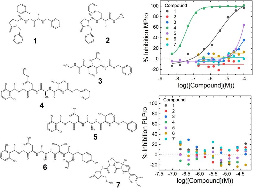
www.nature.com/scientificreports/
play a role suppressing viral proliferation in patients already infected at an early stage in disease and thus reduce
overall disease burden6 and have so far shown broad spectrum activity against several coronavirus variants7,8.
Existing small molecules for treating COVID19 include the anti-viral RNA polymerase inhibitor remdesivir9
(originally developed for Hepatitis C Virus) as well as compounds with anti-inflammatory and immunosuppres-
sant effects such as dexamethasone10 and b aricitinib11. Unfortunately, none of these medicines have delivered
meaningful benefits to patient populations, execpt dexamethasone which provides benefits in only the most
advanced stages of the disease10, although combinations of remdesivir and baricitinib show promising results11.
Thus there is strong motivation for the discovery of new therapeutics not only for the treatment of acute disease.
Antivirals offer the potential of prophylaxis, reduction of transmissibility, treatment of unvaccinated patients
and suppression of emergent coronaviruses. Considering the lengthy timescales required to develop and approve
new therapeutic agents, repurposing of known drugs can potentially reduce the time to develop new treatments.
The SARS-CoV-2 genome encodes small molecule druggable targets including the main protease12 (Mpro)
encoded by non-structural protein 5 (nsp5) and the papain-like protease8 (PLpro) which is part of non-structural
protein 3 (nsp3)13. As both proteins are essential for viral replication they present attractive targets for drug
repurposing efforts. These cysteine proteases are encoded along with the other 14 nsps by the 5′-terminal open
reading frame 1a/b (ORF1a/b), which takes up approximately two-thirds of the viral genome. This leads to the
expression of the two large replicase polyproteins pp1a and pp1ab. Mpro and P Lpro are responsible for the pro-
teolytic cleavage of pp1a and pp1ab which consist of nsps 1–11 and 1–16, r espectively14. Mpro cleaves at 11 sites
releasing the functional nsps 4–16, while PLpro cleaves at 3 sites releasing nsps 1–315. Additionally, PLpro acts as a
deubiquitinase and deISGylase which may modulate the host anti-viral response via suppression of type-I inter-
feron production16. Recent developments have seen a rationally designed Mpro inhibitor enter clinical testing17.
To rapidly identify potential anti–coronavirus therapeutics, we undertook extensive experimental screens
of the drug repurposing ReFRAME library, that consists of 12,000 therapeutic agents against M pro and P Lpro to
identify clinically-viable agents that may be repositioned to treat COVID-19 and future outbreaks. One com-
pound, SDZ 224015, is a potent irreversible inhibitor of Mpro, whilst a second, Tarloxotinib, is a sub micromolar
Lpro.
inhibitor of P
Results
ReFRAME library screening and hit triage. The ReFRAME library comprises 12,000 molecules that
have been previously approved for clinical investigation in humans, including all currently approved medicines18.
Despite the overall quality and relevance of this library, it contains some older compounds with properties less
attractive for modern drug discovery, such as polyphenol groups, flavonoids and catechols (sennosides)19, reac-
tive Michael acceptors (oxantel)20, unattractive molecular weights and poor solubility, many of which can also
cause assay interference21.
Mpro and P Lpro have the potential to exist in several conformationally distinct states, each of which may
favour the binding of different i nhibitors22. Mpro substrate velocity titrations revealed evidence for catalytically
distinct monomeric and dimeric f orms23. Dimer dependent catalysis manifests as the observation of enzyme
concentration dependent sigmoidal substrate velocity plots (Fig. 1a) as observed for Mpro from SARS-CoV-124.
The parameters describing the midpoint or slope of the sigmodal substrate velocity plots displayed a bell-shaped
relationship with enzyme concertation (Fig. 1b,c), whereas the maximum velocity displayed a sigmoidal relation-
ship with enzyme concentration (Fig. 1d). These results strongly indicate dimerization may be induced either by
increasing enzyme or substrate concentration, at high concentrations (> 300 nM) M pro spontaneously dimerizes
and low concentration (< 3 nM) M pro behaves as a monomer. These data indicate the potential for inhibitors to
bind to these catalytically distinct forms25 as well as the potential for binding at the dimer interface24. Conse-
quently, the M pro HTS assay was designed to balance these forms.
pro
PL was found to require the presence of high concentrations of anionic Hoffmeister salts to catalyse hydroly-
sis of small peptide substrates, but not large ubiquitin mimetics (Fig. 2). This indicated that the P Lpro active site is
pro26
not accessible in isotonic buffer, as previously observed for SARS-CoV-1 P L . This requirement may relate to
the subcellular location of nsp3 expressed during viral infection. Nsp3 is expressed on the surface of the endoplas-
mic reticulum and in combination with nsp4 creates double membraned vesicles27. For the papain-like protease
of the betacoronavirus murine hepatitis virus, P Lpro did not process the viral polyprotein unless expressed on the
28
ER membrane . Thus, to maximise the discovery of inhibitors, the HTS was run in the presence of 0.8 M citrate.
In order to minimise the number of HTS false positives, confirmatory screens were run, followed by a counter
screen of M pro and PLpro hits against one another, taking advantage that despite minimal homology both have
nucleophilic cysteine containing active sites but recognise distinct peptide substrate sequences.
An initial single point screen of the ReFRAME library performed well for M pro and adequately for P Lpro (Sup-
plementary Fig. 1). Following single point screening, a hit triage consisting of repeat confirmation and selectivity
counter screening (Supplementary Fig. 2), twenty-one M pro and thirty-five P Lpro compounds remained (Sup-
plementary Table 1). After filtering to remove undesirable chemical structures, such as pan assay interference
compounds etc., two novel M pro and a single P
Lpro hit were selected for further analysis.
SARS‑CoV‑2 Mpro inhibitors. The two selected Mpro hits from the ReFRAME screen are compounds 1 and
4 (Fig. 3). 4 has an IC50 of 30 nM (the biochemical limit of the assay), however contains a less attractive peptide
backbone scaffold, whilst 1 exhibits a weaker IC50 of 3 µM and structure consistent with drug-like absorption,
distribution, metabolism, and excretion (ADME) p roperties29. 1 is a derivative of the investigational compound
ABT-95730 (2, Fig. 3, Supplementary Table 1) a calpain 1 & 2 inhibitor, differing only by the presence of a pyridi-
nyl group instead of the ABT-957 cyclopropyl-group. This substitution is responsible for the Mpro inhibition
potency of 1 compared to 2 (IC50 > 100 µM, Fig. 3). Inhibition of Mpro does not seem to be a general property
Scientific Reports | (2021) 11:13208 | https://doi.org/10.1038/s41598-021-92416-4 2
Vol:.(1234567890)
www.nature.com/scientificreports/
Figure 1. Mpro enzymology. Panel (a) shows the initial rate of substrate cleavage by Mpro at different enzyme
concentrations. Data are plotted as an average of four replicates, shown as black circles with error bars
representing the standard deviation; a fit of an allosteric sigmoidal model is shown as a black line. Panels (b)
and (c) show a bell-shaped relationship for both the Hill-factor and Khalf obtained for M pro-substrate kinetics at
pro, fitted results are shown as black circles, with lines between the points. Panel (d)
differing concentrations of M
shows a log–log plot of [Mpro] vs Vmax; circles show the fitted results.
Figure 2. PLpro enzymology. The upper two panels show the initial rate of reaction for ubiquitin-rhodamine
cleavage by PLpro in either tris-saline buffer or 0.8 M citrate. The lower two panels show the initial rate of
substrate cleavage of a 7-mer peptide corresponding to the C-terminal of ubiquitin in either tris-saline buffer or
0.8 M citrate. The data points show the average of 4-replicates with error bars showing the standard deviation,
with either a straight line or Michaelis–Menten fit shown as a solid black line.
Scientific Reports | (2021) 11:13208 | https://doi.org/10.1038/s41598-021-92416-4 3
Vol.:(0123456789)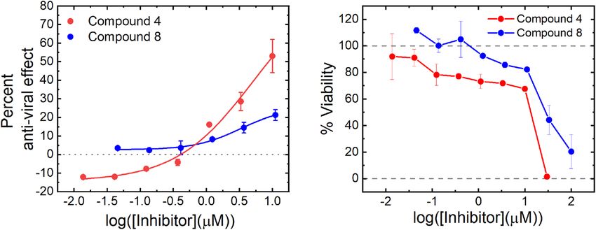
www.nature.com/scientificreports/
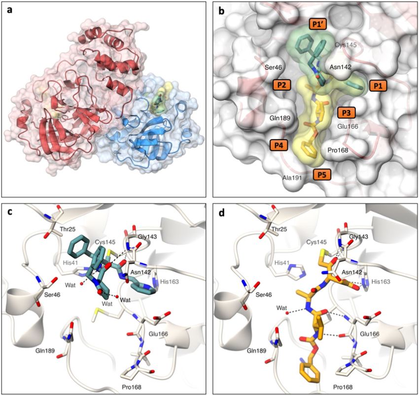
Figure 3. Potent inhibitors of SARS-CoV-2 Mpro. Structures of the compounds tested against M pro are shown
to the left of the graphs. Compounds 1–3 are calpain inhibitors and 4–7 are caspase-1 inhibitors. 1 is a pyridine
analogue of ABT-957 (2), 4 is the ester prodrug SDZ 224015 with 5 Caspase-1 active acid version of 4. The
upper graph shows titrations of the compounds plotted against inhibition of M pro, and the lower graph shows
the same compounds against PLpro. Data are singlicate and representative of at least four repeats on separate
occasions.
of calpain inhibitors, as the tool calpain inhibitor Z-L-Abu-CONH-ethyl (3, Fig. 3) does not inhibit. 1 was
confirmed to bind to M pro in a SPR assay, giving a K D of 1 µM (Supplementary Fig. 3). Furthermore, in HUH7
cells 1 was non cytotoxic at concentrations up to 100 µM and showed an antiviral effect of 75% at 100 µM (Sup-
plementary Fig. 5).
Compound 4 is the investigational caspase 1 inhibitor prodrug SDZ-22401531, which is cleaved by esterases
in vivo to yield 5, where the free aspartic acid is revealed (Fig. 3). In contrast to the exquisite potency of 4, 5 has
limited potency for Mpro, yielding only 50% inhibition at 100 µM. Inhibition of M pro did not seem to be a general
property of caspase 1 inhibitors, as the tool tetrapeptide Ac-YVAD-AOM (6, Fig. 3) and the investigational cas-
pase 1 drug belnecasan (7, Fig. 3) did not substantially inhibit M pro. 4 was confirmed to bind to M pro in an SPR
assay, although due to the mechanism of action a K D cannot be reported (Supplementary Fig. 3). Compound 4
is a suicide inhibitor which is cleaved by M pro, releasing a dichlorobenozic acid leaving group and forming an
irreversible covalent adduct by reaction with the nucleophilic cysteine (Supplementary Fig. 4).
Due to the presence of three esters, 4 is unstable in aqueous media so is unsuitable for the long incubations
used in antiviral assays. However, when the dose was refreshed daily, 4 was found to be non-cytotoxic in HUH7
cells at concentrations at or below 10 µM and showed an antiviral effect of 50% at 10 µM (Fig. 4). Refreshing the
dose more frequently is likely to further increase the apparent potency.
SARS CoV‑2 PLpro inhibitors. The ReFRAME screen revealed a single inhibitor, tarloxotinib as a potent
Lpro inhibitor (Table 1, 8) which has an IC50 of 300 nM. 8 is also a prodrug, activated in hypoxic conditions
P
in vivo by S TEAP432 to yield the equipotent compound 9 (Table 1) which was designed to target the kinase
domain of EGFR33. Whilst both 8 and 9 contain a 4-anilinoquinazoline core that is present in several approved
drugs34, the tested related molecules proved to be less potent than tarloxotinib (Table 1, 10–13).
Analysis of the other 4-anilinoquinazoline approved medicines revealed the presence of the α,β-unsaturated
amide warhead on 8 and 9 was not necessary or sufficient for activity, as 13 achieved weak activity without the
warhead whilst the presence of a nitrile on the 3 position of 10 completely removes potency despite the presence
of the warhead. These observations show that inhibition is not solely due to intrinsic reactivity of the molecules
but requires specific molecular recognition.
Scientific Reports | (2021) 11:13208 | https://doi.org/10.1038/s41598-021-92416-4 4
Vol:.(1234567890)www.nature.com/scientificreports/
Figure 4. Anti-viral effect of compounds 4 and 8. The left hand chart shows anti-viral effect of compounds 4
and 8 in HUH7_mCherry cells. An anti-viral effect was established after a 4-day incubation with SARS CoV-2
virus at concentrations which did not cause significant cytotoxic effects. The anti-viral effect was established
by an increase in fluorescent cells counted compared to an untreated control. The right-hand chart shows cell
viability after 4-days treatment with compounds 4 and 8 in HUH7_mCherry cells. Viability was established by
counting the number of fluorescent cells. Data are shown as the average of two technical repeats with error bars
representing the range. Data are representative of two technical repeats.
Compound number Generic name R1 R2 R3 R4 R5 IC50 (µM) (range)
8 Tarloxotinib Br F N N 0.3 (0.1–0.5)
9 Tarloxotinib (ac) Br F N N 0.3 (0.1–0.4)
10 Pelitinib Cl Cl > 100*
11 Afatinib Cl Cl N 12 (11–16)
12 Dacomitinib Cl Cl N 4 (3–5)
13 Gefitinib Cl Cl N 7 (6–11)
Table 1. Structure–activity relationship of 4-aminoquinazoline EGFR inhibitors against SARS-CoV-2 P
Lpro.
*The range could not be established due to lack of potency.
Scientific Reports | (2021) 11:13208 | https://doi.org/10.1038/s41598-021-92416-4 5
Vol.:(0123456789)www.nature.com/scientificreports/
Figure 5. Crystal structures of SARS-CoV-2 Mpro in complex with ReFRAME inhibitors. (a) Ribbon
representation with transparent surface of the Mpro dimer coloured in red and blue to delineate each protomer.
The structures of Mpro in complex with 1 and 5 (sticks with yellow and green transparent surface, respectively)
reveal that both bind in the Mpro substrate binding pocket. (b) Surface representation showing the overall
binding modes of compound 1 and 5 (green and yellow transparent surfaces, respectively). (c) and (d) Stick
representations of compounds 1 and 5 showing interactions (hydrogen bonds as dashed lines) within the Mpro
binding pocket. Structures are deposited in the pdb as 7AEH for 1 and 7AEG for 5. Figure generated with
PyMOL, The PyMOL Molecular Graphics System, Version 2.0 Schrödinger, LLC (https://pymol.org/2/).
8 was found to be competitive with the peptide substrate (Supplementary Fig. 6) and despite the conditions
required for its discovery, the potency of 8 was not dependent on the use of high concentrations of anionic
Hoffmeister salts (Supplementary Fig. 7). In HUH7 cells 8 was non-cytotoxic at concentrations up to 10 µM,
and showed an antiviral effect of 25% at 10 µM (Fig. 4).
Crystallography of the ReFRAME hits. X-ray crystal structures were attempted for compounds 1,4,5
and 8 and obtained for 1 and 5 with M pro (Fig. 5, PDB codes: 7AEH & 7AEG), at 1.3 Å and 1.8 Å resolution,
respectively (Supplementary Table 2). The M pro dimer is shown in ribbon representation with 1 and 5 bound
at the active site (Fig. 5a,b). Both 1 and 5 bind covalently to the catalytic cysteine (Cys145) with well-defined
electron density and form hydrogen bonding networks with the M pro active site (Fig. 5c,d, Supplementary Fig. 8).
Despite both 1 and 5 interacting with Cys145, they have substantially different binding modes. 5 extends from
P1 to P5 of the Mpro active site (Fig. 5b). By contrast 1 binds from P1 to P1′ across the region occupied by the
catalytic cysteine, with the two inhibitor benzyl groups π-stacking together to fill the space of the P1′ pocket
(Fig. 5b). Electron density for the 2,6-dichlorobenzoate leaving group of 5 was not observed, providing evidence
of the proposed mechanism of inhibition (Supplementary Fig. 4 and 8). 1 forms electrostatic interactions with
Gly143, Ser144, Cys145, and His41 as well as a water-mediated interaction with His164 whereas 5 makes electro-
static interactions with Gly143, Cys145, His163, His164, and Glu166 together with a water mediated interaction
with Gln189 (Fig. 5).
Scientific Reports | (2021) 11:13208 | https://doi.org/10.1038/s41598-021-92416-4 6
Vol:.(1234567890)www.nature.com/scientificreports/
Figure 6. Flexibility induced in the active site of SARS-CoV-2 Mpro by compounds 1 and 5 from the ReFRAME
library. Grey–Ligand-free M pro (pdb 5r8t), teal–Mpro in complex with 1, orange–Mpro in complex with 5. Figure
generated with PyMOL, The PyMOL Molecular Graphics System, Version 1.8.0.5 Schrödinger, LLC (https://
pymol.org/2/).
Active site plasticity is important in accommodation of the inhibitors. The P1′ pocket expands on binding of
1, with the alpha carbons of Thr25 and Thr26 shifting by 0.9 Å and 0.7 Å respectively. Similarly, the P5 pocket
expands upon binding to 5, with Pro168 and Thr190 moving by 1.3 Å and 0.8 Å respectively. Additionally, there
is a 1.9 Å shift between Ala191 in the 1 and 5 complexes. In both cases plasticity in response to ligand binding
is also observed for the P2 pocket35 (Fig. 6).
The pyridine of 1 and the aspartate of 5 extend into the P1 pocket and interact with His163 (Fig. 5c,d). Mod-
elling suggests that the cyclopropyl ring sidechain of 2 is unable to make this interaction and as a consequence
does not bind to M pro. Similarly, the acid of the aspartate in 5 is in close proximity to Glu166 residue which may
cause a charge clash explaining the loss in potency of 5 compared to 4.
Prospects for molecular design. Combining the information from the M pro structures of 1 and 5 could be
the starting point to design more potent, drug-like inhibitors. As inhibition of Caspase 1 would inhibit inflam-
mation via suppression of the IL-1β, this could provide additional clinical benefit in the treatment of COVID-
1936,37. Thus inhibitors which possess dual anti-inflammatory and antiviral properties may be desirable. A dock-
ing analysis38,39of the binding pose of 5 in Mpro and the caspase-1 active site reveals multiple shared interactions,
indicating that further, more drug-like molecules could be developed which share the potential dual anti-viral/
anti-inflammatory polypharmacology of SDZ 224015 (Supplementary Fig. 9).
For PLpro, the 4-anilinoquinazoline core is one of the most common scaffolds for generation of tyrosine kinase
inhibitors. Thus, it should be possible to rapidly expand from 8 and 9, to discover new, potentially more potent
PLpro inhibitors with the potential to remove kinase activity all together, whilst retaining drug-like properties.
Discussion
The emergence of COVID-19 has emphasised the need for multiple approaches to tackle viral infections. To
bridge the gap between the need to rapidly address a new disease and the time required to safely develop an
entirely new medicine, repurposing existing drugs is an attractive a lternative40.
There have been several clinical efforts to assess the benefits of existing drugs for COVID-19 treatment. Trials
have broadly focused on repurposed anti-viral drugs to reduce infection such as remdesivir, as well as the use of
existing anti-inflammatory and immunosuppressant compounds to help the body better manage its subsequent
response to the infection. Virtual screening has also been u sed23,35, but has only identified boceprevir, an HCV
protease inhibitor, as an M pro inhibitor35. This molecule was identified in our screen but has disappointing activity
(IC50 of 3 µM) (Supplementary Table 1).
By contrast, the results described here identify highly potent inhibitors of M pro and for the first time a potent
inhibitor of PLpro with drug-like properties. This is also the first description of a non-antiviral molecule to show
repurposed PLpro activity. Neither of the two M pro inhibitors discovered in this study were proposed by docking
efforts. 4 is a suicide inhibitor which uses a complex mechanism that is difficult to predict41. Further, the con-
formation of 1 within the P1’ pocket of M pro which is driven by intramolecular pi-pi stacking (Fig. 3c) is unusual
and not readily predicted by in silico a pproaches42.
A recent crystallographic screen of 5000 compounds discovered several compounds which crystallised with
Mpro43. One such hit was the EGFR inhibitor pelitinib but disappointingly it only subsequently shows micromo-
lar activity in a cellular screen and was not determined to display significant biochemical inhibition of M pro in
this study. In contrast to the M pro crystallographic screen which was also restricted to a single structural form,
we were able to study both M pro and P Lpro in solution where multiple conformations and oligiomeric forms are
Scientific Reports | (2021) 11:13208 | https://doi.org/10.1038/s41598-021-92416-4 7
Vol.:(0123456789)www.nature.com/scientificreports/
present. Pelitinib is compound 10 in our study, but our results show that the 4-aminoquinazoline class of EGFR
inhibitors 8 and 9 are more promising; and importantly operate as potent PLpro, rather than M pro, inhibitors.
By employing optimised screens, we specifically interrogated the two essential SARS-CoV-2 viral proteases,
discovering compounds not identified in previous phenotypic screens despite possessing anti-viral activity. The
ReFRAME collection has been screened in phenotypic viral-replication44 assays. In spite of counter screens,
without deconvolution, the results from phenotypic screens can artificially prioritise highly potent compounds
such as transcription inhibitors and cytotoxic compounds that have undesirable mechanisms of action precluding
therapeutic development45 along with undervaluing the potency of viable compounds. Compounds that require
revised assay protocols to observe activity, such as 4, were therefore not previously identified.
In vitro viral replication assays are of limited value for predicting the in vivo pharmacodynamics of candidate
molecules46 where multi-day assays often underestimate the true in vivo potency. For compounds such as 4,
where a confounding factor is the aqueous stability of the molecule, in vitro data serve to support the mecha-
nism rather than predict in vivo efficacy, where administration frequency, route and immune clearance would
positively influence p otency47,48.
Similarly, potencies of anti-viral activity can vary drastically depending on the methods used. A recent study
Lpro tool inhibitor GRL-06178 saw potency vary by two orders of magnitude between biochemical (IC50 of
of the P
2 µM), cytopathic-effect (30 µM), viral RNA detection (> 50 µM) and FFU (IC50 > 100 µM) assays. Consequently,
there is a prospect that the potencies of both 4 and 8 in vivo may be better than implied by the cytopathic-effect
antiviral measure used in this work.
There have been three widespread outbreaks of fatal novel respiratory coronavirus mediated disease in the
last two d ecades49. Retrospectively, the dangers of further outbreaks were evident following the first50. To avoid
the debilitating effects of future coronavirus pandemics or even escape from immune protection, a range of
treatments are necessary in which effective antiviral drugs will be a critical component. Protease inhibitors have
been highly successful in combating other viral infections51. The high conservation of M pro and P Lpro between
the three strains of coronavirus which cause greatest impact on human health suggest that these are excellent
target opportunities for developing small-molecule anti-viral therapeutics.
Anti-viral efforts aim to treat patients who are already infected and halt progression to severe d isease6. This
serves to reduce the burden of disease on fragile health care systems, but must also be employed alongside
vaccination3 and containment e fforts52. Vaccination and containment serve to prevent the potential for infec-
tion, whereas anti-viral aim to treat those already infected. To be truly useful anti-viral medicines must be broad
spectrum and stockpiled prior to an outbreak as suggested for i nfluenza53.
Our studies describe the discovery of potent, drug-like inhibitors for both M pro and P
Lpro. These inhibitors
display in vitro antiviral activity and have already been shown to be safe for clinical investigation for other
therapeutic areas. Given their existing preclinical safety profiles these compounds have the potential for rapid
progression towards a clinical setting.
Methods
Materials. The ReFRAME library was received from Calibr, Scripps Research, as compounds dissolved to
10 mM in DMSO, spotted in 30 nL volumes in black 384 well plates. All peptides used were prepared with
C-terminal amides from Cambridge Research Biochemicals (Billingham, UK) and provided at > 95% purity.
Cambridge Research Biochemicals (Billingham, UK) synthesized the ester and acid forms of SDZ-224015 (com-
pounds 4 & 5) used in follow-up studies, provided at > 95% purity. Additional compound 4 was synthesised as
described below. pelitinib (10), afatinib (11), dacomitinib (12) and gefitinib (13) were obtained from Tocris
(Bristol, UK). The active form of Tarloxotinib (9) was from Molport (Riga, Latvia). Compound 3 was from Santa
Cruz Biotechnology (Dallas, Texas, USA), Compounds 2, 7 and 8 were from MedChem Express (Sweden); Com-
pound 6 was from Bachem (Bubendorf, Switzerland).
The African monkey kidney cell line Vero E6-GFP was a gift kindly provided by M. van Loock, Janssen
Pharmaceutica, Beerse, Belgium.
The hepatocellular carcinoma cell line Huh7 was a gift kindly provided by Ralf Bartenschlager, University
of Heidelberg, Germany.
All compounds were obtained at a manufacturer specification of > 98% purity.
Unless otherwise stated all other reagents were from Sigma Aldrich (Poole, UK).
Construct design and construction. The Mpro coding sequence was codon optimised for expression in E.
coli and synthesised by Integrated DNA technologies (IDT). The Mpro expression construct used for crystalliza-
tion comprises an N-terminal GST region, an Mpro autocleavage site, the Mpro coding sequence, a hybrid cleavage
site recognizable by 3C HRV protease and a C-terminal 6-Histidine t ag54. The overall construct was flanked by
In-Fusion compatible ends for insertion into BamHI-XhoI cleaved pGEX-6P-1 (Sigma). An additional Mpro
construct was generated with an extended 10-Histidine tag, for enhanced binding to the sensor surface in SPR
assays. This construct was amplified by PCR from the above version, with the C-terminal primer incorporating
a further 4-Histidines. The resulting amplicon was then inserted into BamHI-XhoI cleaved pGEX-6P-1 by In-
Fusion cloning.
The PLpro expression construct was similarly optimised and synthesised and comprised an N-terminal 10
Histidine tag followed by the P Lpro sequence (Nsp3 region E746-K1060). This was then directly inserted into
NcoI-HindIII digested pOPINF via In-Fusion compatible ends. pOPINF was a gift from Ray Owens (University
of Oxford)55 (Addgene plasmid # 26042 ; http://n2t.net/addgene:26042 ; RRID:Addgene_26042).
Scientific Reports | (2021) 11:13208 | https://doi.org/10.1038/s41598-021-92416-4 8
Vol:.(1234567890)www.nature.com/scientificreports/
Protein expression of Mpro with authentic termini. The plasmids were used to transform a competent
E. coli expression cell line based on BL21(DE3)-R3-pRARE. The cells were plated on LB-agar plates containing
50 µg/ml carbenicillin and incubated overnight at 37 °C. The next day multiple colonies were picked and use to
inoculate a series of consecutive starter cultures (LB, 50 µg/ml Carbenicillin) of 1 ml, 10 ml and 100 ml. At each
stage the culture was grown to the exponential phase (OD600 0.6–2, 200 rpm, 37 °C) before using the total vol-
ume of culture to inoculate the next, where the inoculate comprised 10% of the volume of the next culture in the
series. Once 100 ml of exponential culture was achieved, 10 ml of this was used to inoculate 1 L of Auto Induc-
tion medium (Formedium, Terrific broth base including trace elements, prepared to manufacturer’s instructions
with addition of 10 ml glycerol and 50 µg/ml carbenicillin). Cultures were grown for 5 h at 37 °C, 200 rpm, fol-
lowed by 15–20 h at 18 °C, 200 rpm. Cells were harvested by centrifugation and stored at -80 °C.
Protein purification of Mpro with authentic termini for crystallographic analysis. Cells were
resuspended in lysis buffer, 50 mM Tris pH 8, 300 mM NaCl, 10 mM imidazole, 0.03 μg/ml Benzonase, and lysed
using an Emulsiflex homogeniser (3 passes, 30 kpsi, 4 °C). Insoluble material was removed by centrifugation
(50,000 g, 4 °C). Tagged Mpro protein was captured using Nickel-NTA (Takara His60 Superflow Resin) washed
with 50 mM Tris pH 8, 300 mM NaCl, 25 mM imidazole, and eluted with 50 mM Tris pH 8, 300 mM NaCl,
500 mM imidazole. To remove the Mpro poly-histidine tag, N-terminal His tagged HRV 3C protease was added
to the eluted M pro fractions at a ratio of 1 mg 3C protease: 10 mg M
pro. The mixture was dialysed overnight into
50 mM Tris pH 8, 300 mM NaCl, 0.5 mM TCEP at 4 °C and purified by reverse Nickel-NTA. Gel filtration was
performed using a 16/600 Superdex S200 pg column (GE Healthcare) equilibrated in 50 mM Tris pH 8, 300 mM
NaCl buffer. M pro was concentrated to 36 mg/ml using a centrifugal filter device with a 10 kDa molecular weight
cut off prior to flash freezing using liquid nitrogen.
Expression and purification of Mpro‑His10. Mpro-His10 was prepared as for Mpro with authentic ter-
mini with the following modifications. The HRV 3C protease cleavage and reverse Ni–NTA steps were omitted.
Instead, the Ni–NTA purified tagged Mpro was dialysed into 50 mM Tris pH 8.5, 100 mM NaCl, 0.5 mM TCEP
at 4 °C overnight. The dialysed sample was rapidly diluted with 50 mM Tris pH 8.5 buffer to achieve a final NaCl
concentration of 25 mM. The protein was purified by anion exchange chromatography using a 5 ml HiTrap Q
HP column (GE Healthcare) on a NaCl concentration gradient between 25 mM to 0.5 M NaCl. Ion exchange
chromatography was followed by a gel filtration purification step as described above.
Expression and purification of cleaved PLpro. Cleaved PLpro was prepared as for M pro with authentic
termini with the following modifications. A tunable T7 expression strain based on Lemo21 (DE3) was utilised
in the expression of PLpro PLpro and 34 μg/ml chloramphenicol was added to all solid and liquid media to main-
tain its pLemo plasmid. 2 mM Rhamnose was included in the medium in the overnight and sub-culture stages.
0.5 mM Rhamnose was included in the auto induction medium. Instead of the HRV 3C protease, TEV protease
was used to cleave the 10 Histidine tag of PLpro at the same mass ratio as described above. A 16/600 Superdex S75
pg column (GE Healthcare) was used for gel filtration chromatography.
Expression and purification of His10‑PLpro. His10-PLpro was prepared as for cleaved PLpro with the fol-
lowing modifications. The TEV protease cleavage and reverse Ni–NTA steps were omitted. Instead, the Ni–NTA
purified tagged P Lpro was dialysed into 50 mM Tris pH 8.8, 100 mM NaCl, 0.5 mM TCEP at 4 °C overnight.
The dialysed sample was rapidly diluted with 50 mM Tris pH 8.8 buffer to achieve a final NaCl concentration of
25 mM. The protein was purified by anion exchange using a 5 ml HiTrap Q HP column (GE Healthcare) with a
NaCl concentration gradient between 25 mM to 0.5 M NaCl. The ion exchange chromatography was followed by
a gel filtration purification step as described above.
Mpro characterisation. The Mpro substrate, [5-TAMRA]-AVLQSGFR-[Lys(BHQ-2)]-K-amide was disolved
in DMSO (10 mM). This solution was diluted in buffer (150 mM NaCl, 50 mM Tris, 1 mM EDTA, 0.5 mM TCEP,
0.05% (v/v) Triton X-100, pH 7.6), then mixed with Mpro solution, to give a final substrate concentration range
of 0.8–100 µM and Mpro concentration range of 0.3–300 nM. Initial rates were measured using a Pherastar FSX
plate reader equipped with a TAMRA filter set.
Mpro inhibition assays. Compounds were received for ReFRAME screening at 10 mM in DMSO at 30 nL
volumes in black 384 wells plates; otherwise, dilutions were performed in DMSO from 10 mM stock solutions;
300 nL was transferred to a black 384 well plate using a Mosquito liquid handler (SPT Labtech, Melbourne, UK).
10 µL of the Assay Buffer (150 mM NaCl, 50 mM Tris, 1 mM EDTA, 0.5 mM TCEP, 0.05% (v/v) Triton X-100,
pH 7.6) was added to the assay plates to solubilise compounds. 10 µL Mpro, 90 nM in Assay Buffer, was added
to the appropriate wells and incubated with the compounds for 60 min at room temperature. The reaction was
started via the addition of 10 µL of the substrate [5-TAMRA]-AVLQSGFR-[Lys(BHQ-2)]-K-amide (where BHQ
is Black hole quencher 2), 18 µM in assay buffer. This resulted in final assay conditions of 30 nM Mpro and 6 µM
substrate, with either 0.1% or 1% DMSO (v/v). The plates were incubated for a further 60 min at room tempera-
ture; assays employed a Pherastar FSX plate reader (BMG Labtech, Aylesbury, UK) using a TAMRA filter set.
Assay quality was established using Z’, where a low control consisted for substrate alone with balanced DMSO,
and the high control was enzyme and substrate without inhibitors but balanced DMSO. The raw fluorescence
data was converted to percent inhibition using the same controls as for Z’.
Scientific Reports | (2021) 11:13208 | https://doi.org/10.1038/s41598-021-92416-4 9
Vol.:(0123456789)www.nature.com/scientificreports/
Mpro SPR assay. Cytiva Biacore S200 and T200 machines were used for all SPR experiments. Data were
pro-10His was captured on an NTA chip using standard protocols
collected at a constant temperature of 20 °C. M
in running buffer: 20 mM Hepes (pH 7.5), 150 mM NaCl, 50 µM EDTA, 0.05% (v/v) Tween 20 and either 1 or
3% (v/v) DMSO at ~ 8500 RU. The compounds were screened at concentrations ranging from 23 nM to 50 µM
adjusted appropriately for each compound, injecting from the lowest to highest concentrations. Scrubber 2 (Bio-
logic software) was used to process and analyse SPR data. Kinetics were fitted using a 1:1 binding model with
local Rmax for each concentration where required. Data for the inhibitors were referenced to those for a blank
surface and blank injections to normalize for non-specific binding and drift. A DMSO calibration was run to
remove excluded volume effect of binding responses between reference and target surface.
Mpro protein observed mass spectroscopy. Protein MS-analyses were performed as described56 using
a RapidFire RF 365 high-throughput sampling robot (Agilent) attached to an iFunnel Agilent 6550 accurate
mass quadrupole time-of-flight (Q-TOF) mass spectrometer operating in the positive ionization mode with the
parameters: capillary voltage (4000 V), nozzle voltage (1000 V), fragmentor voltage (365 V), gas temperature
(225 °C), gas flow (13 L/min), sheath gas temperature (350 °C), sheath gas flow (12 L/min). The reaction was
initiated either by adding SDZ-224015 (compound 4; 10 mM in DMSO) to a final concentration of 2.5 µM into
reaction buffer (20 mM HEPES, pH 7.5, 50 mM NaCl) containing 2.5 µM Mpro or by adding M pro (15 µM in
20 mM HEPES, pH 7.5, 300 mM NaCl) to a final concentration of 1 µM into reaction buffer (20 mM HEPES, pH
7.5, 50 mM NaCl) containing SDZ-224015 (5 µM). A sample from the reaction mixture was directly aspirated
under vacuum (0.6 s) and loaded onto a C4 solid phase extraction (SPE) cartridge. After loading, the C4 SPE
cartridge was washed with 0.1% (v/v) aqueous formic acid to remove non-volatile buffer salts (5.5 s, 1.5 mL/min)
and the protein was then eluted from the SPE cartridge with 0.1% (v/v) aqueous formic acid in 85/15 (v/v) ace-
tonitrile/water into the mass spectrometer (5.5 s, 1.25 mL/min). The SPE was cartridge re-equilibrated with 0.1%
(v/v) aqueous formic acid (0.5 s, 1.25 mL/min) and a blank water sample was injected before the next reaction
sample was aspirated from the assay mixture. Protein spectra were deconvoluted (mass range: 10–60 kDa, m/z
range: 950–1300 Da, mass step: 1 Da) using the MaxEnt1 function in Agilent MassHunter Version 7 (Agilent),
normalised, and plotted using Graphpad Prism 5.
Crystallisation and structure determination of Mpro in complex with compounds 1 and 5. Mpro
was thawed and diluted to 6 mg/ml using 20 mM Hepes pH 7.5, 50 mM NaCl. The ligand of interest was dis-
solved in DMSO to 10 mM and then diluted into the protein solution to a final concentration of 1 mM. The
ligand was then allowed to incubate with the protein for two hours at room temperature prior to dispensing
plates. The drop composition was 0.15 µL protein ligand solution, 0.3 µL 11% (v/v) PEG 4 K, 0.1 M MES pH 6.5,
and 0.05 µL M pro crystal seed stock. The M
pro crystal seed stock was prepared by crushing M
pro crystals with a
pipette tip, suspending them in 30% PEG 4 K, 5% (v/v) DMSO, 0.1 M MES pH 6.5, and vortexing for 60 s with
approximately 10 glass beads (1.0 mm diameter, BioSpec products). Reservoir solution was 11% (v/v) PEG 4 K,
5% (v/v) DMSO, 0.1 M MES pH 6.5. Crystals were grown using the sitting drop vapor diffusion method at 20 °C
and appeared within 24 h, reaching full size within 36 h.
Data collection and structure determination. All diffraction data were collected from crystals cryo-
cooled to 100 K at Diamond Light Source. X-ray diffraction data for the Mpro compound 1 complex were col-
lected at beamline I04-1 at a wavelength of 0.9126 Å and data for the M pro compound 5 complex were collected
at I24 at 0.9999 Å. Data were processed using Dials57 via Xia258. The datasets were phased using Molrep59 and
the Mpro apo s tructure60. Ligand restraints were generated using GRADE (Global Phasing Ltd) and A ceDRG61.
Crystal structures were manually rebuilt in Coot62 and refined using Refmac63 and B uster64.
PLpro enzyme characterisation. 50 nM PLpro was incubated with titrations of either Ubiquitin-Rhoda-
mine (RnD Systems, Abingdon, UK) (0.1–10 µM) or [5-TAMRA]- VLRLRGG-[Lys(BHQ-2)]-amide (1–100 µM)
in either 150 mM NaCl, 50 mM Tris, 1 mM EDTA, 0.5 mM TCEP, 0.05% (v/v) Triton X-100, pH 7.6 or 800 mM
sodium citrate, 200 mM Tris, 0.5 mM TCEP, 0.05% (v/v) Triton X-100, pH 7.6. Initial rate was established using
a Pherastar FSX plate reader using either a FITC (Ubiquti-Rhodamine) or a TARMA ([5-TAMRA]- VLRLRGG-
[Lys(BHQ-2)]-amide) filter set.
PLpro biochemical assay. Compound plates were prepared as for the Mpro biochemical assay. 10 µL assay
buffer (800 mM sodium citrate, 200 mM Tris, 0.5 mM TCEP, 0.05% (v/v) Triton X-100, pH 7.6) was added to
the assay plates to solubilise the compound. 10 µL of PLpro at 75 nM in assay buffer, was added to the appropri-
ate wells and incubated with the compounds for 60 min at room temperature. The reaction was started via the
addition of 10 µL of the substrate [5-TAMRA]- VLRLRGG-[Lys(BHQ-2)]-amide at 6 µM in assay buffer. This
resulted in final assay conditions of 25 nM PLpro and 2 µM substrate, with either 0.1% or 1% DMSO (v/v). The
plates were incubated for a further 60 min at room temperature and the assay was read on a Pherastar FSX plate
reader (BMG Labtech, Aylesbury, UK) using a TAMRA filter set. Assay quality was established as for the Mpro
biochemical assay. Data were converted to percent inhibition in the same manner as the M pro assay.
PLpro kinetic assays. Assays employed the same conditions as the P Lpro inhibition assays, except the com-
pound and substrate were prepared in the assay plate and the assay was started with an injection of 10 µL of
enzyme using a Pherastar FSX plate reader. Fluorescence was measured every 15 s post injection using a TAMRA
Scientific Reports | (2021) 11:13208 | https://doi.org/10.1038/s41598-021-92416-4 10
Vol:.(1234567890)www.nature.com/scientificreports/
filter set for 60 min. Data were zeroed to the date measured at time of injection and fit to a two-state inhibition
model.
Effect of hoffmeister salt concentration on PLpro activity. [5-TAMRA]- VLRLRGG-[Lys(BHQ-2)]-
amide was dissolved in buffers with increasing concentration of sodium phosphate (0.15–1.5 M sodium phos-
phate, 1 mM EDTA, 0.5 mM TCEP, 0.05% Triton-X100, pH 7.4). This was then mixed with P Lpro in a matched
buffer to give final conditions of: 2 µM substrate and 30 nM enzyme. The initial rate was established using a
Pherastar FSX plate reader using either a FITC (Ubiqutin-Rhodamine) or a TARMA ([5-TAMRA]-VLRLRGG-
[Lys(BHQ-2)]-amide) filter set.
Effect of Hoffmeister salt concentration on tarloxotinib bromide inhibition. This assay was per-
formed in the same manner as the PLpro biochemical assay, except the buffer was either 0.15, 0.75, 1 or 1.5 M
sodium phosphate, 1 mM EDTA, 0.5 mM TCEP, 0.05% Triton-X100 pH7.4.
Cell culture. Huh-7 cells stably expressing H2B-mCherry were generated using lentiviral vectors containing
a CMV-H2B-mCherry-P2A-BlastR cassette. The Huh-7 mCherry cells were maintained in Dulbecco’s modified
Eagle’s medium (DMEM; Gibco) supplemented with 10% v/v fetal calf serum (FCS; Biowest), 10 ml HEPES,
5 ml NEAA, and 1 × Pen-strep (Gibco) and kept under 5% CO2 on 37 °C. Assay medium contained only 4% FCS.
Virus culture. SARS-CoV-2 strain BetaCov/Belgium/GHB-03021/2020 recovered from a nasopharyngeal
swab taken from an asymptomatic patient returning from Wuhan, China at the beginning of February 2020 was
sequenced on a MinION platform (Oxford Nanopore). After serial passaging on Huh7 and Vero E6 cells, infec-
tious content of the virus stock was determined by titration on HUH7 cells using the Spearman-Kärber method.
All virus-related work was carried out in certified, high-containment biosafety level-3 facilities of KU Leuven
Rega institute.
Antiviral assay. To measure inhibition of the SARS-CoV-2 cytopathic effect, 96-well plates (Corning 3300)
were plated with HUH7_mCherry cells at 6000 cells/well in 100 µl. The day after (Day 0), compound was added
in a dilution series for concentration response studies. After two hours, addition of virus dilution (final MOI
0.004) was performed and plates were left for incubation at 37 °C, 5% CO2 for four days.
Cytotoxicity was assessed in parallel using the same protocol, albeit without the addition of virus dilution.
Plates were imaged on an Arrayscan XTI, Thermofisher.
Image acquisition and analysis. At day four post-infection, mCherry signal was captured using wide
field fluorescence imaging by exciting at 560_25 nm and emitting with the BGRFRN filter set. A 5 X objective
sufficed to capture 65–70% of an entire well on a 96well plate (4 pictures in total). The optimal exposure time was
determined based on fluorescence intensity and was set on 0.09 s. A 2 × 2 binning was used and autofocus plane
count was reduced to increase image acquisition speed. An image analysis protocol was developed in-house by
using the SpotDetector bioapplication (Cellomics, Thermofisher). After background reduction on the raw image
files, a fixed fluorescent intensity threshold was determined for the identification of mCherry cells. Afterwards,
the number of fluorescent cells (‘object count’) was calculated per well and compared to the positive (cell control)
and negative (virus) control.
Synthesis of SDZ‑224015 (compound 4). Commercially-sourced reagents (Sigma-Aldrich, Inc.; Flu-
orochem Ltd; Bachem AG) were used as received. Reactions were performed in anhydrous solvents (Sigma-
Aldrich Inc.). Purifications, reaction work-ups, and extractions were performed using HPLC grade solvents
(Sigma-Aldrich Inc.). A Stuart SMP-40 automated melting point apparatus was used to determine melting
points (MP). A Bruker Tensor-27 Fourier transform infrared spectrometer was used for infrared (IR) spec-
troscopy. A Unipol (Schmidt Haensch) polarimeter was used for optical rotation (α) measurements. A Thermo
Scientific Exactive mass spectrometer (ThermoFisher Scientific) operated in the positive ionization mode was
employed for high-resolution mass spectrometry (HRMS) using electrospray ionization (ESI) mass spectrom-
etry (MS); data are presented as a mass-to-charge ratio (m/z). A Bruker AVANCE AVIIIHD 600 spectrometer
equipped with a 5 mm BB-F/1H Prodigy N2 cryoprobe was used for nuclear magnetic resonance (NMR) spec-
troscopy. Proton chemical shifts are reported in parts per million (ppm) downfield from tetramethylsilane, the
residual protium in the NMR solvent is used as a reference (DMSO-d6: δ = 2.49 ppm). Carbon chemical shifts are
reported in parts per million (ppm) in the scale relative to the NMR solvent (DMSO-d6: δ = 39.52 ppm). NMR
data are reported as: chemical shift, multiplicity (m: multiplet, s: singlet, d: doublet, dd: doublet of doublets, t:
triplet, q: quartet), coupling constant (J, Hz), and integration.
Ethyl (5S,8S,11S)-11-(2-((2,6-dichlorobenzoyl)oxy)acetyl)-5-isopropyl-8-methyl-3,6,9-trioxo-1-phenyl-
2-oxa-4,7,10-triazatridecan-13-oate (SDZ-224015, Z-VAD-DCB, compound 4) was synthesized from ethyl
(S)-3-((R)-2,2-dimethyl-1,3-dioxolan-4-yl)-3-(phenylamino)propanoate65 and Z-Val-Ala-OH in five steps as
reported66. However, the final oxidation reaction of the reported synthesis of 466 was modified, due to the insolu-
bility of the starting material in pure dichloromethane, the optimised protocol is given below:
A solution of ethyl (5S,8S,11S)-11-((R)-2-((2,6-dichlorobenzoyl)oxy)-1-hydroxyethyl)-5-isopropyl-8-me-
thyl-3,6,9-trioxo-1-phenyl-2-oxa-4,7,10-triazatridecan-13-oate65 (1.4 g, 2.0 mmol, 1.0 equiv.) in DMSO (3.0 mL)
was diluted with dichloromethane (40 mL). To the resulting clear solution, Dess-Martin p eriodinane67 (3.39 g,
8.0 mmol, 4.0 equiv.) was added at 0° C under ambient atmosphere; the reaction mixture was stirred at 0 °C for
Scientific Reports | (2021) 11:13208 | https://doi.org/10.1038/s41598-021-92416-4 11
Vol.:(0123456789)www.nature.com/scientificreports/
2 h, then for 2 h at ambient temperature, before aqueous phosphate buffer (100 mL, 0.1 M, pH 7) containing
sodium metabisulfite (6 g) was added at 0 °C. The resulting mixture was vigorously stirred at ambient tempera-
ture for 30 min, then five times extracted with dichloromethane. The combined organic extracts were washed
with saturated aqueous N aHCO3 solution, dried over anhydrous N a2SO4, filtered, evaporated; the residue was
then purified by reverse phase HPLC (20 mL/min; linear gradient over 39 min: 2% → 98% acetonitrile in water,
each containing 0.1% (v/v) formic acid; t R = 27.0 min) using a Shimadzu HPLC purification system (composed
of DGU-20A, 2 LC-20AR, CBM-20A, SPD-20A, and FRC-10A units) equipped with a C18 Grace VYDAC
218TP101522 column (Grace Davison Discovery Sciences) to afford 199 mg (15%) of purified SDZ-224015
(compound 4). The analytical data are in agreement with those reported66. White solid, m.p.: 178–180 °C; 1H
NMR (600 MHz, 300 K, DMSO-d6): δ = 8.62 (d, J = 7.5 Hz, 1H), 8.12 (d, J = 6.6 Hz, 1H), 7.60 − 7.59 (m, 2H), 7.55
(dd, J = 9.2, 6.9 Hz, 1H), 7.36 − 7.34 (m, 4H), 7.32 − 7.29 (m, 1H), 7.26 (d, J = 8.7 Hz, 1H), 5.18 (d, J = 17.2 Hz, 1H),
5.14 (d, J = 16.4 Hz, 1H), 5.03 (d, J = 12.7 Hz, 1H), 5.00 (d, J = 12.7 Hz, 1H), 4.64 (q, J = 6.8 Hz, 1H), 4.27 (app.
pent., J = 7.2 Hz, 1H), 4.05 (q, J = 7.1 Hz, 2H), 3.87 (dd, J = 7.9, 7.4 Hz, 1H), 2.85 (dd, J = 16.5, 5.8 Hz, 1H), 2.65
(dd, J = 16.5, 7.2 Hz, 1H), 1.98 − 1.92 (m, 1H), 1.22 (d, J = 7.0 Hz, 3H), 1.16 (t, J = 7.1 Hz, 3H), 0.85 (d, J = 6.8 Hz,
3H), 0.81 ppm (d, J = 6.7 Hz, 3H); 13C NMR (150 MHz, 300 K, DMSO-d6): δ = 199.5, 172.9, 171.0, 170.1, 163.2,
156.1, 137.0, 132.6, 132.0, 130.8, 128.5, 128.3, 127.7, 127.6, 67.7, 65.4, 60.3, 59.8, 52.8, 48.1, 34.2, 30.3, 19.2, 18.0,
17.7, 13.9 ppm; IR (film): ṽ = 3293, 3067, 2964, 2936, 1733, 1689, 1639, 1538, 1434, 1374, 1287, 1247, 1195, 1148,
1040 cm–1; HRMS (ESI): m/z calculated for C30H36O9N3Cl2 [M + H]+: 652.1823, found: 652.1823; [α]25 D = − 47.0
(c = 1.0, acetone).
Hot spot comparison between Mpro and caspase 1. Fragment Hotspot Maps were calculated for
structures 6YB7 and 1SC4 using the Hotspots A PI38,39. The method uses molecular probes, atomic interaction
propensity and a local buriedness measure to highlight key hotspots within the binding site.
Research ethics statement. All methods were carried out in accordance with relevant guidelines and
regulations. All experimental protocols were approved by either, an internal research committee at Exscientia.
Ltd, the ReFRAME committee or the CARE consortium. Viral swabs were obtained with prior patient’s written
informed consent for use in research.
Code availably statement
References pointing the reader towards code used for hotspot mapping are provided in the methods section.
Received: 2 March 2021; Accepted: 10 June 2021
References
1. Coronavirus Disease (COVID-19) Pandemic UNFPA Global Response Plan | UNFPA - United Nations Population Fund. https://
www.unfpa.org/resources/coronavirus-disease-covid-19-pandemic-unfpa-global-response-plan (n.d.).
2. Matsuyama, T., Kubli, S. P., Yoshinaga, S. K., Pfeffer, K. & Mak, T. W. An aberrant STAT pathway is central to COVID-19. Cell
Death Differ. 27, 3209–3225 (2020).
3. Moghadas, S. M. et al. The impact of vaccination on COVID-19 outbreaks in the United States. Medrxiv 2020 https://doi.org/10.
1101/2020.11.27.20240051 (2021).
4. Consortium, C.-19 G. U. (COG-U. et al. SARS-CoV-2 variants, spike mutations and immune escape. Nat. Rev. Microbiol. 1–16
(2021) https://doi.org/10.1038/s41579-021-00573-0.
5. Burton, D. R. & Topol, E. J. Variant-proof vaccines—invest now for the next pandemic. Nature 590, 386–388 (2021).
6. Villamagna, A. H., Gore, S. J., Lewis, J. S. & Doggett, J. S. The need for antiviral drugs for pandemic coronaviruses from a global
health perspective. Front. Med. 7, 596587 (2020).
7. Frecer, V. & Miertus, S. Antiviral agents against COVID-19: structure-based design of specific peptidomimetic inhibitors of SARS-
CoV-2 main protease. Rsc Adv. 10, 40244–40263 (2020).
8. Shin, D. et al. Papain-like protease regulates SARS-CoV-2 viral spread and innate immunity. Nature 587, 657–662 (2020).
9. Beigel, J. H. et al. Remdesivir for the treatment of Covid-19 - full report. New Engl. J. Med. 383, 1813–1826 (2021).
10. Group, R. C. et al. Dexamethasone in hospitalized patients with Covid-19. New Engl. J. Med. 384, 693–704 (2021).
11. Kalil, A. C. et al. Baricitinib plus Remdesivir for hospitalized adults with Covid-19. New Engl. J. Med. 384, 795–807 (2021).
12. Ullrich, S. & Nitsche, C. The SARS-CoV-2 main protease as drug target. Bioorg. Med. Chem. Lett. 30, 127377 (2020).
13. Cárdenas-Conejo, Y., Liñan-Rico, A., García-Rodríguez, D. A., Centeno-Leija, S. & Serrano-Posada, H. An exclusive 42 amino
acid signature in pp1ab protein provides insights into the evolutive history of the 2019 novel human-pathogenic coronavirus
(SARS-CoV-2). J. Med. Virol. 92, 688–692 (2020).
14. Kim, D. et al. The architecture of SARS-CoV-2 transcriptome. Cell 181, 914-921.e10 (2020).
15. Fehr, A. R. & Perlman, S. Coronaviruses, methods and protocols. Methods Mol. Biol. 1282, 1–23 (2015).
16. Lei, J., Kusov, Y. & Hilgenfeld, R. Nsp3 of coronaviruses: Structures and functions of a large multi-domain protein. Antivir Res.
149, 58–74 (2018).
17. Dolgin, E. The race for antiviral drugs to beat COVID—and the next pandemic. Nature 592, 340–343 (2021).
18. Janes, J. et al. The ReFRAME library as a comprehensive drug repurposing library and its application to the treatment of crypto-
sporidiosis. Proc. Natl. Acad. Sci. 115, 201810137 (2018).
19. Jasial, S., Hu, Y. & Bajorath, J. How frequently are pan-assay interference compounds active? Large-scale analysis of screening data
reveals diverse activity profiles, low global hit frequency, and many consistently inactive compounds. J. Med. Chem. 60, 3879–3886
(2017).
20. Matlock, M. K., Hughes, T. B., Dahlin, J. L. & Swamidass, S. J. Modeling small-molecule reactivity identifies promiscuous bioactive
compounds. J. Chem. Inf. Model. 58, 1483–1500 (2018).
21. Baell, J. & Walters, M. A. Chemistry: chemical con artists foil drug discovery. Nat. News 513, 481 (2014).
22. Yang, J., Copeland, R. A. & Lai, Z. Defining balanced conditions for inhibitor screening assays that target bisubstrate enzymes. J.
Biomol. Screen 14, 111–120 (2009).
23. Jin, Z. et al. Structure of Mpro from COVID-19 virus and discovery of its inhibitors. Nature https://doi.org/10.1038/s41586-020-
2223-y (2020).
Scientific Reports | (2021) 11:13208 | https://doi.org/10.1038/s41598-021-92416-4 12
Vol:.(1234567890)You can also read