Chemogenetic and Optogenetic Activation of G αs Signaling in the Basolateral Amygdala Induces Acute and Social Anxiety-Like States - Nature
←
→
Page content transcription
If your browser does not render page correctly, please read the page content below
Neuropsychopharmacology (2016) 41, 2011–2023
                                                                                © 2016 American College of Neuropsychopharmacology. All rights reserved 0893-133X/16

                                                                                www.neuropsychopharmacology.org

Chemogenetic and Optogenetic Activation of Gαs Signaling
in the Basolateral Amygdala Induces Acute and Social
Anxiety-Like States

     Edward R Siuda1,2,3,4, Ream Al-Hasani1,2,4, Jordan G McCall1,2,3,4, Dionnet L Bhatti1 and
     Michael R Bruchas*,1,2,3,4,5
     1
      Department of Anesthesiology, Division of Basic Research, Washington University School of Medicine, St Louis, Missouri, USA; 2Washington
     University Pain Center, Washington University School of Medicine, St Louis, Missouri, USA; 3Division of Biology and Biomedical Sciences, Washington
     University School of Medicine, St Louis, Missouri, USA; 4Department of Neuroscience, Washington University School of Medicine, St Louis, Missouri,
     USA; 5Department of Biomedical Engineering, Washington University in St. Louis, St Louis, Missouri, USA

     Anxiety disorders are debilitating psychiatric illnesses with detrimental effects on human health. These heightened states of arousal are
     often in the absence of obvious threatening cues and are difficult to treat owing to a lack of understanding of the neural circuitry and
     cellular machinery mediating these conditions. Activation of noradrenergic circuitry in the basolateral amygdala is thought to have a role in
     stress, fear, and anxiety, and the specific cell and receptor types responsible is an active area of investigation. Here we take advantage of
     two novel cellular approaches to dissect the contributions of G-protein signaling in acute and social anxiety-like states. We used a
     chemogenetic approach utilizing the Gαs DREADD (rM3Ds) receptor and show that selective activation of generic Gαs signaling is
     sufficient to induce acute and social anxiety-like behavioral states in mice. Second, we use a recently characterized chimeric receptor
     composed of rhodopsin and the β2-adrenergic receptor (Opto-β2AR) with in vivo optogenetic techniques to selectively activate Gαs
     β-adrenergic signaling exclusively within excitatory neurons of the basolateral amygdala. We found that optogenetic induction of
     β-adrenergic signaling in the basolateral amygdala is sufficient to induce acute and social anxiety-like behavior. These findings support the
     conclusion that activation of Gαs signaling in the basolateral amygdala has a role in anxiety. These data also suggest that acute and social
     anxiety-like states may be mediated through signaling pathways identical to β-adrenergic receptors, thus providing support that inhibition of
     this system may be an effective anxiolytic therapy.
     Neuropsychopharmacology (2016) 41, 2011–2023; doi:10.1038/npp.2015.371; published online 27 January 2016

INTRODUCTION                                                                    of action and target a myriad of circuits and systems.
                                                                                Benzodiazepines, SSRIs, atypical antipsychotics and β-block-
Anxiety is a fundamental behavioral response to environ-
                                                                                ers, and antagonists of β-adrenergic receptors (βARs), are
mental threats that prepares the body for immediate action
                                                                                all widely used clinically (Baker et al, 2011; Frishman and
by enhancing arousal. Baseline anxiety levels keep us alert
                                                                                Saunders, 2011). Of these therapies, β-blockers have amassed
and aware of our surroundings. However, anxiety as a
                                                                                a substantial body of work, particularly in peripheral systems,
psychiatric disease is characterized by a heightened state of
                                                                                and are often used to treat social phobias (Schneier, 2011;
arousal, often in the absence of an obvious threat (Lieb,                       Stein et al, 2004). However, there are very few basic
2005). Prolonged anxiety due to psychiatric disorders,                          neuropharmacological reports concerning βAR signaling
traumatic life experience, genetic factors, or sustained stress                 within the central nervous system, specifically the basolateral
is detrimental to physical and mental health.                                   amygdala, leading to debate as to whether the clinical efficacy
   Disorders such as: generalized anxiety disorder, panic                       of β-blockers as anxiolytics is due to receptor signaling
disorder, phobias, obsessive compulsive disorder, and post-                     peripherally, centrally or both.
traumatic stress disorder often require tailored therapeutic                       Norepinephrine (NE) is a critical mediator of the stress
intervention. Current therapies act via different mechanisms                    ‘fight or flight’ response and acts through neural circuits that
                                                                                widely express β- and α-adrenergic receptors of which there
*Correspondence: Dr MR Bruchas, Departments of Anesthesiology and               are nine distinct subtypes (α1a,b,d, α2a,b,c β1–3) throughout
Neuroscience, Washington University School of Medicine, 660 South
Euclid Avenue, Box 8054, St Louis, MO 63110, USA, Tel: +1 314 747               the mammalian brain (Hieble et al, 1995). Release of NE
5754, Fax: +1 314 362 8571, E-mail: bruchasm@wustl.edu                          is further modulated by the presence of presynaptic and
Received 2 October 2015; revised 21 December 2015; accepted 22                  postsynaptic α2 (Gαi) autoreceptors that inhibit NE release
December 2015; accepted article preview online 4 January 2016                   upon binding NE (Arima et al, 1998; Goddard et al, 2010;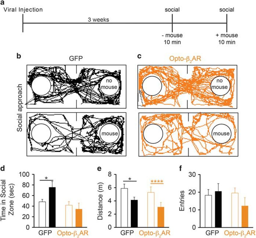
Activation of BLA Gαs signaling is anxiogenic
                                                         ER Siuda et al
2012
       Grigg et al, 1996). Conversely, α1 (Gαq) and β1–3 (Gαs)            demonstrated to mimic β2-adrenergic signaling both in vitro
       adrenergic receptors are expressed postsynaptically causing        and in vivo (Siuda et al, 2015a). Using in vivo optogenetics,
       mobilization of intracellular Ca2+ or cAMP, respectively.          we selectively photostimulated β2AR signaling within the
       Each receptor type mediates disparate downstream effects           BLA and CeA and demonstrated that engagement of β2AR
       and the interplay between them generates norepinephrine’s          signaling selectively in the BLA, but not in the CeA, was
       dynamic range in neural circuits.                                  sufficient to induce acute and social anxiety-like behavior
          Central NE is largely produced in two distinct brain            in mice.
       regions, the locus coeruleus (LC) and A1/A2 cell groups in
       the brain stem. These populations send dense neuronal
       projections to the basolateral (BLA), central (CeA), and           METHODS
       extended amygdala (in particular, the bed nucleus of the stria     Cell Culture
       terminalis (BNST)) (Asan, 1998; Byrum and Guyenet, 1987;
       Woulfe et al, 1990; Zhang et al, 2013), regions known to be        HEK293 cells were grown in Dulbecco’s modified Eagle's
       involved in affective behaviors (Berridge and Waterhouse,          media supplemented with 10% fetal bovine serum containing
       2003; Davis, 1992; Valentino and Aston-Jones, 2010). These         1× pen/strep (Invitrogen) and maintained at 37 °C in a
       regions are also enriched in all the major subtypes of             humidified incubator with 5% CO2. Plasmid containing Gαs
       adrenergic receptors (Lein et al, 2007) making isolation of        DREADD was transfected into HEK293 cells using JetPrime
       their respective contributions in the amygdaloid complex           (Polyplus) reagent per the manufacturer’s instructions.
       pharmacologically challenging. Anatomically speaking, this
       area is further complicated by the adjacent proximity of the       Real-Time cAMP Assay
       BLA and CeA. The predominately glutamatergic (excitatory)
       BLA acts as a relay center, sending projections to the             HEK293 cells were transfected with the pGloSensor-22F
       neighboring GABAergic (inhibitory) CeA, making the role of         cAMP plasmid (Promega E2301) using JetPrime (Polyplus)
       either region in anxiety-like behavior more difficult to dissect   transfection reagent per the manufacturer’s instructions.
       (Lüthi and Lüscher, 2014).                                         The day before an experiment, cells were plated in 96-well
          The basolateral nucleus of the amygdala (BLA) is one key        tissue culture-treated plates (Costar) at 25 000 cells/well and
       region associated with anxiety behavior (Valentino et al,          allowed to recover overnight at 37 °C, 5% CO2. The next day,
       1993), including humans (Feinstein et al, 2013). We know           media was replaced with 2% GloSensor reagent (Promega)
       that, in rodents, βAR antagonists infused into the amygdala        suspended in CO2-independent growth medium (Gibco) and
       block fear memory enhancement following stressful stimuli          incubated for 2 h at 25 °C. For real-time cAMP, baseline
       (Al-Hasani et al, 2013; Mantsch et al, 2010; Quirarte et al,       relative luminescent units (RLUs) were recorded every 6 s for
       1998; Schmidt and Weinshenker, 2014; Vranjkovic et al,             1 min using a SynergyMx microplate reader (BioTek,
       2014); is required for fear conditioning behavior (Cahill et al,   Winooski, VT, USA). Clozapine-N-oxide (CNO; 10 μM;
       1994; Dębiec and Ledoux, 2004; Rogan et al, 1997); and             Sigma C0832) was then used to stimulate the cells, and
       systemic administration of the βAR antagonist propranolol          subsequent RLUs were recorded every 6 s for 5–10 min at
       reduces the spontaneous firing rate of neurons within the          room temperature.
       BLA (Buffalari and Grace, 2007; Ferry et al, 1997; Huang
       et al, 1996). Generally, β-adrenergic receptors have been          Animals
       linked to amygdala-dependent learning of negative affective
       memories, and a predominate focus of this receptor system          Adult (25–35 g) male C57BL/6J mice were group-housed,
       has been examining its role in modulating fear-related             given access to food and water ad libitum, and maintained
       behaviors (McGaugh, 1988, 2004; Strange and Dolan, 2004).          on a 12-h:12-h light:dark cycle. All animals were held in a
       Furthermore, activation of endogenous adrenergic tone              sound attenuated, temperature controlled facility within the
       from the LC has recently been shown to be both                     laboratory 1 week prior to surgery, postsurgery, and
       necessary and sufficient for stress-induced anxiety, and           throughout the duration of the behavioral assays to minimize
       optogenetic-induction of LC-mediated anxiety-like behavior         stress from transportation and disruption from foot traffic.
       is sensitive to systemic blockade of β-adrenergic receptors        All procedures were approved by the Animal Care and Use
       (McCall et al, 2015). However, the putative regions of action      Committee of Washington University in St Louis and
       for these effects or the signaling pathways utilized remain        conformed to US National Institutes of Health guidelines.
       unknown.
          To elucidate the roles of noradrenergic influence on BLA        Viral Preparation
       function and negative affective behavior, we utilized two
       novel approaches of examining receptor function in vivo. We        Plasmids encoding pLenti-CaMKIIα-opto-β2AR-mCherry
       first utilized chemogenetics to selectively control generic Gαs    (final titer 4.8 × 10⁸ IU/ml) and pAAV-hSyn-Optoβ2AR-eYFP
       signaling in excitatory neurons of the BLA through the use of      (final titer 5 × 10¹² vg/ml) were obtained from Deisseroth
       the Gαs DREADD rM3Ds (designer receptors exclusively               Laboratory at Stanford University and then packaged at the
       activated by designer drugs) (Armbruster et al, 2007; Farrell      WUSTL Hope Center Viral Core. Lenti-PGK-GFP (final titer
       et al, 2013; Guettier et al, 2009). Here we show that activation   1.3 × 10⁸ IU/ml) and AAV-EF1α-YFP (final titer 5 × 10¹² virus
       of generic Gαs signaling via within the BLA is sufficient to       molecules/ml) were provided by the WUSTL viral core facility.
       induce acute and social anxiety-like behavioral states. We         AAV5-CaMKIIα-HA-GSD-IRES-mCitrine (final titer 3 × 10¹²
       then utilized a chimeric rhodopsin/β2-adrenergic receptor          virus molecules/ml) and AAV5-CaMKIIα-eGFP (final titer
       (Opto-β2AR; Airan et al, 2009) that has been recently              5 × 10¹² virus molecules/ml) were obtained from University of

       Neuropsychopharmacology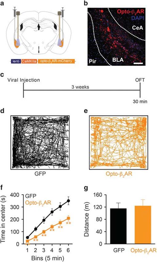
Activation of BLA Gαs signaling is anxiogenic
                                                                     ER Siuda et al
                                                                                                                                       2013
North Carolina Gene Therapy Center Vector Core and Virus             master-9 function generator and placed in the center of the
Vector Core. CaMKIIα and Synapsin promoters are used to              open field and allowed to roam freely for 30 min while
selectively target excitatory neurons and pan-neuronal-specific      receiving 473 nm blue light at 5 s on/off intervals at 1 W/cm2
expression, respectively (Zhang et al, 2010). Elongation factor 1α   power. The center was defined as a square comprising 50%
(EF1α) and phosphoglycerate kinase 1 (PGK) are constitutive          the total area of the OFT. Mean time spent in the center was
promoters commonly used in mammalian systems (Qin et al,             the primary measure of anxiety-like behavior.
2010).
                                                                     Behavior—Social Approach (SA)
Stereotaxic Surgery
                                                                     Social approach behavior was based and modified on
Mice were anesthetized in an induction chamber                       previous studies (Silverman et al, 2010). Lighting was
(5% isoflurane) and placed in a stereotaxic frame (Kopf              measured at ~ 200 lux. Mice were allowed to freely roam a
Instruments, Model 1900) where they were maintained at               two-chambered black, plastic box for 10 min that contained
1–2% isoflurane throughout the procedure. A craniotomy was           an inverted, metal, mesh pencil cup at each end. A novel,
performed, and mice were injected as follows. For basolateral        sex- and age-matched conspecific was then added to one of
amygdala injections, 1.2 μl of either lenti-PGK-GFP or               the inverted cups in a random, counterbalanced manner. The
lenti-CaMKIIα-optoβ2AR-mCherry or 0.5 μl of either AAV5-             social zone is described as a circle that surrounds the
CaMKIIα-HA-GSD-IRES-mCitrine or AAV5-CaMKIIα-eGFP                    inverted cup that is equal to twice its diameter. The test
was injected bilaterally at stereotaxic coordinates from             mouse was then reintroduced to the chamber and the time
bregma: − 1.3 mm (AP), ± 2.9 mm (ML), and − 4.9 mm                   spent in the social zone recorded. For rM3Ds experiments,
(DV). For CeA injections, 1.0 μl of either AAV5-hSyn-opto-           animals were dosed 1 mg/kg (i.p) with CNO dissolved in
β2AR-eYFP or AAV5-EF1α-YFP was injected bilaterally                  saline 30 min prior to the second phase of the assay. For
at stereotaxic coordinates from bregma: − 1.34 mm (AP),              opto-β2AR experiments, animals were connected to fiber
± 2.5 mm (ML), and − 4.6 mm (DV). Mice were then                     optic cables coupled to a master-9 function generator and
implanted with chronic fiber optic implants with coordinates         placed in the center of the chamber and allowed to roam
adjusted from viral injection to 0.00 mm (AP), ± 0.25 mm             freely for 10 min while receiving 473 nm blue light at 5 s on/
(ML), and +1.00 mm (DV). The fiber optic implants were               off intervals at 1 W/cm2 power for the second phase of
secured using two bone screws (CMA, 743102) and affixed              the assay.
with TitanBond (Horizon Dental Products) and dental cement
(Lang Dental) (Al-Hasani et al, 2015; McCall et al, 2013). Mice
                                                                     Behavior—Conditioned Place Aversion (CPA)
were allowed to recover for at least 3 weeks prior to behavioral
testing, allowing for optimal viral expression.                      Mice were trained in an unbiased, balanced three compart-
                                                                     ment conditioning apparatus as previously described
                                                                     (Al-Hasani et al, 2013; McCall et al, 2015). Lighting was
Behavior
                                                                     measured at ~ 200 lux. Animals are placed in a three-
Behavioral assays were performed in a sound attenuated               chambered arena in which two chambers contain visually
room maintained at 23 °C. Lighting was measured and                  disparate contextual cues (horizontal or vertical stripes).
stabilized at ~ 4 lux for anxiety tests and ~ 200 lux for place      Following an initial 30 min pretest, animals are conditioned
testing. All behavioral apparatuses were cleaned with 70%            over a 2-day period (conditioning day 1 (CD1) and
ethanol in between animals. In photostimulation assays, mice         conditioning day 2 (CD2)) with two training sessions
with lenti-PGK-GFP, lenti-CaMKIIα-optoβ2AR-mCherry,                  per day (AM = without light, PM = with light) where the
AAV5-hSyn-opto-β2AR-eYFP or AAV5-EF1α-YFP received                   animal is confined to one chamber or the other. Each
5 s of constant photostimulation (473 nm, 1 W/cm2) followed          training session was separated by at least 4 h. Photostimula-
by 5 s of no light (Airan et al, 2009; Siuda et al, 2015a)           tion (473 nm blue light at 5 s on/off intervals at 1 W/cm2
throughout the trial. In rM3Ds Gαs DREADD assays,                    power) is randomly paired with one chamber, either
mice with AAV5-CaMKIIα-HA-GSD-IRES-mCitrine and                      horizontal or vertical, and the other chamber is paired with
AAV5-CaMKIIα-eGFP received CNO (Sigma; 1 mg/kg,                      no photostimulation. On the fourth day, the animals are
i.p.) in saline 30 min prior to behavioral testing. This dose        given access to all three chambers, and the amount of time
of CNO is standard among users of these DREADD receptor              spent in each chamber is recorded (posttest). Time spent in
types (Farrell et al, 2013; Ferguson et al, 2013; Guettier et al,    each compartment was recorded with a video camera (ZR90;
2009). All behavioral experiments were video recorded and            Canon) and analyzed using Ethovision 8.5 (Noldus). CPA
analyzed using the Ethovision Software (Noldus v8.5).                was assessed on day 4 by allowing the mice to roam freely in
Behavioral outputs included time, distance, and velocity.            all three compartments and recording the time spent in each.
                                                                     Data are expressed as mean ± SEM% time spent in the light
                                                                     stimulus-paired compartment.
Behavior—Open Field Test (OFT)
As previously described (McCall et al, 2015), the open field
                                                                     Behavior—Real-Time Place Aversion (RTPA)
was a 50 × 50 cm2 square plexiglass enclosure. Lighting was
measured and stabilized at ~ 4 lux. For rM3Ds experiments,           Mice were placed into a custom-made unbiased, balanced two-
animals were dosed 1 mg/kg (i.p.) with CNO dissolved in              compartment conditioning apparatus (52.5 × 25.5 × 25.5 cm3)
saline 30 min prior to assay. For opto-β2AR experiments,             as previously described (Al-Hasani et al, 2015; McCall et al,
animals were connected to fiber optic cables coupled to a            2015; Siuda et al, 2015b) and allowed to freely roam the entire

                                                                                                           Neuropsychopharmacology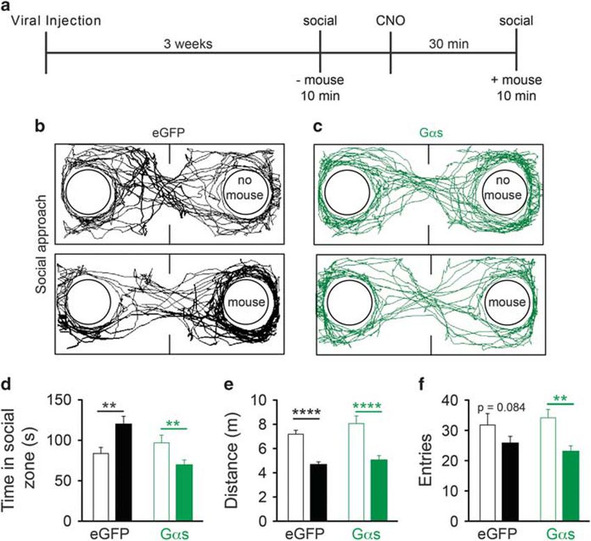
Activation of BLA Gαs signaling is anxiogenic
ER Siuda et al
2014
apparatus for 20 min. Entry into one compartment triggered G-protein coupled receptor (GPCR) types in the BLA that
constant photostimulation (5 s on/off, 473 nm, 1 W/cm2 light couple to diverse G-protein signaling modules, there are nine
power) while the animal remained in the light-paired chamber. total adrenergic receptor types (α1AR–Gαq, α2AR–Gαi, and
Entry into the other chamber ended the photostimulation. The β1–3AR–Gαs). In addition, recent work suggested that
side paired with photostimulation was counterbalanced across β-adrenergic receptors mediate the anxiogenic effects
mice. Time spent in each chamber and total distance traveled following stimulation of locus coeruleus noradrenergic
for the 20-min trial was measured using Ethovision 8.5 neurons (McCall et al, 2015). Thus we first used a global,
(Noldus). Data are expressed as mean ± SEM% time spent in yet region and cell-type selective approach to investigate the
photostimulation-paired chamber. role of Gαs activation signaling within the BLA. We utilized
DREADDS. These receptors are highly selective for coupling
to Gαs pathways over other G-proteins (Ferguson et al, 2013;
Immunohistochemistry
Guettier et al, 2009; Rogan and Roth, 2011). Utilizing the
After the conclusion of behavioral testing, mice were rM3Ds Gαs DREADD receptor packaged in a virus under a
anesthetized with sodium pentobarbital and transcardially CAMKIIα promoter (rM3DsBLA/CaMKIIα), we bilaterally
perfused with ice cold PBS, followed by 4% phosphate- infused the virus or its control empty vector virus into the
buffered paraformaldehyde. Brains were removed, fixed BLA and allowed at least 3 weeks for optimal viral expression
overnight in paraformaldehyde, and saturated in 30% (Figure 1a–c and Supplementary Figure S1a). Using the OFT,
phosphate-buffered sucrose. In all, 30-μm sections were a common assay to investigate anxiety-like behavior in
cut, washed in 0.3% Triton X100/5% normal goat serum in rodents, dosed mice with the DREADD-selective agonist
0.1 M PBS, stained with fluorescent Nissl stain (1 : 400 CNO (1 mg/kg, i.p.) 30 min prior to testing (Figure 1c).
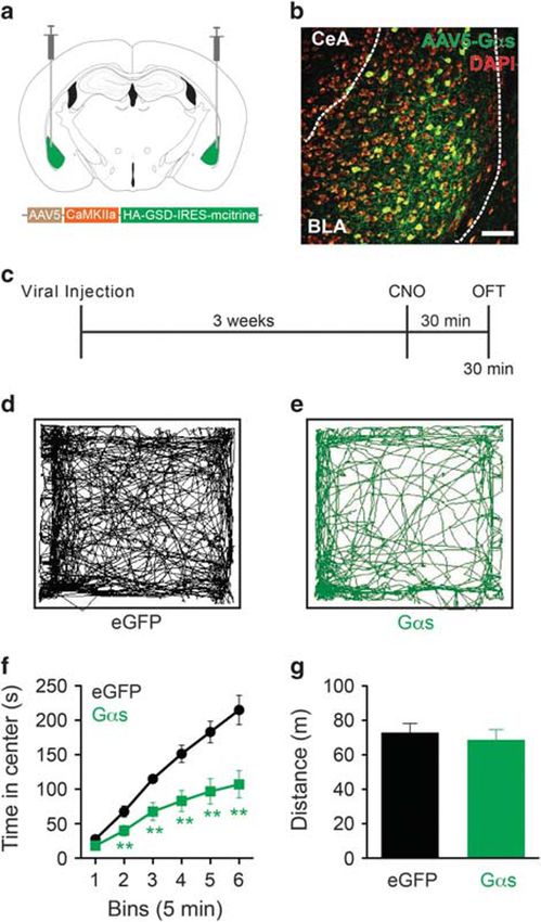
Neurotrace, Invitrogen, Carlsbad, CA) for 1 h, and mounted rM3DsBLA/CaMKIIα animals (green; n = 9) displayed a sig-
onto glass slides with Vectashield (Vector Laboratories, nificant anxiogenic-like response throughout the 30-min
Burlingame, CA). Viral expression was verified using OFT trial compared with control eGFP animals (n = 12)
fluorescence (Olympus, Center Valley, PA) and confocal (Figure 1d–f). This anxiety-like phenotype was evident
microscopy (Leica Microsystems, Bannockburn, IL). Images within the first 10 min of the assay as demonstrated by a
were produced with the Leica Application Suite Advanced cumulative time course (Figure 1f; multiple unpaired
Fluorescence software. Animals that did not show targeted Student’s t-tests; bin 1 t(19) = 1.047, P = 0.3081; bin 2
expression were excluded from analyses. t(19) = 2.881, P = 0.0096; bin 3 t(19) = 3.421, P = 0.0029;
IHC was quantified as previously described (Al-Hasani bin 4 t(19) = 3.689, P = 0.0016; bin 5 t(19) = 3.099, P = 0.0059;
et al, 2013; Kim et al, 2013). 1° Ab rabbit monoclonal bin 6 t(19) = 3.084, P = 0.0061). This anxiogenic-like behavior,
α-CaMKIIα (EPR1828) (Abcam; ab92332; 1 : 500 in blocking however, had no effect on total distance traveled (Figure 1g
buffer) and 2° Ab at 1 : 500 in PBS for opto-β2AR goat unpaired Student’s t-test; t(18) = 0.5084, P = 0.6173) or
α rabbit IgG Alexa Fluor 488 and for GFP goat α rabbit IgG velocity (Supplementary Figure S1b; unpaired Student’s
Alexa Fluor 594. Channels were separated, an exclusive t-test; t(18) = 0.3499, P = 0.7305), suggesting animal mobility
threshold was set, and positive staining for each channel was was not affected. However, there was a reduction in the
counted in a blind-to-treatment manner using Metamorph. number of entries into the center of the open field suggesting
The counts from each channel were then overlaid, and the that mice expressing rM3DsBLA/CaMKIIα made fewer entries
percentage of co-labeled cells were reported. For pLenti- into and spent less time in the center of the open field
CaMKIIα-opto-β2AR-mCherry, four sections from four (Supplementary Figure S1c; unpaired Student’s t-test;
different animals each were used to quantify the percentage t(18) = 2.054, P = 0.0548).
of co-label. For Lenti-PGK-GFP, six sections from three Previous in vitro and in vivo studies using the rM3Ds
different animals each were used to quantify co-label. receptor have suggested that this particular Gαs DREADD
receptor is constitutively active (Guettier et al, 2009). To
address this concern, we used a real-time cAMP assay to
Statistics/Data Analysis examine rM3Ds activity. HEK293 cells expressing the
All data are expressed as mean ± SEM. Statistical GloSensor plasmid (HEK-pGlo) were transfected with high
significance was taken as *Po0.05, **Po0.01, ***Po0.001, levels of Gαs DREADD (Gαs-pGlo). Following an initial
****Po0.0001 as determined by Student’s t-test (paired and 1 min baseline, the Gαs-pGlo-expressing cells show
unpaired). For cumulative, nonlinear, time course data, a significant increase in cAMP following administration of
multiple unpaired t-tests were performed. Statistical analyses CNO (10 μM) in stark contrast to empty HEK-pGlo cells
were performed in GraphPad Prism 5.0. (Supplementary Figure S1d). More importantly, if we expand
the axes before and after addition of CNO (red box
Supplementary Figure S1d), we see that the Gαs-pGlo cells
RESULTS maintain similar baseline cAMP values to HEK-pGlo,
suggesting that there is no constitutive Gαs activity with
Chemogenetic Activation of Gαs Signaling in the BLA is
high DREADD receptor expression in vitro (Supplementary
Sufficient to Induce Anxiogenic Behavior
Figure S1e and f; unpaired Student’s t-test; t(4) = 0.8794,
Excitation of channelrhodopsin expressing BLA neurons has P = 0.4288). Finally, if we examine the initial 5 min of the
been previously shown to cause anxiogenic behavior OFT data (Figure 1e), we see that there are no statistical
(Felix-Ortiz et al, 2013; Tye et al, 2011), although the differences between controls and Gαs-expressing animals
specific signaling pathways that mediate these effects are not (unpaired Student’s t-test; bin 1 t(19) = 1.047, P = 0.3081),
known. Furthermore, in addition to numerous other suggesting again that there is no constitutive activity in vivo.
Neuropsychopharmacology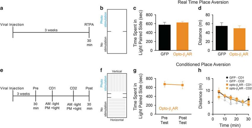
Activation of BLA Gαs signaling is anxiogenic
                                                                              ER Siuda et al
                                                                                                                                                  2015
                                                                              anxiolytics, such as diazepam (Stemmelin et al, 2007). We
                                                                              first demonstrated that animals expressing the empty vector
                                                                              eGFP control virus (n = 15) or rM3DsBLA/CaMKIIα (n = 11)
                                                                              spent similar amounts of time investigating the empty cup
                                                                              (Figure 2b and c, top panels and Figure 2d; unpaired
                                                                              Student’s t-test; t(24) = 1.109, P = 0.2785). Thirty minutes
                                                                              following CNO (1 mg/kg, i.p.) administration, control
                                                                              animals spent significantly more time in the social zone in
                                                                              the presence of a novel conspecific (Figure 2b, bottom panel
                                                                              and Figure 2d; paired Student’s t-test; t(14) = 3.427,
                                                                              P = 0.0041). In contrast, chemogenetic activation of Gαs
                                                                              signaling in the BLA following CNO administration induced
                                                                              a significant reduction in the time spent in the presence of a
                                                                              novel conspecific (Figure 2c, bottom panel and Figure 2d;
                                                                              paired Student’s t-test; t(10) = 4.042, P = 0.0024), suggesting
                                                                              that activation of Gαs-signaling in CamKIIα-positive
                                                                              BLA neurons is sufficient to promote social anxiety-like
                                                                              behavioral states. Furthermore, we found that, for both
                                                                              treatment groups, animals traveled significantly less total
                                                                              distance in the presence of a conspecific (Figure 2e; eGFP
                                                                              paired Student’s t-test; t(14) = 6.882, Po0.0001 and
                                                                              rM3DsBLA/CaMKIIα paired Student’s t-test; t(10) = 7.624,
                                                                              Po0.0001) with neither group significantly different than
                                                                              the other (eGFP vs rM3DsBLA/CaMKIIα without mouse
                                                                              unpaired Student’s t-test; t(24) = 1.336, P = 0.1939 and with
                                                                              mouse unpaired Student’s t-test; t(24) = 0.8214, P = 0.4195).
                                                                              Finally, rM3DsBLA/CaMKIIα animals made fewer entries into
                                                                              the social zone in the presence of a conspecific, suggesting
                                                                              that in addition to less time there were fewer entries into the
                                                                              social zone (Figure 2f; paired Student’s t-test; t(10) = 4.312,
                                                                              P = 0.0015). Control animals also trended to make fewer
                                                                              entries into the social zone consistent with animals spending
                                                                              more time with the novel conspecific, albeit this trend is not
                                                                              statistically significant (Figure 2f; paired Student’s t-test;
                                                                              t(14) = 1.857, P = 0.0844). Together, these data suggest that
                                                                              activation of Gαs signaling in excitatory neurons of the BLA
Figure 1 Gαs DREADD signaling in the BLA induces anxiety-like                 is sufficient to promote acute and social anxiety-like states.
behavior. (a) Bilateral viral injection sites in the BLA. (b) AAV5-CaMKIIα-
HA-GSD-IRES-mCitrine (rM3Ds; green) expression in BLA (DAPI = red;
scale bar = 50 μm). (c) Behavior diary. Representative open field traces of   Photoactivation of β-Adrenergic Signaling In vivo
(d) control and (e) rM3DsBLA/CaMKIIα mice. (f) rM3DsBLA/CaMKIIα mice          Within the BLA Promotes Anxiety-Like Behavior
(green) show cumulatively less time in the center of the open field as
compared with controls (black) following CNO administration (1 mg/kg, i.p.)   The rM3DsBLA/CaMKIIα data suggest that Gαs signaling within
(**Po0.01, multiple unpaired Student’s t-tests). (g) Distance traveled of     the BLA elicits anxiogenic behavioral phenotypes. Among
rM3DsBLA/CaMKIIα and control mice.
                                                                              the GPCRs present in the BLA, adrenergic receptors, and in
                                                                              particular β-adrenergic receptors (βARs), that signal via Gαs
                                                                              signaling are highly expressed (Lein et al, 2007). Previous
Taken together, these data support the conclusion that                        reports have also shown that NE release in the BLA decreases
activation of Gαs signaling within the BLA induces an acute                   neuronal firing, an effect predominately mediated via
anxiogenic state.                                                             α2-adrenergic receptors (Gαi), whereas activation of βAR
                                                                              (Gαs) results in excitation (Buffalari and Grace, 2007; Davis
                                                                              et al, 1994; Menard and Treit, 1999). The central CeA, BLA
Chemogenetic Activation of Gαs Signaling in the BLA is
                                                                              and BNST are enriched in all the major subtypes of
Sufficient to Produce Social Anxiety-Like Behavior
                                                                              adrenergic receptors (Lein et al, 2007), making isolation of
In addition to acute anxiety, we next examined the effects of                 their respective contributions in the amygdaloid complex
Gαs activation in the BLA in a mouse behavioral model of                      pharmacologically challenging. Thus, to gain better spatio-
social anxiety. Adapted from similar behavioral assays that                   temporal isolation of adrenergic receptor pathways, we
model social anxiety-like states (Bailey and Crawley, 2009;                   utilized the chimeric rhodopsin/β2-adrenergic receptor,
Bruchas et al, 2011; Felix-Ortiz and Tye, 2014; Silverman                     opto-β2AR (Figure 3a). Opto-β2AR has been shown to
et al, 2010), the social approach assay measures the time a                   activate cAMP, presumably through Gαs-mediated signaling,
mouse spends in the presence of a novel conspecific (social                   and to modulate neuronal excitability (Airan et al, 2009;
zone) (Figure 2a). Less time spent in the social zone indicates               Bailes et al, 2012; Kim et al, 2005; Siuda et al, 2015a), as such
a social anxiety-like state that is reversible using traditional              this chimeric receptor offers advantages over traditional

                                                                                                                     Neuropsychopharmacology
Activation of BLA Gαs signaling is anxiogenic
ER Siuda et al
2016
Figure 2 Gαs DREADD signaling in the BLA alters social interaction. (a) Behavior diary. Representative traces of (b) control and (c) rM3DsBLA/CaMKIIα mice
in the absence (top) and presence (bottom) of a conspecific. (d) Control animals (black) spend more time in the social zone in the presence of a conspecific
(solid bars) than in its absence (empty bars) (**Po0.01, paired Student’s t-test), while rM3DsBLA/CaMKIIα (green;) animals spend less time in the social zone in
the presence of a conspecific (**Po0.01, paired Student’s t-test). (e) Both control (black) and rM3DsBLA/CaMKIIα (green) mice travel less distance in the
presence of a novel mouse (solid bars) than in its absence (empty bars) (****Po0.0001, paired Student’s t-test). (f) Both control (black) and rM3DsBLA/CaMKIIα
(green) mice travel less distance in the presence of a novel mouse (solid bars) than in its absence (empty bars) (****Po0.0001, paired Student’s t-test).
(f) Control (black; P = 0.084) and rM3DsBLA/CaMKIIα (green; **Po0.01 paired Student’s t-test) mice make fewer entries into the social zone in the presence
of a novel mouse (solid bars) than in its absence (empty bars).
pharmacological approaches in allowing cell-type specificity opto-β2ARBLA/CaMKIIα animals showed no differences in total
and greater spatiotemporal control over βAR signaling. distance traveled (Figure 3h; unpaired Student’s t-test;
We next explored whether selective activation of t(13) = 0.3221, P = 0.7525) or velocity (Supplementary Figure
β-adrenergic signaling in BLA excitatory neurons using S2a; unpaired Student’s t-test; t(13) = 0.5324, P = 0.6035).
optogenetic approaches was sufficient to promote anxiety- Interestingly there were no differences in number of entries
like behavior. We first packaged opto-β2AR in a lentivirus into the center of the open field, suggesting that opto-
under a CaMKIIα promoter and then bilaterally injected the β2ARBLA/CaMKIIα-expressing animals entered the open area
virus (opto-β2ARBLA/CaMKIIα) or empty vector control virus but did not remain there for a long period of time
into the BLA (Figure 3b). Following viral injection, we then (Supplementary Figure S2b; unpaired Student’s t-test;
implanted permanent optic ferrules slightly dorsal to t(13) = 0.7522, P = 0.4653).
the injection coordinates in the BLA (Figure 3c). We utilized These data suggest that activation of β-adrenergic signaling
the OFT as a model of acute anxiety-like behavior and in BLA excitatory neurons is anxiogenic. Given that the BLA
photoactivated (473 nm, 5 s on/off, 1 W/cm2) animals via is composed predominately of excitatory neurons (Carlsen,
fiber optic cables throughout the assay. Photoactivation of 1988; Smith and Paré, 1994), we believe that a CaMKIIα
opto-β2ARBLA/CaMKIIα (n = 7) in the BLA produced rapid and promoter should label the vast majority of neurons within
sustained anxiogenic-like behavior with mice spending the BLA. To verify this, viral expression of lenti-CaMKIIα-
significantly less cumulative time in the center of the OFT opto-β2AR-mCherry and lenti-PGK-GFP was confirmed in a
than GFP controls (n = 8) (Figure 3d–f). The anxiogenic subset of animals and stained for CaMKIIα. Approximately
effect was seen almost immediately and lasted throughout 96% of lenti-opto-β2AR-mCherry+ cells and lenti-PGK-GFP
(Figure 3f; multiple unpaired Student’s t-tests; bin 1 co-labeled with CaMKIIα+ cells in the BLA suggesting
t(13) = 3.472, P = 0.0041; bin 2 t(13) = 3.367, P = 0.0051; bin 3 expression driven under the CaMKIIα promoter was efficient
t(13) = 3.169, P = 0.0074; bin 4 t(13) = 3.725, P = 0.0025; bin 5 (Supplementary Figure S2c and d; unpaired Student’s t-test;
t(13) = 3.452, P = 0.0043; bin 6 t(13) = 3.463, P = 0.0042). We t(8) = 0.02041, P = 0.9842). These data corroborate others
observed no effect on animal mobility as both controls and using CaMKIIα co-labeling in the BLA (Johansen et al,
NeuropsychopharmacologyActivation of BLA Gαs signaling is anxiogenic
ER Siuda et al
2017
opto-β2ARCeA/hSyn (n = 6) did not result in an anxiogenic
phenotype (Supplementary Figure S3d–f; multiple unpaired
Student’s t-tests; bin 1 t(10) = 0.1040, P = 0.9192; bin 2
t(10) = 0.09359, P = 0.9273; bin 3 t(10) = 0.05840, P = 0.9546;
bin 4 t(10) = 0.05844, P = 0.9546; bin 5 t(10) = 0.08324,
P = 0.9353; bin 6 t(10) = 0.01982, P = 0.9846). and had no
effect on animal mobility (Supplementary Figure S3g–i;
distance t(9) = 1.771, P = 0.1103; velocity t(8) = 1.717,
P = 0.1243; entries t(10) = 0.6925, P = 0.5044) in comparison
to YFP controls (n = 6). These data suggest that β-adrenergic
signaling in the BLA, but not in the CeA, is important in
acute anxiety-like states.
Photoactivation of β-Adrenergic Signaling Within the
BLA Does not Promote Real-Time or Conditioned
Aversion
Previous studies have shown that activation of LC cell bodies
via channelrhodopsin induces an aversive behavioral phe-
notype in a RTPA assay, which was reversed with
antagonism at α1-adrenergic receptors (prazosin) but not
β-adrenergic (propranolol) (McCall et al, 2015). However,
anxiogenic behavior induced by tonic optogenetic activation
of the LC was indeed sensitive to systemic β-adrenergic
receptor blockade. We used two models of assessing aversion
in mice: RTPA and CPA (Al-Hasani et al, 2015; McCall et al,
2015). Opto-β2ARBLA/CaMKIIα- (n = 7) and viral GFP control-
expressing mice (n = 5) were photostimulated (473 nm, 5 sec
on/off, 1 W/cm2) in real time when entering a chamber
randomly paired to photostimulation (Figure 4a and b).
Photostimulation was terminated when the animal left the
chamber. Consistent with recent reports suggesting that
β-adrenergic receptors do not mediate noradrenergic-
dependent real-time aversion (McCall et al, 2015), during
this 20-min trial opto-β2ARBLA/CaMKIIα- and viral control-
Figure 3 Photoactivation of Opto-β2AR in the BLA promotes
anxiety-like behavior. (a) Bilateral viral injection sites and optic fiber implants. expressing mice showed no differences in the amount of time
(b) Viral expression of lenti-CaMKIIα-opto-β2AR-mCherry in the BLA (scale spent in the photostimulation-paired chamber (Figure 4c;
bar = 50 μm). (c) Behavior diary. Representative traces of control (d) and unpaired Student’s t-test; t(10) = 0.9059, P = 0.3863) or in
opto-β2ARBLA/CaMKIIα (e) mice in OFT. (f) Cumulative time course shows general mobility (Figure 4d; unpaired Student’s t-test;
opto-β2ARBLA/CaMKIIα (orange) mice spend less time in the center of an t(10) = 0.5893, P = 0.5687). These data suggest that activation
open field compared with controls (black) while receiving light stimulation of βAR signaling within excitatory neurons of the BLA does
(473 nm, 5 s off/on; **Po0.01, via multiple unpaired Student’s t-tests). (g) In
the OFT, viral control (black) and opto-β2ARBLA/CaMKIIα (orange) mice do
not induce an acute aversive state.
not differ in total distance traveled. Previous studies have also shown that β-adrenergic activity
within the amygdala has a key role in fear memory and fear
conditioning, both of which possess a learning component
(Cahill et al, 1994; Dębiec and Ledoux, 2004; Quirarte et al,
2010). Similar results were obtained with lenti-PGK-GFP+ 1998; Rogan et al, 1997). To determine whether βAR
cells as PGK is a constitutive and fairly ubiquitous promoter activation in the BLA alters aversive learning, we
(Qin et al, 2010). photostimulated opto-β2ARBLA/CaMKIIα during the CPA assay
To test for regional specificity of viral expression, photo- (Figure 4e and f). Opto-β2ARBLA/CaMKIIα-expressing mice
illumination, and functional isolation of this β-adrenergic (n = 7) showed no difference in the amount of time spent in
signaling response, we also examined the effects of the photostimulation-paired chamber following 2 days of
opto-β2AR stimulation when expressed in neurons of the conditioning (Figure 4g; paired Student’s t-test; t(6) = 0.4447,
CeA (opto-β2ARCeA/hSyn) (Supplementary Figure S3a and b). P = 0.6721) and showed no differences in mobility compared
As the cytoarchitecture of the CeA is more heterogeneous with empty vector viral GFP controls (Figure 4h; two-way
than the BLA and is predominately GABAergic (Lüthi and ANOVA for repeated measurements, time F(5,25) = 6.10,
Lüscher, 2014), opto-β2AR was packaged under the pan- P = 0.0008, virus F(1,5) = 3.88, P = 0.1060, interaction
neuronal human synapsin promoter as previously used in the F(5,25) = 1.16, P = 0.3560). These data suggest that activation
CeA for optogenetic studies (Robinson et al, 2014). Using of βAR signaling within excitatory neurons of the BLA does
the same experimental paradigm (Supplementary Figure not effect conditioned aversion. In summary, these data
S3c), photostimulation (473 nm, 5 s on/off, 1 W/cm2) of suggest that activation of βAR signaling within the BLA has
NeuropsychopharmacologyActivation of BLA Gαs signaling is anxiogenic
ER Siuda et al
2018
Figure 4 Photoactivation of Opto-β2AR does not alter RPTA or CPA. (a) Behavior diary and (b) chamber schematic. (c) Opto-β2ARBLA/CaMKIIα (orange)
and control mice (black) spend similar amounts of time in the photostimulation-paired chamber in real-time place aversion (RTPA) and show no differences in
total distance traveled (d). (e) Behavior diary and (f) chamber schematic. (g) Opto-β2ARBLA/CaMKIIα (orange) do not show an aversive response to the
condition-photostimulation chamber. (h) Opto-β2ARBLA/CaMKIIα (orange) and control animals (black) show no differences in distances traveled during
conditioning day 1 (CD1) and or conditioning day 2 (CD2) in CPA.
effects on acute anxiety-like behavior but not in either acute Po0.0001) with neither group significantly different than
or conditioned aversive states. the other (GFP vs opto-β2ARBLA/CaMKIIα without mouse
unpaired Student’s t-test; t(13) = 0.5590, P = 0.5857 and with
mouse unpaired Student’s t-test; t(13) = 1.238, P = 0.2376).
Optogenetic Activation of Opto-β2AR Signaling Within
Both groups also had similar entries into the social zone
the BLA is Sufficient to Induce Social Anxiety-Like
(Figure 5f; GFP paired Student’s t-test; t(6) = 0.7285,
Behavior
P = 0.4937 and opto-β2ARBLA/CaMKIIα paired Student’s t-test;
β-Adrenergic receptors have also been implicated in social t(6) = 2.213, P = 0.0689). These data suggest that activation of
anxiety disorders (Liebowitz, 1987) and β-blockers are opto-β2AR signaling in the BLA is sufficient to induce acute
commonly prescribed as anxiolytics clinically (Baker et al, social anxiety-like states.
2011). Having shown that activation of generic Gαs signaling Taken together, these data demonstrate that in vivo
within the BLA disrupted social behavior via DREADD photoactivation of opto-β2ARBLA/CaMKIIα in the BLA induces
receptors (Figure 2d), and knowing prior studies have shown an anxiety-like behavioral phenotype. We also report that
that the BLA is critical for these behaviors (Felix-Ortiz and this effect is selective to the BLA, as animals transduced with
Tye, 2014), we posited that activation of βAR signaling would opto-β2AR in the neighboring CeA showed no acute anxiety-
have similar effects. Utilizing the social approach assay like behavior. In addition, we found that social interaction
(Figure 5a), we show that both opto-β2ARBLA/CaMKIIα- (n = 8) time in photoactivated opto-β2ARBLA/CaMKIIα animals is
and viral control GFP-expressing mice (n = 7) spend similar diminished in comparison to controls, suggesting an
amounts of time in the social zone in the absence of a novel important role of this Gαs-signaling pathway in mediating
conspecific (Figure 5b and c, top panels and Figure 5d; social behavior.
unpaired Student’s t-test; t(13) = 0.7423, P = 0.4711).
However, in the presence of a novel conspecific and while
receiving photostimulation, GFP control animals showed a
DISCUSSION
significant increase in the time spent in the social zone
(Figure 5b, bottom panel and Figure 5d; paired Student’s The BLA is one key region particularly associated with
t-test; t(6) = 3.567, P = 0.0118), whereas opto-β2ARBLA/CaMKIIα anxiety and mood (Feinstein et al, 2013; Valentino et al,
animals did not display this social interaction behavior 1993). The BLA receives noradrenergic input from the LC
(Figure 5c, bottom panel and Figure 5d; paired Student’s (Asan, 1998; Fallon et al, 1978) and sends projections to the
t-test; t(7) = 0.4419, P = 0.6719). Both groups showed a CeA, the prefrontal cortex, nucleus accumbens, and BNST,
significant reduction in the total distance traveled in regions linked to mood regulation, and some with very
the presence of the novel conspecific (Figure 5e; GFP disparate effects (Tye and Deisseroth, 2012). For example,
paired Student’s t-test; t(6) = 3.253, P = 0.0174 and in vivo photostimulation of BNST glutamatergic projections
opto-β2ARBLA/CaMKIIα paired Student’s t-test; t(7) = 10.26, is both aversive and anxiogenic (Jennings et al, 2013), while
NeuropsychopharmacologyActivation of BLA Gαs signaling is anxiogenic
ER Siuda et al
2019
Figure 5 Photoactivation of Opto-β2AR in the BLA alters social behavior. (a) Behavior diary. Representative traces of (b) control and (c) opto-β2ARBLA/CaMKIIα
in the absence (top) and presence (bottom) of a novel conspecific. (d) Control animals (black) spend more time in the social zone in the presence of a novel
conspecific (solid bars) than in its absence (empty bars) (*Po0.05, paired Student’s t-test), while opto-β2ARBLA/CaMKIIα (orange) animals show no increased
time spent in the social zone in the presence of a conspecific. (e) Both control (black; *Po0.05 paired Student’s t-test) and opto-β2ARBLA/CaMKIIα (orange;
****Po0.0001, paired Student’s t-test) mice travel less distance in the presence of a novel mouse (solid bars) than in its absence (empty bars). (f) Both control
(black) and opto-β2ARBLA/CaMKIIα (orange) animals show similar number of entries into the social zone in the presence (solid bars) or absence (empty bars) of
a novel mouse.
activation of BLA terminals specifically in the CeA is CNO produces not just acute anxiety but also induces deficits
anxiolytic (Tye et al, 2011). Thus teasing out the relative in social interaction between conspecifics. These results
contributions of each region has been historically challenging corroborate previous studies that photostimulated
with traditional pharmacological techniques. channelrhodopsin-expressing BLA terminals in the ventral
The advent of chemogenetic and optogenetic techniques hippocampus, which produced reduced social behaviors in
has enabled the selective targeting and manipulation of the resident-juvenile intruder procedure and decreased time
specific cell types and brain regions (Namburi et al, 2015). in the social zone in the three chamber sociability test
Previous studies have shown that photostimulation of (Felix-Ortiz et al, 2013). While in agreement with other
CaMKIIα+ channelrhodopsin-expressing cell bodies in the studies, our results are unique in that they dissect the cellular
BLA produced an anxiogenic phenotype (Tye et al, 2011). signaling mechanisms and are less binary (on/off) than the
Although highlighting the important role of the BLA in previous studies using channelrhodopsin. However, Gαs
anxiogenesis, the use of channelrhodopsin itself limits the signaling via chemogenetic approaches comes with some
unraveling of potential neuromodulatory mechanisms of limitations. While still activating intracellular cAMP, Gαs
action. In the present study, we utilized chemogenetic and DREADDs act as a ‘generic’ Gαs GPCR, it is currently
optogenetic approaches to manipulate G-protein coupled unclear whether this tool exhibits identical intracellular
cellular activity through more endogenous neuromodulatory signaling cascades comparable to the endogenous Gαs
mechanisms known to regulate the gain of these neural receptors expressed within the BLA. We now know, for
circuits. instance, that all pools of Gαs are not identical and that
The rM3Ds DREADD receptor mimics generic Gαs receptors signal to G-proteins in a very selective
intracellular signaling (Guettier et al, 2009). The BLA microdomain-dependent manner that is limited by receptor
endogenously expresses a host of G-protein coupled subtype (Irannejad et al, 2013; Puthenveedu et al, 2010).
receptors, many via Gαs mechanisms (Lein et al, 2007). Studies thoroughly comparing the pharmacodynamic
Here we show that, when localized to CaMKIIα+ neurons of properties of Gαs DREADDs to canonical GPCRs,
the BLA, systemic administration of the DREADD agonist such as β2AR, are needed to provide further validation.
NeuropsychopharmacologyActivation of BLA Gαs signaling is anxiogenic
ER Siuda et al
2020
Chemogenetic approaches are also faced with similar inhibitory circuitry (Ciocchi et al, 2010; Haubensak et al,
pharmacokinetic and pharmacodynamic issues as more 2010; Wilensky et al, 2006). It is possible that ubiquitous
traditional pharmacological approaches, thus highlighting expression of our construct was not sufficient to yield an
the need to complement multiple techniques and alternative obvious behavioral phenotype and may have affected the
approaches to fully examine the signaling and neurocircuitry microcircuitry within the CeA itself.
mediating affective behavioral states. In this case, the use of Previous studies show that activation of the lateral
chemogenetic technology narrowed the window of receptor amygdala is involved in fear learning (Johansen et al,
systems involved to Gαs signaling, likely to βAR signaling 2010). Here we show that activating opto-β2AR in the
pathways. However, we do not rule out the possible CaMKIIα+ neurons of the BLA had no effect on CPA or
contribution of other Gαs GPCRs expressed in the BLA RTPA. Although consistent with recent reports suggesting
and future studies utilizing other DREADD receptors, such that β-adrenergic receptors do not mediate noradrenergic
as Gαi and Gαq, would complement our findings and further dependent real-time aversion (McCall et al, 2015), it is
narrow down potential targets mediating acute and social somewhat surprising that there was no effect in the CPA
anxiety-like states. assay. Given the role of the BLA and β-adrenergic receptors
Manipulation of endogenous intracellular signaling in vivo in emotional and stressful memory (Bernardi et al, 2009;
has historically required pharmacological techniques. The McGaugh, 2004; Wu et al, 2014), it is possible that our
use of optically active rhodopsin chimeric receptors, stimulus was not aversive or salient enough. Future studies
however, has allowed us to manipulate neurocircuitry could expand on the photostimulation parameters, and other
in vivo, with higher specificity to endogenous mechanisms learning assays, such as fear conditioning, may be more
(Airan et al, 2009). Currently, there are several chimeric suited.
receptors that upon photoactivation are able to signal Our results suggest that noradrenergic influence in the
intracellularly via G-protein- and arrestin-mediated cascades: BLA is mediated via activation of neuromodulatory
α1-adrenergic, β2-adrenergic, adenosine A2A, 5HT1A, β-adrenergic Gαs-signaling pathways that may ultimately
μ-opioid receptor, and the D1 dopamine receptor (Airan promote both acute and social anxiogenic-like behavioral
et al, 2009; Bailes et al, 2012; Barish et al, 2013; Franke et al, states. Although these data were collected from male mice
1992; Gunaydin et al, 2014; Kim et al, 2005; Li et al, 2015; Oh only, recent evidence suggests structural sexual dimorphisms
et al, 2010; Siuda et al, 2015a, b). Here we incorporate the of the LC and sensitivity of female LC neurons may
chimeric rhodopsin/β2-adrenergic receptor (opto-β2AR) and contribute to higher susceptibility rates to mood and anxiety
show that activation of CaMKIIα+ neurons in the BLA via disorders in females (Bangasser and Valentino, 2014;
simulated β-adrenergic activation induces both acute and Bangasser et al, 2015). We can only speculate that our data
social anxiety-like states. These findings are in agreement can be extrapolated to females, and we believe this is an
with our chemogenetic data presented here and the recent important area for future investigation.
channelrhodopsin data from other groups (Felix-Ortiz et al, In summary, we show here a role of Gαs signaling within
2013; Tye et al, 2011). Our stimulation paradigm (5 s on/off) the BLA in mediating acute and social anxiety-like behavioral
was based on previous studies that resulted in a robust states. These results suggest that noradrenergic influence on
behavioral phenotype (Siuda et al, 2015a). Other studies with signaling into the BLA may have important consequences for
this chimeric receptor used a 10-Hz (50 ms) stimulation generating anxiogenic behaviors; however, further studies of
paradigm that resulted in no significant behavioral output in these receptors, circuits, and pathways are required. These
the nucleus accumbens, a region with low β-adrenergic results provide new insights into the receptors, cells, and
receptor expression (Airan et al, 2009). We know that NE circuits that mediate anxiety-like behavior and extend our
release in the BLA is predicated on inputs from the LC, understanding of the development of therapeutics for
whose activity contributes to an animal’s arousal state treating anxiety and stress disorders.
(Aston-Jones and Cohen, 2005; Berridge and Waterhouse,
2003; Bouret and Sara, 2005; Chang and Grace, 2013). At
rest, the LC is spontaneously active, while acute stress shifts
FUNDING AND DISCLOSURE
the firing patterns to increased tonic activity (5–8 Hz) or
initiates phasic bursting to cause temporally distinct release This work is supported by NIDA R01DA037152 (to MRB),
of NE (Abercrombie and Jacobs, 1987a, b; Galvez et al, 1996; R21DA035144 (to MRB), R00DA025182 (to MRB), NIMH
Mana and Grace, 1997; Quirarte et al, 1998). The kinetics of F31MH101956 (to JGM), TR01NS081707, and the McDon-
NE release, degradation, and uptake have been loosely nell Center for Systems Neuroscience (to MRB). The authors
examined (Iversen, 1971; Pelton et al, 1981), and as such it is declare no conflict of interest.
unclear whether our stimulation paradigm truly mimics
endogenous NE release. Thus it is possible that utilizing a
different stimulation paradigm could result in different
ACKNOWLEDGMENTS
behavioral outputs and could represent different elements of
noradrenergic tone into the BLA. We thank The HOPE Center viral vector core (NINDS,
While anxiogenic when expressed in the BLA, opto-β2AR P30NS057105), Bakewell Imaging Center, and Karl Deisser-
stimulation had no effect when expressed in neurons of the oth for the opto-β2AR cDNA. Finally, we also thank the
CeA. Others have shown that stimulating channelrhodopsin- members of the Bruchas laboratory, Robert Gereau IV
expressing BLA terminals in the CeA produced an anxiolytic (WUSTL), Thomas Baranski (WUSTL), Joe Henry Steinbach
effect (Tye et al, 2011). The CeA is known to mediate (WUSTL), and N Gautam (WUSTL) for helpful discussion
conditioned fear and the acquisition of fear conditioning via and technical assistance.
NeuropsychopharmacologyActivation of BLA Gαs signaling is anxiogenic
ER Siuda et al
2021
AUTHOR CONTRIBUTIONS Bouret S, Sara SJ (2005). Network reset: a simplified overarching
theory of locus coeruleus noradrenaline function. Trends Neurosci
ERS designed and performed experiments, collected and 28: 574–582.
analyzed data, and wrote the manuscript. RA, JGM, and DLB Bruchas MR, Schindler AG, Shankar H, Messinger DI, Miyatake M,
performed experiments and collected data. MRB helped Land BB et al (2011). Selective p38α MAPK deletion in
design and oversee experiments and wrote the manuscript. serotonergic neurons produces stress resilience in models of
depression and addiction. Neuron 71: 498–511.
Buffalari DM, Grace AA (2007). Noradrenergic modulation of
basolateral amygdala neuronal activity: opposing influences of α-2
and β receptor activation. J Neurosci 27: 12358–12366.
REFERENCES Byrum CE, Guyenet PG (1987). Afferent and efferent connections
Abercrombie ED, Jacobs BL (1987a). Single-unit response of of the A5 noradrenergic cell group in the rat. J Comp Neurol 261:
noradrenergic neurons in the locus coeruleus of freely moving 529–542.
cats. I. Acutely presented stressful and nonstressful stimuli. Cahill L, Prins B, Weber M, McGaugh JL (1994). β-Adrenergic
J Neurosci 7: 2837–2843. activation and memory for emotional events. Nature 371:
Abercrombie ED, Jacobs BL (1987b). Single-unit response of 702–704.
noradrenergic neurons in the locus coeruleus of freely moving Carlsen J (1988). Immunocytochemical localization of glutamate
cats. II. Adaptation to chronically presented stressful stimuli. decarboxylase in the rat basolateral amygdaloid nucleus, with
J Neurosci 7: 2844–2848. special reference to GABAergic innervation of amygdalostriatal
Airan RD, Thompson KR, Fenno LE, Bernstein H, Deisseroth K projection neurons. J Comp Neurol 273: 513–526.
(2009). Temporally precise in vivo control of intracellular Chang C, Grace AA (2013). Amygdala β-noradrenergic receptors
signalling. Nature 458: 1025–1029. modulate delayed downregulation of dopamine activity following
Al-Hasani R, McCall JG, Foshage AM, Bruchas MR (2013). Locus restraint. J Neurosci 33: 1441–1450.
coeruleus kappa-opioid receptors modulate reinstatement of Ciocchi S, Herry C, Grenier F, Wolff SBE, Letzkus JJ, Vlachos I et al
cocaine place preference through a noradrenergic mechanism. (2010). Encoding of conditioned fear in central amygdala
Neuropsychopharmacology 38: 2484–2497. inhibitory circuits. Nature 468: 277–282.
Al-Hasani R, McCall JG, Shin G, Gomez AM, Schmitz GP, Davis M (1992). The role of the amygdala in fear and anxiety. Annu
Bernardi JM et al (2015). Distinct subpopulations of nucleus Rev Neurosci 15: 353–375.
accumbens dynorphin neurons drive aversion and reward. Davis M, Rainnie D, Cassell M (1994). Neurotransmission in the rat
Neuron 87: 1063–1077. amygdala related to fear and anxiety. Trends Neurosci 17: 208–214.
Arima J, Kubo C, Ishibashi H, Akaike N (1998). alpha2- Dębiec J, Ledoux JE (2004). Disruption of reconsolidation but not
Adrenoceptor-mediated potassium currents in acutely dissociated consolidation of auditory fear conditioning by noradrenergic
rat locus coeruleus neurones. J Physiol 508 Pt 1: 57–66. blockade in the amygdala. Neuroscience 129: 267–272.
Armbruster BN, Li X, Pausch MH, Herlitze S, Roth BL (2007). Fallon JH, Koziell DA, Moore RY (1978). Catecholamine innerva-
Evolving the lock to fit the key to create a family of G protein- tion of the basal forebrain II. Amygdala, suprarhinal cortex and
coupled receptors potently activated by an inert ligand. Proc Natl entorhinal cortex. J Comp Neurol 180: 509–531.
Acad Sci 104: 5163–5168. Farrell MS, Pei Y, Wan Y, Yadav PN, Daigle TL, Urban DJ et al
Asan E (1998). The catecholaminergic innervation of the rat (2013). A Gαs DREADD mouse for selective modulation of
amygdala. Adv Anat Embryol Cell Biol 142: 1–118. cAMP production in striatopallidal neurons. Neuropsychophar-
Aston-Jones G, Cohen JD (2005). An integrative theory of locus macology 38: 854–862.
coeruleus-norepinephrine function: adaptive gain and optimal Feinstein JS, Buzza C, Hurlemann R, Follmer RL, Dahdaleh NS,
performance. Annu Rev Neurosci 28: 403–450. Coryell WH et al (2013). Fear and panic in humans with bilateral
Bailes HJ, Zhuang L-Y, Lucas RJ (2012). Reproducible and sustained amygdala damage. Nat Neurosci 16: 270–272.
regulation of gαs signalling using a metazoan opsin as an Felix-Ortiz AC, Beyeler A, Seo C, Leppla CA, Wildes CP, Tye KM
optogenetic tool. PLoS One 7: e30774. (2013). BLA to vHPC inputs modulate anxiety-related behaviors.
Bailey KR, Crawley JN. Anxiety-Related Behaviors in Mice. In: Neuron 79: 658–664.
Buccafusco JJ (ed). Methods of Behavioral Analysis in Neuro- Felix-Ortiz AC, Tye KM (2014). Amygdala inputs to the ventral
science. 2nd edn. Chapter 5. CRC Press: Boca Raton (FL), 2009. hippocampus bidirectionally modulate social behavior. J Neurosci
Baker JG, Hill SJ, Summers RJ (2011). Evolution of β-blockers: from 34: 586–595.
anti-anginal drugs to ligand-directed signalling. Trends Pharma- Ferguson SM, Phillips PEM, Roth BL, Wess J, Neumaier JF (2013).
col Sci 32: 227–234. Direct-pathway striatal neurons regulate the retention of
Bangasser DA, Valentino RJ (2014). Sex differences in stress-related decision-making strategies. J Neurosci 33: 11668–11676.
psychiatric disorders: neurobiological perspectives. Front Neu- Ferry B, Magistretti PJ, Pralong E (1997). Noradrenaline modulates
roendocrinol 35: 303–319. glutamate-mediated neurotransmission in the rat basolateral
Bangasser DA, Wiersielis KR, Khantsis SM (2015). Sex differences amygdala in vitro. Eur J Neurosci 9: 1356–1364.
in the locus coeruleus-norepinephrine system and its regulation Franke RR, Sakmar TP, Graham RM, Khorana HG (1992).
by stress. Brain Res (doi:10.1016/j.brainres.2015.11.021). Structure and function in rhodopsin. Studies of the interaction
Barish PA, Xu Y, Li J, Sun J, Jarajapu YPR, Ogle WO (2013). Design between the rhodopsin cytoplasmic domain and transducin. J Biol
and functional evaluation of an optically active μ-opioid receptor. Chem 267: 14767–14774.
Eur J Pharmacol 705: 42–48. Frishman WH, Saunders E (2011). β-Adrenergic blockers. J Clin
Bernardi RE, Ryabinin AE, Berger SP, Lattal KM (2009). Hypertens 13: 649–653.
Post-retrieval disruption of a cocaine conditioned place Galvez R, Mesches MH, Mcgaugh JL (1996). Norepinephrine release
preference by systemic and intrabasolateral amygdala beta2- in the amygdala in response to footshock stimulation. Neurobiol
and alpha1-adrenergic antagonists. Learn Mem 16: 777–789. Learn Mem 66: 253–257.
Berridge CW, Waterhouse BD (2003). The locus coeruleus- Goddard AW, Ball SG, Martinez J, Robinson MJ, Yang CR,
noradrenergic system: modulation of behavioral state and Russell JM et al (2010). Current perspectives of the roles of the
state-dependent cognitive processes. Brain Res Brain Res Rev 42: central norepinephrine system in anxiety and depression. Depress
33–84. Anxiety 27: 339–350.
NeuropsychopharmacologyYou can also read