Description and etiology of paleopathological lesions in the type specimen of Parasaurolophus walkeri (Dinosauria: Hadrosauridae), with proposed ...
←
→
Page content transcription
If your browser does not render page correctly, please read the page content below
Received: 25 August 2020      |   Revised: 23 October 2020   |   Accepted: 30 October 2020

DOI: 10.1111/joa.13363

ORIG INAL PAPER

Description and etiology of paleopathological
lesions in the type specimen of Parasaurolophus walkeri
(Dinosauria: Hadrosauridae), with proposed reconstructions
of the nuchal ligament

Filippo Bertozzo1,2  | Fabio Manucci3 | Matthew Dempsey4 | Darren H. Tanke5 |
David C. Evans6,7 | Alastair Ruffell1 | Eileen Murphy1

1
  School of Natural and Built Environment,
Queen’s University Belfast, Belfast, UK            Abstract
2
 CI2Paleo, Sociedade de Historia Natural,          Paleopathology, or the study of ancient injuries and diseases, can enable the ecol-
Torres Vedras, Portugal
3
                                                   ogy and life history of extinct taxa to be deciphered. Large-bodied ornithopods are
 Associazione Paleontologica
Paleoartistica Italiana, Parma, Italy              the dinosaurs with the highest frequencies of paleopathology reported to-date.
4
 Department of Musculoskeletal &                   Among these, the crested hadrosaurid Parasaurolophus walkeri is one of the most fa-
Ageing Science, University of Liverpool,
                                                   mous, largely due to its dramatic elongated and tubular nasal crest. The holotype of
Liverpool, UK
5
 Royal Tyrrell Museum of Paleontology,             Parasaurolophus walkeri at the Royal Ontario Museum, Canada, displays several paleo-
Drumheller, AB, Canada                             pathologies that have not been discussed in detail previously: a dental lesion in the
6
 Royal Ontario Museum, Toronto, ON,
                                                   left maxilla, perhaps related to periodontal disease; callus formation associated with
Canada
7
  Ecology and Evolutionary Biology,
                                                   fractures in three dorsal ribs; a discoidal overgrowth above dorsal neural spines six
University of Toronto, Toronto, ON,                and seven; a cranially oriented spine in dorsal seven, that merges distally with spine
Canada
                                                   six; a V-shaped gap between dorsal spines seven and eight; and a ventral projection
Correspondence                                     of the pubic process of the ilium which covers, and is fused with, the lateral side of
Filippo Bertozzo, School of Natural and
Built Environment, Queen’s University
                                                   the iliac process of the pubis. These lesions suggest that the animal suffered from
Belfast, Belfast, Northern Ireland, UK.            one or more traumatic events, with the main one causing a suite of injuries to the
Email: f.bertozzo@qub.ac.uk
                                                   anterior aspect of the thorax. The presence of several lesions in a single individual
Funding information                                is a rare observation and, in comparison with a substantial database of hadrosaur
Dinosaur Research Institute Student
Project, Grant/Award Number: 2018;
                                                   paleopathological lesions, has the potential to reveal new information about the biol-
Horizon 2020 research and innovation               ogy and behavior of these ornithopods. The precise etiology of the iliac abnormality
programme under the MSCA grant
agreement, Grant/Award Number:
                                                   is still unclear, although it is thought to have been an indirect consequence of the an-
754507                                             terior trauma. The discoidal overgrowth above the two neural spines also seems to be
                                                   secondary to the severe trauma inflicted on the ribs and dorsal spines, and probably
                                                   represents post-traumatic ossification of the base of the nuchal ligament. The exist-
                                                   ence of this structure has previously been considered in hadrosaurs and dinosaurs
                                                   more generally through comparison of origin and insertion sites in modern diapsids
                                                   (Rhea americana, Alligator mississippiensis, Iguana iguana), but its presence, structure,
                                                   and origin-attachment sites are still debated. The V-shaped gap is hypothesized as

This is an open access article under the terms of the Creative Commons Attribution-NonCommercial License, which permits use, distribution and reproduction
in any medium, provided the original work is properly cited and is not used for commercial purposes.
© 2020 The Authors. Journal of Anatomy published by John Wiley & Sons Ltd on behalf of Anatomical Society

Journal of Anatomy. 2020;00:1–15.                                                                                    wileyonlinelibrary.com/journal/joa   |   1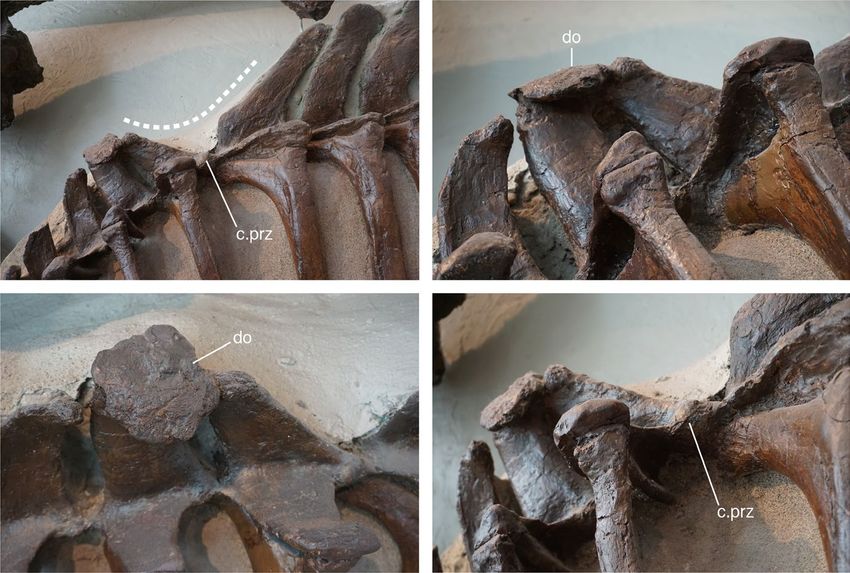
2   |                                                                                                                            BERTOZZO et al.

                                                representing the point between the stresses of the nuchal ligament, pulling the an-
                                                terior neural spines forward, and the ossified tendons pulling the posterior neural
                                                spines backward. Different reconstructions of the morphology of the structure based
                                                on the pathological conditions affecting the neural spines of ROM 768 are proposed.
                                                Finally, we review the history of reconstructions for Parasaurolophus walkeri showing
                                                how erroneous misconceptions have been perpetuated over time or have led to the
                                                development of new hypotheses, including the wide neck model supported in the
                                                current research.

                                                KEYWORDS
                                                Alberta, Cretaceous, nuchal ligament, Ornithopoda, trauma

1       |   I NTRO D U C TI O N                                              (a)

Among         hadrosaurs     (a.k.a.   the   “duck-billed”   dinosaurs),
Parasaurolophus walkeri (Parks, 1922) is one of the genera best-
known among the general public iconically due to its posteriorly and
downward directed, hollow, tubular crest. The genoholotypic spec-
imen was described by Parks (1922), and comprised a nearly com-
plete skeleton (ROM 768) discovered in the Dinosaur Park Formation
in today's Dinosaur Provincial Park (DPP, Alberta, Canada) in 1920
by a team from the University of Toronto. The specimen is lacking
the caudal series, hindlimbs (only the left femur is preserved), and
distal ends of the ischia (Parks, 1922; Figure 1a). Unfortunately, no
                                                                             (b)
other complete or articulated skeletons have been confirmed in this
area since, making it one of the rarest and most enigmatic hadro-
saur species from the DPP (Evans et al., 2007, 2009; Ryan & Evans,
2005). Additional Parasaurolophus specimens have been discovered
in the Fruitland Formation (P.cyrtocristatus; Ostrom, 1961) and the
Kirtland Formation (Sullivan & Williamson, 1999; Wiman, 1931) in
New Mexico, USA. Recently, several specimens have been collected
from the Kaiparowits Formation of Utah (Gates et al., 2013), includ-
ing an exquisite near-complete juvenile skeleton (Farke et al., 2013).
Isolated skeletal material is often found scattered in these regions,
but complete specimens are limited to those reported so far. The           F I G U R E 1 (a) The type specimen of Parasaurolophus walkeri
paleobiology of Parasaurolophus has attracted the attention of re-         (ROM 768) exhibited at the ROM in the opisthotonic “death pose”
searchers, largely because of its exaggerated and unusual crest. As        position as it was found in 1920; (b) simplified skeletal drawing
for other lambeosaurine dinosaurs, it has been variously proposed          of ROM 768, in which the red circles indicate the positions of the
                                                                           paleopathological lesions: (a) dental disease, (b) V-shaped gap of
that, in addition to visual display, the crest facilitated underwater
                                                                           neural spines (“saddle”) and discoidal overgrowth, (c) fractures of
feeding, was involved with thermoregulation or improving olfaction,
                                                                           the ribs, and (d) irregular overgrowth of the pubic peduncle of the
or that its internal structures functioned as resonating chambers,         ilium
which is the most widely accepted hypothesis (see review by Evans,
2006, and an exhaustive list of references in Weishampel, 1981).           of the neural spines of dorsal vertebrae six and seven; a V-shaped
        The holotype of P.walkeri reveals further information about the    deformation of the neural spines of dorsal vertebrae seven and
biology and life history of the individual itself. The specimen displays   eight; a bony spur on the fourth dorsal rib; callus formation on the
a wide number of fossilized injuries in the postcranial skeleton, as       fifth and sixth dorsal ribs; and a ventral projection of the pubic pe-
noted by Parks (1922), as well as a previously undescribed dental          duncle of the left ilium (Figure 1b). During Parks’ time, interest in
pathology in the upper jaw. Parks (1922) did not extensively describe      dinosaur paleopathology was in its infancy (see Moodie, 1923), and
the lesions and their possible causes in detail; however, doing so is      no proper references or comparative material were available. Today,
one of the aims of the present paper. The lesions comprise dental          based on subsequent large collections of skeletons and skeletal ma-
disease in the left maxilla; a roughened bony plate on the distal tips     terial, we know that large ornithopod dinosaurs have the highest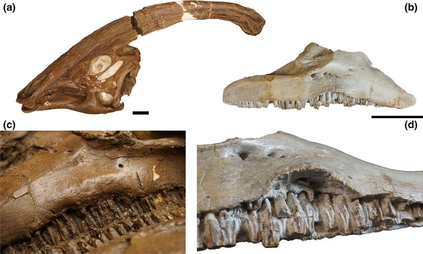
BERTOZZO et al.                                                                                                                                  |   3

frequency of fossilized injuries and diseases among the dinosaurian        next to the bones where possible, and measurements were taken with
fossil record (Bertozzo et al. in prep; Rothschild et al., 2003; Siviero   a 1-m tape. The morphological analysis of the pathologies aimed to
et al., 2020; Tanke & Rothschild, 2002, 2014). In addition, new re-        recognize: (a) reactive bone surfaces; (b) new bone formation; (c) the
search and technologies over the past 30 years have provided a             presence of underlying original bone tissue; and (d) drainage sinuses
larger sample of pathological specimens for comparative purposes,          for the release of infectious material. The identified lesions were com-
as well as enabling a better understanding to be gained of the ex-         pared with another specimen, referred to as cf. Parasaurolophus (TMP
ternal and internal structure of fossilized lesions and diseases in di-    1992.053.0021), as well as those included in the increasing database
nosaurs. Among the injuries recorded in ROM 768, those affecting           of fossilized injuries and diseases of dinosaurs and other extinct verte-
the neural spines of the dorsal vertebrae have been the subject of         brates (e.g., Bertozzo et al. in prep; Barbosa et al., 2016, 2018; Molnar,
much attention over the decades, especially among non-specialist           2001; Tanke & Rothschild, 2002, 2014; Senter & Juengst, 2016; Hunt
publications for the general public. At first sight, this “opening” of     et al., 2019). To facilitate description of the presence of the nuchal
the neural spines is located virtually below the distal end of the nasal   ligament in P.walkeri, and its connection to the purported pathologi-
crest, hence Andrew Milner hypothesized that this represented a            cal neural spines, the origins, insertions, and structure of the nuchal
“saddle”-like concavity on the anterior torso midline to receive the       ligament were created based on reconstructions for extant crocodiles
crest when the animal was moving its head from side-to-side (see           and birds (Tsuihiji, 2004; Yasuda, 2002), sauropods (Woodruff, 2017),
Norman, 1985, p. 126). Initially, Parks thought that the neural spine      and mammals (Bertram & Gellman, 2002; Fielding et al., 1976; Kadri &
configuration in P.walkeri was pathological but, in the same paper, he     Al-Mefty, 2007; May-Davis & Kleine, 2014).
then revised this view to suggest that the odd discoidal overgrowth            A visual representation of the traumatic event that may have
on top of the rostrally facing neural spines served as an attachment       been responsible for causing some of the injuries in ROM 768 is pro-
point for muscles or tendons linked directly to the distal crest (Parks,   vided later, in Figure 6. We further reviewed the history of scien-
1922, p. 18). Later, Russell (1946) suggested the fusion of the neu-       tific reconstructions of Parasaurolophus to highlight the anatomical
ral spines would have supported strong cervical musculature for the        changes and the mistakes made in depictions of the species over the
large head during underwater feeding. No further research focused          years. The importance of the relationship between the reconstruc-
on this feature, and the debate about its pathological nature has          tions and the paleopathological lesions is emphasized (following
never been fully resolved, despite some researchers agreeing with          Bertozzo et al., 2017).
Parks’ initial pathological hypothesis (see Lull & Wright, 1942; Naish,
2008; Ostrom, 1962). In this paper, we provide a detailed morpho-
logical description of the injuries in the P.walkeri holotype and com-     2.1 | Institutional Abbreviations
pare them with examples previously identified in Ornithopoda and
other dinosaurs. The possible nature of the “saddle” and bone over-        AMNH, American Museum of Natural History, New York, USA;
growth on the dorsal spines as a ligament attachment site for the          CMN, Canadian Museum of Nature, Ottawa, Canada; CUST,
crest are reviewed. Finally, an ecological explanation for the causes      Museum of Natural History, Jilin University, Changchun, P.R. China;
of the injuries, which we suggest may have happened in one to per-         DPP, Dinosaur Provincial Park, Alberta, Canada; FMNH, Field
haps three event(s) during the lifetime of the individual, is provided.    Museum of Natural History, Chicago, Illinois, USA; GMH, Geological
                                                                           Museum of Heilongjiang, Heilongjiang Province, P.R. China; GPDM,
                                                                           Great Plains Dinosaur Museum, Malta, USA; LACM, Los Angeles
2   |   M ATE R I A L S A N D M E TH O DS                                  County Museum, Los Angeles, California, USA; MCD, Museu de la
                                                                           Conca Dellà, Isona, Spain; MOR, Museum of the Rockies, Bozeman,
Parasaurolophus walkeri (ROM 768) is panel-mounted at Royal Ontario        Montana, USA; MPZ, Museo Paleontológico de la Universidad de
Museum (ROM) in an articulated pose, with the left side exposed,           Zaragoza, Zaragoza, Spain; ROM, Royal Ontario Museum, Toronto,
whilst the original skull is kept in the collection and a cast completes   Ontario, Canada; TMP, Royal Tyrrell Museum of Palaeontology,
the mount. The axial skeleton is rigidly fixed to the panel mount, and     Drumheller, Alberta, Canada; ZPAL, Institute of Paleobiology, Polish
the postcranial elements could not be removed to facilitate an inter-      Academy of Sciences, Warsaw, Poland.
nal analysis via CT scanning. As reported by several authors (Anné
et al., 2015, 2016; Ekhtiari et al., 2020; Hedrik et al., 2016; Jentgen-
Ceschino et al., 2020; Redelstorff et al., 2015), diagnoses of fossil-     3    |   R E S U LT S A N D D I S CU S S I O N
ized injuries and diseases can be improved through the use of internal
analytical techniques such as histology, (micro)CT scans, and syn-         The paleopathological lesions evident in ROM 768 are of one to
chrotron. Unfortunately, ROM 768 cannot be disassembled from the           three putative different etiologies: the skull suffered from periodon-
mount and, therefore, this study solely focuses on the external mor-       tal disease in the alveolar bone of a group of maxillary teeth; the
phology while bearing in mind the difficulties of providing detailed       thorax was hit at least once by an external object, resulting in local-
diagnoses using such an approach. The elements were photographed           ized injuries; and the ilium displays a hypertrophic extension of the
with a Sony Mirrorless a5100 camera, a 10-cm scale bar was placed          pubic peduncle that covered a small portion of the underlying pubic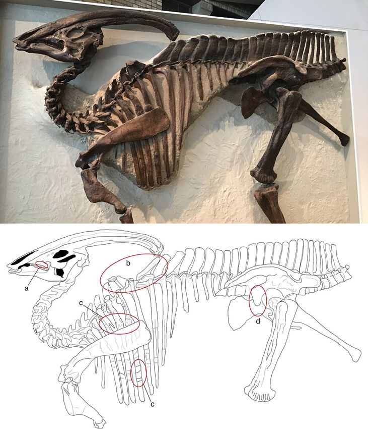
4   |                                                                                                                              BERTOZZO et al.

blade and may have been secondary to the injuries of the thorax.           margin of the spines to the articulation between the rib capitulum/
Each group of lesions will be described in detail below.                   prezygapophysis point; Figure 3) is 110°. Dorsal spine eight bears
                                                                           a small knob anteriorly and, from this point up to the distal tip of
                                                                           the spine, the anterior margin is slightly concave, a shape not visible
3.1 | Maxilla                                                              in any other dorsal spines. The three dorsal spines succeeding the
                                                                           deformed area are angled slightly caudally. In all other hadrosaurid
The margins of the teeth display a swollen area around the midpoint        taxa, the dorsal spines are angled caudally in cranial dorsal vertebrae
of the external side of the left maxilla, slightly inferior to the max-    and gradually become more vertical caudally along the mid-dorsal
illary facet of the jugal. The affected area has rounded and smooth        and posterior dorsal vertebrae (e.g., Campione, 2014; Godefroit
margins, suggestive of a healed and long-standing nature, and extend       et al., 2012). A marked difference in the length and width of the two
for the length of (approximately) seven teeth at their eruption point      spines in ROM 768 is also evident. In the hadrosaur dorsal series,
(Figure 2a). Similar conditions have been recognized in the maxillae of    the length of the spines usually increases regularly from the cervi-
CMN 362 (“Stephanosaurus;” Tanke & Rothschild, 2014; Figure 2b) and        codorsal transition to the more posterior dorsals, without marked
AMNH FARB 6390 (Bactrosaurus johnsoni; FB, pers. obs.). Although           deviations in height (at least, in the dorsal series; hadrosaurid taxa
the latter specimen shows a milder condition with just an upturn of        show different lengths of vertebral spines in the sacral and proxi-
the maxillary margins of the teeth, the lesion in CMN 362 had a more       mal caudal regions; Horner et al., 2004). The spines are straight, and
severe impact on the bone (FB, pers. obs.), although a more detailed       no apparent major fracturing, truncation or deflection are visible on
study is needed to assess the potential impact on the lifestyle of the     their bodies. However, on dorsals nine, 10 and perhaps 11, there
individual. Differential diagnosis for the lesion in ROM 768, and con-     seems to be a horizontal “fascia” of deformation, and the lower half
sequently in CMN 362 and AMNH FARB 6390, includes periodontal              of those spines are slightly swollen.
disease, infection (maxillary abscess), and traumatic injury. However,         A small, enlarged area (callus?) is visible emerging from the right
internal analysis through CT and microCT scans is required in the fu-      prezygapophysis of dorsal eight (Figure 3d); Lull and Wright (1942)
ture to enable a more reliable description of the bone tissue, and pos-    previously reported on this modified zygapophyseal articulation.
sibly determine the chronicity of the condition.                           The rostral-most margin of the neural spine of dorsal seven is fused
                                                                           with the caudal margin of the spine of dorsal six via an abnormal
                                                                           overgrowth of bone (Figure 3b). This overgrowth has the shape of
3.2 | Dorsal vertebrae                                                     a flat disk of consistent thickness with a highly remodeled and po-
                                                                           rotic surface (Figure 3c). Parks (1922) described it as: “[…] a discoidal
The lesions in the dorsal vertebrae are the most striking pathologi-       expansion or co-ossified separate bone about 100 mm in diameter
cal feature present. The neural spines of dorsal seven and eight are       and 20 mm thick.” The “dorsal” surface is subcircular and more de-
spread apart, facing rostrally and caudally respectively. The angle        veloped on the right side and covers the entire dorsal surface of the
of this deformation (calculated from the central point of the distal       anterior neural spine.

F I G U R E 2 (a) skull of Parasaurolophus walkeri ROM 768 in left lateral view; (b), isolated right maxilla of "Stephanosaurus" (CMN 362) in
lateral view; (c), detail of the ventral margin of the left side of the maxilla, highlighting the disease of the central portion of the alveolar
margin; (d), detail of the maxilla of CMN 362, highlighting the everted ventral margin, with the exposed alveolar base of the maxillary teeth.
Scale bar =10 cm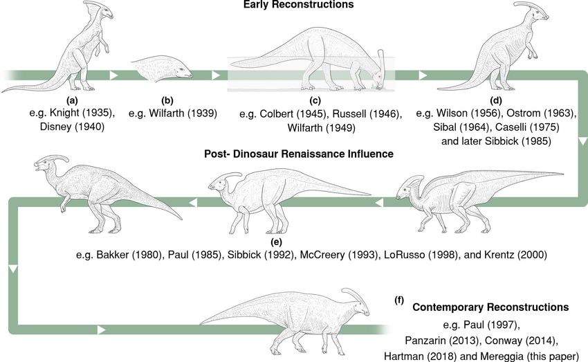
BERTOZZO et al. | 5
F I G U R E 3 (a) anterior dorsal
(a) (b)
vertebrae of ROM 768, showing the
V-like deformation between the neural
spines of vertebrae seven and eight. The
dotted line demarcates the “saddle” as
described in the text; (b), the discoidal
overgrowth on top of dorsal vertebra
six, fusing at the apical portion with that
of dorsal vertebra seven; (c), dorsal view
of the discoidal overgrowth; (d), latero-
caudal view of the articulation between
dorsal vertebrae six and seven, focusing (c) (d)
on the enlarged, callus-like appearance
of the prezygapophysis. Abbreviations:
do, discoidal overgrowth; c.prz, callus on
prezygapophysis
F I G U R E 4 (a) ROM 768 dorsal ribs (a) (b)
connected to dorsal vertebrae five, six and
seven. The first injury is associated with
a bony spur, while the shaft is enlarged
by callus formation on the fifth and sixth
rib. (b), two small calluses at the distal
region of the sixth rib, separated by a
shallow surface. (c), TMP 1992.053.0021,
cranial view showing callus next to the
rib neck, with a detail of the medial view
(d). (e) TMP 1992.053.0021, cranial
view of a rib showing callus formation
on the upper third of the shaft, with a
detail of the medial view (f). (g), TMP
1992.053.0021, cranial view of a shorter
(c) (d) (e) (g) (h)
rib showing callus at the neck, with a
detail of the medial view (h). The arrows
point to the pathological areas. Scale bar
in a, b, c, e, and g = 10 cm; scale bar in d, f,
h = 5 cm
(f)
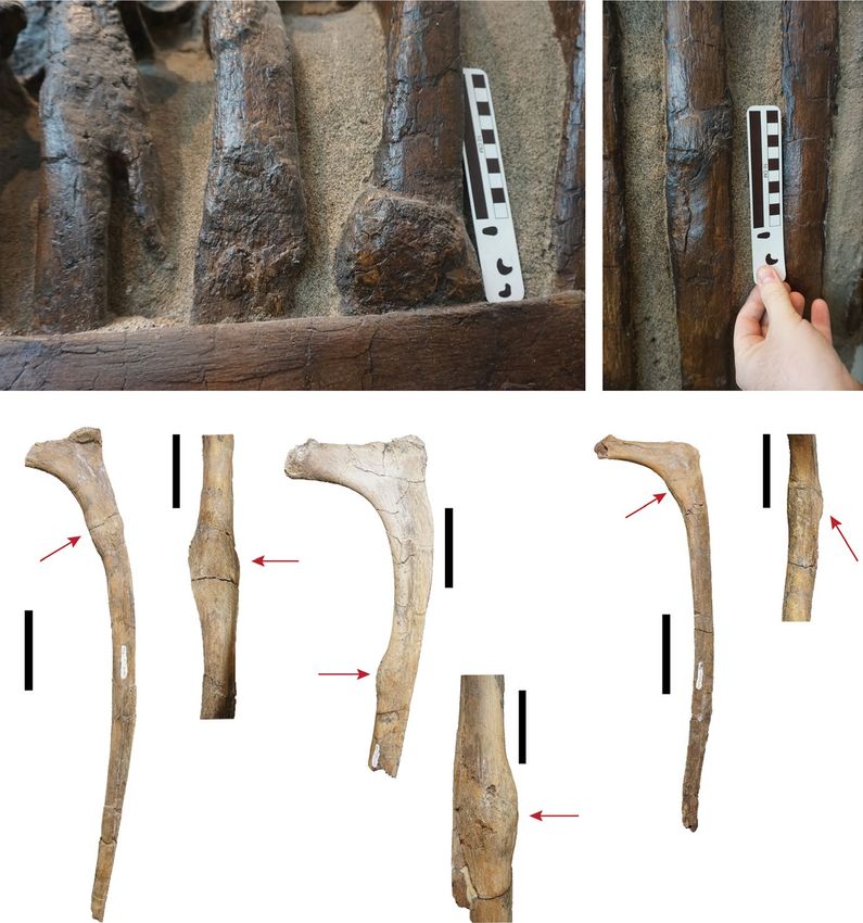
3.3 | Ribs an angle of 15°. The bone surface at the base of this overgrowth
is roughly remodeled, but no resorption or recesses (draining chan-
The fourth, fifth, and sixth dorsal ribs in ROM 768 show signs of nels or small cavities) are evident. The spur has a straight outline.
simple fractures on their shafts (Figure 4a). The fourth rib bears a The fifth rib has a modest cranio-caudal enlargement, attribut-
pointed, elongated spur that diverges from the main rib shaft by able to callus formation. A more expanded and developed area of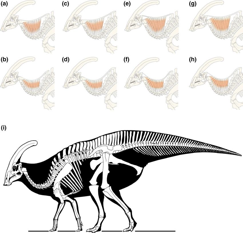
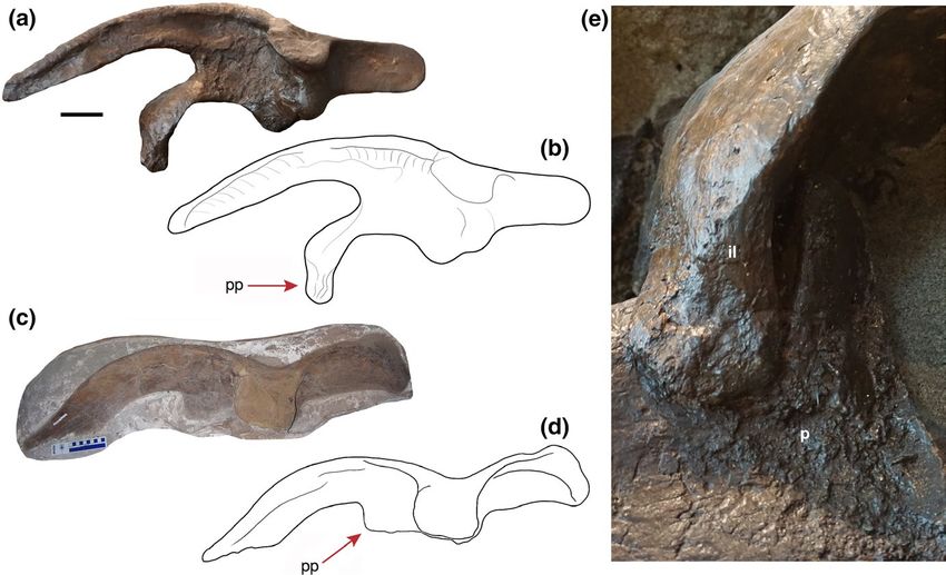
6   |                                                                                                                               BERTOZZO et al.

callus is evident in the sixth rib. These areas have a rougher tex-          been attained during a single traumatic event. In comparison to the
ture than the “healthy” portions of the bone, but the appearance             fractures of ROM 768, the injuries in the ribs of TMP 1992.053.0021
of the callus is an indication of the advanced state of healing of the       are at different locations and the callus is smoother in appearance,
bones. In addition, the sixth rib bears two, less distinct calluses at       thereby suggesting a more advanced state of healing. Another ex-
the distal region (Figure 4b). The scapula shows no evidence of in-          ample of a healing fractured rib was found in MOR 548, a hatchling
juries, despite scapular trauma having been previously identified in         Hypacrosaurus from Montana, with a small callus in the shaft (FB,
hadrosaurs (Tanke & Rothschild, 2002), and it may physically cover           pers. obs.), suggesting that hadrosaurs were able to survive thoracic
additional rib fractures. The morphology of the callus formation as-         injuries attained at a relatively young age. The etiology of the heal-
sociated with the rib injuries indicates that they had all healed to a       ing rib fractures in these hadrosaurs cannot be confirmed with cer-
similar extent when the animal died, suggesting they were probably           tainty but, given their location (most are located on the shaft rather
attained at the same time. Previous research has identified a variety        than the head or neck areas of the ribs), it is considered feasible that
of injuries in the rib cage of hadrosaurs (Tanke & Rothschild, 2014;         they might have resulted from intraspecific fighting (Bertozzo et al.,
Bertozzo et al. in prep), thereby demonstrating how the lateral sides        2017); failed predation (Tanke & Rothschild, 2014); impacts from
of the trunk in these dinosaurs were susceptible to trauma. Bertozzo         falling objects, such as trees or rocks, or crashing against hard ob-
et al. (2017) and Tanke and Rothschild (2014) have suggested that            jects, such as an uneven ground surface.
such injuries can be a byproduct of intraspecific fighting, a behav-
ior also witnessed in modern mammals and varanid lizards. To ex-
tend the current knowledge of pathological ribs in Parasaurolophus,          3.4 | Ilium
and for comparison with ROM 768, three further pathological ribs
that belong to another cf. Parasaurolophus sp., but in an incomplete         The left ilium of ROM 768 bears an abnormal, ventrally elongated
and disarticulated condition, are reported here (Figure 4c–h). TMP           pubic peduncle, which overlaps the iliac peduncle of the pubis
1992.053.0021 was discovered by one of the authors (DHT) in                  (Figure 5). Parks (1922) noted the remarkable nature of this fea-
1992 in the Dinosaur Park Formation (DPP, Alberta, Canada). The              ture and, in later studies, it has been referred to for comparison
three pathological ribs are mostly complete, although one of them is         (e.g., Godefroit et al., 2012). The dorsal portion of the elongation
missing half of the shaft (Figure 4e). Callus formation is evident on        seems to have been artificially constructed during restoration of the
each rib, two of which are located beneath the neck of the capitu-           skeleton, but the section attaching to the pubis, as well as the as-
lum along the curved portion of the proximal region (Figure 4c–d,g–          sociated pubic surface, is original. Therefore, it seems unlikely that
h), while the larger rib displays callus at its midshaft (Figure 4e,f).      the elongated feature arose during the preparation or restoration
Similar to ROM 768, the calluses all present the same state of heal-         of the skeleton. The pubic peduncles of the ilia in Parasaurolophus
ing with irregular and remodeled surfaces, suggesting they had all           cyrtocristatus (FMNH P 27393), in the juvenile Parasaurolophus sp.

F I G U R E 5 (a) Left ilium of ROM 768, with sketch of the same (b); (c) right ilium TMP 1992.053.0021 referred to as cf. Parasaurolophus
sp., with sketch of the same (d). The picture of the latter specimen and its drawing are mirrored for a better comparison with the left ilium
of ROM 768. The red arrows point towards the pubic peduncle of the ilium, which is ventrally elongated in ROM 768 and flat and parallel
in TMP 1992.053.0021; (e), close-up of the pathological articular surface in ROM 768 between the abnormally developed pubic peduncle
of the ilium and the iliac process of the pubis. Abbreviation: il, ilium; p, pubis; pp, pubic peduncle of the ilium. Scale bar in A and C = 10 cm
BERTOZZO et al.                                                                                                                                 |   7

(RAM 14000), and in Charonosaurus jiaynensis (CUST J-V1251-57)              archosaurs, oral infections occur in the form of osteomyelitis caused
are shorter and less pointed, and similar to Hypacrosaurus altispinus       by Staphylococcus aureus, streptococci, enterococci, Pseudomonas,
(AMNH FARB 5204; Brown, 1913), an indeterminate lambeosaurine               Enterobacter, Proteus, Escherichia coli, Serrotia and anaerobic bacteria
MCD 4791 (Prieto-Márquez et al., 2013), Sahaliyania elunchuno-              (Huchzermeyer, 2002; Rothschild & Martin, 2006). Once affected by
rum (GMH W103; Godefroit et al., 2008), Corythosaurus casuarius             an external agent, the infected area increases in dimension due to
(AMNH FARB 5240; Brown, 1916), Barsboldia sicinskii (ZPAL MgD-              elevation of the cortical bone, as seen in ROM 768. In infections, this
I/110; Maryanska & Osmólska, 1981), Magnapaulia laticaudus (LACM            results in the development of sequestra, bone fragments entrapped
20874; Prieto-Márquez et al., 2012), and an indeterminate lambeo-           within the involucrum (a sheet of granulose tissue) and separated
saurine MPZ 2005/90 (Prieto-Márquez et al., 2013). In Olorotitan            from the original bone. In the maxilla of ROM 768, however, there
arharensis (Godefroit et al., 2012), the pubic peduncle is longer, but      are no signs of an active infection and drainage channels are absent.
not as extended as in ROM 768. Prieto-Márquez (2010) assigns two            The lesion could either be in an advanced state of healing when the
states for the pubic peduncle morphology in Hadrosauridae: “rela-           individual died (with the bone undergoing the final stages of remod-
tively large and dorsoventrally deep (longer than wide), subconi-           eling, obscuring any evidence of infection) or in an early stage infec-
cal, with a proximal region that is only slightly craniocaudally wider      tion. Another explanation is that the alveolar margin of the maxillary
than the distal end of the process” as the plesiomorphic state and          dentition was affected by a local inflammation or periodontal dis-
“relatively shorter (wider or as wide as long) and triangular, with         ease, generally caused by bacteria in the mouth infecting the tissue
a proximal region that is much craniocaudally wider than the dis-           around the teeth. Future internal analysis via computerized tomog-
tal end” as the derived state. The author scores the derived state          raphy (CT) scanning will hopefully reveal more information about the
for Parasaurolophus cyrtocristatus and Charonosaurus jiaynensis, but        definitive nature of the maxillary condition of both ROM 768 and
also for Parasaurolophus walkeri. The derived morphology is verified        CMN 362.
in a cf. Parasaurolophus sp. ilium from DPP (TMP 1992.053.0021,                The injuries in the trunk, both in the dorsal vertebrae and the
Figure 5c,d; Prieto-Márquez, pers. comm. 2019). Thus, either the            ribs, are here suggested to have been caused by a single trau-
morphology in ROM 768 is due to individual variation or, given the          matic event. The V-shaped gap, or “saddle” of the dorsal neural
concurrence of injuries in the postcranial skeleton, it is another pa-      spines, appears to have resulted from a vertically directed impact.
thology. The second hypothesis seems to be supported by the na-             Furthermore, the succeeding three dorsal neural spines are also an-
ture of the contact surface between the ilium and pubis. The pubic          gled caudally, which is a different morphology to that observed in
peduncle extends ventrally but, near the point of contact with the          other hadrosaurids (e.g., Campione, 2014; Godefroit et al., 2012).
distal tip of the iliac peduncle, it develops further curving by approxi-   This, the possible callus on the prezygapophysis, the V-shaped gap,
mately 150° and overlapping the lateral side of the iliac peduncle.         the abnormal morphology of spine eight with the anterior knob and
Here it is pressed over the surface of the latter, forming compressive      the anterior slightly concave curvature, and the presence of a dis-
propagation “waves” on the contact surface (Figure 5e). Usually, in         coidal overgrowth on the sixth and seventh dorsal spines together
hadrosaurs, pelvic elements are not fused, but rather connected via         suggest a traumatic cause. However, the shaft of spines seven to
masses of ligamentous connective tissue (Horner et al., 2004), mak-
ing the morphology in ROM 768 extremely abnormal. The surface
of the overgrowth of the pubic peduncle is slightly rugose, but no
evidence of infection or callus formation is evident.

3.5 | Possible etiologies of the pathological lesions

Although the maxillary lesion can probably be regarded as an iso-
lated condition related to oral health status, the pathologies in
the postcranial skeleton, while separated physically, may be inter-
connected to life event(s). The condition associated with the max-
illary teeth could have been induced by inflammation (periodontal
disease), external trauma, or localized infection. As various hadro-
saur species were herding animals (Fiorillo et al., 2014; Hone et al.,
2014; Horner et al., 2004; Lockley et al., 1983), injurious conspe-         F I G U R E 6 Paleoart reconstruction of a plausible scenario
cific interactions would be expected. A maxillary trauma might              explaining the fossilized injuries in the thorax of ROM 768. In a
                                                                            violent rain and windstorm, a large tree (Platanaceae) falls on an
have resulted from a lateral impact from a conspecific animal and/
                                                                            adult Parasaurolophus walkeri, while the group is escaping. The tree
or an external object (perhaps, the same incident that caused the
                                                                            falls vertically on the back of the animal, hitting the rib cage and the
injuries in the trunk), or by chewing upon a hard object that dam-          neural spines of the anterior dorsal vertebrae. Artwork by Marzio
aged soft tissues and (possibly) resulted in a local infection. In living   Mereggia
8 | BERTOZZO et al.
11 do not show abnormal fractures or truncations, except for the 3.6 | Nuchal ligament reconstruction
slightly swollen lower half of dorsal spines nine, 10 and 11, making
the nature of the impact difficult to determine. It could potentially According to Barkow (1856), in birds the nuchal ligament extends
have involved a fallen tree, a large rock, or another animal of similar from the dorsal midline in the posterior cervical region and attaches
size and mass, striking vertically or diagonally the trunk of the ani- to the dorsal edge of the neural spines, while its anterior most in-
mal, without causing marked fractures of the spines but rather in- sertion varies across avian taxa. This structure is weakly developed
ducing plastic deformation of the spines in the direct location of the or absent in birds, but is more robust in Rhea americana, where it
injuries. Another possible explanation is that the strong posterior inserts on the neural spine of the axis (Tsuihiji, 2004). Later, the
orientation of dorsal spine eight was caused by the pulling action of nuchal ligament was also described in Struthio camelus (Dzemski &
the ossified tendons (see below), and the absence of clear fractures Christian, 2007). Based on the terminology of Baumel and Raikow
in dorsal spine seven is due to a weaker impact than previously as- (1993), Tsuihiji (2004) refers to the ventrally branched median cervi-
sumed. Whatever the cause, the wounded area has undergone ad- cal ligament of Rhea americana as ligamentum elasticum interspinale,
vanced healing, which has eliminated some of the finer details of reserving the term ligamentum nuchae to the bifurcating ligamen-
the original injuries. The hypothetical scenario suggested in Figure 6 tous sheath enclosing it. However, in some avian literature, the
summarizes the data acquired from the holotype—a large object non-bifurcated median cervical ligament of other birds is referred
(here imagined as a tree trunk) fell on the individual, hitting first the to as ligamentum nuchae (Barkow, 1856; Yasuda, 2002). A similar,
lateral side of the trunk, and then the apical region of the back, in and potentially homologous, median ligamentous structure is also
line with the fractured ribs. The absence of the “saddle” in the nearly present in non-avian diapsids and inserts to the occiput in Alligator
complete skeleton of Parasaurolophus cyrtocristatus (Ostrom, 1963) mississippiensis and Iguana iguana (Tsuihiji, 2004). In other crocodile-
further supports the pathological interpretation of the V-shaped related taxa such as dyrosaurids, the supraspinal ligament is consid-
gap in ROM 768. ered to have developed into the nuchal ligament, expanding from
The abnormal overgrowth of the pubic peduncle of the ilium in the anterior thoracic vertebrae to the occipital region of the skull,
ROM 768 is more difficult to diagnose. It could have resulted from while ventrally branching onto each of the cervical neural spines
a direct blow or, alternatively, was a secondary injury attained be- (Schwartz-Wings, 2014). The function of the structure is to support
cause of the difficulties the individual had to endure after the trau- and strengthen the neck, by connecting each cervical vertebra in
matic event as it attempted to regain normal movement. An unusual a single contiguous tensile “beam.” The presence of the ligament
increase of bone material can be a heterotopic ossification derived in dinosaurs has been theorized through the application of extant
from a trauma (myositis ossificans traumatica; Chang et al., 2018; phylogenetic bracketing (sensu Witmer, 1995; Tsuihiji, 2004). In sau-
Davies et al., 2017; Kransdorf & Meis, 1993) or a tumoral growth ropods, the ligament occupies an important point of discussion for
following an injury (i.e., Uda et al., 2002; Urist, 1957). If we assume the biomechanics of their extremely long and well-developed neck.
the abnormal morphology is related to an external trauma, it is pos- Based on R.americana, Tsuihiji (2004) suggested that the nuchal
sible the anterior region of the ilium might have suffered from the ligament attached above and to both neural spines of each cervi-
same impact as the thorax, or perhaps a second, separate knock. cal vertebra (in a branching pattern), originating from the first non-
The absence of any signs of trauma in neighboring elements, such bifurcated dorsal vertebra (dorsal eight) in Camarasaurus grandis and
as the pubis itself, the ischium, the femur, and the posterior dorsal on dorsal seven in Apatosaurus. Further variations have also been
vertebrae and their associated ribs, however, makes this suggestion proposed by Schwarz et al. (2007) and Woodruff (2017). In tyranno-
somewhat problematic. The growth of tumoral bone usually hap- saurid theropods, the cervical neural spines are connected through
pens at the point of injury (see Uda et al., 2002), whereas no re- the M. transversospinalis capitis (Snively & Russell, 2007), whereas
ports exist of tumoral masses occurring in a different site following the nuchal ligament (“supraspinal ligament”) would have inserted
a trauma. In fact, the growth of the peduncle in ROM 768 is limited onto the midline of the supraoccipital (Tsuihiji, 2010). Organ (2006)
only to the peduncle itself. The articular point between the ilium and reconstructed the iguanodontian epaxial musculature following a
pubis comprises cartilage and, as such, a hypertrophic ossification crocodile model, but he did not discuss the nuchal ligament in orni-
of the ligamentous connective tissue could have occurred (myositis thopods. In Parasaurolophus, the occurrence of the elongated nasal
ossificans traumatica; Kransdorf & Meis, 1993). The implication of crest has raised questions about the possible presence of such a lig-
this is that a major trauma had resulted in torn muscles/ligaments, ament in the species, and more generally, within hadrosaurids. The
possibly as a direct consequence of the impact or perhaps due to the crest was a hollow structure, formed internally by three dorsal pairs
body trying to re-establish a proper balance for locomotion in the of tube (median, lateral and dorsal), rising from the external nares
subsequent period after the injury. The manner in which the holo- to ascend to the tip of the crest where they loop ventrally to return
type of P.walkeri is mounted means that it is not possible to analyse inside the ventral margin of the crest to terminate in a large chamber
the inner structure of the ilium and, as such, interpretations of the (Sullivan & Williamson, 1999). Overall, the crest was a relatively light
pelvic lesion must remain tentative. Another possible suggestion is structure but, because of its large size, it probably required a strong
a genetic defect but, given the concurrence of injuries on the same and muscular neck. The nuchal ligament was first proposed by
side of the body, this seems less likely. Brown (1912) as a means of connecting the anterior dorsal vertebraeBERTOZZO et al. | 9 to the base of the neck, while Parks (1922) even suggested it was between the third and sixth dorsal spines in B.canadensis (Prieto- attached to the distal region of the crest (this last assumption can Márquez, 2007), might suggest that (a) the nuchal ligament and a be discarded since in living archosaurs the ligament attaches to the simple set of ossified tendons coexisted in one, perhaps two con- axis or the occiput). The discoidal overgrowth on the neural spines secutive vertebrae, or (b) the origin site of the nuchal ligament dif- of dorsal vertebrae six and seven in ROM 768 might be the first fered between hadrosaurid taxa. Further histological analysis on the osteological correlate to infer the origin site of the nuchal ligament. neural spines of these dorsal vertebrae are needed in future, aided Both in sauropods (Tsuihiji, 2004) and dyrosaurids (Schwarz-Wings, by a possible reexamination of “mummified” specimens. 2014), the ligament could have originated in the anterior dorsals at In summary, it seems possible that the pathological structure that the same level as the impact injuries in ROM 768. Woodruff (2017) merges the spines of dorsal vertebrae six and seven is a secondary theorized that, in sauropods, the nuchal ligament (referred to as hypertrophic ossification of the base of the nuchal ligament. In hu- nuchal ligament and supraspinous ligament altogether in Woodruff mans, trauma can result in a hypertrophic growth of bone material, (2014)) extended from the occipital region of the cranium to the a benign process characterized by heterotopic ossification usually tail, thereby spanning the entire axial series. In Parasaurolophus, and within large muscles and known as myositis ossificans traumatica. most likely all hadrosaurids, this might not have occurred because of Heterotopic ossification is defined as the formation of bone at atyp- the presence of the overlapping and latticework of ossified epaxial ical sites, in which ectopic lamellar bone forms within a tendon, tendons that stiffened the spine from the central and posterior dor- muscle, or other soft tissue (Chang et al., 2018; Davies et al., 2017; sal vertebrae to the mid-caudal vertebrae, perhaps reducing the role Kransdorf & Meis, 1993). Myositis ossificans traumatica seems to of the supraspinal ligament across the axial series. The beginning explain the ossification of the discoidal overgrowth on the neural of the interlaced tendons in the anterior region of the body coin- spine, as well as at the pubic peduncle of the ilium (given the absence cides with the range of the seventh to the ninth dorsal vertebrae of internal analysis). Therefore, it seems feasible that the discoidal in other hadrosaurids. In Brachylophosaurus canadensis (MOR 794) overgrowth does represent a potential candidate for the first oste- the first tendon appears on the third dorsal neural spine, and the ological/pathological correlate for the origin site of the ligamentum number of tendon rods increase to three on the fifth dorsal spine nuchae in hadrosaurids. This strong fibrous structure appears to and five on the sixth dorsal spine (Prieto-Márquez, 2007). However, have originated from the anterior dorsal vertebrae (six or seven), de- the proper lattice (well visible in Gryposaurus notabilis ROM 764; scended over the length of the neck and branched into ventral con- Parks, 1920, pl. 1) begins to form at the caudal portion of the ninth nections to the cervical neural spines, before finally inserting in the dorsal neural spine (Prieto-Márquez, 2007). In Prosaurolophus maxi- occipital region of the cranium or the axial spine (Figure 7). Ostrom mus (ROM 787), ossified tendons start on the fifth dorsal vertebra (1961) suggested that in Corythosaurus casuarius the nuchal ligament (Parks, 1924), developing an X-shaped lattice in the more posterior would have attached to the groove rising from the cross bar formed vertebrae. In Gryposaurus notabilis (ROM 764, “G.incurvimanus”), in- by the supraoccipitals, exoccipitals, opisthotics, and parietals, al- sertions of tendons start in dorsal seven and extend to caudal 19, though in “Procheneosaurus erectofrons” (AMNH 5461) the groove is ranging over the entire axial series (Parks, 1920). In Corythosarus narrower and shorter, likely linked to the young ontogenetic stage of casuarius (AMNH 5338), Brown (1916, pg. 712) reports that “[ten- the individual. On the other hand, the anterior half of the axis neural dons] are developed chiefly overlying the posterior dorsals, sacrals spine in hadrosaurids is generally thickened transversely along its and anterior caudals, diminishing posteriorly and do not appear to dorsal margin and roughened dorsally, perhaps indicating another have been present toward the distal end of the tail”. Based on the possible insertion area for the nuchal ligament (DCE, pers. obs.). skeletal drawings published by Brown (1916, plate XIV, Figure 4), the When present in extant tetrapods, the ligamentum nuchae (or the tendons appear to start on dorsal eight (counting 15 cervical verte- homologue) can typically be differentiated into two parts – a long brae starting from the axis). In conclusion, the neural spines where fibrous component corresponding to the dorsal border of the liga- the overlapping lattice of epaxial tendons originate are usually those ment, and a ventrally branched series of fibers or layered fiber bun- posterior to dorsals six and seven. These are the same vertebrae dles that attach to multiple cervical vertebrae. These are referred (serially) that bear the discoidal shape in ROM 768, thereby ena- to as the funicular parts and lamellar (or laminar) parts, respectively, bling a new depiction of the tendon system of hadrosaurids to be in clinical and veterinary literature (i.e. Bertram & Gellman, 2002; determined. The ossified tendons reinforced the dorso-sacro-cau- Fielding et al., 1976; Kadri & Al-Mefty, 2007; Mary-Davis & Kleine, dal vertebrae, functioning as a solid and fixed “beam” which could 2014). In our reconstruction, we keep a similar structure, although have been adapted for increasing spinal rigidity (Organ, 2006). This more comparative analyses are required to enable a more definitive would also suggest that the caudal inclination of the three succeed- and realistic reconstruction. Two different origin sites and two dif- ing dorsal spines could have resulted from a “pulling” action of the ferent insertions were considered, based on current knowledge of ossified tendons to counterbalance the spinal rigidity after the trau- the location of the nuchal ligament in modern taxa and the myo- matic event. On the other hand, the neck necessitated a less rigid sitis ossificans traumatica “shared” between the sixth and seventh structure with a non-ossified, yet strong, fibrous structure such as neural spines (Figure 7a,c,e,g). Furthermore, we added a second set the nuchal ligament. The position of few, horizontal epaxial ossified of reconstructions, reducing the contribution of the lamellar part to tendons in the fifth dorsal vertebra in ROM 787 (Parks, 1924), and the general depth of the neck, since the extension of the lamellar
10   |                                                                                                                         BERTOZZO et al.

                                                                                                  F I G U R E 7 Musculoskeletal
                                                                                                  representation of the nuchal ligament
                                                                                                  in Parasaurolophus walkeri, based on
                                                                                                  the paleopathological lesions evident
                                                                                                  in ROM 768, with both long (first row)
                                                                                                  and short (second row) extension of the
                                                                                                  lamellar parts. (a,b) the nuchal ligament
                                                                                                  originates on dorsal six, and attaches
                                                                                                  to the axis; (c,d) it attaches to the axis
                                                                                                  while originating on dorsal seven; (e,f)
                                                                                                  the nuchal ligament originates on dorsal
                                                                                                  six, and attaches to the occipital region,
                                                                                                  while in (g,h), it originates from dorsal
                                                                                                  seven attaching to the occipital region;
                                                                                                  (i), skeletal reconstruction of P. walkeri
                                                                                                  by Marco Auditore, with the neck depth
                                                                                                  based on 7e

part varies between closely-related taxa (Figure 7b,d,f,h). Woodruff    portrayed in books, movies, and exhibitions. Disney's Fantasia
(2014, fig. 11) showed such variation in the Ankole-Watusi, cow,        of 1940 is an important exception, where the representation of
horse, and giraffe, and it might be linked to the development and       P.walkeri is based upon the work of Charles Knight and, in particu-
elongation of the cervical neural spines. In Parasaurolophus walkeri,   lar, a painted scene for the Field Museum in 1931 with an Albertan
the anterior cervical spines are low and weakly developed, whereas      Campanian vertebrate fauna. In the movie, Parasaurolophus has a
those in the last five cervicals (thereby next to the cervicodorsal     well-developed skin frill that connects the skull crest to the back, a
transition) start to elongate, perhaps suggesting a greater contribu-   trait that became popular amongst various illustrators, even today.
tion of the lamellar sections of the nuchal ligament. A thoughtful      This reconstruction was based on early research by Barnum Brown.
quantification and comparison with other hadrosaurids and extant        In fact, Brown (1912, pg. 133) advanced that “[in Saurolophus] the
mammals are imperative to better estimate the development of            crest near the posterior end on the dorsal face carries a series of
these structures in the future. The resultant thickened neck depth      fine ridges and in life it probably bore a frill as in the living lizard
(Figure 7i) corresponds most closely to the morphology illustrated      Basiliscus. This comparison is further borne out by the high spines
by Gregory S. Paul in recent years (Brett-Surman, 1997) and is in       of the mid-dorsal vertebrae which, like Basiliscus, probably car-
contrast with the initial swan-like neck suggested by traditional au-   ried a high median dorsal frill”. The later discovery of a mummy of
thors (i.e. Charles Knight; see below).                                 Corythosaurus casuarius (Brown, 1914; AMNH 5240) strengthened
                                                                        Brown's interpretation. The specimen shows a wide fleshy area that
                                                                        connects the skull to the back, considered as evidence of Brown’s
3.7 | Paleoart history of Parasaurolophus                               (1912) frill. A similar structure was also considered to be present for
                                                                        AMNH 5060 (Edmontosaurus mummy; Osborn, 1912), which had
Consideration of the iconographic history is essential to enable a      preserved skin on the dorsal region of the neck. The area is also
better understanding of the scientific assumptions and subjective       characterized by marked skin folds, interpreted by Paul (1987) as
opinions that lay behind recurring “models” (Figure 8). If these are    natural structures instead of taphonomic artifacts. This considera-
not reviewed a priori, continuous misunderstanding can lead to          tion helps to understand the point of origin for the Parasaurolophus
repetitive inaccuracies. Today, Parasaurolophus is one of the most      frill, as most of the reconstructions (i.e. by Charles Knight and
famous dinosaurs outside the academic world. Reconstructions of         Gregory Paul) connect the frill to the anterior region of the back,
this taxon were rare during the first half of the 20th century, com-    following Parks’ ideas. Parks (1922) went even further by assuming
pared to other hadrosaurids, such as Edmontosaurus, which is widely     a muscular and tendinous connection between the “saddle” and the
BERTOZZO et al. | 11 F I G U R E 8 “Evolution” of the restorations of Parasaurolophus through history, starting from top left. The drawings of each “morphology” represent the general overview of the taxon as understood at that time, together with the external features usually depicted. (a) Knight- influenced: tripodal stance, lizard like anatomy with a large neck frill; (b) crest used as muscular attachment; (c) the aquatic Parasaurolophus: the crest is reconstructed as functionally related to aquatic behavior (e.g. air store, water trap or snorkel) for feeding on soft sub-aqueous vegetation; (d) Pre-Dinosaur Renaissance influence: tripodal stance, bulkier anatomy, usually terrestrial, but aquatic depictions still prosper; (e) Post-Dinosaur Renaissance influence: skeletal proportions and overall posture more consistently correct, overly pronated forelimbs, slender anatomy, neck frill and “saddle” variably present; (f) Contemporary reconstructions: bulkier anatomy, thicker neck increasingly common, correctly configured forelimbs and pectoral girdles increasingly common, neck frill and “saddle” generally absent, larger rhamphotheca, speculative soft tissue increasingly common. The years within brackets refer to the year of the published image by the illustrator caudal-most surface of the crest itself, proposing an extremely wide extended the frill from the crest to the back, whereas Mark Hallett neck for Parasaurolophus. This configuration has since been rejected (Wexo, 1985), Luis Rey (Mayes, 1993), John Sibbick (Gardom & Milner, because of the absence of clear muscle markings on the crest, and 1993) and Graham Rosewarne (Lambert, 1990) reduced its exten- the lack of similar features when considering muscle insertion sites sion, in a similar manner to that of Basiliscus plumifrons. Although the in modern Diapsida (Tsuhiji, 2004). Instead, the crest was believed pathological conditions had not been properly analyzed before this to serve as a snorkeling structure to facilitate a more amphibious study, Paul (1987) proposed a skeletal reconstruction with a modi- lifestyle (see references in Weishampel, 1981), following the early fied, normal pattern of dorsal spines – more similar to that of other erroneous reconstructions of hadrosaurids based on the putative hadrosaurids – used later for other paleontological illustrations and “webbed-feet” of the mummies (Osborn, 1912). While the exact de- even for movie productions. The same non-pathological configura- tails of Parks’ reconstruction do not appear to have been plausible, tion portrayed by Hartman (2020) is largely used as a modern refer- his appreciation of the importance of cervical musculature seems to ence since he proposed the same version in 2004. Interestingly, the correspond well with more contemporary knowledge of terrestrial pathological “saddle” was kept in reconstructions of the taxon with- hadrosaurs. out the skin frill (Barrett, 2001; Dixon et al., 1988; Norman, 1985). The previous reconstructions of the frill have drawn attention A case of both “saddle” and skin frill seems to have been presented away from the pathological nature of the discoidal overgrowth and in the Parasaurolophus that appeared in Disney's Dinosaur movie. For the “saddle”, and perhaps early artists decided to portray a frill to the movie, several paleoartists (David Krentz, Mark Hallett, Gregory avoid this ambiguous feature. This situation arose despite the fact Paul, Douglas Henderson, and Ricardo Delgado) collaborated, pro- that Parks (1922) and Lull and Wright (1942) had already suggested ducing one of the best animated models to date, based on the origi- that these features in ROM 768 were pathological. Paul (1987) nal skeleton with the addition of the Fantasia-referenced frill.
12   |                                                                                                                                 BERTOZZO et al.

     The presence of the nuchal ligament and its pathological correlate         lesions were all in an advanced state of healing at the time of death,
in ROM 768 highlights another unresolved question – how deep was                it seems likely the animal survived for at least one to four months
the neck? Early reconstructions of Parasaurolophus and other hadro-             following the traumatic event. The injuries do not appear to have
saurids depict them with a swan-like, pencil-shaped neck suitable for           been a direct cause of the animal's death. This finding, together
grazing in an amphibious environment (Figure 8). Czerkas (1993) was             with the increasing observations and reports of paleopathologies
the first to suggest the presence of a large nuchal ligament on top of          in hadrosaurids, might suggest that these dinosaurs were able to
massive neck musculature connected to the first dorsal vertebrae. Paul          overcome and survive such massive injuries. To substantiate this as-
(Brett-Surman, 1997) shared this interpretation, reconstructing a more          sertion, however, further analysis is required, especially a statistical
bull-like neck in his hadrosaurids, however this was further modified           approach based on remains from large bonebed and museum col-
and resized after the discovery of GPDM 115, a Brachylophosaurus                lections that enable the frequencies of pathological and non-patho-
canadensis “mummy” (Bell, 2014; Murphy et al., 2007). Unfortunately,            logical individuals to be determined; such work is already underway
this mummy has not yet been studied extensively, an unfortunate trend           (Bertozzo et al., in prep).
that affects other skin specimens and mummies, which are sometimes                  The injuries in the spines of dorsal vertebrae six and seven may
only briefly discussed. Hartman (2002) and Ford (2003) proposed an              have corresponded to the original site of the nuchal ligament, a
intermediate form that lay in between the swan-like and bull-like neck.         major tendon inserting either on the neural spine of the axis or the
In recent years, more provocative and extreme reconstructions have              occipital region, and likely branching on the cervical neural spines of
been proposed, such as the “fat” Parasaurolophus by Conway (2012),              the neck. It is interesting to note that hadrosaurs and other ornithis-
to highlight the range of variability and unpredictability of soft tissue       chians show a complex array of ossified tendons along the vertebral
reconstruction. The authors “explore the possibility that [the vertical         column. The starting point is located posterior to the cervicodorsal
shoulder folds that cover the upper arm and shoulder region] are actu-          transition, but it changes through taxa, as a first line of horizontal
ally artifacts of desiccation, and that they supported a heavy padding          tendons, developing into an overlapping X-like pattern generally
of fat and muscles in real life” (Conway, 2012, pg. 50). Finally, Bell et al.   after dorsal seven, and corresponding to the area posterior to the
(2014) reported a mummified specimen of Edmontosaurus regalis with              origin site of the nuchal ligament. This research proposes an indirect
preserved fossilized skin and a fleshy cranial crest. Based on the figures      osteological correlative for the origin of the nuchal ligament in had-
provided, the neck looks wide and thick dorsally (as seen in other spec-        rosaurids, although more in-depth studies are required to confirm
imens as well, such as Corythosaurus casuarius) perhaps supporting our          this assertion, particularly histological analyses as previously under-
Parasaurolophus reconstruction.                                                 taken by Woodruff (2017). The study highlights the necessity of ex-
                                                                                amining complete or near-complete hadrosaurid skeletons, with the
                                                                                cervicodorsal transition preserved, to confirm the origin of this im-
4        |   CO N C LU S I O N S                                                portant structure. This would enable more insights to be gained con-
                                                                                cerning the biomechanics of hadrosaurids and other ornithischians.
The holotype of Parasaurolophus walkeri is here reported showing
several major pathologies: a dental lesion in the left maxilla; myosi-          AC K N OW L E D G E M E N T S
tis ossificans traumatica in the pubic peduncle of the left ilium and           We thank Shino Sugimoto and the staff of the ROM for the help
in dorsal spines six and seven; a V-shaped gap or “saddle” between              provided during the study of the holotype of Parasaurolophus.
dorsal spines seven and eight; and a series of callus formations due            We are grateful to Dr. Caleb Brown (TMP), and Brandon Strilisky
to healing simple fractures on three ribs. The lesions evident in               (TMP) for access to the pathological ribs in the TMP collection,
P.walkeri increase understanding of the pathological bone response              and a special thanks are due to the staff of the TMP for the help
to external trauma, adding further data to the expanding knowledge              provided during the study. We thank Dr. Jordan Mallon (CMN)
of hadrosaurid paleopathology (Tanke & Rothschild, 2014; Bertozzo               and the staff of CMN for providing access and help to study the
et al., in prep). The lesions in ROM 768 appear to have been caused             ornithopod bone collection, especially CMN 362. Information
by a single to perhaps three distinct events (maxilla, thorax, pelvis).         about hadrosaur anatomy and muscular structure have been
It is proposed here that the rib and vertebral injuries may have oc-            discussed with Dr. Albert Prieto-Márquez (Istitut Català de
curred as a result of a large object, such as a tree, falling onto the          Paleontologia Miquel Crusafort, Sabadell, Spain), who also pro-
back of ROM 768, with the pelvic injuries perhaps occurring simulta-            vided comparative pictures, and Dr. Simone Maganuco (Museo
neously or developing as a secondary response to the initial trauma.            di Storia Naturale, Milan, Italy). We thank Marzio Mereggia
It is possible that the dental lesion was also secondary to the trauma          (Accademia di Belle Arti, Bologna, Italy) for the wonderful in-vivo
although it could equally have occurred in isolation. The injured               reconstruction of the Parasaurolophus herd (Figure 6), and Marco
bones are well remodeled and do not show any signs of ongoing in-               Auditore for the skeletal reconstruction in Figure 7i. The project
fection. Usually, a callus appears after two-three weeks following              was partially funded by the 2018 Dinosaur Research Institute
the injury (Lovell, 1997; Marsell & Einhorn, 2011), and its resorp-             Student Project (Alberta, Canada), and it is part of FB’s doctoral
tion is advanced, with progressive obliteration of the fracture line,           dissertation within the Horizon 2020 research and innovation
by the 16th week (Rothschild & Martin, 2006). Since the pathological            programme under the MSCA grant agreement no 754507. All the
You can also read