Design of a Cantilever Biosensor for Tuberculosis Detection
←
→
Page content transcription
If your browser does not render page correctly, please read the page content below
Annals of R.S.C.B., ISSN:1583-6258, Vol. 25, Issue 4, 2021, Pages. 710 - 715
      Received 05 March 2021; Accepted 01 April 2021.

             Design of a Cantilever Biosensor for Tuberculosis Detection
                                       Bhenesha shree R P1 and Shanmugaraja P2
                                      1
                                       Research scholar, Annamalai university, Chidambaram.
                                         2
                                          Professor, Annamalai university, Chidambaram.

Abstract
In this paper, various materials are used in the cantilever biosensor to review the characteristics of the
structure and analyse the better material to be used up for the functionality of the biosensor. The biosensor is
simulated using Comsol multiphysics 4.3b. This structure is responsible for the detection of tuberculosis
with its displacement value. This type of device provides better accuracy. Sensitivity is the main parameter
used in biosensor to detect the tuberculosis. Sensitivity of a cantilever based biosensor is measured using the
defection in the cantilever occuring as a result of interaction of analyte with bioreceptor molecules.
Keywords: Cantilever, biosensor, displacement, Bioreceptor

I. INTRODUCTION
MEMS i.e. Micro-electromechanical system is a process technology that is used to create very small
integrated devices or systems combining an electrical and mechanical component. Its range is usually from
micro to few millimeters.
It has an ability of sensing, controlling as well as actuating on the micro scale, also generating the effect on
macro scale [1]. Bio-MEMS are a micro technology of operating in the field of biological and biomedical
applications that may or may not contain electronics and mechanical function. It is an integral combination
of many fields of engineering. It includes applications like proteomics, genomics, diagnostics etc.
Biosensor is the device which is used to convert biorecognition analysis event into measurable signal [2]. In
biomedical equipments, bio-receptor plays a vital role in the detection of target molecules.

II. CANTILEVER BASED BIOSENSOR
Cantilever biosensor is used as a sensing device because of high throughput. In the cantilever structure, the
surface is made sensitive by depositing sensing layer that contains bioreceptors covalently bonded together
onto the surface.The reaction exist between analyte and bioreceptor molecules.
Fig. 1 shows that bioreceptor absorb the analyte molecules in the surface of the cantilever creating stress in
the surface to bend the cantilever beam. The deflection depends on the concentration of the analyte. The
induced stress on the beam gets converted as concentrated load[9] that can provide the surface stress induced
by the absorption of bio-molecules on the cantilever surface.It can be equivalent to the concentrated load at
free end of a cantilever [2][3].

http://annalsofrscb.ro

                                                                                                            710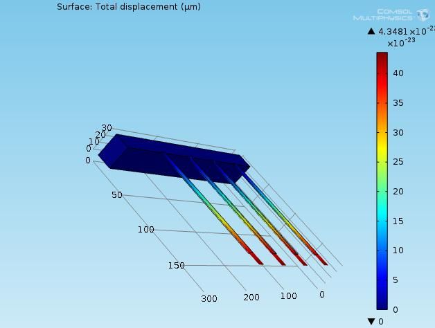
Annals of R.S.C.B., ISSN:1583-6258, Vol. 25, Issue 4, 2021, Pages. 710 - 715
      Received 05 March 2021; Accepted 01 April 2021.

                                      Fig. 1. Cantilever before and after binding[4]
III. CANTILEVER STRUCTURE
   An array of four beams in the cantilever are constructed by fixed dimensions with length 150 μm, width
10 μm and thickness 1000 nm in all the cases. A pressure of 3.146167743*10-12 Pa as input in the surface is
given and the deflection is observed. A total pressure of 5.749892771*10-12 Pa including the antibody and
antigen binding is applied in the surface of the cantilever again. It is assumed that antigens interact with all
the antibodies of the cantilever. Therefore the displacement of the cantilever will be equal to the deflection
caused     by    the    pressure     of    the     antigen    antibody      binding    of    the     cantilever.

                                               Fig 2 Structure of the Cantilever
By varying the material of the array, performance of the biosensor has been checked for maximum
displacement.
The difference in surface stress[6][7] due to molecular adsorption is given by,
Δg = (E. Δh.t2)/ (4(1-v) L2) [7]                             (1)
Where
http://annalsofrscb.ro

                                                                                                            711
Annals of R.S.C.B., ISSN:1583-6258, Vol. 25, Issue 4, 2021, Pages. 710 - 715
Received 05 March 2021; Accepted 01 April 2021.
Δg = surface stress change(N/m)
Δh = Cantilever deflection (m)
E = Young’s modulus (Pa)
v = Poisson’s ratio
t = Thickness of the cantilever (m)
L = Length of the cantilever (m)
The deflection of the cantilever varies with length, width, thickness and various other properties of the
material. The cantilever's stiffness is determined by the geometric shape and the material used to build the
cantilever.
Sensitivity of the cantilever explained in terms of the spring constant is given by the following equation.
k = (E.W.t3)/ (4L3) [8] (2)
Where,
k = Spring constant (N/m)
w = Width of the cantilever (m)
IV. SIMULATION RESULTS
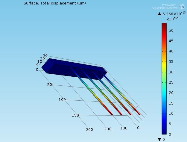
IV.I. Si
Fig 3 Si Displacement in antigen antibody binding
It displays the displacement of Si in antigen antibody binding.
http://annalsofrscb.ro
712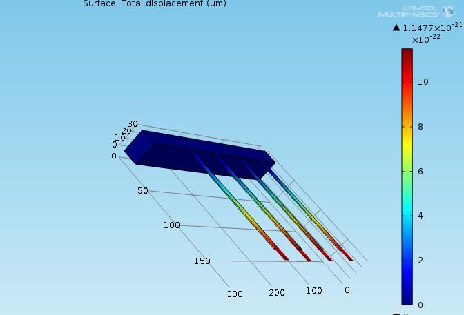
Annals of R.S.C.B., ISSN:1583-6258, Vol. 25, Issue 4, 2021, Pages. 710 - 715
      Received 05 March 2021; Accepted 01 April 2021.

IV.II. SiO2

                                Fig 4 SiO2 Displacement in antigen antibody binding
It displays the displacement of SiO2 in antigen antibody binding.

IV.III. SiC

                                 Fig 5 SiC Displacement in antigen antibody binding
It displays the displacement of SiC in antigen antibody binding.

http://annalsofrscb.ro

                                                                                      713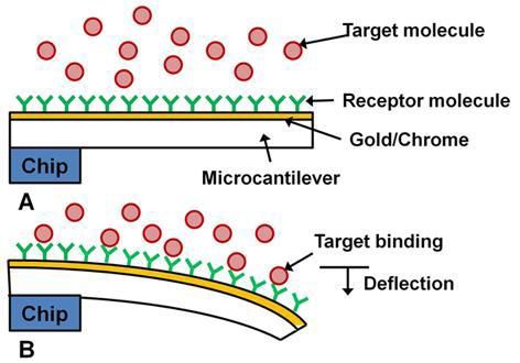
Annals of R.S.C.B., ISSN:1583-6258, Vol. 25, Issue 4, 2021, Pages. 710 - 715
        Received 05 March 2021; Accepted 01 April 2021.

IV.IV. Si3N4

                                  Fig 6 Si3N4 Displacement in antigen antibody binding
It displays the displacement of Si3N4 in antigen antibody binding.

V. DISCUSSION
From the simulation results, following displacement values have been obtained

                                      MATERIAL                            DISPLACEMENT
                                                                          (μm)
                                      Si                                  5.356 E-13
                                      SiO2                                4.3481E-22
                                      SiC                                 2.881E-22
                                      Si3N4                               1.1477E-21

Since SiO2 has the lowest Young’s Modulus of 70GPa among the four materials, the antigen-antibody
binding led to a maximum displacement in the surface thus proving the best material for sensitivity. When
the length L of the cantilever beam is increased, the spring constant k decreases and the SiO2 cantilever
beam shows increased deflection.

VI. CONCLUSION
The cantilever provides greater displacement which results in higher sensitivity when compared to the
conventional devices.Sensitivity is directly proportional to the length of cantilever surface.It is easy to detect
tuberculosis with greater accuracy.

REFERENCES
[1]   Julian W. Gardner, Vijay K. Varadan, Osama O.                             Awadelkarim, “Microsensor MEMS and Smart
      Devices” Wiley publication.

http://annalsofrscb.ro

                                                                                                                     714
Annals of R.S.C.B., ISSN:1583-6258, Vol. 25, Issue 4, 2021, Pages. 710 - 715
Received 05 March 2021; Accepted 01 April 2021.
[2] G. Wu, H. Ji, K. Hansen, T. Thundat, R. Datar, R. Cote, M. F.Hagan,A. K. Chakraborty, and
A.Majumdar, “Origin of nanomechanical cantilever motion generated from biomolecular interactions,”
Proc. Nat.Acad. Sci., vol. 98, pp. 1560–1564, 2001.
[3] Scott MacKay, David Wishart, James Z. Xing, and Jie Chen,“Developing Trends in Aptamer-Based
Biosensor Devices and Their Applications”, IEEE Transaction on biomedical circuits and
systems,vol.8,No.1,February 2014.
[4] Nireekshith yarraguntla, Hari venkata sai ram Kalla, Sai Mounika draksharapu, syed shameem,
“Detection Of Hepatitis b Virus in Serum Using Mems Based Biosensor”, International Journal of pure
and applied mathematics in vol115, nov 17.
[5] Gimzewski J K, Gerber Ch, Meyer E and Schlittler R R, Observation of the chemical reaction using a
nanomechanical sensor, Chem. Phys. Lett. 217 (1994) 589–94.
[6] Berger R, Delamarche E, Lang H P, Gerber Ch, Gimzewski J K, Meyer E and Guntherodt H-J, Surface
Stress in the Self-Assembly ofAlkanethiols on Gold, Science 276 (1997)2021–4.
[7] Ram Datar, Cantilever Sensors: NanomechanicalTools for Diagnostics, MRS Bulletin, 34, (2009).
[8] SuryanshArora, Sumati, ArtiArora, P.J.George, Design Of Mems Based Microcantilever Using
ComsolMultiphysics, International Journal of Applied Engineering Research, 17 (2012).
[9] Gulsan v , Thakare , anup nage,”Design and analysis of high sensitive biosensor using MEMS” in
IJISET ,vol2 June 2015.
http://annalsofrscb.ro
715You can also read