Evaluation of Anti-Biofilm Activity of Mouthrinses Containing Tannic Acid or Chitosan on Dentin In Situ
←
→
Page content transcription
If your browser does not render page correctly, please read the page content below
molecules
Article
Evaluation of Anti-Biofilm Activity of Mouthrinses Containing
Tannic Acid or Chitosan on Dentin In Situ
Anton Schestakow *, Moritz S. Guth, Tobias A. Eisenmenger and Matthias Hannig

                                          Clinic of Operative Dentistry, Periodontology and Preventive Dentistry, University Hospital, Saarland University,
                                          Building 73, 66421 Homburg/Saar, Germany; moritzguth@gmx.de (M.S.G.); ft.eisenmenger@web.de (T.A.E.);
                                          matthias.hannig@uks.eu (M.H.)
                                          * Correspondence: anton.schestakow@uks.eu; Tel.: +49-157-50637041

                                          Abstract: In contrast to enamel, dentin surfaces have been rarely used as substrates for studies evalu-
                                          ating the effects of experimental rinsing solutions on oral biofilm formation. The aim of the present
                                          in situ study was to investigate the effects of tannic acid and chitosan on 48-h biofilm formation on
                                          dentin surfaces. Biofilm was formed intraorally on dentin specimens, while six subjects rinsed with
                                          experimental solutions containing tannic acid, chitosan and water as negative or chlorhexidine as
                                          positive control. After 48 h of biofilm formation, specimens were evaluated for biofilm coverage and
                                          for viability of bacteria by fluorescence and scanning electron microscopy. In addition, saliva samples
                                          were collected after rinsing and analyzed by fluorescence (five subjects) and transmission electron
                                          microscopy (two subjects) in order to investigate the antibacterial effect on bacteria in a planktonic
                                          state and to visualize effects of the rinsing agents on salivary proteins. After rinsing with water,
                                          dentin specimens were covered by a multiple-layered biofilm with predominantly vital bacteria. In
                                          contrast, chlorhexidine led to dentin surfaces covered only by few and avital bacteria. By rinsing with

                                   tannic acid both strong anti-adherent and antibacterial effects were observed, but the effects declined
Citation: Schestakow, A.; Guth, M.S.;
                                          in a time-dependent manner. Transmission electron micrographs of salivary samples indicated that
Eisenmenger, T.A.; Hannig, M.             aggregation of proteins and bacteria might explain the antiadhesion effects of tannic acid. Chitosan
Evaluation of Anti-Biofilm Activity of    showed antibacterial effects on bacteria in saliva, while biofilm viability was only slightly reduced
Mouthrinses Containing Tannic Acid        and no effects on bacterial adherence on dentin were observed, despite proteins being aggregated
or Chitosan on Dentin In Situ.            in saliva after rinsing with chitosan. Tannic acid is a promising anti-biofilm agent even on dentin
Molecules 2021, 26, 1351. https://        surfaces, while rinsing with chitosan could not sufficiently prevent biofilm formation on dentin.
doi.org/10.3390/molecules26051351

                                          Keywords: tannic acid; chitosan; preventive dentistry; biofilm
Academic Editor: Beata Sadowska

Received: 4 February 2021
Accepted: 28 February 2021
                                          1. Introduction
Published: 3 March 2021
                                                Dental caries is the most prevalent chronic disease worldwide [1]. Even though caries
Publisher’s Note: MDPI stays neutral
                                          is rarely life threatening, operative treatment of carious lesions is expensive and untreated
with regard to jurisdictional claims in
                                          caries can have further impact on oral health [2,3]. Therefore, the trend is moving towards
published maps and institutional affil-   prevention now and, since caries is a multifactorial disease, there are several targets where
iations.                                  prophylaxis can take effect [4].
                                                Caries is defined as the destruction of dental hard tissues due to acids made by
                                          bacteria within a biofilm when frequently exposed to sugars [4]. Hence, biofilm control,
                                          mechanically or chemically, is an important part of caries prophylaxis [5].
Copyright: © 2021 by the authors.
                                                For mechanical biofilm control, teeth brushing with fluoridated toothpaste has been
Licensee MDPI, Basel, Switzerland.
                                          established. Both teeth brushing at high frequency and usage of higher fluoride concen-
This article is an open access article
                                          trations can reduce the incidence of caries [5,6]. It is not necessary to change established
distributed under the terms and           preventive measurements such as teeth brushing twice a day with a fluoridated toothpaste.
conditions of the Creative Commons        However, mechanical biofilm control requires compliance and manual skills, and conse-
Attribution (CC BY) license (https://     quently, it is difficult for incompliant people and mentally or physically impaired people to
creativecommons.org/licenses/by/          maintain high oral hygiene standards with mechanical biofilm control alone [7]. In this
4.0/).                                    case, chemical biofilm control can meet the demands.

Molecules 2021, 26, 1351. https://doi.org/10.3390/molecules26051351                                          https://www.mdpi.com/journal/molecules
Molecules 2021, 26, 1351                                                                                             2 of 14

                                For chemical biofilm control, chlorhexidine (CHX) is considered the gold standard [8].
                           CHX can prevent biofilm formation by a prolonged antibacterial and anti-adherent effect,
                           which is the result of CHX’s retention in the oral cavity [9–11]. However, side effects such
                           as tooth discoloration, taste irritation and irritation of the oral mucosa limit the use of
                           CHX [12]. Some alternates to CHX have already been investigated [13], of which natural
                           products have become more and more popular [14].
                                Natural products have always been of importance for drug discovery, and they have
                           been used to treat oral diseases for thousands of years [15,16]. They can inhibit caries
                           progression through an antibacterial or anti-adherent effect or by inhibition of the polysac-
                           charide synthesis. Due to technological advances, it has also become easier to identify and
                           examine active components of natural products [17], which are often polyphenols [18]. In
                           the present study, the natural substances tannic acid and chitosan were investigated.
                                Tannic acid belongs to the tannins, a group of water-soluble polyphenols that are
                           often found in plants, protecting them from herbivores and decay. Therefore, it is not
                           surprising that oak or chestnut trees, which are known for their high durability, are rich
                           in tannins [19,20]. Due to astringent properties, tannins can not only inhibit proteins that
                           are necessary for bacterial adherence [21], they also have an antibacterial effect through
                           their chelating properties [22]. In addition, tannins may also interact with membranes,
                           leading to leakage of internal contents or even to bursting of cells [23]. With regard to
                           dental biofilm formation, several in situ studies have already investigated the effects of
                           tannins showing anti-adherent, antibacterial and anti-erosive properties of tannic acid in
                           particular [24,25].
                                In contrast to tannic acid, the other test substance, chitosan, is a semi-synthetic material
                           made from chitin [26]. Chitin is a very common natural polymer and can be obtained from
                           fungi, especially from arthropods [27]. Since chitin is highly insoluble, the soluble deacety-
                           lated derivate chitosan is used for practical applications [28]. Chitosan has antibacterial
                           properties thanks to its positively charged amino groups in acidic aqueous solutions [29].
                           In the literature, chitosan is often used in different application forms or derivatives [26,30].
                           Nevertheless, mouthrinses containing water-soluble chitosan can reduce plaque indices as
                           well as bacterial viability [31,32].
                                The present study is a follow-up of the previously published study by Schestakow
                           and Hannig [33] regarding the effects of tannic acid and chitosan on biofilm formation on
                           enamel in situ. Since dentin exposure to the oral cavity is more common as the population
                           ages [34], the anti-adherent and antibacterial effect of tannic acid and chitosan was further
                           investigated on biofilm formation on dentin. For this purpose, bovine dentin specimens
                           were placed in the oral cavity for 48 h to enable biofilm formation while subjects rinsed
                           with solutions containing tannic acid or chitosan as well as water or CHX as controls. Two
                           rinsing protocols were applied, which differed in the number of rinses and thus the time
                           interval between the last rinse and the ex vivo examination allowing the investigation of
                           immediate and long-term effects by fluorescence microscopy (FM) and scanning electron
                           microscopy (SEM). In order to investigate the mode of action with regard to anti-adherent
                           and antibacterial properties, the interaction of test substances with non-adherent bacteria
                           in saliva was examined by using FM and transmission electron microscopy (TEM).

                           2. Results
                           2.1. FM Analysis of the Biofilm
                                When subjects rinsed with the negative control water, specimens were predomi-
                           nantly covered by a multiple-layered biofilm regardless of the rinsing protocol (about 4.6)
                           (Figure 1, Figure S1). Bacteria were mainly cocci; rods were less common. After rinsing
                           with tannic acid according to rinsing protocol 1, specimens were significantly less covered
                           by bacteria (2.8 ± 1), which were scattered or aggregated. Using rinsing protocol 2, spec-
                           imens were more covered by biofilm (3.8 ± 0.8) after rinsing with tannic acid compared
                           to applying rinsing protocol 1. After rinsing with chitosan, specimens were covered by
                           a multiple-layered biofilm, which did not differ significantly from the negative control,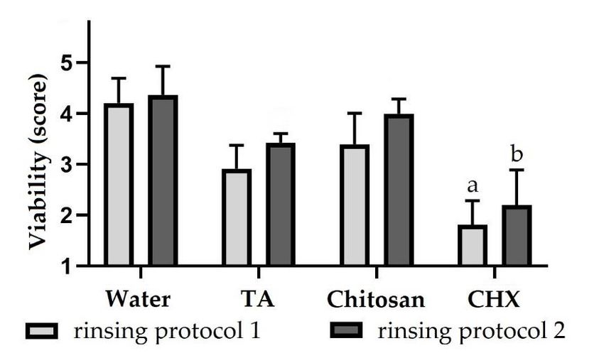
Molecules 2021, 26, x FOR PEER REVIEW 3 of 15
by bacteria (2.8 ± 1), which were scattered or aggregated. Using rinsing protocol 2, speci-
Molecules 2021, 26, 1351 mens were more covered by biofilm (3.8 ± 0.8) after rinsing with tannic acid compared 3 of to
14
applying rinsing protocol 1. After rinsing with chitosan, specimens were covered by a
multiple-layered biofilm, which did not differ significantly from the negative control, re-
gardless of the rinsing protocol (about 4.4). Both when using rinsing protocol 1 (1.9 ± 1)
regardless of the rinsing protocol (about 4.4). Both when using rinsing protocol 1 (1.9 ± 1)
and rinsing protocol 2 (1.4 ± 0.3), after rinsing with CHX, specimens were only covered by
and rinsing protocol 2 (1.4 ± 0.3), after rinsing with CHX, specimens were only covered
scattered bacteria or small bacterial aggregations. The biofilm coverage was significantly
by scattered bacteria or small bacterial aggregations. The biofilm coverage was signifi-
reduced by rinsing
cantly reduced with CHX
by rinsing withaccording to bothtorinsing
CHX according protocols
both rinsing compared
protocols to the nega-
compared to the
tive control.
negative control.
Biofilm
Figure 1.1.Biofilm
Figure coverage
coverage (score
(score 1–5)
1–5) of dentin
of dentin specimens
specimens afterafter rinsing
rinsing with with different
different rinsingrinsing
solu-
solutions.
tions. SubjectsSubjects
carriedcarried intraoral
intraoral splints
splints with with specimens
specimens for 48
for 48 h and h and
rinsed withrinsed withexperi-
different different
experimental
mental solutions.
solutions. In rinsing
In rinsing protocolprotocol 1, rinsing
1, rinsing was performed
was performed after 3after
min,312
min, 12 h,
h, 24 h, 36
24 hh,and
36 h47.5
and
h47.5
andhin
andrinsing protocol
in rinsing 2 after2 3after
protocol min,312 h, 24
min, 12 hh,and
24 h36and
h. The biofilm
36 h. formedformed
The biofilm on specimens was
on specimens
stained with with
was stained LIVE/DEAD
LIVE/DEAD® Baclight™ and evaluated
® Baclight™ usingusing
and evaluated a scoring system.
a scoring The height
system. of theofbars
The height the
corresponds to mean values and the line applied to standard deviations. Friedman
bars corresponds to mean values and the line applied to standard deviations. Friedman test followedtest followed
by Dunn’s multiple comparison test: significant differences (p < 0.05) to water are marked with ‘a’
by Dunn’s multiple comparison test: significant differences (p < 0.05) to water are marked with ‘a’ for
for rinsing protocol 1 and with ‘b’ for rinsing protocol 2. TA = tannic acid, CHX = chlorhexidine.
rinsing protocol 1 and with ‘b’ for rinsing protocol 2. TA = tannic acid, CHX = chlorhexidine.
Bacteria
Bacteria in inthe
thebiofilm
biofilmwerewerealso
alsoevaluated
evaluatedfor forviability
viabilityininorder
ordertotoinvestigate
investigate anti-
an-
bacterial
tibacterialproperties
propertiesofoftesttestsubstances.
substances.AfterAfterrinsing
rinsingwith
withwater
wateraccording
accordingto toprotocol
protocol 11
(4.2 ±±0.5)
(4.2 0.5)and (4.4 ±±0.6),
and22 (4.4 0.6),most
mostbacteria
bacteriawere
werevital
vitalin
in the
the biofilm
biofilm (Figure
(Figure 2).
2). By
By rinsing
rinsing
with
with tannic acid applying protocol 1, the viability was reduced so that the
acid applying protocol 1, the viability was reduced so that the biofilm contained biofilm con-
vital and
tained avital
vital and bacteria in equal
avital bacteria in amounts (2.9 ± (2.9
equal amounts 0.5).± However, the biofilm
0.5). However, contained
the biofilm con-
more vital
tained morebacteria after rinsing
vital bacteria with tannic
after rinsing acid according
with tannic to protocol
acid according 2 (3.42±(3.4
to protocol 0.2). The
± 0.2).
samesame
The applies to rinsing
applies with with
to rinsing chitosan (3.4 ±(3.4
chitosan 0.6 ±and ± 0.3).
0.64and For the
4 ± 0.3). Forpositive control
the positive CHX,
control
however,
CHX, the biofilm
however, consisted
the biofilm mainlymainly
consisted of avitalofbacteria, regardless
avital bacteria, of the rinsing
regardless of theprotocol
rinsing
(about 2). The viability was significantly reduced in both rinsing protocols.
protocol (about 2). The viability was significantly reduced in both rinsing protocols. The full dataset
The
of the biofilm analysis can be seen in the supplementary material (Table
full dataset of the biofilm analysis can be seen in the supplementary material (Table S1). S1).
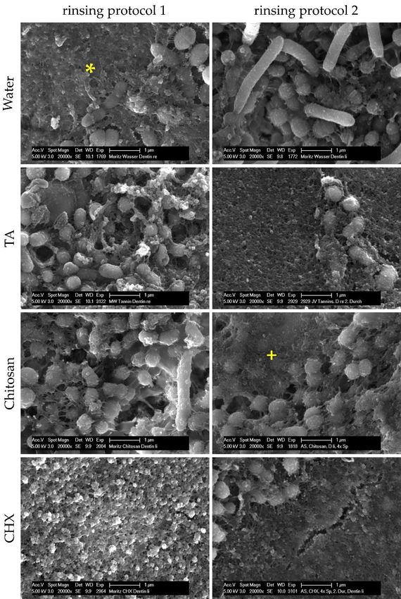
2.2. SEM Analysis of the Biofilm
Considering the results of FEM analyses, test substances have anti-adherent effects on
dental biofilm formation in situ. In order to examine how test substances exert their anti-
adherent effects, SEM was applied to investigate bacterial adherence under the influence of
test substances. After 48 h of biofilm formation and four or five rinses, specimens were
either covered by the pellicle that appeared as a layer of globular aggregates 100–200 nm
in size, or by bacteria, which were mainly cocci and a few rods (Figure 3). After rinsing
with water, tannic acid or chitosan, bacteria had an intact morphology with a globular
structured surface representing the glycocalyx or the pellicle covering bacteria. Some
bacteria had fimbriae that were linked to other bacteria, the biofilm matrix or the pellicle.
In comparison to rinsing with water or chitosan, specimens were less covered by bacteria
when subjects were rinsed with tannic acid. After rinsing with CHX, specimens were
predominantly covered by bacteria-free pellicles with an altered structure consisting of
globular agglomerates with a size of 200–500 nm. The few bacteria were isolated or in
colonies. As a result, dentinal tubules were visible more frequently than after rinsing with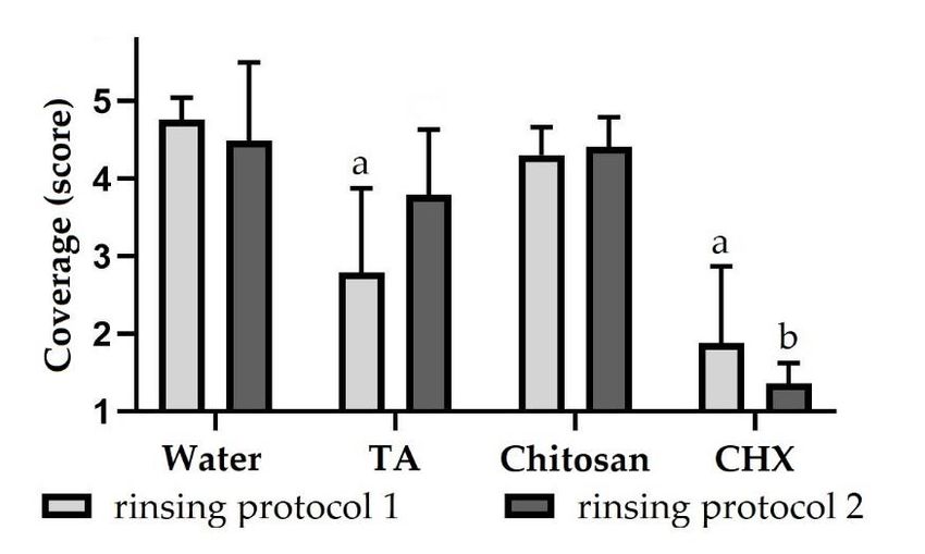
Molecules 2021, 26, 1351 4 of 14
Molecules 2021, 26, x FOR PEER REVIEW 4 of 15
water, chitosan or tannic acid, which contained thicker biofilms with characteristic water
channels. Adherent bacteria also appeared in dentinal tubules.
Figure 2.
Figure Bacterial viability
2. Bacterial viability(score
(score1–5)
1–5)ofofbiofilms
biofilmsformed
formedon ondentin
dentinspecimens
specimensafter after rinsing
rinsing with
with
rinsing solutions.
different rinsing solutions.Subjects
Subjectscarried
carried intraoral
intraoral splints
splints with
with specimens
specimens forfor
48 h48andh and rinsed
rinsed
different experimental
with different experimental solutions.
solutions. In
In rinsing
rinsing protocol
protocol1,1,rinsing
rinsingwas wasperformed
performedafter after33min,
min,12
12h,
h,
24 24
h, h,
36 36 h and
h and 47.5
47.5 h and
h and in in rinsing
rinsing protocol
protocol 2 after
2 after 3 3min,
min,1212h,h,2424h hand
and3636h.h.The
Thebiofilm
biofilmformed
formed on specimens
on specimens was stained
was stained with LIVE/DEAD
with LIVE/DEAD
® Baclight™ and evaluated using a scoring
® Baclight™ and evaluated using a scoring system.
system. The height of the bars corresponds to mean
The height of the bars corresponds to mean values and the values andlinethe applied
line applied to standard
to standard devia-
deviations.
tions. Friedman test followed by Dunn’s multiple comparison test: significant differences (p < 0.05)
Friedman test followed by Dunn’s multiple comparison test: significant differences (p < 0.05) to
to water are marked with a for rinsing protocol 1 and with b for rinsing protocol 2. TA = tannic
water are marked with a for rinsing protocol 1 and with b for rinsing protocol 2. TA = tannic acid,
acid, CHX = chlorhexidine.
CHX = chlorhexidine.
2.2.
2.3. SEM AnalysisofofSaliva
FM Analysis the Biofilm
Samples
Considering
In order to further clarifyofthe
the results FEM analyses, test
antibacterial substances
effects of tannichave
acid,anti-adherent effects
chitosan or CHX, as
on dental biofilm formation in situ. In order to examine how test substances
observed on biofilms by FM, saliva samples with non-adherent bacteria were investigated exert their
anti-adherent
for viability aftereffects, SEMwith
rinsing was different
applied totest
investigate bacterial
substances. Afteradherence
rinsing withunder
the the influ-
negative
ence of water,
control test substances. After
the viability 48 h of biofilm
of bacteria was over formation and fouroforwhether
70%, regardless five rinses, specimens
saliva samples
were either
were covered
collected 1 min,by30theminpellicle
or 2 hthat appeared
after rinsing as a layer
(Figure 4, of globular
Figure S2). aggregates
Viability of100–
the
200 nm in size, or by bacteria, which were mainly cocci and a few rods (Figure
salivary bacteria was reduced 1 min after rinsing with tannic acid (47 ± 6). The antibacterial 3). After
rinsing with water,
effect declined aftertannic
30 minacid
(64or±chitosan, bacteria the
22) and reached hadvalue
an intact morphology
of the with a glob-
negative control after
ular structured
2 h (75 ± 13). The surface
samerepresenting the glycocalyx
applies to rinsing or the(42
with chitosan pellicle covering
± 17 and 61 ± bacteria.
14 and 75Some± 9)
bacteria
with thehad fimbriae that
antibacterial were
effect linked tobeing
of chitosan otherslightly
bacteria,stronger
the biofilmthanmatrix or the
of tannic pellicle.
acid. One
In
mincomparison
(33 ± 8) and to rinsing
30 minwith(33 ±water or chitosan,
8) after specimens
rinsing with were lesswere
CHX, bacteria covered by bacteria
predominantly
when
avital.subjects
After 2 h,were
aboutrinsed
half with
of thetannic acid.
bacteria After
were rinsing
vital againwith
(53 ± CHX, specimens
12). In comparisonwere topre-
the
dominantly covered
negative control, CHX bysignificantly
bacteria-freereduced
pelliclesthe
with an altered
viability afterstructure
1 min, 30consisting
min and 2ofh.glob-
The
ular agglomerates
full dataset with aanalysis
of the saliva size of 200–500 nm. The
can be seen few
in the bacteria were material
supplementary isolated or in colonies.
(Table S2).
As a result, dentinal tubules were visible more frequently than after rinsing with water,
2.4. TEMor
chitosan Analysis
tannic of Saliva
acid, Samples
which contained thicker biofilms with characteristic water chan-
nels. An antibacterial
Adherent bacteria effect
alsoof tannic acid,
appeared chitosantubules.
in dentinal and CHX on non-adherent bacteria in
saliva was examined quantitatively by FM. In comparison to FM, ultrastructural alterations
of both bacteria and saliva can be visualized with TEM and, therefore, TEM was used to
clarify the mechanism of action. One min, 30 min and 2 h after rinsing with the negative
control water, intact bacteria were present, which were mainly cocci and a few rods with
fimbriae covering the bacterial surface (Figure 5). Cleavage furrows were also visible,
suggesting that bacteria were undergoing cell division at the time of fixation. In addition
to bacteria, loose filamentous structures were detected representing salivary proteins. The
bacteria were often found close to proteins or adsorbed to epithelial cells. When subjects
rinsed with tannic acid, globular electron-dense structures appeared, which is the result
of protein aggregation or formation of tannic acid-protein complexes. In particular, 1 min
after rinsing with tannic acid, irregular shapes of bacteria were detected in contrast to the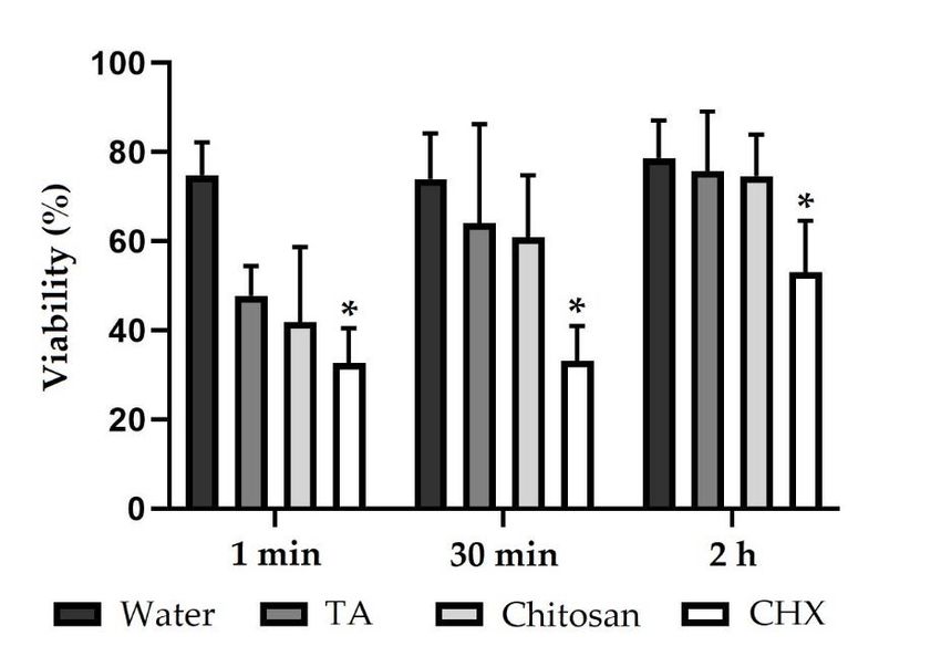
Molecules 2021, 26, 1351                                                                                                                          5 of 14

                                       round and plump shape representing intact morphology. Furthermore, the bacterial cell
                                      wall had a higher electron density. Similar to tannic acid, protein aggregates were also
                                       visible with rinsing solutions containing chitosan, especially 1 min after rinsing. However,
                                       proteins were not aggregated into dense clusters as they were with tannic acid. Occasionally,
                                       cell remnants of bacteria were found within the protein networks. When subjects rinsed
                                      with the positive control CHX, cell lysis was frequently observed 1 min and 30 min after
                                       rinsing. CHX agglomerates appeared as globular electron-dense structures 5predominantly
     Molecules 2021, 26, x FOR PEER REVIEW                                                                             of 15
                                       adsorbing and covering bacterial surfaces.

                                 Figure 3. SEM images of dentin specimens at 20,000-fold magnification. Subjects carried intraoral
                                Figure
                                 splints 3.
                                         withSEM   images
                                                dentin       of dentin
                                                       specimens   for 48specimens
                                                                          h and rinsedatwith
                                                                                          20,000-fold    magnification.
                                                                                              different experimental          Subjects
                                                                                                                        solutions.       carried intraoral
                                                                                                                                    In rinsing
                                splints
                                 protocolwith    dentin
                                           1, rinsing wasspecimens
                                                           performedfor    483hmin,
                                                                        after     and12rinsed
                                                                                        h, 24 h,with  different
                                                                                                 36 h and  47.5 h experimental     solutions.
                                                                                                                  and in rinsing protocol    2 In rinsing
                                 after 3 min,
                                protocol       12 h, 24 hwas
                                            1, rinsing    andperformed
                                                               36 h. Specimens
                                                                             afterwere either
                                                                                   3 min,   12covered
                                                                                               h, 24 h,by36the
                                                                                                             h pellicle
                                                                                                               and 47.5 (*) h
                                                                                                                            orand
                                                                                                                               by bacteria
                                                                                                                                   in rinsing protocol 2
                                 that were mostly cocci and a few rods. The bacteria were partially covered by the pellicle (+). Al-
                                after 3 min, 12 h, 24 h and 36 h. Specimens were either covered by the pellicle (*) or by bacteria that
                                 terations of the pellicle structure occurred after rinsing with chlorhexidine.
                                were mostly cocci and a few rods. The bacteria were partially covered by the pellicle (+). Alterations
                                of2.3.
                                    theFM
                                        pellicle  structure
                                           Analysis           occurred
                                                      of Saliva Samples after rinsing with chlorhexidine.
                                      In order to further clarify the antibacterial effects of tannic acid, chitosan or CHX, as
                                 observed on biofilms by FM, saliva samples with non-adherent bacteria were investigated
                                 for viability after rinsing with different test substances. After rinsing with the negative
2 h (75 ± 13). The same applies to rinsing with chitosan (42 ± 17 and 61 ± 14 and 75 ± 9)
with the antibacterial effect of chitosan being slightly stronger than of tannic acid. One
min (33 ± 8) and 30 min (33 ± 8) after rinsing with CHX, bacteria were predominantly
avital. After 2 h, about half of the bacteria were vital again (53 ± 12). In comparison to the
Molecules 2021, 26, 1351 negative control, CHX significantly reduced the viability after 1 min, 30 min and 2 6h.ofThe
14
full dataset of the saliva analysis can be seen in the supplementary material (Table S2).
Figure 4. Viability (%) of bacteria in saliva samples. Subjects rinsed with different experimental
Figure 4. Viability (%) of bacteria in saliva samples. Subjects rinsed with different experimental
solutions.
solutions.Saliva
Salivasamples
sampleswere
were collected
collected 11 min,
min, 30 min and
30 min and 22 hh after
afterrinsing.
rinsing.Saliva
Salivabacteria
bacteriawere
were
cules 2021, 26, x FOR PEER REVIEW ® Baclight™, and viability was evaluated using ImageJ 7 of 15
stained
stained with LIVE/DEAD® Baclight™, and viability was evaluated using ImageJ software. TheThe
with LIVE/DEAD software.
height
heightofofthe
thebars
barscorresponds
corresponds toto
mean
mean values and
values thethe
and line applied
line appliedto standard deviations.
to standard Friedman
deviations. Fried-
test
man followed by Dunn’s
test followed multiple
by Dunn’s comparison
multiple test: test:
comparison significant differences
significant (pMolecules 2021, 26, 1351 7 of 14
3. Discussion
Considering fluorescence microscopic analysis of the intraorally formed biofilm, rins-
ing solutions containing tannic acid, chitosan or CHX have an anti-adherent and antibacte-
rial effect compared to the negative control water, with CHX showing the strongest and
chitosan the weakest anti-biofilm effect. The antibacterial effect is the result of a disruption
of membrane integrity, as shown by TEM. Furthermore, after rinsing with tannic acid,
chitosan or CHX, ultrastructural alterations appeared in terms of protein aggregations and
complexes that were also shown by SEM when rinsing with CHX caused alterations of the
pellicle structure.
In the present study, tannic acid and native chitosan were tested for their anti-adherent
and antibacterial effect on dental biofilm formation, since chitosan and polyphenols in
general have often been the subject of medical research.
Tannic acid belongs to tannins, which are a subgroup of polyphenols that are known
for their antibacterial activity due to chelating properties or interactions with the bacterial
cell membrane when applied in high concentrations [22,35]. The antibacterial effect of
tannic acid in particular was already investigated on dental biofilm formation. However,
biofilm was either formed on enamel or biofilm formation time was low [24,25,33]. Since
dentin is increasingly exposed to the oral cavity due to the aging population and the decline
in edentulous adults [34], dentin specimens were used in the present study. When subjects
rinsed with tannic acid, the viability of both non-adherent bacteria in the planktonic state
and bacteria in biofilm was reduced. In order to investigate the duration of action, saliva
samples were collected 1 min, 30 min and 2 h after rinsing with experimental solutions and
for biofilm formation experiments two different rinsing protocols were applied. According
to rinsing protocol 1, the last rinse was shortly before the ex vivo examination, and thus, the
immediate effect on biofilm was investigated. For rinsing protocol 2, the last rinse occurred
12 h prior to the ex vivo examination, and thus, the long-term effect was examined. In
view of this, the antibacterial effect decreased in a time-dependent manner, indicating poor
retention of tannic acid in the oral cavity. Considering TEM analyses of saliva, tannic acid
led to alterations of bacterial morphology, and thus, the antibacterial effect may be due
to interaction of tannins with the bacterial membrane, as suggested by Tamba et al. [23],
resulting in osmotic dysregulation and finally in cell death.
In addition to the morphological alterations, the formation of globular and electron-
dense protein aggregations in saliva was observed by TEM, which occurred after 1 min
and 30 min after rinsing with tannic acid. Since salivary proteins are used as receptors
for initial bacterial adherence, precipitation of those proteins with the so-called tanning
effect can explain the anti-adherent properties of tannic acid [36–38]. Tannins can also
inhibit glycosyl-transferase, and thereby, glucan synthesis, which is used for bacterial
adherence [39]. The same applies to bacterial fimbriae with which tannins can interact, as
reported by Sakanaka et al. [40]. Furthermore, aggregations of bacteria can be observed in
the presence of tannins [41], and therefore, bacteria may no longer adsorb to oral surfaces
and are swallowed instead. Regarding the anti-adherent properties of tannic acid in
particular, so far, one study showed an effect on biofilm formation on dentin that was
exposed to the oral cavity [25]. In a study by Xi et al. [25], participants rinsed with a
solution containing tannic acid (1%) twice a day; the biofilm was formed for 24 h. In the
present study, when subjects rinsed with tannic acid (5%) and the biofilm formation time
was 48 h, the anti-adherent effect of tannic acid was confirmed. However, 12 h after the last
rinsing, as simulated by rinsing protocol 2, the effect was lower than in rinsing protocol 1,
indicating a low substantivity of tannic acid.
In summary, rinsing with experimental solutions containing tannic acid inhibits
biofilm formation. However, it is unclear to what extent tannic acid can disrupt an estab-
lished biofilm. Furthermore, the exact mechanism of action of antibacterial effects of tannic
acid in particular is still not fully clarified and should be further investigated.
The other test substance, chitosan, has been supposed as a promising anti-biofilm
agent according to the literature [31,42–46], in which different chitosan derivatives orMolecules 2021, 26, 1351 8 of 14
different application forms were investigated. However, the effect of native chitosan
on intraoral biofilm formation on dentin has not been investigated yet. Chitosan has
antibacterial properties due to positively charged groups. As a result, chitosan can disrupt
the membrane integrity of bacteria or chelate metal ions [29,47]. Although chitosan did
not lead to visible membrane interactions in the present study as shown by TEM, an
antibacterial effect on non-adherent bacteria in saliva was observed especially 1 min after
subjects rinsed with chitosan. The same applies to bacteria in the biofilm, but compared to
the negative control, the antibacterial effect was very low and was only present in rinsing
protocol 1. Applying rinsing protocol 2, on the other hand, no antibacterial effects were
observed, which is in accordance with the results on bacteria in saliva 30 min and 2 h after
rinsing with chitosan speaking for a low retention of chitosan in the oral cavity. Although
a previous study on enamel led to similar results [33], the short duration of action of
chitosan was not expected, since chitosan can adsorb to both buccal cells and the dental
pellicle [46,48]. There are several factors that may lead to the limited activity of chitosan.
The low pH value needed to dissolve chitosan and chitosan itself impart a positive charge
to the pellicle in vitro [49,50] and, as suggested by Rehage et al. [51], this observation may
inhibit further the accumulation of chitosan on dental surfaces. Furthermore, the salivary
protein lysozyme, which is present in the pellicle maintaining its enzymatic activity, can
degrade chitosan, and thus, inhibit its antibiofilm properties [52–55].
Considering transmission electron micrographs in the present study, rinsing agents
containing chitosan led to the aggregation of proteins and bacteria due to the polycationic
nature of chitosan [29,50]. Although the treatment of the pellicle with chitosan showed
an anti-adherent effect in vitro [46,50], the anti-adherent properties of chitosan were not
confirmed in the present in situ study, which is in accordance with a previous in situ study
on enamel [33].
In contrast to tannic acid and chitosan, rinsing with the positive control CHX resulted
in significant anti-adherent and antibacterial effects on biofilm formation on dentin regard-
less of the rinsing protocol as well as significant reduction of viability of non-adherent
bacteria in saliva 1 min, 30 min and 2 h after rinsing with CHX. As a polycation, CHX
can interact with the bacterial membrane, and thus, disrupt membrane integrity and cell
metabolism [12]. The antibacterial effects were only observed by FM, but neither by SEM
nor TEM. In the in vitro study by Vitkov et al. [56] when CHX was applied to saliva samples
for 1 or 5 min, a loss of bacterial membrane integrity was visualized. In the present in situ
study, however, no alterations occurred as shown by TEM, which may be due to the low
concentration or the short rinsing time of only 30 s.
Unlike the other rinsing agents tested, alterations of the pellicle structure were detected
by SEM. After rinsing with CHX, globular agglomerates with a size of 200–500 nm appeared.
Since the polycation CHX can bind to negatively charged groups of salivary proteins, it
is suggested that these agglomerates represent chlorhexidine-protein complexes in the
pellicle leading to reduction of bacterial adherence [12,57]. In addition to adsorption to the
pellicle, CHX also absorbs to other oral surfaces resulting in a high substantivity of CHX
in the oral cavity [9–11]. When assessing significance, primarily CHX showed significant
results. According to G*Power software, at least 12 subjects would be required to detect an
80% reduction in biofilm coverage or viability with a power of 80%. In the present study, six
subjects participated. Subjects would be easier to hire when the number of rinsing solutions
is reduced. The aim of the present study was also to show which substances actually
work on dentin specimens. With the new findings, follow-up experiments concentrating
specifically on one solution with a higher number of participants can be carried out.
4. Materials and Methods
4.1. Subjects and Test Substances
Six volunteers (aged 24–30 years) participated in the present study, which uses a
cross-over design. All subjects were dental students who neither had caries nor periodontalMolecules 2021, 26, 1351 9 of 14
diseases; they did not smoke nor take any drugs. The study was approved by the Medical
Ethic Committee of the Medical Association of Saarland (238/03, 2016).
Subjects rinsed with four different mouth rinses. The washout phase was at least
one whole day for all experiments, according to the substantivity of the positive control
and the previous study on enamel [12,33,58]. Sterile water (Ampuwa® , Fresenius Kabi,
Bad Homburg, Germany) and chlorhexidine-digluconat (0.2%) (Apotheke des Universität-
sklinikum des Saarlandes, Homburg, Germany) were used as negative and positive control.
Both tannic acid (Tannic Acid, Sigma® , Saint Louis, USA) and chitosan (Chitosan 95/3000,
Heppe Medical Chitosan GmbH, Halle, Germany) were solids and had to be dissolved
first. For 100 mL of a tannic acid solution (5%), sterile water was added to 5 g of tannic
acid. To dissolve chitosan, sterile water was added to 5 g of chitosan and 3.5 mL of acetic
acid to get a 1000 mL solution (0.5%). The chitosan used had a degree of deacetylation of
≥ 92.6% and a molecular weight of 300–700 kDa.
4.2. Specimens for Biofilm Formation
To investigate the effect on biofilm formation, six subjects carried upper jaw splints
(DURAN® , Scheu Dental GmbH, Iserlohn, Deutschland) with dentin specimens that were
fixed buccally with silicone impression material (PRESIDENT light body, Coltène/Whaledent
GmbH + Co. KG, Langenau, Germany). Dentin specimens were made from bovine teeth
from two-year-old cattle from the slaughterhouse in Zweibrücken by using a cut-off and
wet grinding machine. They had a rectangular form with a surface of 5 × 5 mm2 and
thickness of 1 mm and were ground and polished up to 2500 grit. The superficial smear
layer was removed by ultrasonication with NaOCl (3%) for 30 s. Then, specimens were
cleaned with distilled water and disinfected with isopropyl alcohol (70%) for 15 min before
rehydrating in sterile water for 6 h [25].
4.3. Biofilm Formation In Situ
Four specimens were fixed to the splints, which were carried in the oral cavity to allow
biofilm formation. Splints were in situ for 48 h, since biofilm thickness and viability are
less susceptible to intraindividual differences in terms of the location of specimens [59,60].
Subjects rinsed four or five times with 10 mL of the different test substances for 30 s, as
generally recommended for dental prophylaxis [61]. Two rinsing protocols were applied.
In rinsing protocol 1, rinsing occurred 3 min, 12 h, 24 h, 36 h and 47.5 h after insertion of
the splints. The last rinse was shortly before the ex vivo examination. In rinsing protocol
2, subjects rinsed only after 3 min, 12 h, 24 h and 36 h. During the trial, subjects had to
temporarily take off the splints when they wanted to eat or brush their teeth, but usage of
toothpaste or other mouth rinses were not allowed. After 48 h, splints were removed from
the oral cavity and specimens were dismounted and rinsed with sterile water in order to
remove non-adherent bacteria and salivary remnants. Specimens were then prepared for
FM and SEM (Figure 6).
4.4. FM Analysis of the Biofilm
Two of the four specimens were stained with LIVE/DEAD® BacLight™ Bacterial
Viability Kit L7012 (Invitrogen, Molecular Probes, Eugene, OR, USA) for 10 min and then
examined with FM (Axio Imager.M2, CarlZeiss Microscopy GmbH, Jena, Deutschland)
using a fluorescein diacetate (Sigma, St. Louis, MO, USA) and an ethidium bromide filter
(Roth, Mannheim, Deutschland) [33]. Six pictures of each specimen were taken, which
were evaluated by two investigators for coverage and viability using a scoring system
(Tables 1 and 2).had to temporarily take off the splints when they wanted to eat or brush their teeth, but
usage of toothpaste or other mouth rinses were not allowed. After 48 h, splints were re-
moved from the oral cavity and specimens were dismounted and rinsed with sterile water
in order to remove non-adherent bacteria and salivary remnants. Specimens were then
Molecules 2021, 26, 1351 prepared for FM and SEM (Figure 6). 10 of 14
Figure6.6.Flow
Figure Flowchart
chartof
ofthe
thein
insitu
situexperiments.
experiments.
4.4. FM Analysis of the Biofilm
Table 1. Modified scoring for biofilm coverage according to Xi et al. [25].
Score Definition
1 Pellicle with no or scattered bacteria
2 Few and small bacterial aggregations, dozens of bacteria
3 Multiple bacterial aggregations, hundreds of bacteria
4 Monolayer biofilm or biofilm covering 50% of the surface
Table 2. Scoring for biofilm viability according to Nobre et al. [62].
Score Definition
Primarily red fluorescent bacteria,
1
Ratio of red to green fluorescent bacteria is 90:10 or more
2 Ratio of red to green fluorescent bacteria is about 75:25
3 Ratio of red to green fluorescent bacteria is about 50:50
4 Ratio of red to green fluorescent bacteria is about 25:75
Primarily green fluorescent bacteria,
5
ratio of red to green fluorescent bacteria 10:90 or lowerMolecules 2021, 26, 1351 11 of 14
4.5. SEM Analysis of the Biofilm
The other two specimens were prepared for SEM. First, specimens were fixed in a
solution consisting of 2% glutaraldehyde and 0.1 M cacodylate buffer for at least 1 h. Then,
specimens were washed in cacodylate buffer, dehydrated in an ascending alcohol series
and dried with hexamethyldisilazane. After air drying overnight, specimens’ surface was
coated with carbon and examined for its morphology with a magnification of up to 20,000
using SEM (XL 30 ESEM FEG, FEI Company, Eindhoven, The Netherlands).
4.6. FM Analysis of Saliva Samples
Five subjects rinsed with 10 mL of the different test substances for 30 s, and the
unstimulated saliva was collected after 1 min, 30 min and 2 h in an Eppendorf tube.
Samples were centrifuged for 10 min at 1000 rpm and the supernatant was centrifuged
again for 10 min at 10,000 rpm. The bacterial pellet was stained with LIVE/DEAD®
BacLight™ for 15 min and examined with FM. Eight pictures were taken, and the viability
of bacteria was evaluated using the software ImageJ 1.52 (NIH, Bethesda, MD, USA).
4.7. TEM Analysis of Saliva Samples
In order to visualize the interaction of test substances with bacteria, the saliva of two
subjects was additionally examined with TEM. The subjects rinsed for 30 s with 10 mL of
a test substance and their unstimulated saliva was collected after 1 min, 30 min and 2 h
in an Eppendorf tube. The samples were centrifuged at 5000 rpm and the bacterial pellet
was fixed in a fixing solution consisting of 1% formaldehyde, 1% glutaraldehyde and 0.1 M
cacodylate buffer for 90 min. Then, samples were postfixed with 2% osmium for 1 h and
pre-embedded in low-melting agarose. After dehydration in an ascending alcohol series,
samples were embedded in araldite (Araldit CY212, Agar Scientific Ltd., Stansted, UK).
Ultrathin sections of the embedded samples were cut in an ultramicrotome (Leica EM UC7,
Leica Microsystems, Wetzlar, Germany). The sections were contrasted with UranyLess
(UranyLess EM Stain, Delta Microscopies, Mauressac, France) and 3% lead citrate before
investigated by transmission electron microscopy (TEM Tecnai 12 BioTwin, FEI Company,
Eindhoven, Netherlands) at magnifications of up to 68,000-fold.
4.8. Statistical Analysis
The results of FM analyses were tested statistically. First, data were examined for
normal distribution using the Shapiro-Wilk test. They were not normally distributed
(p < 0.05). Statistical differences of the test substances to the negative control were tested
with the Friedmann test (p = 0.05) followed by Dunn’s multiple comparison test. Differences
between both rinsing protocols were tested with the Wilcoxon test (one-tailed). Bonferroni
adjustments were conducted (p = 0.05/4 = 0.0125). Statistical analyses were performed
with the GraphPad Prism 8 software (GraphPad Software, San Diego, CA, USA).
5. Conclusions
In conclusion, rinsing agents containing tannic acid reduced the bacterial viability
and adherence to dentin specimens in situ due to interactions with bacterial membranes
and proteins. Therefore, tannic acid is a promising anti-biofilm agent. On the other hand,
rinsing with chitosan resulted in antibacterial effects on non-adherent bacteria in saliva
and bacteria in the biofilm, but the antibacterial effect on biofilm formation was low and
no anti-adherent properties were observed.
Supplementary Materials: The following are available online. Figure S1: fluorescence micrographs
biofilm, Figure S2: fluorescence micrographs saliva, Table S1: dataset biofilm, Table S2: dataset saliva.
Author Contributions: Conceptualization, M.H.; methodology, M.H.; validation, A.S., M.S.G. and
T.A.E.; formal analysis, A.S.; investigation, A.S., M.S.G. and T.A.E.; writing—original draft prepa-
ration, A.S.; writing—review and editing, M.H.; visualization, A.S.; supervision, M.H.; projectMolecules 2021, 26, 1351 12 of 14
administration, M.H.; funding acquisition, M.H. All authors have read and agreed to the published
version of the manuscript.
Funding: This research was funded by Deutsche Forschungsgemeinschaft, grant number SFB 1027.
Institutional Review Board Statement: The study was approved by the Medical Ethic Committee
of the Medical Association of Saarland (238/03, 2016).
Informed Consent Statement: Informed consent was obtained from all subjects involved in the study.
Data Availability Statement: The data presented in this study are available in the
supplementary materials.
Acknowledgments: The authors would like to thank S. König and N. Pütz for the excellent support
in the laboratory.
Conflicts of Interest: The authors declare no conflict of interest. The funders had no role in the design
of the study; in the collection, analyses or interpretation of data; in the writing of the manuscript, or
in the decision to publish the results.
Sample Availability: Samples for fluorescence microscopy can only be investigated once and are dis-
carded afterwards. Samples for scanning electron microscopy and transmission electron microscopy
presented in this study are available on request from the corresponding author.
References
1. Kassebaum, N.J.; Smith, A.G.C.; Bernabé, E.; Fleming, T.D.; Reynolds, A.E.; Vos, T.; Murray, C.J.L.; Marcenes, W. Global, Regional,
and National Prevalence, Incidence, and Disability-Adjusted Life Years for Oral Conditions for 195 Countries, 1990–2015: A
Systematic Analysis for the Global Burden of Diseases, Injuries, and Risk Factors. J. Dent. Res. 2017, 96, 380–387. [CrossRef]
2. Hu, D.; Hong, X.; Li, X. Oral Health in China—Trends and Challenges. Int. J. Oral Sci. 2011, 3, 7–12. [CrossRef] [PubMed]
3. Niessen, L.C.; Weyant, R.J. Causes of Tooth Loss in a Veteran Population. J. Public Health Dent. 1989, 49, 19–23. [CrossRef]
[PubMed]
4. Selwitz, R.H.; Ismail, A.I.; Pitts, N.B. Dental Caries. Lancet 2007, 369, 51–59. [CrossRef]
5. Figuero, E.; Nobrega, D.F.; García-Gargallo, M.; Tenuta, L.M.; Herrera, D.; Carvalho, J.C. Mechanical and Chemical Plaque
Control in the Simultaneous Management of Gingivitis and Caries: A Systematic Review. J. Clin. Periodontol. 2017, 44, S116–S134.
[CrossRef]
6. Marinho, V.C.; Higgins, J.; Logan, S.; Sheiham, A. Fluoride Toothpastes for Preventing Dental Caries in Children and Adolescents.
Cochrane Database Syst. Rev. 2003. [CrossRef] [PubMed]
7. Löe, H. Oral Hygiene in the Prevention of Caries and Periodontal Disease. Int. Dent. J. 2000, 50, 129–139. [CrossRef] [PubMed]
8. Kour, K.; Kaur, S.; Singh, P. Comparative Evaluation of the Efficacy of Chlorohexidine Mouthwash as a Supplement to Regular
Tooth Brushing. Int. J. Oral Health Dent. 2019, 5, 97–103. [CrossRef]
9. Bonesvoll, P.; Lökken, P.; Rölla, G.; Paus, P.N. Retention of Chlorhexidine in the Human Oral Cavity after Mouth Rinses. Arch.
Oral Biol. 1974, 19, 209–212. [CrossRef]
10. Rölla, G.; Melsen, B. On the Mechanism of the Plaque Inhibition by Chlorhexidine. J. Dent. Res. 1975, 54, 57–62. [CrossRef]
11. Reda, B.; Hollemeyer, K.; Trautmann, S.; Hannig, M.; Volmer, D.A. Determination of Chlorhexidine Retention in Different Oral
Sites Using Matrix-Assisted Laser Desorption/Ionization-Time of Flight Mass Spectrometry. Arch. Oral Biol. 2020, 110, 104623.
[CrossRef]
12. Mathur, S.; Mathur, T.; Srivastava, R.; Khatri, R. Chlorhexidine: The Gold Standard in Chemical Plaque Control. Natl. J. Physiol.
Pharm. Pharmacol. 2011, 1, 45.
13. Marsh, P.D. Controlling the Oral Biofilm with Antimicrobials. J. Dent. 2010, 38, S11–S15. [CrossRef]
14. Groppo, F.C.; Bergamaschi, C.d.C.; Cogo, K.; Franz-Montan, M.; Motta, R.H.L.; Andrade, E.D. Use of Phytotherapy in Dentistry.
Phytother. Res. 2008, 22, 993–998. [CrossRef]
15. Newman, D.J.; Cragg, G.M. Drugs and Drug Candidates from Marine Sources: An Assessment of the Current “State of Play”.
Planta Med. 2016, 82, 775–789. [CrossRef] [PubMed]
16. Lewis, W.H.; Elvin-Lewis, M.P. Medical Botany: Plants Affecting Human Health, 2nd ed.; John Wiley & Sons: Hoboken, NJ, USA,
2003; ISBN 0-471-62882-4.
17. Jeon, J.-G.; Rosalen, P.; Falsetta, M.; Koo, H. Natural Products in Caries Research: Current (Limited) Knowledge, Challenges and
Future Perspective. Caries Res. 2011, 45, 243–263. [CrossRef]
18. Cheng, L.; Li, J.; He, L.; Zhou, X. Natural Products and Caries Prevention. Caries Res. 2015, 49, 38–45. [CrossRef] [PubMed]
19. Haslam, E. Natural Polyphenols (Vegetable Tannins) as Drugs: Possible Modes of Action. J. Nat. Prod. 1996, 59, 205–215.
[CrossRef] [PubMed]
20. Scalbert, A. Antimicrobial Properties of Tannins. Phytochemistry 1991, 30, 3875–3883. [CrossRef]Molecules 2021, 26, 1351 13 of 14
21. Wu-Yuan, C.D.; Chen, C.Y.; Wu, R.T. Gallotannins Inhibit Growth, Water-Insoluble Glucan Synthesis, and Aggregation of Mutans
Streptococci. J. Dent. Res. 1988, 67, 51–55. [CrossRef]
22. Chung, K.-T.; Wong, T.Y.; Wei, C.-I.; Huang, Y.-W.; Lin, Y. Tannins and Human Health: A Review. Crit. Rev. Food Sci. Nutr. 1998,
38, 421–464. [CrossRef]
23. Tamba, Y.; Ohba, S.; Kubota, M.; Yoshioka, H.; Yoshioka, H.; Yamazaki, M. Single GUV Method Reveals Interaction of Tea
Catechin (−)-Epigallocatechin Gallate with Lipid Membranes. Biophys. J. 2007, 92, 3178–3194. [CrossRef] [PubMed]
24. Hertel, S.; Pötschke, S.; Basche, S.; Delius, J.; Hoth-Hannig, W.; Hannig, M.; Hannig, C. Effect of Tannic Acid on the Protective
Properties of the in Situ Formed Pellicle. Caries Res. 2017, 51, 34–45. [CrossRef] [PubMed]
25. Xi, Q.; Hoth-Hannig, W.; Deng, S.; Jin, X.; Fu, B.; Hannig, M. The Effect of Polyphenol-Containing Solutions on in Situ Biofilm
Formation on Enamel and Dentin. J. Dent. 2020, 102, 103482. [CrossRef]
26. Kumar, M.N.V.R. A Review of Chitin and Chitosan Applications. React. Funct. Polym. 2000, 46, 1–27. [CrossRef]
27. Periayah, M.H.; Halim, A.S.; Saad, A.Z.M. Chitosan: A Promising Marine Polysaccharide for Biomedical Research. Pharmacogn.
Rev. 2016, 10, 39. [CrossRef]
28. Wieckiewicz, M.; Boening, K.W.; Grychowska, N.; Paradowska-Stolarz, A. Clinical Application of Chitosan in Dental Specialities.
Mini Rev. Med. Chem. 2017, 17, 401–409. [CrossRef]
29. Fei Liu, X.; Lin Guan, Y.; Zhi Yang, D.; Li, Z.; de Yao, K. Antibacterial Action of Chitosan and Carboxymethylated Chitosan. J.
Appl. Polym. Sci. 2001, 79, 1324–1335. [CrossRef]
30. Dash, M.; Chiellini, F.; Ottenbrite, R.M.; Chiellini, E. Chitosan—A Versatile Semi-Synthetic Polymer in Biomedical Applications.
Prog. Polym. Sci. 2011, 36, 981–1014. [CrossRef]
31. Bae, K.; Jun, E.J.; Lee, S.M.; Paik, D.I.; Kim, J.B. Effect of Water-Soluble Reduced Chitosan on Streptococcus Mutans, Plaque
Regrowth and Biofilm Vitality. Clin. Oral Investig. 2006, 10, 102. [CrossRef] [PubMed]
32. Pasquantonio, G.; Greco, C.; Prenna, M.; Ripa, C.; Vitali, L.A.; Petrelli, D.; di Luca, M.C.; Ripa, S. Antibacterial Activity and
Anti-Biofilm Effect of Chitosan against Strains of Streptococcus Mutans Isolated in Dental Plaque. Int. J. Immunopathol. Pharmacol.
2008, 21, 993–997. [CrossRef] [PubMed]
33. Schestakow, A.; Hannig, M. Effects of Experimental Agents Containing Tannic Acid or Chitosan on the Bacterial Biofilm Formation
in Situ. Biomolecules 2020, 10, 1315. [CrossRef] [PubMed]
34. McKenna, G.; Tsakos, G.; Burke, F.; Brocklehurst, P. Managing an Ageing Population: Challenging Oral Epidemiology. Prim.
Dent. J. 2020, 9, 14–17. [CrossRef]
35. Petti, S.; Scully, C. Polyphenols, Oral Health and Disease: A Review. J. Dent. 2009, 37, 413–423. [CrossRef]
36. Joiner, A.; Muller, D.; Elofsson, U.M.; Arnebrant, T. Ellipsometry Analysis of the in Vitro Adsorption of Tea Polyphenols onto
Salivary Pellicles. Eur. J. Oral Sci. 2004, 112, 510–515. [CrossRef]
37. Hannig, M.; Joiner, A. The Structure, Function and Properties of the Acquired Pellicle. In Monographs in Oral Science; S. Karger
AG: Basel, Switzerland, 2006; Volume 19, pp. 29–64. ISBN 0077-0892. [CrossRef]
38. Yan, Q.; Bennick, A. Identification of Histatins as Tannin-Binding Proteins in Human Saliva. Biochem. J. 1995, 311, 341–347.
[CrossRef]
39. Schilling, K.M.; Bowen, W.H. Glucans Synthesized in Situ in Experimental Salivary Pellicle Function as Specific Binding Sites for
Streptococcus Mutans. Infect. Immun. 1992, 60, 284. [CrossRef] [PubMed]
40. Sakanaka, S.; Aizawa, M.; Kim, M.; Yamamoto, T. Inhibitory Effects of Green Tea Polyphenols on Growth and Cellular Adherence
of an Oral Bacterium, Porphyromonas Gingivalis. Biosci. Biotechnol. Biochem. 1996, 60, 745–749. [CrossRef]
41. Wolinsky, L.; Mania, S.; Nachnani, S.; Ling, S. The Inhibiting Effect of Aqueous Azadirachta Indica (Neem) Extract upon Bacterial
Properties Influencing in Vitro Plaque Formation. J. Dent. Res. 1996, 75, 816–822. [CrossRef] [PubMed]
42. Archana, D.; Upadhyay, L.; Tewari, R.; Dutta, J.; Huang, Y.; Dutta, P. Chitosan-Pectin-Alginate as a Novel Scaffold for Tissue
Engineering Applications. Indian J. Biotechnol. 2013, 475–482.
43. Hayashi, Y.; Ohara, N.; Ganno, T.; Yamaguchi, K.; Ishizaki, T.; Nakamura, T.; Sato, M. Chewing Chitosan-Containing Gum
Effectively Inhibits the Growth of Cariogenic Bacteria. Arch. Oral Biol. 2007, 52, 290–294. [CrossRef]
44. Uraz, A.; Boynueğri, D.; Özcan, G.; Karaduman, B.; Uç, D.; Şenel, S.; Pehlivan, S.; Öğüs, E.; Sultan, N. Two Percent Chitosan
Mouthwash: A Microbiological and Clinical Comparative Study. J. Dent. Sci. 2012, 7, 342–349. [CrossRef]
45. del Carpio-Perochena, A.; Bramante, C.M.; Duarte, M.A.H.; de Moura, M.R.; Aouada, F.A.; Kishen, A. Chelating and Antibacterial
Properties of Chitosan Nanoparticles on Dentin. Restor. Dent. Endod. 2015, 40, 195–201. [CrossRef] [PubMed]
46. Busscher, H.J.; Engels, E.; Dijkstra, R.J.B.; Van Der Mei, H.C. Influence of a Chitosan on Oral Bacterial Adhesion and Growth in
Vitro. Eur. J. Oral Sci. 2008, 116, 493–495. [CrossRef] [PubMed]
47. Sahariah, P.; Másson, M. Antimicrobial Chitosan and Chitosan Derivatives: A Review of the Structure–Activity Relationship.
Biomacromolecules 2017, 18, 3846–3868. [CrossRef]
48. Kockisch, S.; Rees, G.D.; Young, S.A.; Tsibouklis, J.; Smart, J.D. A Direct-Staining Method to Evaluate the Mucoadhesion of
Polymers from Aqueous Dispersion. J. Control. Release 2001, 77, 1–6. [CrossRef]
49. Zimmermann, R.; Delius, J.; Friedrichs, J.; Stehl, S.; Hofmann, T.; Hannig, C.; Rehage, M.; Werner, C.; Hannig, M. Impact of Oral
Astringent Stimuli on Surface Charge and Morphology of the Protein-Rich Pellicle at the Tooth–Saliva Interphase. Colloids Surf. B
Biointerfaces 2019, 174, 451–458. [CrossRef] [PubMed]Molecules 2021, 26, 1351 14 of 14
50. Van Der Mei, H.C.; Engels, E.; De Vries, J.; Dijkstra, R.J.B.; Busscher, H.J. Chitosan Adsorption to Salivary Pellicles. Eur. J. Oral Sci.
2007, 115, 303–307. [CrossRef] [PubMed]
51. Rehage, M.; Delius, J.; Hofmann, T.; Hannig, M. Oral Astringent Stimuli Alter the Enamel Pellicle’s Ultrastructure as Revealed by
Electron Microscopy. J. Dent. 2017, 63, 21–29. [CrossRef] [PubMed]
52. Tomihata, K.; Ikada, Y. In Vitro and in Vivo Degradation of Films of Chitin and Its Deacetylated Derivatives. Biomaterials 1997, 18,
567–575. [CrossRef]
53. Hannig, C.; Hannig, M.; Attin, T. Enzymes in the Acquired Enamel Pellicle. Eur. J. Oral Sci. 2005, 113, 2–13. [CrossRef]
54. Hannig, C.; Spitzmüller, B.; Hoth-Hannig, W.; Hannig, M. Targeted Immobilisation of Lysozyme in the Enamel Pellicle from
Different Solutions. Clin. Oral Investig. 2011, 15, 65–73. [CrossRef]
55. Hannig, C.; Spitzmüller, B.; Al-Ahmad, A.; Hannig, M. Effects of Cistus-Tea on Bacterial Colonization and Enzyme Activities of
the in Situ Pellicle. J. Dent. 2008, 36, 540–545. [CrossRef] [PubMed]
56. Vitkov, L.; Hermann, A.; Krautgartner, W.D.; Herrmann, M.; Fuchs, K.; Klappacher, M.; Hannig, M. Chlorhexidine-Induced
Ultrastructural Alterations in Oral Biofilm. Microsc. Res. Tech. 2005, 68, 85–89. [CrossRef]
57. Brecx, M.; Theilade, J. Effect of Chlorhexidine Rinses on the Morphology of Early Dental Plaque Formed on Plastic Film. J. Clin.
Periodontol. 1984, 11, 553–564. [CrossRef]
58. Rindom Schiøtt, C.; Löe, H.; Børglum Jensen, S.; Kilian, M.; Davies, R.M.; Glavind, K. The Effect of Chlorhexidine Mouthrinses on
the Human Oral Flora. J. Periodontal Res. 1970, 5, 84–89. [CrossRef]
59. Auschill, T.M.; Hellwig, E.; Sculean, A.; Hein, N.; Arweiler, N.B. Impact of the Intraoral Location on the Rate of Biofilm Growth.
Clin. Oral Investig. 2004, 8, 97–101. [CrossRef] [PubMed]
60. Arweiler, N.B.; Hellwig, E.; Sculean, A.; Hein, N.; Auschill, T.M. Individual Vitality Pattern of in Situ Dental Biofilms at Different
Locations in the Oral Cavity. Caries Res. 2004, 38, 442–447. [CrossRef]
61. Osso, D.; Kanani, N. Antiseptic Mouth Rinses: An Update on Comparative Effectiveness, Risks and Recommendations. J. Am.
Dent. Hyg. Assoc. 2013, 87, 10–18.
62. Nobre, C.M.; Pütz, N.; König, B.; Rupf, S.; Hannig, M. Modification of in Situ Biofilm Formation on Titanium by a Hydroxyapatite
Nanoparticle-Based Solution. Front. Bioeng. Biotechnol. 2020, 8, 1384. [CrossRef] [PubMed]You can also read


