Evolutionary Interpretations of Nicotinic Acetylcholine Receptor Targeting Venom Effects by a Clade of Asian Viperidae Snakes - venom doc
←
→
Page content transcription
If your browser does not render page correctly, please read the page content below
Neurotoxicity Research
https://doi.org/10.1007/s12640-020-00211-2

    ORIGINAL ARTICLE

Evolutionary Interpretations of Nicotinic Acetylcholine Receptor
Targeting Venom Effects by a Clade of Asian Viperidae Snakes
Richard J. Harris 1       &   Christina N. Zdenek 1       &   Jordan Debono 1        &   David Harrich 2   &   Bryan G. Fry 1

Received: 23 January 2020 / Revised: 29 March 2020 / Accepted: 17 April 2020
# Springer Science+Business Media, LLC, part of Springer Nature 2020

Abstract
Ecological variability among closely related species provides an opportunity for evolutionary comparative studies. Therefore, to
investigate the origin and evolution of neurotoxicity in Asian viperid snakes, we tested the venoms of Azemiops feae,
Calloselasma rhodostoma, Deinagkistrodon acutus, Tropidolaeums subannulatus, and T. wagleri for their relative specificity
and potency upon the amphibian, lizard, bird, rodent, and human α-1 (neuromuscular) nicotinic acetylcholine receptors. We
utilised a biolayer interferometry assay to test the binding affinity of these pit viper venoms to orthosteric mimotopes of nicotinic
acetylcholine receptors binding region from a diversity of potential prey types. The Tropidolaemus venoms were much more
potent than the other species tested, which is consistent with the greater prey escape potential in arboreal niches. Intriguingly, the
venom of C. rhodostoma showed neurotoxic binding to the α-1 mimotopes, a feature not known previously for this species. The
lack of prior knowledge of neurotoxicity in this species is consistent with our results due to the bias in rodent studies and human
bite reports, whilst this venom had a greater binding affinity toward amphibian and diapsid α-1 targets. The other large terrestrial
species, D. acutus, did not display any meaningful levels of neurotoxicity. These results demonstrate that whilst small peptide
neurotoxins are a basal trait of these snakes, it has been independently amplified on two separate occasions, once in Azemiops and
again in Tropidolaemus, and with Calloselasma representing a third possible amplification of this trait. These results also point to
broader sources of novel neuroactive peptides with the potential for use as lead compounds in drug design and discovery.

Keywords Viperidae . Crotalinae . Pit vipers . Prey specificity . Evolution . Nicotinic acetylcholine receptor . Neurotoxins

Introduction                                                                   present in their last common ancestor but was independently
                                                                               amplified in each of the two genera. Supporting the latter
Within venomous snakes, a generalisation exists that the                       hypothesis are the sequences of the neurotoxins themselves,
Elapidae family are neurotoxic, whilst those within the                        which in both cases are small, proline-rich peptides that bind
Viperidae family are coagulotoxic. However, as with any rule                   to the orthosteric sites of the nicotinic acetylcholine α-1 sub-
there are always exceptions. In the Asian vipers, two genera                   units at the neuromuscular junction (Hsiao et al. 1996; Lin
have been documented as being notably neurotoxic: Azemiops                     et al. 1995; McArdle et al. 1999; Molles et al. 2002a, b; Tsai
and Tropidolaemus (Hsiao et al. 1996; Lin et al. 1995;                         et al. 1995; Utkin et al. 2012b). These neurotoxic peptides are
McArdle et al. 1999; Molles et al. 2002a, b; Tsai et al. 1995;                 the result of de novo evolution from within the propeptide
Utkin et al. 2012b). As these genera are not sister to each                    region of the C-type natriuretic peptide, whereby the ancestral
other, this suggests two competing hypotheses: that neurotox-                  gene not only expresses the natriuretic peptide gene but also
icity evolved convergently between the two lineages, or it was                 expresses additional new peptides which are post-
                                                                               translationally liberated to exert their neurotoxic function
                                                                               (Brust et al. 2013; Debono et al. 2017).
* Bryan G. Fry                                                                    There is evidence, however, of diversification in the neu-
  bgfry@uq.edu.au
                                                                               rotoxic peptides between the two lineages, with those of
1
     Venom Evolution Lab, University of Queensland, School of
                                                                               Azemiops (azemiospin peptide) remaining in the basal linear
     Biological Sciences, Brisbane, Queensland 4072, Australia                 form, with the functional residues presented via proline
2
     QIMR Berghofer, Royal Brisbane Hospital, Brisbane, QLD 4029,
                                                                               bracketing, whilst those of Tropidolaemus (waglerin peptides)
     Australia                                                                 are more derived, possessing a newly evolved cysteine bond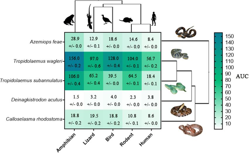
Neurotox Res

that present the functional residues as part of a constrained     about the ecology and evolution of this basal clade regard-
loop (Brust et al. 2013; Debono et al. 2017). Due to the small    ing their unique neurotoxins.
size of these viper neurotoxins, having simple structures that
lack specific requirements to maintain critical folding that
larger and more complex toxins require, they would be more        Results and Discussion
easily subject to positive selection pressures to more potent
forms (Schmidt et al. 1992) or for prey-specific targeting, as    Results from the functional testing of these venoms support
seen in some elapid and colubrid three-finger toxins (3FTxs)      previous data indicating that waglerins are a more potently
(da Silva Jr and Aird 2001; Pawlak et al. 2006; Pawlak et al.     derived form of the azemiopsin peptide (Debono et al.
2009). However, there have been few studies that have func-       2017). This is indicated by the extreme increase in relative
tionally tested the nAChR potency of these basal viper            binding affinity from A. feae to both T. wagleri and
venoms to ascertain if prey-specific targeting of their neuro-    T. subannulatus (Fig. 1), and is likely due to the addition of
toxins occurs. Consequently, in both cases the relative potency   the cysteines in waglerins presenting the bioactive residues in
against prey-specific lineages or humans is unknown, with         a more stable conformation than in the linear forms present in
testing only having been undertaken on a few taxonomical          Azemiops venom (Debono et al. 2017). The greater prey es-
lineages such as birds or rodents (Hsiao et al. 1996; Lin         cape potential in an arboreal niche, such as occupied by the
et al. 1995; McArdle et al. 1999; Molles et al. 2002a, b; Tsai    Tropidolaemus genus, would provide a strong selection pres-
et al. 1995; Utkin et al. 2012b). Therefore, interpretations of   sure for faster acting and more potent toxins. This further
these venoms in their evolutionary or clinical context have       supports the idea that toxins can evolve under positive selec-
been lacking and their role in viper venom evolution is unclear   tion pressures to a more potent form (Fry et al. 2003).
as well as their hazard to humans.                                    With further regard to the waglerins, the waglerin 1 and 3
   The Viperidae family of snakes contains many species of        peptides from T. wagleri share a very similar homology to
medical importance and consists of three subfamilies;             waglerin 2 and 4 as well as the waglerin-Ts-1 from
Crotalinae (pit vipers), Viperinae (true vipers), and             T. subannulatus (Debono et al. 2017). However, they differ
Azemiopinae. Species within the Crotalinae subfamily inhabit      by small amino acid changes from Y to H (tyrosine to histi-
numerous habitats across large ranges globally, from Asia         dine), with waglerin 1/3 having a -CHPPC- motif whilst
through to the Americas (Alencar et al. 2016), and as such        waglerin 2/4 and waglerin-Ts-1 have a -CYPPC- (Debono
possess vast variability regarding their morphology and ecol-     et al. 2017). This amino acid change led other studies to sug-
ogy. For example, a basal clade within Crotalinae consists of     gest that the -CYPPC- motif is the plesiotypic form and that
both terrestrial (Calloselasma, Deinagkistrodon, and Hypnale      the waglerin 1/3 toxins of T. wagleri have evolved under pos-
spp.) and arboreal (Tropidolaemus spp.) Asian genera. In con-     itive selection pressures to increase the potency of these toxins
trast to the neurotoxicity of Azemiops and Tropidolaemus          (Fry 2005). Furthermore, this amino acid change has been
(Debono et al. 2017; Lin et al. 1995; Schmidt and Weinstein       shown to increase the functional neurotoxic activity, causing
1995; Schmidt et al. 1992; Tan et al. 2017; Utkin et al. 2012a,   a greater LD50 (Schmidt et al. 1992). Thus, our data also
b), the venoms of Asian Viperidae species are predominantly       support this finding, as T. wagleri indeed displayed an overall
coagulotoxic (Debono et al. 2019; Nielsen 2016; Nielsen and       greater binding affinity to all mimotopes than that of
Frank 2018; Tang et al. 2016; Withana et al. 2014). In align-     T. subannulatus (Fig. 1). Other small nAChR targeting toxins
ment with variable ecology and morphology within this             such as 3FTxs have been suggested to act under similar selec-
group, one study observed significant inter-genus differences     tion pressures (Dashevsky and Fry 2018; Jackson et al. 2013).
in venom coagulation within the basal clade (Debono et al.        Future work should endeavour to examine the structure-
2019). Such diversity in niche partitioning among closely re-     function relationship of waglerin peptides and their binding
lated genera provides an ideal opportunity for evolutionary       activity to the mimotopes using synthetic waglerin analogues
comparison studies. The closely related monotypic sister taxa     with amino acid changes at key positions.
Azemiopinae, consisting of the semi-fossorial Azemiops feae,          Our data also suggests that Calloselasma rhodostoma may
can also provide a further point of evolutionary comparison,      contain peptides similar to that of the plesiotypic azemiopsin
as it is thought to be an intermediate between the basal          peptide, by showing low binding affinity similar to A. feae
Viperinae and the derived Crotalinae.                             (Fig. 1). The most parsimonious explanation here is that these
   Using a range of taxon-specific mimotopes on a                 neurotoxic peptides evolved in an earlier ancestor of
biolayer interferometry (BLI) assay, we determined the            Azemiops/Calloselasma, yet no such peptides have been iso-
prey specificity of crude venoms from species within a            lated from C. rhodostoma venom (Ali et al. 2013; Tang et al.
basal pit viper clade to the orthosteric site of α-1              2016). One explanation for this discrepancy may be that the
nAChRs. Understanding the unique viper neurotoxicity              proteomic analyses conducted might not have be able to detect
within this clade will help answer fundamental questions          or identify these small azemiopsin- or waglerin-like peptides
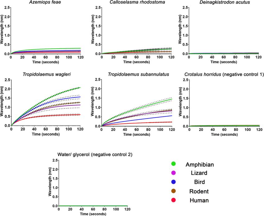
Neurotox Res

Fig. 1 A heatmap comparison of basal viper species Tropidolaemus           AUC ± SEM with N = 3. Image credits : Tom C harlton—
wagleri, T. subannulatus, Deinagkistrodon acutus, and Calloselasma         ecoanimalencounters.co.uk (A. feae); Rushden—(thainationalparks.
rhodostoma including the closely related monotypic sister taxa             com) via flikr.com CC BY-SA 2.0 (T. wagleri and C. rhodostoma);
Azemiops feae. The heat map is based on the relative potency (AUC)         Bernard Dupont via flickr.com CC BY-SA 2.0 (T. subannulatus); Alex
ranking system—derived from Ka (binding rate) curves in triplicate—        White via flickr.com CC BY-NC-SA 2.0 (D. acutus). Blacked-out images
with darker green indicating greater affinity and vice versa. Values are   are public domain CC0 1.0 via phylopic.org

on 1D or 2D gels (Ali et al. 2013) or chromatography studies               neuromuscular junction with all other subunits being neuronal
may not have returned significant database search hits due to              and mostly located within the central nervous system (CNS)
the lack of a C. rhodostoma transcriptome. Thus, future anal-              (Gotti and Clementi 2004; Le Novere and Changeux 1995), it
yses should aim to further isolate, characterise, and assess               is unlikely that the toxins have been evolutionarily selected to
what toxins within C. rhodostoma venom are binding to the                  target any other nAChR subunit. This is due to the strict bio-
nAChR mimotopes. Such investigations would aid conclu-                     chemical and physiological mechanisms of the blood-brain
sions regarding the evolutionary history of the toxins within              barrier controlling the passage of endo- and exogenous sub-
this basal clade.                                                          stances across (Abbott et al. 2010); thus, unlikely venom
   Interestingly, D. acutus showed no binding to the                       toxins can reach the CNS. Therefore, the α-1 seems the most
mimotopes, as is evidenced by the small AUC values (Fig.                   likely intended evolutionary target since it is easily reached
1) and the flat-line wavelength data paralleling both negative             via the blood stream and located at the neuromuscular junction
controls (Fig. 2). Crotalus horridus was chosen as a negative              where other nAChR targeting neurotoxins have been shown to
control because there is no evidence that this species utilises            target, causing flaccid paralysis (Barber et al. 2013; Nirthanan
nAChR targeting neurotoxins, but also to give a comparison                 and Gwee 2004). Future work including functional testing on
of a venom that is rich in other non-binding toxin types (e.g.             chick biventer cervicis muscle preparations would be neces-
PLA2s, SVSPs and SVMPs) (Rokyta et al. 2013; Rokyta et al.                 sary to ascertain fully the lack of nAChR binding (regardless
2015). We suspect that the small AUC values compared with                  of orthosteric vs. allosteric binding).
water/glycerol negative control are due to large non-binding                   Venom from both species of Tropidolaemus showed a
toxins such as PLA2s having negligible steric interferences                stronger affinity toward the amphibian than other mimotopes
with the mimotopes (Fig. 2). Thus, these data suggest that                 (Figs. 1 and 2). This result is surprising in that the literature
D. acutus has subsequently lost the ancestral neurotoxic pep-              reports that adults of both Tropidolaemus species feed primar-
tide after branching off from C. rhodostoma. Alternatively, a              ily on rodents and birds (Das and Charles 2015; Orlov et al.
less parsimonious explanation is that selection pressures have             2002). Although bird and rodent mimotopes were still potent-
evolved the toxin to become an allosteric binder of nAChRs,                ly targeted (Figs. 1 and 2), the literature only suggests amphib-
which would not be detected in this assay (Zdenek et al.                   ians as main prey items for T. wagleri in juveniles and small
2019). Another explanation may be that the toxins have                     males (Orlov et al. 2002), and no amphibians noted in the diet
evolved to target other nAChR subunits rather than the α-1.                for T. subannulatus (Das and Charles 2015). Thus, other
However, since the α-1 is the only subunit located at the                  toxins with different functions within the venom (e.g.
Neurotox Res

Fig. 2 A comparison of the wavelength (nm) curves of the association     water/glycerol (negative control 2). All venoms were tested against am-
step (ka binding step) over a 120-s assay run period of Azemiops feae,   phibian, lizard, bird, and rodent mimotopes in triplicate. The dots sur-
Calloselasma rhodostoma, Deinagkistrodon acutus, Tropidolaemus           rounding the curve lines are error bars based on SEM values with N = 3
wagleri, T. subannulatus, Crotalus horridus (negative control 1) and

coagulotoxic and myotoxic PLA2s) are likely to play an equal-            specificity since the Tropidolaemus venoms were more potent
ly vital role in prey immobilisation of different taxa types,            and target specific toward amphibian than the A. feae venom
which has been suggested in other studies (Harris et al.                 (Figs. 1 and 2). The low levels of neurotoxicity for
2020; Lyons et al. 2020), with a certain prey types possibly             C. rhodostoma and lack of postsynaptic neurotoxicity in
being immobilised more effectively by different pathophysio-             D. acutus would suggest their venom is more suited to
logical functions and molecular constituents of the venom.               targeting other pathophysiological functions such as the blood
   Azemiops feae showed its highest binding preference to-               coagulation of prey and that C. rhodostoma is likely evolving
ward amphibian mimotope, although with much less disparity               to lose the nAChR targeting components of its venom and
between the taxa mimotopes than what both Tropidolaemus                  shifting to a more coagulotoxin-rich composition (Ali et al.
species displayed. This result was interesting as the                    2013; Debono et al. 2019). Not surprisingly, functional studies
azemiopsin peptide is thought to be a plesiotypic form of the            on the coagulation of C. rhodostoma and D. acutus venom
derived waglerin peptides (Debono et al. 2017), thus further             have indicated that they display strong coagulotoxic effects:
supporting the idea that toxins can evolve under selection               with C. rhodostoma being procoagulant through Factor X
pressures toward a greater potency and possibly prey                     activation whilst conversely D. acutus has a pseudo-
Neurotox Res

procoagulant mechanism resulting in a net anticoagulant state      Spectrophotometer (Thermofisher, Sydney, NSW, Australia)
due to fibrinogen being cleaved to form weak, unstable clots       at an absorbance wavelength of 280 nm.
(Debono et al. 2019).
   In regard to human effects, proportionally larger amounts       Mimotope Production and Preparation
of venom would be required to produce neurotoxic symptoms
in humans than in prey animals; this is indicated by human         Expanding upon previous research (Bracci et al. 2001; Bracci
being the lowest of all binding across the mimotopes (Figs. 1      et al. 2002; Chiappinelli et al. 1996; Katchalski-Katzir et al.
and 2). Thus, this emphasises that using human bite reports to     2002) a 13–14 amino acid mimotope of ACh orthosteric site
predict prey effects is a poor methodological strategy to ascer-   of vertebrate α-1 nAChR subunit was developed by GenicBio
tain ecological and evolutionary inferences (Davies and            Ltd. (Shanghai, China) designed upon specifications which
Arbuckle 2019) as prey-specific effects may not be captured        were adapted from publicly available sequences of cholinergic
in clinical observations of envenomation to a much larger          receptors (Chrna1) from GenBank.
organism who are less sensitive to a particular venom effect           The amino acid sequences for the α-1 orthosteric site for
than would be native prey animal. Thus, evolutionary inter-        each taxon were obtained with the following accession codes:
pretations regarding venom diversification patterns can only       amphibian α-1 (uniprot F6RLA9), lizard α-1 (genbank
be elucidated through evidence obtained by testing on prey-        XM_015426640), avian α-1 (uniprot E1BT92), rodent α-1
relevant assay platforms and not clinical human                    (uniprot P25108), human α-1 (uniprot G5E9G9).
envenomations.                                                         The Cys-Cys of the native mimotope is replaced during
   This research has highlighted many important and previ-         peptide synthesis with Ser-Ser to avoid uncontrolled
ously unknown facets about the neurotoxins of this basal viper     postsynthetic thiol oxidation. The Cys-Cys bond in the
group. Our results corroborate previous research suggesting        nAChR binding region does not participate directly in
that the Tropidolaemus waglerin neurotoxins are a more po-         analyte-ligand binding (McLane et al. 1994; McLane et al.
tent and derived form of the Azemiopsin peptide from               1991; Tzartos and Remoundos 1990); thus, replacement to
Azemiops feae (Debono et al. 2017). We also show that the          Ser-Ser is not expected to have any effect on the analyte-
venom of C. rhodostoma seems to contain nAChR targeting            ligand complex formation. However, the presence of the
neurotoxins, whilst D. acutus lack this neurotoxic action, both    Cys-Cys bridge is key in the conformation of the interaction
of which have never been tested for functional neurotoxic          site of whole receptors (Testai et al. 2000). As such, we sug-
activity. Further to this, we showed that T. wagleri and           gest direct comparisons of kinetics data, such as Ka or KD,
T. subannulatus venom was more selective toward the am-            between nAChR mimotopes and whole receptor testing
phibian mimotope than the other taxa mimotopes tested,             should be avoided, or at least approached with caution.
which seems inconsistent with their preferred prey types; thus,    Mimotopes were further synthesised to a biotin linker bound
it would seem that other venom functions will likely play an       to two aminohexanoic acid (Ahx) spacers, forming a 30 Å
equally vital role in prey immobilisation of different taxa        linker.
types. Future research should endeavour to understand the              Mimotope dried stocks were solubilised in 100% dimethyl
evolutionary history of this basal group in terms of venom         sulfoxide (DMSO) and diluted in deionised water at 1:10 di-
composition, function, and evolutionary ecology.                   lution to create a working stock of 50 μg/mL. All stocks were
                                                                   stored at − 80 °C until use and limited to three freeze-thaw
                                                                   cycles.

Methods                                                            Bio-layer Interferometry

Venom Collection and Preparation                                   The bio-layer interferometry (BLI) assay was performed on
                                                                   the Octet RED 96 system (ForteBio). The assay used, includ-
Venoms were obtained from pooled snake venom extractions           ing all methodology and data analysis, was based upon a val-
of multiple individuals (captive and wild-caught) from either      idated protocol (Zdenek et al. 2019).
the long-term cryogenic collection of the Venom Evolution
Laboratory.                                                        Data Processing and Statistical Analyses
   All venom samples were lyophilised and reconstituted in
double deionised water (ddH2O) then centrifuged (4 °C,             All data obtained from BLI on Octet RED 96 system
10 min at 14,000 RCF). The supernatant was then made into          (ForteBio) were processed in exact accordance to the valida-
a working stock (1 mg/mL) in 50% glycerol to prevent freez-        tion of this assay (Zdenek et al. 2019). The association step
ing at − 20 °C. The concentrations of working stocks were          data (in triplicate) were obtained in an Excel .csv file extracted
determined in triplicate using a NanoDrop 2000 UV-Vis              from raw outputs of the Octet Red 96 system and then
Neurotox Res
imported into Prism 7.0 software (GraphPad Software Inc., La analysis of toxin sequences and related body proteins. Genome
Res 15:403–420
Jolla, CA, USA) where area under the curve (AUC) calcula-
Fry BG, Wüster W, Ryan Ramjan SF, Jackson T, Martelli P, Kini RM
tions were made and graphs produced. Phylogenetic trees (2003) Analysis of Colubroidea snake venoms by liquid chromatog-
were obtained from timetree.org and then further manually raphy with mass spectrometry: evolutionary and toxinological im-
recreated using Mesquite software (version 3.2). plications. Rapid Commun Mass Spectrom 17:2047–2062
Gotti C, Clementi F (2004) Neuronal nicotinic receptors: from structure to
pathology. Prog Neurobiol 74:363–396
Author Contributions Conceptualisation: BGF; methodology: RJH,
Harris RJ, Zdenek CN, Harrich D, Frank N, Fry BG (2020) An appetite
CNZ, BGF; investigation: RJH, JD; resources: JD, DH, BGF; writing
for destruction: Detecting prey-selective binding of α-neurotoxins in
of first draft: RJH; editing subsequent drafts; RJH, CNZ, BGF; project
the venom of Afro-Asian elapids. Toxins 12(3):205
administration: BGF.
Hsiao Y-M, Chuang C-C, Chuang L-C, Yu H-M, Wang K-T, Chiou S-H,
Wu S-H (1996) Protein engineering of venom toxins by synthetic
Funding Information RJH and CNZ was supported by the University of approach and NMR dynamic simulation: status of basic amino acid
Queensland International PhD scholarship fund. BGF was funded by residues in waglerin I. Biochem Biophys Res Commun 227:59–63
Australian Research Council Discovery Project DP190100304. Jackson T et al (2013) Venom down under: dynamic evolution of
Australian elapid snake toxins. Toxins 5:2621–2655
Katchalski-Katzir E, Kasher R, Balass M, Scherf T, Harel M, Fridkin M,
Sussman JL, Fuchs S (2002) Design and synthesis of peptides that
References bind α-bungarotoxin with high affinity and mimic the three-
dimensional structure of the binding-site of acetylcholine receptor.
Abbott NJ, Patabendige AA, Dolman DE, Yusof SR, Begley DJ (2010) Biophys Chem 100:293–305
Structure and function of the blood–brain barrier. Neurobiol Dis 37: Le Novere N, Changeux J-P (1995) Molecular evolution of the nicotinic
13–25 acetylcholine receptor: an example of multigene family in excitable
Alencar LR, Quental TB, Grazziotin FG, Alfaro ML, Martins M, Venzon cells. J Mol Evol 40:155–172
M, Zaher H (2016) Diversification in vipers: phylogenetic relation- Lin W, Smith L, Lee C (1995) A study on the cause of death due to
ships, time of divergence and shifts in speciation rates. Mol waglerin-I, a toxin from Trimeresurus wagleri. Toxicon 33:111–114
Phylogenet Evol 105:50–62 Lyons K, Dugon MM, Healy K (2020) Diet breadth mediates the prey
Ali SA et al (2013) Proteomic comparison of Hypnale hypnale (hump- specificity of venom potency in snakes. Toxins 12(2):74
nosed pit-viper) and Calloselasma rhodostoma (Malayan pit-viper) McArdle JJ, Lentz TL, Witzemann V, Schwarz H, Weinstein SA, Schmidt
venoms. J Proteome 91:338–343 JJ (1999) Waglerin-1 selectively blocks the epsilon form of the mus-
Barber CM, Isbister GK, Hodgson WC (2013) Alpha neurotoxins. cle nicotinic acetylcholine receptor. J Pharmacol Exp Ther 289:543–
Toxicon 66:47–58 550
Bracci L, Lozzi L, Lelli B, Pini A, Neri P (2001) Mimotopes of the McLane KE, Wu X, Diethelm B, Conti-Tronconi BM (1991) Structural
nicotinic receptor binding site selected by a combinatorial peptide determinants of α-bungarotoxin binding to the sequence segment
library. Biochemistry 40:6611–6619 181-200 of the muscle nicotinic acetylcholine receptor. α-subunit:
Bracci L et al (2002) A branched peptide mimotope of the nicotinic effects of cysteine/cystine modification and species-specific amino
receptor binding site is a potent synthetic antidote against the snake acid substitutions. Biochemistry 30:4925–4934
neurotoxin α-bungarotoxin. Biochemistry 41:10194–10199 McLane KE, Wu X, Conti-Tronconi BM (1994) An α-bungarotoxin-
Brust A et al (2013) Differential evolution and neofunctionalization of binding sequence on the Torpedo nicotinic acetylcholine receptor
snake venom metalloprotease domains. Mol Cell Probes 12:651– α-subunit: conservative amino acid substitutions reveal side-chain
663 specific interactions. Biochemistry 33:2576–2585
Chiappinelli VA, Weaver WR, McLane KE, Conti-Fine BM, Fiordalisi JJ, Molles BE, Rezai P, Kline EF, McArdle JJ, Sine SM, Taylor P (2002a)
Grant GA (1996) Binding of native κ-neurotoxins and site-directed Identification of residues at the α and ε subunit interfaces mediating
mutants to nicotinic acetylcholine receptors. Toxicon 34:1243–1256 species selectivity of Waglerin-1 for nicotinic acetylcholine recep-
tors. J Biol Chem 277:5433–5440
da Silva NJ Jr, Aird SD (2001) Prey specificity, comparative lethality and
Molles BE, Tsigelny I, Nguyen PD, Gao SX, Sine SM, Taylor P (2002b)
compositional differences of coral snake venoms. Comp Biochem
Residues in the ε subunit of the nicotinic acetylcholine receptor
Physiol, Part C: Toxicol Pharmacol 128:425–456
interact to confer selectivity of Waglerin-1 for the α− ε subunit
Das I, Charles JK (2015) Venomous snakes and envenomation in Brunei.
interface site. Biochemistry 41:7895–7906
In: Gopalakrishnakone P (ed) Clinical Toxinology in Asia Pacific
Nielsen VG (2016) Ancrod revisited: viscoelastic analyses of the effects
and Africa. Springer Science, pp 103–114
of Calloselasma rhodostoma venom on plasma coagulation and
Dashevsky D, Fry BG (2018) Ancient diversification of three-finger fibrinolysis. J Thromb Thrombolysis 42:288–293
toxins in Micrurus coral snakes. J Mol Evol 86:58–67 Nielsen VG, Frank N (2018) Differential heme-mediated modulation of
Davies E-L, Arbuckle K (2019) Coevolution of snake venom toxic activ- Deinagkistrodon, Dispholidus, Protobothrops and Pseudonaja
ities and diet: evidence that ecological generalism favours toxico- hemotoxic venom activity in human plasma. Biometals 31:951–959
logical diversity. Toxins 11:711 Nirthanan S, Gwee MC (2004) Three-finger α-neurotoxins and the nic-
Debono J, Xie B, Violette A, Fourmy R, Jaeger M, Fry BG (2017) Viper otinic acetylcholine receptor, forty years on. J Pharmacol Sci 94:1–
venom botox: the molecular origin and evolution of the waglerin 17
peptides used in anti-wrinkle skin cream. J Mol Evol 84:8–11 Orlov N, Ananjeva N, Khalikov R (2002) Natural history of pitvipers in
Debono J, Bos MH, Coimbra F, Ge L, Frank N, Kwok HF, Fry BG (2019) Eastern and Southeastern Asia. Biol Vipers 345–361
Basal but divergent: clinical implications of differential Pawlak J, Mackessy SP, Fry BG, Bhatia M, Mourier G, Fruchart-Gaillard
coagulotoxicity in a clade of Asian vipers. Toxicol in Vitro 58: C, Servent D, Ménez R, Stura E, Ménez A, Kini RM (2006)
195–206 Denmotoxin, a three-finger toxin from the colubrid snake Boiga
Fry BG (2005) From genome to “venome”: molecular origin and evolu- dendrophila (mangrove catsnake) with bird-specific activity. J Biol
tion of the snake venom proteome inferred from phylogenetic Chem 281:29030–29041Neurotox Res
Pawlak J et al (2009) Irditoxin, a novel covalently linked heterodimeric Tsai M-C, Hsieh W, Smith L, Lee C (1995) Effects of waglerin-I on
three-finger toxin with high taxon-specific neurotoxicity. FASEB J neuromuscular transmission of mouse nerve-muscle preparations.
23:534–545 Toxicon 33:363–371
Rokyta DR, Wray KP, Margres MJ (2013) The genesis of an exception- Tzartos S, Remoundos MS (1990) Fine localization of the major alpha-
ally lethal venom in the timber rattlesnake (Crotalus horridus) re- bungarotoxin binding site to residues alpha 189-195 of the Torpedo
vealed through comparative venom-gland transcriptomics. BMC acetylcholine receptor. Residues 189, 190, and 195 are indispens-
Genomics 14:394 able for binding. J Biol Chem 265:21462–21467
Rokyta DR, Wray KP, McGivern JJ, Margres MJ (2015) The Utkin YN, Weise C, Anh HN, Kasheverov I, Starkov V, Tsetlin V (2012a)
transcriptomic and proteomic basis for the evolution of a novel ven- The new peptide from the Fea’s viper Azemiops feae venom interacts
om phenotype within the timber rattlesnake (Crotalus horridus). with nicotinic acetylcholine receptors. In: Doklady biochemistry and
Toxicon 98:34–48 biophysics, vol 442. vol 1. Springer Science, pp 33–35
Schmidt JJ, Weinstein SA (1995) Structure-function studies of waglerin I, Utkin YN, Weise C, Kasheverov IE, Andreeva TV, Kryukova EV, Zhmak
a lethal peptide from the venom of Wagler’s pit viper, Trimeresurus MN, Starkov VG, Hoang NA, Bertrand D, Ramerstorfer J, Sieghart
wagleri. Toxicon 33:1043–1049 W, Thompson AJ, Lummis SCR, Tsetlin VI (2012b) Azemiopsin
Schmidt JJ, Weinstein SA, Smith LA (1992) Molecular properties and from Azemiops feae viper venom, a novel polypeptide ligand of
structure-function relationships of lethal peptides from venom of nicotinic acetylcholine receptor. J Biol Chem 287:27079–27086
Wagler’s pit viper, Trimeresurus wagleri. Toxicon 30:1027–1036 Withana M, Rodrigo C, Gnanathasan A, Gooneratne L (2014)
Tan CH, Tan KY, Yap MKK, Tan NH (2017) Venomics of Tropidolaemus Presumptive thrombotic thrombocytopenic purpura following a
wagleri, the sexually dimorphic temple pit viper: unveiling a deeply hump-nosed viper (Hypnale hypnale) bite: a case report. J
conserved atypical toxin arsenal. Sci Rep 7:43237 Venomous Anim Toxins Incl Trop Dis 20:26
Tang ELH, Tan CH, Fung SY, Tan NH (2016) Venomics of Calloselasma
Zdenek CN et al (2019) A taxon-specific and high-throughput method for
rhodostoma, the Malayan pit viper: a complex toxin arsenal
measuring ligand binding to nicotinic acetylcholine receptors.
unraveled. J Proteome 148:44–56
Toxins 11:600
Testai FD, Venera GD, Peña C, de Jiménez Bonino MJB (2000) Histidine
186 of the nicotinic acetylcholine receptor α subunit requires the
presence of the 192–193 disulfide bridge to interact with α- Publisher’s Note Springer Nature remains neutral with regard to jurisdic-
bungarotoxin. Neurochem Int 36:27–33 tional claims in published maps and institutional affiliations.You can also read