FACILE, LOW-COST AND RAPID PHYTOSYNTHESIS OF STABLE AND ECO-FRIENDLY GOLD NANOPARTICLES USING GREEN WALNUT SHELL AND STUDY OF THEIR ANTICANCER ...
←
→
Page content transcription
If your browser does not render page correctly, please read the page content below

WCRJ 2021; 8: e2037

FACILE, LOW-COST AND RAPID
PHYTOSYNTHESIS OF STABLE
AND ECO-FRIENDLY GOLD NANOPARTICLES
USING GREEN WALNUT SHELL AND STUDY
OF THEIR ANTICANCER POTENTIAL
M. SALANDARI RABORI1, M. NOROOZI KARIMABAD1, M. REZA HAJIZADEH1,2
1
 Molecular Medicine Research Center, Research Institute of Basic Medical Sciences, Rafsanjan University of
 Medical Sciences, Rafsanjan, Iran
2
  Department of Clinical Biochemistry, Faculty of Medicine, Rafsanjan University of Medical Sciences,
 Rafsanjan, Iran

    Abstract – Objective: Nowadays, gold nanoparticles (GNPs) are used in targeted nano photo-
thermal cancer therapy. Considerable interest has been dedicated to gold nanoparticles because
of their characteristics which are controllable and unique. Different synthesis methods have been
proposed to produce these nanoparticles, which often require elevated temperatures/pressures or
toxic solvents. Therefore, green synthesis would be a substitution selection as an environmentally
friendly, economically viable and simple alternative method for the gold nanoparticles synthesis.
    Materials and Methods: In this study, using walnut green external shell, GNPs have been syn-
thesized by green chemistry method. In this reaction, walnut green shell is a reducer and stabilizing
factor for preparing GNPs. In this work, after extracting walnut, 2 ml of extract was added to 4 ml
of Au+3 solution ( ), purple color indicates synthesis of GNPs. For NPs synthesis with an appropriate
size, some factors like pH, extract volume, gold salt concentration and reaction temperature were
surveyed and by using UV-Visible (UV-Vis) absorption spectroscopy, optimum conditions were se-
lected for preparing NPs. In addition, prepared NPs have been tested by x-ray diffraction (XRD) and
transmission electron microscopy (TEM) to determine structure, size and their shape. Afterward,
the synthesized GNPs were determined by 3- (4, 5-dimethylthiazolyl)-2, 5-diphenyl-tetrazolium bro-
mide (MTT) to assay their antitumor, anti-oxidant properties.
    Results: The results of these measurements show the spherical and triangular GNPs with different
sizes between 10-50 nm were produced. There is an important development in the antioxidant and
cytotoxicity characteristics of GNPs which are green synthesized ones. Statistical analysis also found a
significant difference among various GNPs concentrations on declined cell viability of Michigan Can-
cer Foundation-7 (MCF7) cells in 24 h in concentration-dependent fashion. The IC50 value evaluated
after 24 h of GNPs against MCF7 cells caused 52% cell death at the concentration of 3 mM.
    Conclusions: The above-mentioned results suggest that synthesized nanoparticles employing
green nanotechnology is a suitable technique to fight against infectious diseases and cancer.

KEYWORDS: Anticancer potential, Green synthesis, Gold nanoparticles, Phytosynthesis, Selective
cytotoxicity.

          This work is licensed under a Creative Commons Attribution-NonCommercial-ShareAlike 4.0 International License

Corresponding Author: Mojgan Noroozi Karimabad, PhD; e-mail: mojgan.noroozi@yahoo.com                                1
INTRODUCTION                                                drugs that have the most effective and the lowest-side
                                                            effects. Even though in the diagnosis and treatment of
Nanotechnology, in developing countries, is be-             cancer, considerable progress has been made, but the
ing applied to assist disease treatment and is a kind       eradication of it is limited. As a result, finding nov-
of prevention for health concerns. For that sort of nan-    el treatment and diagnosis approaches is crucial12,13.
otechnology, nanomedicine is the umbrella term. Ad-         Medically, nanotechnology for cancer is an emerging
ditionally, nanotechnology is being used or improved        field that aims to develop advancements toward both
toward industrial diversity, and purified NP is applied     the treatment and diagnosis of cancer. Much attention
in the various manufacturing products and process-          has been paid to gold nanoparticles, because of the
es1,2, and healthcare involving lubricant additives, fil-   biocompatibility of them, their better optical prop-
ters, paints, and insulation. The novel drugs for age       erties, and they have capability to chemically modi-
are the NPs of ceramics, metals, or polymers that can       fy the surface of them via the addition of numerous
fight conditions such as cancer3 and combat human           types of ligands14. Concerning the importance of gold
pathogens such as bacteria4. Nanotechnology usages          NPs in cancer therapy, in this research, experimental
in the control, diagnosis, treatment, and monitoring        studies were done to synthesize gold NPs of walnut
of diseases have been attributed to nanomedicine.           green shells in order to cure breast cancer. Walnuts
NPs are positive and helpful agents in cancer therapy       are a rich source of multiple nutrients and are culti-
and are analyzed as radio-sensitizers, drug carriers,       vated in different areas of the world15. In this study,
contrast agents, and photo-thermal agents4. Also, as        the walnut green shell was used to synthesize gold
regards the high activity of NPs and their tendency to      NPs and their anticancer and antioxidant properties
connect with biomolecules and macromolecules, the           were recognized by MTT assay and these NPs were
NPs linked to peptides can be used as detectors inside      applied in cancer treatment.
molecules, which is a good and important method in
cellular pictures, drug delivery and biomolecules rec-
ognition5. Gold NPs are functionalized by different         MATERIALS AND METHODS
biological factors and are recognized and detected af-
ter interring in the body and transferring to the target    Extraction
section. Toward the areas of special disease, GNPs ex-
ternal function for biomedical application is essential     First, a walnut green shell was prepared which is a
for targeting them and allows interacting selectively       disposable part of the plant, the shells were washed
with biomolecules or cells. In cancer therapy, one of       and dried in a dark room. After this, they were
the main and significant uses is the gold NPs. Studies      ground and weighted to 1 gr of dried powder and
indicate solid gold nanospheres selectively kill can-       dissolved in 100 mL deionized water. This solution
cer cells and have no adverse effects on the healthy        was placed on the shaker at 50°C for 30 min. After
cells6,7. NPs are synthesized by two methods: 1)            cooling the solution, it was filtrated using Whatman
Chemical and physical methods that have more yield,         filter paper No. 42. Finally, for deletion of suspended
produced NPs in this method are purer, but chemical         particle, the extract was put in centrifuge with 1000
materials in synthetic NPs are toxic and pollute the        revolutions per min for 30 min. The extract was pre-
environment. 2) Green method that uses biological           pared and kept at a temperature of 4°C16.
factors to synthesize NPs. This method uses plants
to produce NPs and it is a safe and suitable method.
Cells are randomly divided by cancer. This damages          Synthesis of gold NPs
the immune system, further impairment, and tumors
which are fatal. In many developed countries among          For the synthesis of gold NPs, the first 1 mM con-
females8, breast cancer has been recognized as the          centration of HAuCl4.3H2O was prepared and 4 ml
conducting cause for cancer death. Also, in less devel-     of this solution was added to 2 ml walnut extract at
oped countries, among females, it remains the leading       room temperature. The measured pH was 5.4. Color
cause of cancer death. Every year 1.7 million people        solution change from green to purple showed syn-
are afflicted with breast cancer9. The most common          thesized gold NPs.
cause of death is a metastatic disease to various or-
gans10. In general, most of the chemical drugs that are
used in cancer therapy change the meiosis process           Optimum conditions for producing
and stop cancer cell diagnosis. This happens in differ-     bigger gold NPs
ent ways such as apoptosis induction, DNA structure
alteration, tyrosine kinase, and so on11. Regarding the     In order to make gold NPs with high quality and bigger
fact that chemical drugs have high toxicity and de-         size, we changed some factors like pH, extract volume,
stroy safe cells, many efforts have been made to use        gold solution concentration, and temperature.

2
SYNTHESIS OF GNPS AND THEIR POTENTIAL ANTICANCER EFFECT

Optimum pH                                                 to allow cells to be added into the wells. After 24 h,
                                                           cells were subjected to differing concentrations for
We prepared six solutions (2 ml walnut green shell         the GNPs (0.5-5 mM) and were incubated at 37°C
extract + 4 ml gold solution 1 mM) with different          for 24 h. The cells with MTT were treated after
pH between 3-8. pH setting was done by 0.1 M (Hy-          exposure to the drug dissolved in PBS and incubat-
drogen chloride) HCl and 0.1 M (Sodium hydroxide)          ed at 37°C for more than 4 h. Afterwards, with a
NaOH. By comparing the spectrum of these solu-             syringe and needle, the whole liquid was extracted;
tions, optimum pH was selected.                            and to each well, dimethyl sulfoxide was attached
                                                           to dissolve the crystals of MTT-formazan. The
                                                           plates were placed in a spectrophotometer which is
Optimization extracts volume                               a multi-well scanning. Then, every plate was quan-
                                                           tified photo-metrically after stirring for the 30s,
In the next step, to select the best walnut extract vol-   at 570 nm absorbance by ELISA reader (BioTek;
ume, 4 ml of gold solution was added to four tubes         Winoosky, VT, USA). All experiments were re-
and 1-4 ml of extract was added to each tube, re-          peated 3 times. The purple formazan amount
spectively. pH in each tube was optimum pH. Based          generated through treated cells was compared to
on the absorption spectrum of different solutions,         that of cells that were untreated control, and in the
volume was selected.                                       treated cells, the absorbance was declared as a con-
                                                           trol percentage17,18.

Optimization of gold salt
concentration                                              Instrumental analysis

In this step, five different concentrations of gold        Excel and SPSS software version 18 (SPSS Inc., Chi-
solution (0.5, 1, 2, 3, 4 mM) were prepared and at         cago, IL, USA) was used to analyze the data statis-
optimum pH, optimum volume of extract was added            tically. All experiments were executed in triplicate
to each tube. The maximum absorption shows the             and all attained consequences, as the mean ± scan-
optimum concentration of the gold solution.                ning electron microscope (SEM) (Germany). Also,
                                                           data were investigated using one way ANOVA and
                                                           (p ≤ 0.05) was considered significant. To measure
Optimum temperature                                        the size of synthesized NPs and study their shape,
                                                           Transmission Electron Microscopy (TEM), (model
In the final step, four solutions with the mentioned       Zeiss-EM10C-100 KV; Germany) was used. The re-
optimum conditions were prepared and one of them           sults confirm that spherical and triangular gold NPs
was placed at room temperature (25°C), the other           with the size between 10-50 nm have been synthe-
solutions were placed in a heat bath at different tem-     sized. Furthermore, in order to identify the phase of
peratures (35, 55, 75°C). Analyzing the solutions,         crystalline NPs and for provision of information on
the optimum temperature was determined.                    unit cell dimensions, x-ray diffraction ((X-ray pow-
                                                           der diffraction) XRD (Shimadzu; Vietnam Tube:
                                                           Cu (Kα = 1.54 Å) X’Pert Pro MPD (PANalytical))
Culture of MCF7 cells                                      was applied.

A sample of MCF7 cells was purchased (from the
National Cell Bank of Iran, Pasteur Institute). Cells      RESULTS
were seeded into 90% RPMI-1640 which was inoc-
ulated with 10% heat-inactivated fetal bovine serum        Study of absorption spectroscopy
(Gibco Laboratories; Waltham, MA, USA) and con-            of the Au NPs
tained Penicillin 100 IU/ml and streptomycin 100
μg/mL, with 5% CO2 - 95% O2 in a 37ºC humidified           Gold NPs were prepared by adding 4 ml of 1mM
incubator17.                                               Au3+ solution to 2 ml walnut extract. After some
                                                           minutes, the green-color solution changed to purple
                                                           color that shows synthesized gold NPs, purple col-
MTT assay                                                  or is related to surface Plasmon resonance of gold
                                                           NPs. Then, absorption spectrum of NPs and walnut
In medium culture, seeding density cells were plat-        extract were studied. Using visible-ultraviolet spec-
ed on 96-well plates, and incubated at 37°C in hu-         trophotometer in 200-800 nm gold NPs spectrum,
midified atmosphere including 5% carbon dioxide            maximum was determined 480 nm (Figure 1).

                                                                                                               3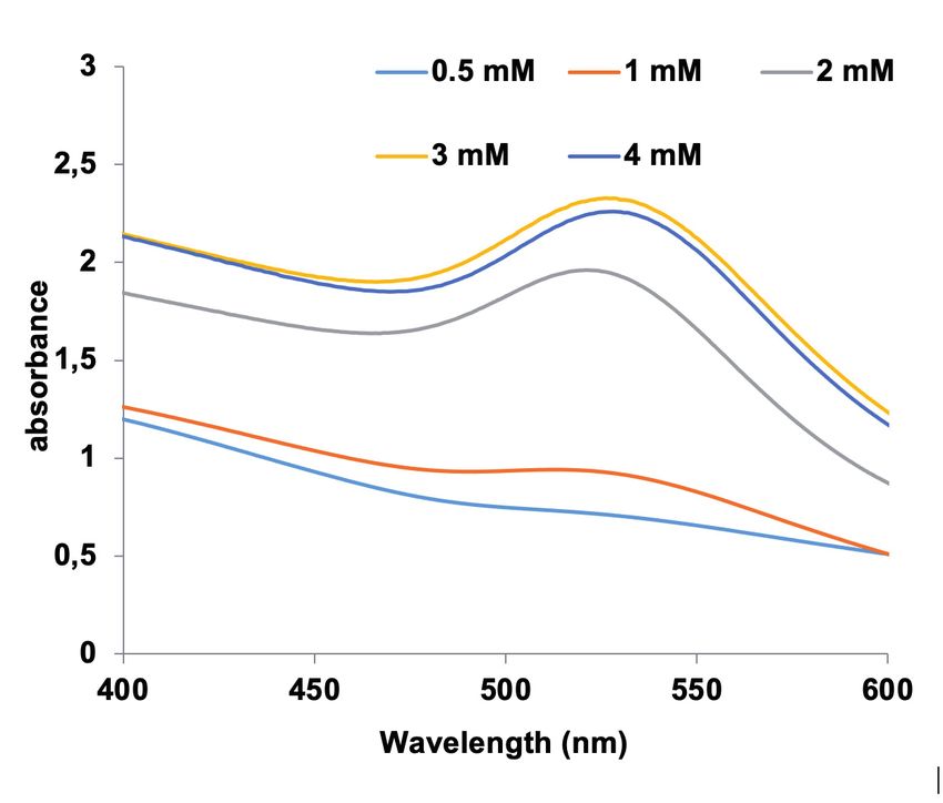
Fig. 1. UV- Vis absorption spectroscopy a) Au NPs and b)       Fig. 3. UV-Vis absorption spectroscopy GNPs in various
walnut green shell extract without optimum conditions.         volumes of extract.

Results of optimum pH                                          2 ml extract was added to each tube. The maximum
                                                               absorption is related to gold salt with 3 mM concen-
Six solutions with different pH between 3-8 were stud-         tration (Figure 4).
ied. By comparing the spectrum of these solutions, it
was demonstrated that solution with pH=6 has maxi-
mum concentration. Thus, optimum pH is 6 (Figure 2).           Results of optimum temperature

                                                               Finally, for studying optimum temperature, solutions
Results of optimum extract volume                              with mentioned optimum conditions were prepared
                                                               and were studied at different temperatures (25, 35,
As mentioned before, to study optimum extract                  55, 75°C). Absorption spectrum of solutions showed
volume, 4 ml of gold solution was added to four                no remarkable difference in these temperatures but at
tubes and extract volume was set between 1-4 ml                55°C there was higher adsorption spectrum. So, 55°C
for each tube, respectively. pH in each tube was 6             was the optimum temperature (Figure 5).
(optimum pH). Absorption spectrum of different
solutions showed that more gold NPs were made in
2 ml extract volume. Thus, 2 ml was considered as              Characterization of the Au NPs
optimum volume of extract (Figure 3).                          by XRD and TEM

                                                               TEM was used to measure the size of synthesized
Results of optimization                                        NPs and to investigate their shape. The results con-
of gold salt concentration

In this step, five different concentrations of gold solu-
tion (0.5, 1, 2, 3, 4 mM) were prepared with pH=6 and

                                                               Fig. 4. UV-Vis absorption spectroscopy GNPs in various
Fig. 2. UV-Vis absorption spectroscopy GNPs in different pH.   gold salt concentration.

4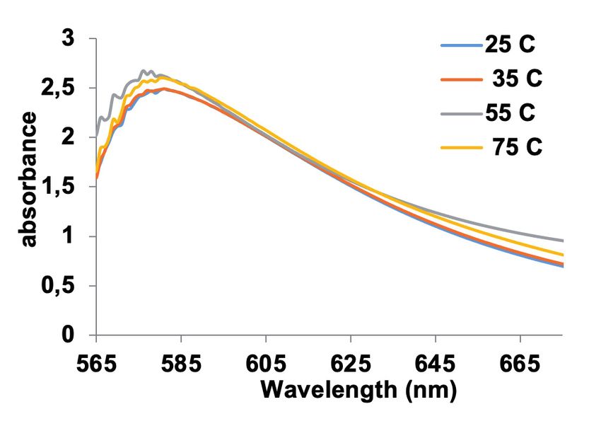
SYNTHESIS OF GNPS AND THEIR POTENTIAL ANTICANCER EFFECT

                                                             Furthermore, in order to identify the phase of
                                                         crystalline NPs and to provide information on unit
                                                         cell dimensions, XRD was applied (Figure 6b). The
                                                         average size of crystalline particles was determined
                                                         by measuring the width of the peaks formed in the
                                                         samples using the Debye- Scherrer equation:

                                                             Equation 1: D = 0.9λ / β cos θ

                                                             Where β is the peak width at half the maximum
                                                         height, λ, the wavelength of the X-rays is 1.4 nm, θ is
                                                         the angle between the radiation beam and reflection,
                                                         and D is the size of the crystalline particles. In areas
                                                         of 2θ=38.21, 44.51, 64.90, 77.90, GNPs show sharp
Fig. 5. UV-Vis absorption spectroscopy GNPs at various   peaks, demonstrating the successful synthesis of
temperatures.
                                                         NPs. The structural analysis shows that GNPs have
                                                         a crystalline structure with Miller indexes (111),
firmed that spherical and triangular gold NPs with       (200), (220) and (311) in a cubic network. The pres-
the size of between 10-50 nm were synthesized (Fig-      ence of sharp peaks in patterns shows a high degree
ure 6a).                                                 of crystallinity for NPs.

Fig. 6. A, The TEM picture of syn-
thesized GNPs by walnut green
shell. B, The XRD picture of synthe-
sized GNPs by walnut green shell.

                                                                                                               5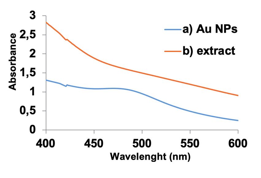
Results of the cell viability assay                      decreasing agent. In this research, a different size
                                                         and shape for the nanoparticles of gold were built
The changes in cell viability of MCF7 treated with       whenever the reactants ratio was changed regard-
different doses of GNPs at 24h were determined           ing chloroauric acid 1.0 mM solution. GNPs have
using MTT method. Statistical analysis also found        been prepared by Elia et al11 applying four different
a significant difference among various GNPs dos-         plants (Punica granatum, Lippia citriodora, Salvia
es on decreased cell viability of MCF7 cells in 24       officinalis, and Pelargonium graveolens) as stabiliz-
h in dose-dependent fashion (p < 0.05) (Figure 7).       ing and decreasing agents. By applying three var-
The 50% inhibition (IC50) value evaluated after 24       ious approaches the GNPs size distributions were
h of GNPs against Michigan Cancer Foundation-7           measured: nanoparticle-tracking analysis, the im-
(MCF7) cells was 3 mM (p ≤ 0.05).                        ages of scanning electron microscopy analysis, and
                                                         the dynamic light scattering. Two novel glycosides
                                                         of α-tetralonyl were isolated by Wang et al19 from
DISCUSSION                                               the Juglans mandshurica green walnut husks. The
                                                         entire compounds structural characterization was
Much attention has been paid to gold nanoparticles       carried out via spectroscopic analysis, containing
because of the biocompatibility, better optical prop-    HR-ESI-MS, 2D NMR and 1D experiment. The
erties, and their capability to chemically modify        isolated compounds were assayed for their cytotox-
their surface via addition of several kinds of ligands   icity against two lines of human cancer cell, HeLa
14
   . In this study, we used walnut green shell to syn-   and A549. Inhibitory influences were displayed by
thesize gold NPs and characterized their anticancer,     four compounds7-10 against two lines of human can-
antioxidant properties using MTT assay and applied       cer cell with the values of GI50 between 5.8 and
these NPs for cancer treatment. Marshall et al8 re-      1.3 µM19. Proteins from Juglans regia were isolated
corded gold NPs accumulation into Brassica Juncea        by Carrillo et al20 and were hydrolyzed with vari-
with diameters of 5-50 nm, as displayed via trans-       ous enzymes. A high anti-proliferative activity was
mission electron microscopy (TEM), at 1120 and           indicated by the proteins of prolamin and Glutelin
760 ppm Au concentrations. The gold nanoparticles        against the K-562 (leukemia) and PC-3 (prostate)
ability for biosynthesizing the flower of pharma-        cancer cells with 84.4 and 43.9 μg/mL values, re-
cologically significant tree Couroupita guianensis       spectively. The observed influence of highest in-
has been shown by Geetha et al9. The synthesis           hibition, at 0.25 μg/mL concentration, was 50% in
process, which is one-step, fast and cost-effective,     gastrointestinal digestion with Corolase PP and pep-
has been obtained. The gold nanoparticles biologi-       sin in a dose dependent manner against UACC62
cal synthesis has been reported by Sujitha and Kan-      (melanoma) cancer cells. Inhibitory impacts were
nan 10 through the HAuCl4 decline via applying the       indicated by pepsin hydrolysate against UACC62
extract of citrus fruit juice as the stabilizing and     cancer cells cancer at concentration of 71.0 μg/mL.

                                                                                  Fig. 7. The changes in viability
                                                                                  of MCF7 cell lines treated with
                                                                                  various doses of GNPs after 24
                                                                                  h, by MTT assay (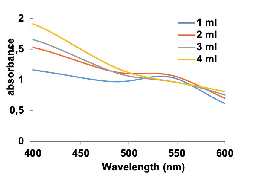
In a dose-dependent manner, the influences were            ion. The IC50 value evaluated after 24 h of GNPs
discussed. Neutrase enzyme inhibitory impacts              against MCF7 cells caused 52% cell death at the
were presented by the attained hydrolysate against         concentration of 3 mM. Consequently, in addition
UACC62 (melanoma) cancer cell at concentration of          to its many nutritional values, walnut can be used in
25 μg/mL. The cytotoxicity was not indicated, nei-         medicine and pharmacology research.
ther by protein hydrolysates nor by proteins against
VERO assay with normal cell (epithelial) 20. Wang          Acknowledgments
et al19 improved a hybrid nanocomposite which is a         This project was supported by a grant from the Rafsanjan
new kind of thermal-fluorescent core-shell that in-        University of Medical Sciences.
dicates whether the composite has the effectiveness
of cancer therapy in in vivo and in vitro assays and       Conflict    of   Interest:
also has remarkable bio-compatibility21. A one-step        The authors declare no conflict of interest
and green technique was reported by Liang et al22
for synthesizing the gold fluorescent nanoclusters         REFERENCES
via applying a cyclic commercialized acid peptide,
arginine-glycine-aspartic as the template that can           1. Poole Jr CP, Owens FJ. Introduction to Nanotechnol:
be used as fluorescent for staining the αvβ3 integ-              John Wiley & Sons 1st ed 2003.
rin-positive cancer cells, as well as radio sensitizing      2. Murray H. Nanotechnology, Gregrory Timp, Bell Lab
agents for increasing the effectiveness of killing ra-           1st ed 1999; NJ07974-0636.
                                                              3. Farokhzad OC, Cheng J, Teply BA, Sherifi I, Jon S, Kan-
diation therapy22. The nanostructures of core-shell              toff PW, Richie JP, Langer R. Targeted nanoparticle-
Fe3O4/Au were constructed by Izadian et al23 ap-                 aptamer bioconjugates for cancer chemotherapy in
plying a progressive synthesis approach which has                vivo. Proc Natl Acad Sci 2006; 103: 6315-6320.
two-steps, from the green husk extract of Juglans             4. Jin R, Cao Y, Mirkin CA, Kelly KL, Schatz GC, Zheng
regia (walnut). This compound is a good candidate                J. Photoinduced conversion of silver nanospheres to
                                                                 nanoprisms. Sci 2001; 294: 1901-1903.
for further biomedical applications and cancer treat-         5. Dykman L, Khlebtsov N. Gold nanoparticles in biology
ment23. Research studies show pH is one of the im-               and medicine: recent advances and prospects. Acta
portant parameters affecting NPs synthesis 24. This              Naturae 2011; 3: 34-55.
effect is mostly on NPs sizes, not their shapes25. In        6. Qian X, Peng XH, Ansari DO, Yin-Goen Q, Chen
addition, studies show in acidic pH, larger sizes (25-           GZ, Shin DM, Yang L, Young AN, Wang MD, Nie S.
                                                                 In vivo tumor targeting and spectroscopic detection
85 nm) of NPs and, in contrast, in alkali pH small-              with surface-enhanced Raman nanoparticle tags. Nat
er sizes (5-20 nm), are made26. In NPs biosynthesis              Biotechnol 2008; 26: 83-90.
methods by plants, essence plays a reductant and             7. Daraee H, Eatemadi A, Abbasi E, Fekri Aval S, Kouhi
stabilizing role27.                                              M, Akbarzadeh A. Application of gold nanoparticles
                                                                 in biomedical and drug delivery. AArtif Cells Nanomed
                                                                 Biotechnol 2016; 44: 410-422.
                                                             8. Marshall AT, Haverkamp RG, Davies CE, Parsons JG,
CONCLUSIONS                                                      Gardea-Torresdey JL, van Agterveld D. Accumulation
                                                                 of gold nanoparticles in Brassic juncea. Int J Phytore-
In this study, a green method was used to synthesize             mediation 2007; 9: 197-206.
Au NPs from J. regia green husk and the forma-                9. Geetha R, Ashokkumar T, Tamilselvan S, Govindaraju
                                                                 K, Sadiq M, Singaravelu G. Green synthesis of gold
tion of Au structure was studied by TEM and XRD.                 nanoparticles and their anticancer activity. Cancer
Since many factors are effective in NPs synthesis,               Nanotechnol 2013; 4: 91-98.
some of them studied in this paper show that at             10. Sujitha MV, Kannan S. Green synthesis of gold na-
high volume, larger NPs probably lead to absorp-                 noparticles using Citrus fruits (Citrus limon, Citrus
tion reduction (Figure 3). Furthermore, absorption               reticulata and Citrus sinensis) aqueous extract and
                                                                 its characterization. Spectrochimica Acta Part A. Mol
increases by increasing gold solution concentration              Biomol Spectrosc 2013; 102: 15-23.
that is due to rapid reactions between gold solution        11. Elia P, Zach R, Hazan S, Kolusheva S, Porat Ze, Zeiri
and extract. But, by exceeding gold optimum con-                 Y. Green synthesis of gold nanoparticles using plant
centration a little reduction is seen in absorption that         extracts as reducing agents. Int J Nanomedicine 2014;
can probably be attributed to NPs sizes. This study              9: 4007.
                                                            12. Owen JA SP, Kuby A. Immunol Lett 2013; 7th Ed 627-
reveals walnut’s high potential in synthesis of metal            629.
gold NPs and its effects on cancer cells and there          13. Karimabad MN, Mahmoodi M, Jafarzadeh A, Darekor-
is an important development in the antioxidant and               di A, Hajizadeh MR, Hassanshahi G. Molecular targets,
cytotoxicity characteristics of gold nanoparticles               anti-cancer properties and potency of synthetic indole-
which are green synthesized. Statistical analysis                3-carbinol derivatives. Mini Rev Med Chem 2019; 19:
                                                                 540-554.
also found a significant difference among various           14. Torre LA, Bray F, Siegel RL, Ferlay J, Lortet-Tieulent J,
GNPs doses on decreased cell viability of MCF7                   Jemal A. Global cancer statistics, 2012. CA Cancer J
cell lines in 24 h in concentration-dependent fash-              Clin 2015; 65: 87-108.

                                                                                                                       7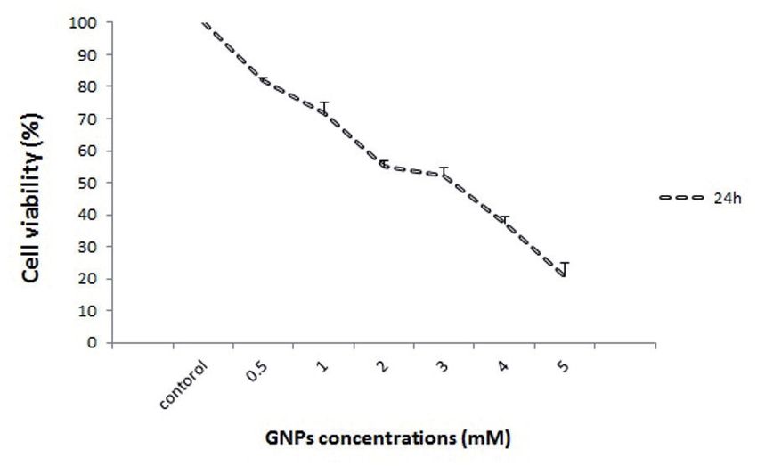
15. Shah UN, Mir JI, Ahmed N, Jan S, Fazili KM. Bioeffica-     21. Wang C, Xu L, Xu J, Yang D, Liu B, Gai S, He F and
    cy potential of different genotypes of walnut Juglans          Yang P. Multimodal imaging and photothermal therapy
    regia L. J Food Sci Technol 2018; 55: 605-618.                 were simultaneously achieved in the core–shell UCNR
16. Azizian-Shermeh O, Valizadeh M, Taherizadeh M, Bei-            structure by using single near-infrared light. Dalton
    gomi M. Phytochemical investigation and phytosyn-              Trans 2017; 46: 12147-12157.
    thesis of eco-friendly stable bioactive gold and silver    22. Liang G, Jin X, Zhang S, Xing D. RGD peptide-modified fluo-
    nanoparticles using petal extract of saffron (Crocus           rescent gold nanoclusters as highly efficient tumor-targeted
    sativus L.) and study of their antimicrobial activities.       radiotherapy sensitizers. Biomater 2017; 144: 95-104.
    Appl Nano Sci 2019; 1-14.                                  23. Izadiyan Z, Shameli K, Miyake M, Teow SY, Peh SC,
17. Karimabad MN, Mahmoodi M, Jafarzadeh A, Da-                    Mohamad SE, Mohd Taib SH. Green fabrication of
    rehkordi A, Hajizadeh MR, Khorramdelazad H, Sayadi             biologically active magnetic core-shell Fe3O4/Au na-
    AR, Rahmani F, Hassanshahi G. Evaluating of OCT-4              noparticles and their potential anticancer effect. Mater
    and NANOG was differentially regulated by a new                Sci Eng C Mater Biol Appl 2019; 96: 51-57.
    derivative indole in leukemia cell line. Immunol Lett      24. Waghmare SS, Deshmukh AM, Sadowski Z. Biosynthesis,
    2017; 190: 7-14.                                               optimization, purification and characterization of gold
18. Akbarpoor V, Karimabad MN, Mahmoodi M, Mirzaei                 nanoparticles. Afr J Microbiol Res 2014; 8: 138-146.
    MR. The saffron effects on expression pattern of cri-      25. Gardea-Torresdey J, Tiemann K, Gamez G, Dokken K,
    tical self-renewal genes in adenocarcinoma tumor cell          Tehuacanero S, Jose-Yacaman M. Gold nanoparticles
    line (AGS). Gene Rep 2020; 19: 100629.                         obtained by bio-precipitation from gold (III) solutions.
19. Wang AD, Xie CJ, Zhang YQ, Li MC, Wang X, Liu JY,              J Nanopart Res 1999; 1: 397-404.
    Xu YN. α-tetralonyl glucosides from the green walnut       26. Armendariz V, Herrera I, Jose-yacaman M, Troiani H, San-
    husks of juglans mandshurica and their antiproliferati-        tiago P, Gardea-Torresdey JL. Size controlled gold nano-
    ve effects. Planta Med 2019; 85: 335-339.                      particle formation by Avena sativa biomass: use of plants
20. Carrillo W, Gómez-Ruiz JA, Ruiz AL, Carvalho JE.               in nanobiotechnology. J Nanopart Res 2004; 6: 377-382.
    Antiproliferative activity of walnut (Juglans regia L.)    27. Philip D. Green synthesis of gold and silver nanoparticles
    proteins and walnut protein hydrolysates. J Med Food           using Hibiscus rosa sinensis. Phys E: Low-Dimensional
    2017; 20: 1063-1067.                                           Systems and Nanostructures 2010; 42: 1417-1424.

8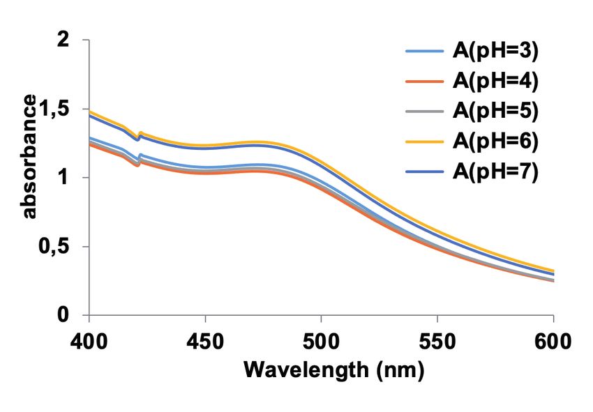
You can also read