HAGLR PROMOTES NEURON DIFFERENTIATION THROUGH THE MIR-130A-3P-MECP2 AXIS
←
→
Page content transcription
If your browser does not render page correctly, please read the page content below
Open Medicine 2021; 16: 1121–1131
Research Article
Bo Wei, Gui-rong Xiao, Cheng-long Wu, Yi-qin Xu*
HAGLR promotes neuron differentiation through
the miR-130a-3p-MeCP2 axis
https://doi.org/10.1515/med-2021-0301
received November 19, 2020; accepted April 28, 2021
1 Introduction
Abstract: Parkinson’s disease (PD) is a prevalent neuro- Parkinson’s disease (PD), a neurodegenerative disorder,
degenerative disease. Currently, the molecular mechan- is one of the most prevalent neurodegenerative diseases
isms underlying the progressions of PD are not fully worldwide [1]. Symptoms of PD generally develop over
understood. The human neuroblastoma cell line SH- years, resulting in socioeconomic burden [1,2]. The mole-
SY5Y has been widely used as an in vitro model for PD. cular mechanisms underlying PD and the effective disease-
This study aims to investigate the molecular mechanisms modifying treatments are still under clinical investigation
of the non-coding RNA-mediated SH-SY5Y differentiation [3]. The human neuroblastoma SH-SY5Y cell line is an
induced by retinoic acid (RA). By microArray analysis, in vitro model for PD research since it synthesizes both
lncRNA HAGLR was observed to be significantly upregulated dopamine (DA) and noradrenaline (NA) [4]. In addition,
during the RA-induced SH-SY5Y differentiation. Silencing retinoic acid (RA) has been used to induce the in vitro
HAGLR blocked the RA-induced SH-SY5Y differentiation. neuronal phenotypes of SH-SY5Y cells by inducing term-
Moreover, bioinformatical analysis illustrated that miR- inal neural differentiation [5]. However, the precise mole-
130a-3p contains binding sites for HAGLR. The RNA-pull cular mechanisms for the RA-induced SH-SY5Y differen-
down assay and luciferase assay demonstrated that tiation are not fully understood.
HAGLR functioned as a ceRNA of miR-130a-3p in SH-SY5Y lncRNAs, which are non-coding RNAs with relatively
cells. Overexpression of miR-130a-3p effectively inhibited larger sizes (>200 nucleotides), have been extensively stu-
SH-SY5Y differentiation. We identified MeCP2, a vital mole- died as important regulators for diverse diseases [6,7].
cule in neuronal diseases, to be a direct target of miR-130a-3p Accumulating evidence revealed that lncRNAs function
in SH-SY5Y cells by western blot and luciferase assays. The by specifically binding to DNA, RNA, or proteins to reg-
rescue experiments verified that recovery of miR-130a-3p in ulate gene expressions [8]. Moreover, studies uncovered
HAGLR-overexpressing SH-SY5Y cells could successfully that lncRNAs act as competitive endogenous RNAs (ceRNA)
overcome the RA-induced SH-SY5Y differentiation by tar- of miRNAs by sponging them to de-repress the expressions
geting MeCP2. In summary, this study reveals a potential of miRNA target genes [9]. lncRNA HAGLR (HOXD antisense
molecular mechanism for the lncRNA-HAGLR-promoted growth-associated long non-coding RNA) has been reported
in vitro neuron differentiation by targeting the miR-130a- to be positively associated with various diseases [10–13].
3p-MeCP2 axis, contributing to the understanding of the Currently, the biological roles of HAGLR in neuronal dis-
pathogenesis and progression of PD. eases such as PD have not been elucidated.
Methyl CpG binding protein 2 (MeCP2), which specifi-
Keywords: PD, neuron differentiation, lncRNA-HAGLR, cally binds to methylated cytosines on DNA, is a member of
MeCP2, miR-130a-3p the methyl-CpG-binding domain (MBD) protein family [14].
Studies reported that MeCP2 was the most abundant mole-
cule in the adult brain and tightly correlated with diverse
neuronal processes during neurodevelopment [15]. More-
over, mutations in the MeCP2 gene cause the Rett syndrome
(RTT), which is a neurologic condition affecting young girls
[16] and lead to the MeCP2 duplication syndrome (MDS) [17].
* Corresponding author: Yi-qin Xu, Department of Neurology,
These studies suggest MeCP2 is an important diagnostic bio-
Shaoxing People’s Hospital, Shaoxing, Zhejiang, 312000, China,
e-mail: tky0f888@163.com, tel: +86-18557949316 marker and therapeutic target for neuronal diseases. How-
Bo Wei, Gui-rong Xiao, Cheng-long Wu: Department of Neurology, ever, the precise roles and molecular mechanisms of MeCP2
Shaoxing People’s Hospital, Shaoxing, Zhejiang, 312000, China in PD are still under investigation.
Open Access. © 2021 Bo Wei et al., published by De Gruyter. This work is licensed under the Creative Commons Attribution 4.0 International
License.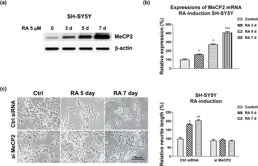
1122 Bo Wei et al.
In this study, we investigated the biological functions reports [19]. The online non-coding RNA service predicts
of HAGLR and MeCP2 during the RA-induced neuron binging of miRNA with lncRNAs or mRNA targets of miRNA
differentiation of the SH-SY5Y neuroblastoma cell line. by searching for the presence of conserved 8mer, 7mer, and
From lncRNA-microarray analysis, for the first time, we 6mer sites that match the seed region of each miRNA. Pre-
discovered HAGLR was upregulated from the in vitro neu- diction of starBase was from 108 CLIP-Seq (PAR-CLIP, HITS-
ronal differentiation model. The lncRNA–miRNA ceRNA CLIP, iCLIP, and CLASH) data sets generated by 37 indepen-
network and the targets of miRNA will be identified. dent studies.
2.4 Quantitative RT-PCR
2 Materials and methods
Total RNA was extracted from SH-SY5Y cells using the
2.1 Cell culture and treatment RNeasy Mini Kit (Qiagen, Shanghai, China) according to
the manufacturer’s instructions. The quality and concentra-
Human SH-SY5Y and SK-N-MC cells were cultured in tion of RNA samples were determined using a Nanodrop 2000
Dulbecco’s modified Eagle’s medium/ F12 medium (DMEM/ spectrophotometer (Thermo Fisher Scientific, Shanghai,
F12) (Thermo Fisher Scientific, Shanghai, China) supple- China). Total RNA was treated with DNase I, followed by
mented with 10% fetal bovine serum (FBS) (Thermo Fisher reverse transcription into cDNA using the PrimeScript RT
Scientific, Shanghai, China) with 100 units/mL penicillin Master Mix (TaKaRa, Dalian, China) according to the manu-
and 100 μg/mL streptomycin (Thermo Fisher Scientific, facturer’s instructions. The qRT-PCRs were performed using
Shanghai, China) at 37°C in a humidified atmosphere the SYBR Green qPCR Master Mix (ThermoFisher Scientific,
with 5% CO2. RA induction was performed as previously Waltham, MA, USA) in an ABI PRISM 7500 Real-Time
described [18]. Cells were treated with 5 µM RA for 3–7 System. A total of 40 amplification cycles were applied
days. Morphological changes were monitored under a with the following settings: denaturation at 95°C for 15 s,
brightfield microscope. RA was purchased from Sigma- annealing at 58°C for 30 s, and extension at 72°C for 42 s.
Aldrich (Shanghai, China). Mouse anti-MeCP2 (#M6818) The relative gene expression was determined by the 2−ΔΔCt
and β-actin antibodies (#A5441) were purchased from method. β-Actin was an internal control for lncRNA and
Sigma-Aldrich (Shanghai, China). MeCP2 mRNA detection, and U6 was an internal control
for miRNA detection. Experiments were performed in tripli-
cate and repeated three times.
2.2 Transfections of shRNA, miRNA, and
plasmid DNA 2.5 RNA pull-down assay
Transfections were performed using Lipofectamine 2000 The RNA pull-down assay was performed as described pre-
(Invitrogen, Carlsbad, CA, USA) according to the manu- viously. Briefly, biotin-labeled scramble, sense, or antisense
facturer’s protocols. SH-SY5Y cells (1 × 10⁶) were plated in lncRNA HAGLR DNA oligomers (RiboBio) were incubated
60 mm wells for 24 h. siRNA, miRNAs, shRNA, and their with the SH-SY5Y cell lysate followed by incubation for 1 h;
negative controls were synthesized by GenePharma Inc. then streptavidin-coupled agarose beads (Thermo Fisher
(Shanghai, China) and transfected at 50 nM for 48 h. Scientific, Shanghai, China) were added to pull down the
Plasmid DNA was transfected at 2 µg/well for 48 h. Cells RNA–RNA complexes. The miR-130a-3p expressions were
were then treated for 48 h with RA. Experiments were analyzed from the RNA–RNA complexes by quantitative
conducted in triplicate and repeated at least three times. RT-PCR (qRT-PCR). Experiments were repeated three times.
2.3 lncRNA–miRNA and miRNA–mRNA 2.6 Luciferase assay
interaction analysis
The wild-type (WT) and mutated (Mut) HAGLR or MeCP2
The HAGLR-miR-130a-3p and miR-130a-3p-MeCP2 3′UTR 3′UTR were amplified and cloned into the pGL3-control
bindings were predicted from Targetscan.org and starBase luciferase reporter vector system (Promega, Madison, WI,
of ENCORI http://starbase.sysu.edu.cn/ according to previous USA). SH-SY5Y cells were co-transfected with control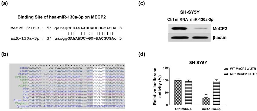
HAGLR-miR-130a-3p promotes neuron differentiation 1123
miRNA or miR-130a-3p with WT or Mut HAGLR or MeCP2 3′ times. Independent unpaired Student’s t-test was applied to
UTR using Lipofectamine 2000 (Invitrogen, Carlsbad, CA, compare differences between two groups. Values of p < 0.05
USA) for 72 h. Luciferase activity was measured using a were considered statistically significant.
Dual-Luciferase reporter assay system (Promega, Madison,
WI, USA) according to the protocol from the kit. The experi-
ment was performed in triplicate and repeated three times.
3 Results
2.7 Microarray analysis 3.1 HAGLR is upregulated during the RA-
Total RNA was extracted from SH-SY5Y cells using the
induced neuron differentiation
RNeasy Mini Kit (Qiagen, Shanghai, China) according to
We evaluated the cellular mechanisms of neuron differ-
the manufacturer’s instructions. RNA was quantified using
entiation using RA-induction of SH-SY5Y cells as an in
a Nanodrop 2000 spectrophotometer (Thermo Fisher Scientific,
vitro model, which has been widely used in PD research
Shanghai, China). Integrities of RNA samples were assessed
[4]. The results showed that the RA treatment (5 μM) of
using an Agilent Bioanalyzer 2100 (Agilent Technologies,
SH-SY5Y cells for 4 days induced remarkable morpho-
Santa Clara, CA, USA). RNA was amplified and labeled.
logic differentiation with neurite extension compared
The Cy3-labeled complementary RNA was hybridized using
with control cells that showed no morphological differentia-
the Gene Expression Hybridization Kit (Agilent Technologies,
tion (Figure 1a and Figure S1a). Moreover, the neuron differ-
Santa Clara, CA, USA). Slides were scanned using an Agilent
entiation marker, GAP43, was induced under RA treatments
Microarray Scanner (Agilent Technologies, Santa Clara, CA,
(Figure S1b), indicating successful neuronal induction by
USA). The MicroArray results were analyzed using Agilent
RA. Accumulating studies revealed that non-coding RNAs
Feature Extraction software (Agilent Technologies, Inc.) and
play vital roles during neuron differentiation [7,8]. To inves-
GeneSpring GX v11.5.1 software (Agilent Technologies, Inc.).
tigate the molecular mechanisms, we performed a lncRNA
microArray analysis to profile lncRNA expressions using
SH-SY5Y cells with or without RA induction. Among the
2.8 Western blot
differentially expressed lncRNAs in SH-SY5Y cells with RA
induction, we observed that lncRNA HAGLR was signifi-
Proteins from SH-SY5Y cells were extracted using RIPA
cantly upregulated by the RA treatment (Figure 1b), sug-
buffer (ThermoFisher Scientific, Inc.) with a 1× protease
gesting that HALGR is positively associated with neuron
inhibitor cocktail (Thermo Fisher Scientific Inc.). After
differentiation. The results from microArray were further
20 min incubation on ice, lysates were centrifuged at
validated by qRT-PCR in SH-SY5Y cells with RA induction
10,000 g for 10 min at 4°C. An equal amount of protein
at 3, 5, and 7 days. In addition, the RA-induced HAGLR
from each sample was separated by SDS-PAGE and trans-
upregulation at 5 and 7 days was observed in another neuro-
ferred to polyvinylidene difluoride membranes (PVDFs)
blastoma cell line, SK-N-MC (Figure S2a). To assess the bio-
(Millipore, Bedford, MA, USA) followed by blockage for
logical functions of HAGLR in neuron differentiation, HAGLR
1 h at room temperature with 4% bovine serum albumin
was knocked down in SH-SY5Y cells (Figure 1d). As we
(BSA). Membranes were incubated with primary antibo-
expected, SH-SY5Y and SK-N-MC cells with HAGLR silencing
dies at 1:1,000 overnight at 4°C. After washing with phos-
displayed little response to RA treatments compared with
phate buffered saline with tween 20 (PBST), membranes
the control shRNA transfected cells (Figure 1e and Figure
were incubated with secondary antibody (1:3,000) for 1 h at
S2b). Taken together, these results consistently demon-
room temperature. Blots were visualized with enhanced chemi-
strated that HAGLR promotes neuron differentiation from
luminescence (Millipore, Bedford, MA, USA). β-Actin was used
an RA-induced in vitro SH-SY5Y model.
as a loading control. Experiments were repeated three times.
2.9 Statistical analysis 3.2 HAGLR sponges miR-130a-3p as a ceRNA
in SH-SY5Y cells
Data were analyzed using Prism version 7.0 (GraphPad
Software, La Jolla, CA, USA). Data were expressed as mean We next explored the molecular mechanisms underlying
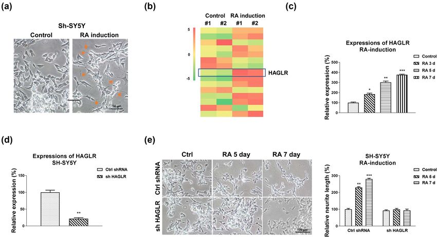
± standard deviation (SD). Experiments were repeated three the HAGLR-promoted neuron differentiation. Previous1124 Bo Wei et al. Figure 1: Roles of HAGLR in RA-induced neuron differentiation. (a) Effects of RA treatment on cell morphology. SH-SY5Y cells were treated with 5 µM RA for 5 days. (b) SH-SY5Y cells were treated with RA using the above protocol. RNAs were isolated and lncRNA-microArray analysis was performed in duplicate. (c) SH-SY5Y cells were treated with 5 µM RA for 3, 5, and 7 days. HAGLR expressions were examined by qRT-PCR. (d) SH-SY5Y cells were transfected with control shRNA or HAGLR shRNA for 48 h, and expression of HAGLR was determined by qRT- PCR. (e) SH-SY5Y cells were transfected with control shRNA or HAGLR shRNA for 48 h, followed by treatments with 5 µM RA for 5 and 7 days. Cell morphologies were examined under a microscope, and the neurite length was measured. *p < 0.05; **p < 0.01; and ***p < 0.001. studies uncovered that lncRNAs function by interfering downregulates miR-130a-3p, HAGLR was overexpressed with target miRNAs as molecular sponges [9]. The down- or silenced in SH-SY5Y cells. Expectedly, overexpression regulation of miRNAs by lncRNA leads to de-repression of HAGLR effectively blocked the miR-130a-3p expres- of their target mRNAs [20]. To identify the potential sions, while knockdown of HAGLR significantly upregu- miRNA targets of HAGLR, we analyzed the HAGLR– lated miR-130a-3p expressions (Figure 2d). To validate miRNA interaction through the non-coding RNA data- the binding of miR-130a-3p on HAGLR, we performed base, starBase2.0. We observed miR-130a-3p, which plays an RNA pull-down assay. The biotin-labeled scramble, an important role during neuron development and neu- sense, or antisense DNA probe of HAGLR was incubated ronal diseases [21], contains HAGLR binding sites (Figure 2a). with SH-SY5Y cell lysates. The qRT-PCR results demon- We then evaluated the functional roles of miR-130a-3p strated that only the antisense HAGLR DNA probe pulled during RA-induced neuron differentiation. qPCR results down the enriched miR-130a-3p (Figure 2e). The endo- remarkedly showed a reverse phenotype between HAGLR genous miR-130a-3p could not be effectively pulled down and miR-130a-3p, which was significantly downregulated by the scramble or sense HAGLR probe (Figure 2e), sug- in SH-SY5Y and SK-N-MC cells with RA induction at 3, gesting that HAGLR specifically interacts with miR-130a- 5, and 7 days (Figure 2b and Figure S3a). Moreover, 3p in neuroblastoma cells. To validate whether HAGLR although RA treatments suppressed miR-130a-3p expres- directly binds on the seeding region of miR-130a-3p, SH- sions, exogenous overexpression of miR-130a-3p (Figure SY5Y cells were co-transfected with a luciferase vector S3b and c) rendered cells to maintain relatively high miR- containing the wild-type HAGLR (WT-HAGLR) or the 130a-3p expression levels (Figure S3d and e). Expectedly, binding site mutant HAGLR (Mut-HAGLR) and control SH-SY5Y and SK-N-MC cells with exogenous overexpres- miRNA or miR-130a-3p. Luciferase activity of cells with sion of miR-130a-3p showed no effective morphological WT-HAGLR and miR-130a-3p co-transfection was signifi- changes in the neurite extension under RA treatments cantly suppressed compared with that from Mut-HAGLR (Figure 2c and Figure S3f). To assess whether HAGLR and miRNA-130a-3p co-transfection (Figure 2f). In
HAGLR-miR-130a-3p promotes neuron differentiation 1125
Figure 2: HAGLR sponges miR-130a-3p to negatively regulate its expression. (a) Bioinformatics analysis of potential binding of HAGLR on
miR-130a-3p. (b) SH-SY5Y cells were treated with 5 µM RA for 3, 5, and 7 days. miR-130a-3p expressions were examined by qRT-PCR. (c) SH-
SY5Y cells were transfected with control miRNA or miR-130a-3p for 48 h, followed by treatments with 5 µM RA for 5 and 7 days. Cell
morphologies were examined under a microscope, and the neurite length was measured. (d) SH-SY5Y cells were transfected with control,
the HAGLR overexpression plasmid, or HAGLR shRNA for 48 h; miR-130a-3p expressions were examined by qRT-PCR. (e) SH-SY5Y cell lysates
were incubated with biotin-labeled scramble, sense, or antisense HAGLR DNA probes. Biotin pull-down assay was performed, and
miR-130a-3p expression was assessed by qRT-PCR. (f) Dual-luciferase reporter assay was performed in SH-SY5Y cells with co-transfection
of WT-HAGLR or Mut-HAGLR with control miRNA, or miR-130a-3p. *p < 0.05; **p < 0.01; and ***p < 0.001.
summary, these results demonstrated that HAGLR sponges luciferase vector containing WT-MeCP2 3′UTR or the miR-
miR-130a-3p by forming a ceRNA network. 130a-3p binding site mutant MeCP2 3′UTR (Mut-MeCP2) and
control miRNA or miR-130a-3p. Luciferase activity of the
vector containing WT-MeCP2 was remarkedly blocked by
miR-130a-3p (Figure 3d). Expectedly, the luciferase activity
3.3 miR-130a-3p directly targets MeCP2 and of the Mut-MeCP2 vector has no significant change by miR-
inhibits the RA-induced neuron 130a-3p overexpression (Figure 3d). Together, these results
differentiation demonstrated that miR-130a-3p could directly target 3′UTR
of MeCP2.
Since miRNAs function via targeting mRNA 3′UTR to down-
regulate target mRNA expressions [20], we performed miR-
NA–mRNA interaction analysis from the non-coding RNA
database, starBase 2.0. Interestingly, the 3′UTR of MeCP2, 3.4 MeCP2 is induced and promotes the
which is an important molecule in neuron differentiation RA-induced neuron differentiation
and neuronal diseases, contains conserved miR-130a-3p
binding sites through multiple species (Figure 3a and b). We continue to evaluate the roles of MeCP2 in RA-
To examine whether miR-130a-3p could suppress protein induced neuron differentiation. Under RA treatments,
expression of MeCP2, miR-130a-3p or control miRNA was protein and mRNA expressions of MeCP2 were signifi-
transfected into SH-SY5Y cells. The results from western cantly upregulated (Figure 4a and b and Figure S4a).
blot showed that overexpression of miR-130a-3p effectively Furthermore, MeCP2 was knocked down by siRNA in
blocked MeCP2 protein expression in SH-SY5Y cells (Figure SH-SY5Y cells. The results in Figure 4c and Figure S4b
3d). To validate the direct binding of miR-130a-3p on 3′UTR illustrate that SH-SY5Y cells with downregulated MeCP2
of MeCP2 mRNA, SH-SY5Y cells were co-transfected with expression exhibited unchanged neurite extension in1126 Bo Wei et al. Figure 3: MeCP2 is directly targeted by miR-130a-3p. (a) Bioinformatics analysis of potential binding of miR-130a-3p on 3′UTR of MeCP2. (b) The conserved binding sites of miR-130a-3p on 3′UTR of MeCP2 in multiple species. (c) SH-SY5Y cells were transfected with control or miR-130a-3p for 48 h; the protein expression of MeCP2 was detected by western blot. (d) Dual-luciferase reporter assay was performed in SH-SY5Y cells with co-transfection of WT-MeCP2 or Mut-MeCP2 with control miRNA, or miR-130a-3p. **p < 0.01. Figure 4: Roles of MeCP2 in the RA-induced neuron differentiation. (a) SH-SY5Y cells were treated with 5 µM RA for 3, 5, and 7 days. Protein and (b) mRNA expressions of MeCP2 were detected. (c) SH-SY5Y cells were transfected with control siRNA or MeCP2 siRNA for 48 h, followed by treatments with 5 µM RA for 5 and 7 days. Cell morphologies were examined under a microscope, and the neurite length was measured. *p < 0.05; **p < 0.01; and ***p < 0.001.
HAGLR-miR-130a-3p promotes neuron differentiation 1127
response to RA induction compared with control cells, SY5Y cells were transfected with control, HAGLR over-
indicating that MeCP2 promotes the process of neuron expression plasmid alone, or with miR-130a-3p. qRT-PCR
differentiation. and western blot results demonstrated that co-transfection
of HAGLR with miR-130a-3p successfully restored miR-130a-3p
and MeCP2 protein levels compared with HAGLR over-
expressing cells (Figure 6a and b). As expected, SH-SY5Y
3.5 Restoration of MeCP2 in miR-130a-3p cells with co-transfection of HAGLR and miR-130a-3p re-
overexpressing SH-SY5Y cells rescues suppressed the RA-induced neuron differentiation pheno-
the RA-induced neuron differentiation type compared with that from HAGLR overexpressing cells
(Figure 6c). In summary, these mechanism rescue experi-
Rescue experiments were performed to validate whether ments consolidated a HAGLR-miR-130a-3p-MeCP2 axis that
miR-130a-3p blocked the RA-induced neuron differentia- contributes to the processes of neuron differentiation.
tion through targeting MeCP2. SH-SY5Y cells were trans-
fected with control miRNAs, miR-130a-3p alone or with
MeCP2 overexpression plasmid. Co-transfection of miR-
130a-3p with MeCP2 successfully recovered MeCP2 pro- 4 Discussion
tein levels compared with miR-130a-3p overexpression
cells (Figure 5a). Moreover, the expected results demon- PD is one of the most prevalent neurodegenerative dis-
strated that SH-SY5Y cells with the restoration of MeCP2 eases worldwide [1,2]. Currently, the molecular mechan-
rescued the RA-induced neuron differentiation pheno- isms underlying PD and the effective disease-modifying
type (Figure 5b). These rescue experiments further con- approaches are not fully understood. The human neuro-
firmed that miR-130a-3p directly targets MeCP2 to inhibit blastoma SH-SY5Y cell line is widely studied for investi-
neuron differentiation. gating the molecular and cellular mechanisms underlying
PD pathogenesis and progression since it synthesizes both
DA and NA [4]. Moreover, although the SH-SY5Y cell line
displays a number of cancerous characteristics, most
genes regulations and signal pathways in PD pathogenesis
3.6 HAGLR promoted neuron differentiation are intact [4]. It is difficult to obtain and maintain human
by targeting the miR-130a-3p- dopaminergic neurons as primary cells, which are mainly
MeCP2 axis affected in PD [4]. Thus, the neuroblastoma SH-SY5Y cell
line is a widely used in vitro model for PD research. Cur-
Finally, we assessed whether neuron differentiation was rently, the biological roles of lncRNA HAGLR in neuronal
regulated by the HAGLR-miR-130a-3p-MeCP2 axis. SH- diseases remain unclear. In this study, we first reported the
Figure 5: Restoration of MeCP2 rescues the miR-130a-3p suppressed neuron differentiation. (a) SH-SY5Y cells were transfected with control,
miR-130a-3p alone, or with MeCP2 overexpression plasmid for 48 h; protein expression of MeCP2 was determined by western blot. (b) The
above transfected cells were treated with 5 µM RA for 5 and 7 days. Cell morphologies were examined under a microscope, and the neurite
length was measured. *p < 0.05 and **p < 0.01.1128 Bo Wei et al.
Figure 6: The HAGLR-miR-130a-3p-MeCP2 axis in neuron differentiation. (a) SH-SY5Y cells were transfected with control, HAGLR alone, or
with miR-130a-3p for 48 h; expression of miR-130a-3p was detected by qRT-PCR and (b) protein expression of MeCP2 was determined by
western blot. (c) The above transfected cells were treated with 5 µM RA for 5 and 7 days. Cell morphologies were examined under a
microscope, and the neurite length was measured. *p < 0.05 and **p < 0.01.
non-coding RNA-regulated neuron differentiation using MeCP2 gene cause Rett syndrome (RTT), a neurologic
SH-SY5Y cells as an in vitro model. Under RA induction, condition affecting primarily young girls [16]. Interestingly,
we detected that HAGLR was significantly upregulated in girls with RTT exhibiting motor deficits showed similar pheno-
SH-SY5Y cells from both microArray analysis and qPCR types to those in PD [24], suggesting MeCP2 is involved in
results. In addition, silencing HAGLR effectively blocked the defects of the nigrostriatal pathway. A recent study used
the RA-induced SH-SY5Y differentiation based on the the 6-hydroxydopamine-induced human neuroblastoma
observations that the neurite extension of SH-SY5Y cells cell (SH-SY5Y cell) injury as a cell model of PD [25]. They
with lower HAGLR expression showed little response to described that overexpression of MeCP2 was able to ame-
RA treatments, suggesting an essential role of HAGLR in liorate the 6-hydroxydopamine-induced apoptosis of SH-
SH-SY5Y cell differentiation. SY5Y cells, suggesting that MeCP2 is a potential therapeutic
Accumulating studies demonstrated that miRNAs target for the treatment of PD [25]. Currently, the precise
play regulatory roles in neuron development and neu- roles and molecular mechanisms of MeCP2 in PD have not
ronal diseases [20]. Moreover, the lncRNA–miRNA inter- been elucidated. Here, we described that the expressions of
action has been shown to control vital functions during MeCP2 were significantly upregulated in SH-SY5Y cells by
the molecular and cellular processes of human malignan- RA induction. One advantage of this study is that we illu-
cies, including neuronal diseases [8,9]. A recent study strated the direct binding of miR-130a-3p on 3′UTR of
reported that miR-130a-3p regulates VEGFR-2 expression MeCP2 in SH-SY5Y cells. Furthermore, the rescue experi-
in sensory and motor neurons during development [22]. ments validated that the miR-130a-3p-inhibited SH-SY5Y
Moreover, the miR-130a-3p/DAPK1 axis was known to cell differentiation was by direct targeting of MeCP2. Thus,
regulate the pathophysiology of neonatal hypoxic-ischemia it was possible to target the above signaling pathways in
encephalopathy [23], suggesting miR-130-3p to be a potential neuronal disease to improve the clinical therapeutic out-
therapeutic target for the hypoxic ischemia encephalopathy comes. These strategies still have limitations owing to the
treatment. However, the miR-130a-3p-mediated neuron dif- fact that the neuroblastoma cell line is not purely neuron
ferentiation has not been investigated. Here, we show that cells since it is oncogenically transformed with catechola-
miR-130a-3p was remarkedly downregulated in SH-SY5Y minergic, resulting in different physiological characteristics
cells under RA induction. Overexpression of miR-130a-3p compared to those from the normal DAergic neuronal fea-
inhibited the morphological changes of SH-SY5Y cells under tures. In addition, the above in vitro signaling pathway
RA treatments. Bioinformatics analysis revealed that HAGLR needs to be verified in animal models.
contains miR-130a-3p binding sites. The predicted ceRNA In summary, this study reports a non-coding RNA-
network was further validated by the RNA pull-down assay based molecular mechanism for the PD using SH-SY5Y
and luciferase assay. cells as an in vitro model. HAGLR positively regulates SH-
MeCP2 is the most abundant methyl-DNA binding SY5Y cell differentiation, which is induced by RA via tar-
domain family member in the adult brain and is tightly geting the miR-130a-3p-MeCP2 axis, contributing to an
correlated with diverse neuronal processes during neuro- extensive understanding of the pathogenesis and pro-
development [15]. It was known that mutations in the gression of PD.HAGLR-miR-130a-3p promotes neuron differentiation 1129
Acknowledgement: The authors appreciate the contribu- regulate epithelial-mesenchymal transition and metastatic
tions from all medical doctors and research scientists potential in esophageal cancer by regulating LAMP3. FASEB J.
from the Department of Neurology, Shaoxing People’s 2019;33:10490–504.
[11] Lu C, Ma J, Cai D. Increased HAGLR expression promotes non-
Hospital, for generously supporting this study.
small cell lung cancer proliferation and invasion via enhanced
de novo lipogenesis. Tumour Biol. 2017;
Funding information: No funds. 39:1010428317697574.
[12] Sun W, Nie W, Wang Z, Zhang H, Li Y, Fang X. Lnc HAGLR
Author contributions: B.W. and Y.Q.X. designed the experi- promotes colon cancer progression through sponging miR-
185-5p and activating CDK4 and CDK6 in vitro and in vivo. Onco
ments and wrote the manuscript. B.W., G.R.X., Ch.L.W.,
Targets Ther. 2020;13:5913–25.
and Y.Q.X. carried out the experiments and analyzed [13] Zakany J, Darbellay F, Mascrez B, Necsulea A, Duboule D.
the data. Control of growth and gut maturation by HoxD genes and the
associated lncRNA Haglr. Proc Natl Acad Sci USA.
Conflict of interest: The authors declare that there are no 2017;114:E9290–9.
[14] Horvath PM, Monteggia LM. MeCP2 as an activator of gene
conflicts of interest.
expression. Trends Neurosci. 2018;41:72–4.
[15] Sharma K, Singh J, Frost EE, Pillai PP. MeCP2 in central nervous
Data availability statement: The datasets used and/or system glial cells: current updates. Acta Neurobiol Exp (Wars).
analyzed during the current study are available from 2018;78:30–40.
the corresponding author on reasonable request. [16] Shah RR, Bird AP. MeCP2 mutations: progress towards under-
standing and treating Rett syndrome. Genome Med. 2017;9:17.
[17] Gulmez Karaca K, Brito DVC, Oliveira AMM. MeCP2: A critical
regulator of chromatin in neurodevelopment and adult brain
function. Int J Mol Sci. 2019;20:4577.
References [18] Teppola H, Sarkanen JR, Jalonen TO, Linne ML. Morphological
differentiation towards neuronal phenotype of SH-SY5Y neuro-
[1] Reich SG, Savitt JM. Parkinson’s disease. Med Clin North Am. blastoma cells by estradiol, retinoic acid and cholesterol.
2019;103:337–50. Neurochem Res. 2016;41:731–47.
[2] Ascherio A, Schwarzschild MA. The epidemiology of [19] Li JH, Liu S, Zhou H, Qu LH, Yang JH. StarBase v2.0: decoding
Parkinson’s disease: risk factors and prevention. Lancet miRNA-ceRNA, miRNA-ncRNA and protein-RNA interaction
Neurol. 2016;15:1257–72. networks from large-scale CLIP-Seq data. Nucleic Acids Res.
[3] Raza C, Anjum R, Shakeel NUA. Parkinson’s disease: 2014;42:D92–7.
mechanisms, translational models and management strate- [20] Mohr AM, Mott JL. Overview of microRNA biology. Semin Liver
gies. Life Sci. 2019;226:77–90. Dis. 2015;35:3–11.
[4] Xicoy H, Wieringa B, Martens GJ. The SH-SY5Y cell line in [21] Glaesel K, May C, Marcus K, Matschke V, Theiss C, Theis V.
Parkinson’s disease research: a systematic review. Mol miR-129-5p and miR-130a-3p regulate VEGFR-2 expression in
Neurodegener. 2017;12:10. sensory and motor neurons during development. Int J Mol Sci.
[5] Gonulalan EM, Bayazeid O, Yalcin FN, Demirezer LO. The roles 2020;21:3839.
of valerenic acid on BDNF expression in the SH-SY5Y cell. [22] Glaesel K, May C, Marcus K, Matschke V, Theiss C, Theis V.
Saudi Pharm J. 2018;26:960–4. miR-129-5p and miR-130a-3p regulate VEGFR-2 expression in
[6] Peng WX, Koirala P, Mo YY. lncRNA-mediated regulation of cell sensory and motor neurons during development. Int J Mol Sci.
signaling in cancer. Oncogene. 2017;36:5661–7. 2020;21:3839.
[7] Lyu Y, Bai L, Qin C. Long noncoding RNAs in neurodevelopment [23] Feng M, Zhu X, Zhuo C. H19/miR-130a-3p/DAPK1 axis regu-
and Parkinson’s disease. Animal Model Exp Med. lates the pathophysiology of neonatal hypoxic-ischemia
2019;2:239–51. encephalopathy. Neurosci Res. 2021;163:52–62.
[8] Riva P, Ratti A, Venturin M. The long non-coding RNAs in [24] Gantz SC, Ford CP, Neve KA, Williams JT. Loss of Mecp2 in
neurodegenerative diseases: novel mechanisms of pathogen- substantia nigra dopamine neurons compromises the nigros-
esis. Curr Alzheimer Res. 2016;13:1219–31. triatal pathway. J Neurosci. 2011;31:12629–37.
[9] Paraskevopoulou MD, Hatzigeorgiou AG. Analyzing MiRNA- [25] Xie T, Zhang J, Yuan X, Yang J, Ding W, Huang X, et al. Is
lncRNA Interactions. Methods Mol Biol. 2016;1402:271–86. X-linked methyl-CpG binding protein 2 a new target for the
[10] Yang C, Shen S, Zheng X, Ye K, Sun Y, Lu Y, et al. Long non- treatment of Parkinson’s disease. Neural Regen Res.
coding RNA HAGLR acts as a microRNA-143-5p sponge to 2013;8:1948–57.1130 Bo Wei et al. Appendix Figure S1: Effects of retinoic acid treatment on neuron differentiation. (a) SH-SY5Y cells were treated with RA at 5 µM for 5 days, the relative neurite length was calculated. (b) Expressions of GAP43 in SH-SY5Y cells under RA treatments at 5 days and 7 days were determined by qRT- PCR. **, p < 0.01; ***, p < 0.001. Figure S2: Effects of retinoic acid treatment on SK-N-MC cells. (a) Expressions of HAGLR in SK-N-MC cells under RA treatments for 3, 5 and 7 days were determined by qRT-PCR. (b) The relative neurite length was calculated in SK-N-MC cells without or with HAGLR silencing under RA treatments for 5 and 7 days. *, p < 0.05; **, p < 0.01; ***, p < 0.001.
HAGLR-miR-130a-3p promotes neuron differentiation 1131 Figure S3: miR-130a-3p suppresses RA-induced neuron differentiation. (a) Expressions of miR-130a-3p in SK-N-MC cells under RA treat- ments at 3, 5 and 7 days were determined by qRT-PCR. (b) Effects of miR-130a-3p overexpression in SH-SY5Y and (c) SK-N-MC cells. (d) Detection of miR-130a-3p expressions in SH-SY5Y and (e) SK-N-MC cells without or with miR-130a-3p overexpression under RA treatments for 5 and 7 days. (f) The relative neurite length was calculated in SK-N-MC cells without or with miR-130a-3p overexpression under RA treatments for 5 and 7 days. *, p < 0.05; **, p < 0.01; ***, p < 0.001. Figure S4: Silencing MeCP2 suppresses RA-induced neuron differentiation. (a) SK-N-MC cells under RA treatments for 3, 5 and 7 days, expressions of MeCP2 were determined by qRT-PCR. (b) The relative neurite length was calculated in SK-N-MC cells without or with MeCP2 silencing under RA treatments for 5 and 7 days. *, p < 0.05; **, p < 0.01; ***, p < 0.001.
You can also read