High-resolution manometry findings with hiatus hernia
←
→
Page content transcription
If your browser does not render page correctly, please read the page content below
Review Article
                                                                                                                                         Page 1 of 10

High-resolution manometry findings with hiatus hernia
Peter J. Kahrilas

Department of Medicine, Northwestern University, Feinberg School of Medicine, Chicago, Illinois, USA
Correspondence to: Peter J. Kahrilas. Department of Medicine, Northwestern University, Feinberg School of Medicine, 676 St Clair St, 14th floor,
Chicago, Illinois 60611-2951, USA. Email: p-kahrilas@northwestern.edu.

                 Abstract: The esophagogastric junction (EGJ) is comprised of both the intrinsic lower esophageal
                 sphincter (LES) and extrinsic crural diaphragm (CD) such that intraluminal pressure represents a composite
                 of the two. With type I hiatal hernias there is weakening of the pharyngoesophageal ligament attaching
                 the esophagus to the CD such that the distal esophagus, including the LES, becomes displaced cephalad.
                 High-resolution manometry (HRM), can accurately detect an axial hiatal hernia as confirmed by studies
                 comparing HRM to barium esophagram, upper endoscopy, or intraoperative findings. In HRM studies,
                 hiatus hernia becomes evident by a spatial separation between the pressure signature of the LES and that
                 of the CD; the operative confirmation of hiatus hernia correlates with this separation exceeding 1 cm.
                 With LES-CD separation of 1 cm and the RIP localized at the CD, and (III) hiatus hernia with LES-
                 CD separation >1 cm and the RIP localized at the LES.

                 Keywords: Hiatal hernia; high-resolution manometry (HRM); gastroesophageal reflux disease (GERD)

                 Received: 09 November 2019. Accepted: 04 March 2020; Published: 20 January 2021.
                 doi: 10.21037/ales.2020.03.08
                 View this article at: http://dx.doi.org/10.21037/ales.2020.03.08

Introduction                                                                 to interpret the complex intraluminal pressure signature
                                                                             of both the normal and anatomically disrupted EGJ.
High-resolution manometry (HRM) is usually
                                                                             Among the technical difficulties encountered are the effects
recommended as a part of the evaluation of gastroesophageal                  of respiratory variation, the effect of movement of the
reflux disease (GERD) when a procedural intervention                         pressure sensor relative to the sphincter with breathing
is under consideration, be that a fundoplication, Linx                       and swallowing, the recording fidelity of the sensor,
implant, or transoral incisionless fundoplication. Among                     distinguishing an intrinsic contraction from extrinsic
the objectives of that evaluation is an assessment of the                    compression, the effects of pulsations from the heart and
integrity of the esophagogastric junction (EGJ) as an                        aorta, and the extreme radial asymmetry and temporal
antireflux barrier including the characterization of an                      variability of the EGJ itself. It is from this vantage point
axial hiatal hernia. However, there are numerous potential                   that the HRM findings of both the normal EGJ and type I
pitfalls in that assessment and consensus is lacking on how                  (sliding) hiatus hernia must be explored.

© Annals of Laparoscopic and Endoscopic Surgery. All rights reserved.     Ann Laparosc Endosc Surg 2021;6:5 | http://dx.doi.org/10.21037/ales.2020.03.08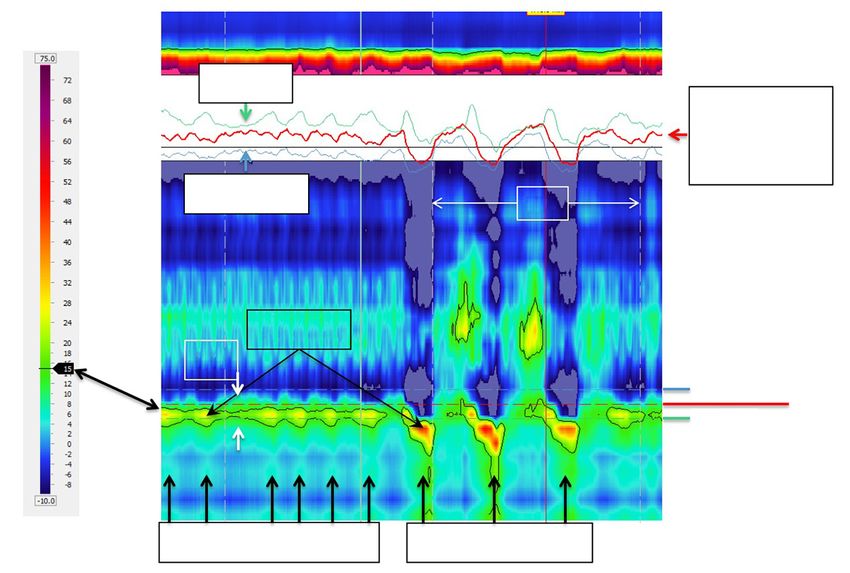
Page 2 of 10                                                                                  Annals of Laparoscopic and Endoscopic Surgery, 2021

                                                                3D-HRM assembly

                                                                        10 mm

                                                                                Averages 12
                                                                                   radial
                             Standard                                            pressures
                               HRM
                              sensor

                                                                                   8 independent
                                                                                       sensors
                                                                                      45° apart

                                                             7.5 mm
                                                                                       3D-HRM segment

Figure 1 Photograph and schematic of a 3D HRM sensor that incorporates a 6 cm 3D segment designed to record the EGJ along with
conventional circumferentially sensitive HRM sensors proximal and distal to the 3D segment for recording within the esophagus and
stomach respectively. Each locus along the 3D segment is comprised of 8 independent sensors oriented at 45° increments circumferentially.
This in contrast to the conventional HRM sensors that contain 12 elements, but they average those signals to record a single pressure.
Figure used with permission from the Esophageal Center at Northwestern.

Normal EGJ pressure morphology: teasing apart                                       profound implications with respect to the EGJ because
the 3D-HRM signal                                                                   that pressure signal is comprised of both an extrinsic crural
                                                                                    diaphragm (CD) and intrinsic lower esophageal sphincter
High among the difficulties encountered in interrogating
                                                                                    (LES) component, each of which is subject to independent
the EGJ pressure profile are limitations of the pressure
                                                                                    physiological control mechanisms and pathophysiology.
sensors themselves. Historically, relatively few sensors
                                                                                    An experimental approach to unraveling this complexity
were employed, and one needed to repeatedly reposition
the manometric catheter or actually pull it across the                              was to develop a 3D HRM device that preserved rather
EGJ to obtain an axial pressure profile. However, with                              than averaged the circumferentially unequal pressures
the advent of HRM and the interpolation paradigms that                              (1,2). Figure 1 is an illustration of that device comprised of
transform HRM into esophageal pressure topography                                   a 3D segment intended to interrogate the EGJ as well as
plots, such things are of historical interest only. There is                        ‘conventional’ HRM sensors both proximal and distal to it
no going back to those archaic techniques. None the less,                           to record esophageal and intragastric pressure respectively.
understanding the HRM pressure signature of the EGJ                                 All told, the 3D-HRM device monitors 128 independent
in a pressure topography format can be quite challenging                            pressure signals in real time, 96 of which are focused on the
as it bears no resemblance to recordings obtained with                              EGJ. Although this device is not commercially available,
line tracings from 3–8 widely spaced pressure sensors.                              recordings obtained from it are very informative with
Adding to the difficulty is that with the current HRM                               respect to understanding the appearance of recordings
catheter designs each of the closely spaced sensors is                              obtained with standard HRM devices.
circumferentially sensitive such that if it is contacted on one                        Figure 2 is a sagittal view of an MRI scan in a normal
side it can yield a pressure signal that is indistinguishable                       individual selected for illustration because the CD and
from the circumferential squeeze of a sphincter. This has                           esophagus outlined in orange and green respectively

© Annals of Laparoscopic and Endoscopic Surgery. All rights reserved.            Ann Laparosc Endosc Surg 2021;6:5 | http://dx.doi.org/10.21037/ales.2020.03.08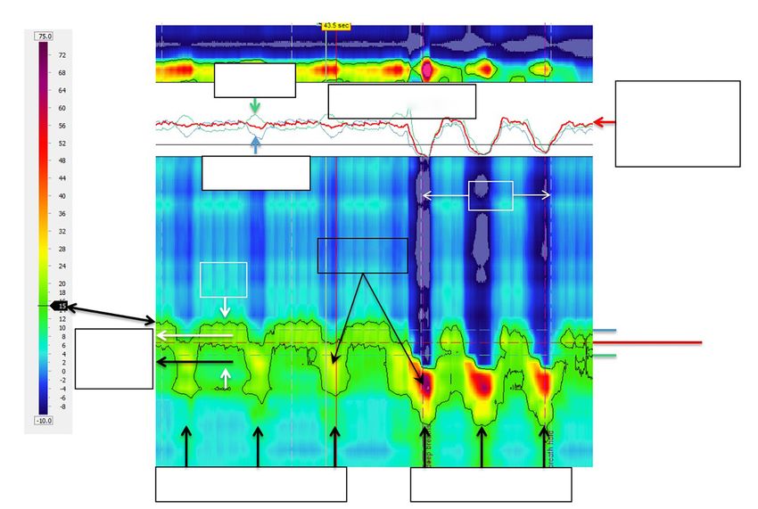
Annals of Laparoscopic and Endoscopic Surgery, 2021                                                                                       Page 3 of 10

                                                                        MRI Median Sagittal Veiw

                                                       Esophagus

                                                                                Heart

                                                                         58°
                                                                                    Axis of CD

                                                              Aorta

                                                                                   Axis of esophagus

                                                                               Rt crus

Figure 2 Mid sagittal MRI image highlighting the EGJ with the 3D HRM placed within the lumen of the esophagus. Note the characteristic
bend imposed on the HRM catheter as it traverses the EGJ. The CD is outlined in orange while the esophagus is outlined in green. Note
that the axis of the CD is not perpendicular to the axis of the esophagus and that wits 3D pressure signature (cylindrical insert) is reflective
of this. The insert, taken at peak inspiration shows the CD apex signal largely restricted to the anterolateral walls of the EGJ. Figure used
with permission from the Esophageal Center at Northwestern.

were both readily identifiable. Note that the axis of the                       unfolding the cylinder and depicting the 3D signal as
CD is not perpendicular to the axis of the esophagus;                           a rectangle centered on the CD apex as defining the 6
rather, it is at about 58° in this example. Consequently,                       o’clock orientation. When imaged in real time, the CD
its pressure signature will be oblique with respect to the                      apex intensifies and then de-intensifies with inspiration
axis of the esophagus. Inserted into the esophagus is a 3D                      and expiration respectively. In fact, the CD signal is so
HRM catheter with the 3D segment depicted in yellow                             dominant that one wonders where the LES is in Figure 3. A
and the remainder in red. Now consider the cylindrical                          key attribute of the LES is that, being a product of circular
representation of the 3D segment recording at inspiration                       (or slightly spiral) muscle contraction, it is a circumferential
inserted onto the MRI. Brighter colors on the cylinder as                       pressure perpendicular to the axis of the esophagus and
well as outward deflection depict greater pressure. Note                        anatomically localized within the hiatus. However, it is
that the CD signal (blue arrow) is the most intense pressure                    impossible to localize the LES in areas where the pressure
recorded but is largely restricted to the anterolateral                         topography is obscured by the superimposed CD, so its
wall of the esophagus. Its counterpart, the green arrow is                      margins can only be identified in the radial sectors not
temporally synchronized with it on the opposite wall and                        affected by the CD pressure recording. The horizontal
likely reflects contact of the HRM device with the opposing                     white dashed lines approximate the margins of the LES in
wall of the esophagus.                                                          Figure 3. Note that with the sensors spaced 7.5 mm apart
   Although the cylindrical representation of the                               this implies that the intrinsic LES is about 2 cm long,
3D-HRM signal is illustrative, it is difficult to interrogate                   which is consistent with physiological data. Support for
quantitatively because half or the signals are hidden and                       this interpretation of the Figure 3 pressure topography is
the whole recording cannot be viewed without rotating                           in Figure 4. The left panel of Figure 4 shows the pressure
the cylinder. Hence, Figure 3 shows the technique of                            topography during the LES after-contraction following

© Annals of Laparoscopic and Endoscopic Surgery. All rights reserved.      Ann Laparosc Endosc Surg 2021;6:5 | http://dx.doi.org/10.21037/ales.2020.03.08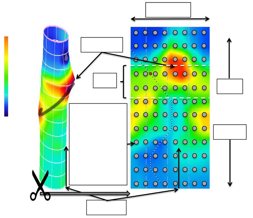
Page 4 of 10                                                                                   Annals of Laparoscopic and Endoscopic Surgery, 2021

                                                    Unfolding the 3D-HRM segment pressure topography

                                                                                           Around

                                          50
                                                                  CD Apex
                                          mmHg

                                          25
                                                                       LES
                                                                                                                Up

                                          0                          96
                                                              simultaneous
                                                                 pressure                                       Down
                                                                recordings
                                                               (one instant
                                                                  in time
                                                                 depicted)

                                                                   6 o'clock

Figure 3 Unfolding the 3D pressure recording from a cylinder to a rectangle. The gray dots signify the locations of the 96 pressure sensors
that contributed to generating this pressure topography plot obtained at peak inspiration. The topography plot is created by interpolating
between pressure sensors both circumferentially and axially. See text for further detail. Figure used with permission from the Esophageal
Center at Northwestern.

                                                     Around                                         Around

                                 Sensor 29                                                                           29 Sensor
                                         30                                    LES                                   30
                                         31                                                                          31
                                         32                                                                          32
                                                                                                    Hernia
                                         33                                  CD Apex                                 33
                                         34                                   50                                     34
                                         35                                                                          35
                                                                               mmHg
                                         36                                                                          36
                                         37                                   25                                     37
                                         38                                                                          38
                                         39                                                      6 o'clock           39
                                                      6 o'clock
                                         40                                   0                                      40
                                               LES after-contraction                            Hiatus hernia

Figure 4 Special circumstances selected to confirm the interpretation in Figure 3 of the CD and LES contributions to the 3D pressure
topography. The left panel illustrates a strong LES after contraction following a test swallow in a person without a hiatal hernia. Note the
bright red circumferential band of pressure contributed by the LES. The CD apex, on the other hand, remains largely restricted to the
anterolateral aspects of the recording intensifying and de-intensifying with respiration (not shown). The right panel illustrates a normal
volunteer who was found to have a hiatus hernia (subsequently confirmed on endoscopy). The CD apex signal is now centered 2.5 cm distal
to the center of the LES with a pressure node between the two. Again, the CD apex remains largely restricted to the anterolateral aspects
of the recording intensifying and de-intensifying with respiration (not shown). Figure used with permission from the Esophageal Center at
Northwestern.

© Annals of Laparoscopic and Endoscopic Surgery. All rights reserved.             Ann Laparosc Endosc Surg 2021;6:5 | http://dx.doi.org/10.21037/ales.2020.03.08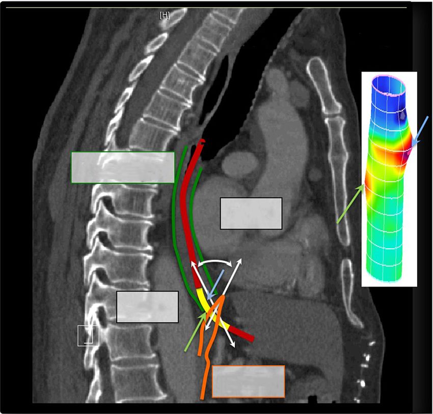
Annals of Laparoscopic and Endoscopic Surgery, 2021                                                                                           Page 5 of 10

                                                                   Around

                                        Sensor   29                                              CD Apex
                                                 30

                                                 31                                             Respiratory
                                                                                                inversion zone
                                                 32                                             using the PIP tool

                                                 33
                                                                                                    50   mmHg
                                                 34

                                                 35

                                                 36
                                                                                                    25
                                                 37

                                                 38

                                                 39
                                                                        6 o'clock
                                                                                                    0
                                                 40

Figure 5 The PIP tool is a feature or Manoview software (Medtronic Inc., Minneapolis, MN) to aid in localizing the RIP, the axial locus at
which the inspiratory effect on EGJ pressure transitions from a pressure increase, characteristic of intra-abdominal pressure to a decrease,
characteristic of intra-thoracic pressure. The PIP tool is better illustrated in Figures 6,7,8, but its output is displayed here to illustrate the
relationship to the RIP and the CD apex pressure signal. With normal anatomy, the RIP is defined by the CD occurring toward its upper
margin. However, there is sufficient movement of structures and imprecision in its localization so that it is more properly thought of as a
respiratory inversion zone. Figure used with permission from the Esophageal Center at Northwestern.

a swallow; this intensity gradually dissipates over the                             The RIP is really more of a zone than an exact point and as
ensuing 5–10 seconds. The panel on the right in Figure 4                            illustrated in the 3D-HRM plot shown in Figure 5, the RIP
is a recording from a subject with a hiatal hernia wherein                          zone normally localizes with the CD-apex signal. This is
the CD is not at all superimposed on the LES and the                                important as we transition to discussing conventional HRM
individual pressure signatures of the LES and CD can be                             because it provides a mechanism for localizing the CD-apex
seen. Both are weak, consistent with this individual having                         even when its radially asymmetric signature is obscured by
an incompetent EGJ, but the pressure signatures of the                              circumferential pressure averaging.
LES and CD are nonetheless preserved.
   Another manometric landmark pertinent to the EGJ
                                                                                    Normal EGJ pressure morphology in HRM
is the respiratory inversion point (RIP), also referred
to as the pressure inversion point (PIP). The RIP is                                While a strength of 3D-HRM lies in its ability to preserve
the location along the axis of the esophagus at which                               circumferential pressure asymmetry, a strength of
the inspiratory signal transitions from being a pressure                            conventional HRM lies in its ability to depict a seamless
increase, characteristic of the abdomen, to a pressure                              longitudinal profile of intraesophageally pressure change
decrease, characteristic of the chest. HRM analysis software                        over time in a single image; this requires a movie with
(Manoview, Medtronic Inc, Minneapolis, MN) localizes                                the 3D format. With conventional HRM, the axis used
the RIP using the ‘PIP tool’ which allows the user to scroll                        in 3D to depict the radial origin of a pressure is used to
up and down over the EGJ pressure complex and find the                              show temporal change of a single pressure sensor over
location at which the pressure signals 1 cm above and 1 cm                          time making respiratory changes and vascular pulsations
below a given location most nearly cancel each other out.                           evident by their synchrony with breathing and pulse rate
In essence, the inspiratory increase one cm below that point                        respectively. Figure 6 illustrates a normal EGJ during
is negated by the inspiratory decrease one cm above it.                             quiet respiration including a segment depicting 3 deep

© Annals of Laparoscopic and Endoscopic Surgery. All rights reserved.          Ann Laparosc Endosc Surg 2021;6:5 | http://dx.doi.org/10.21037/ales.2020.03.08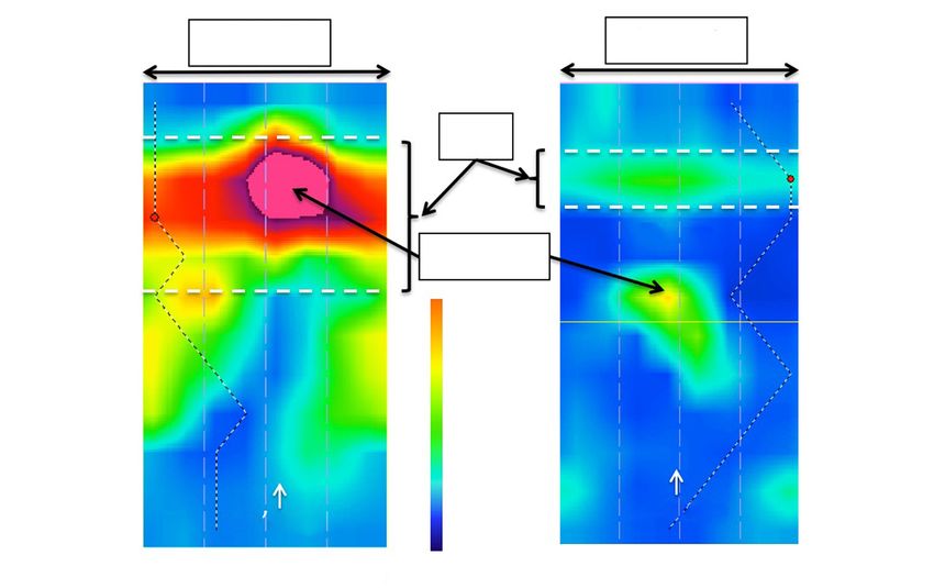
Page 6 of 10 Annals of Laparoscopic and Endoscopic Surgery, 2021
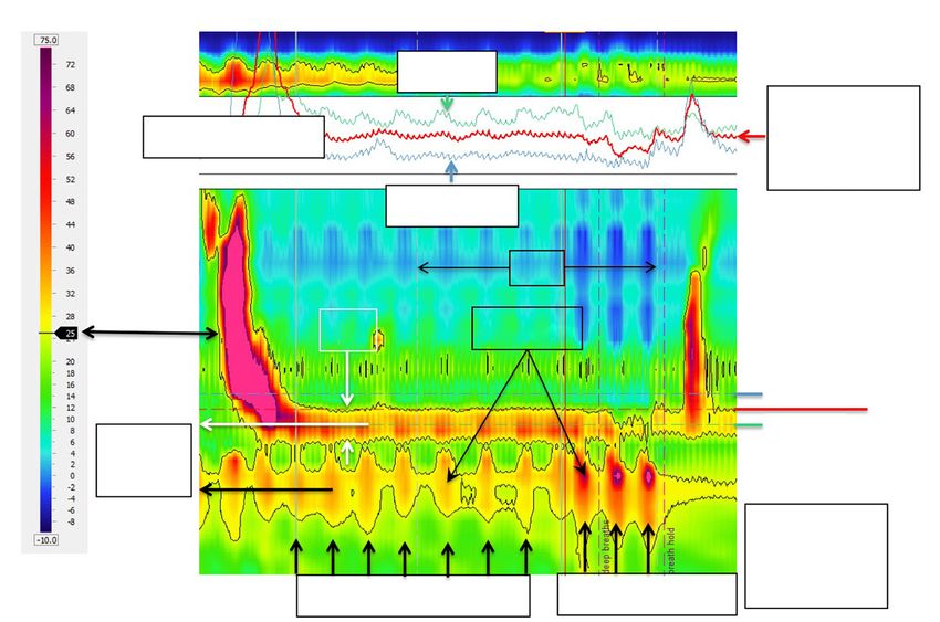
LES-CD separation =0, no hiatal hernia
mmHg
UES
Distal P PIP tool output
Average of
proximal
and distal P
Proximal P 10 s
CD Apex
LES
Proximal
RIP
Distal
Quiet inspirations 3 deep breaths
Figure 6 HRM recording of EGJ pressure in an individual without a hiatal hernia as evident by the CD-apex being completely
superimposed on the LES pressure signature, i.e., the LES-CD separation is 0. Both during quiet respiration and deep breaths, the LES is
only evident between inspirations when the CD signal is minimal. In this example, the PIP tool has been positioned to optimally isolate the
RIP as evident by the PIP tool output shown as an insert. Barely visible on the pressure topography are a horizontal blue dashed line and
green dashed line indicating the locations of the proximal and distal pressure (P) recordings shown in the PIP tool output. The red line in
the PIP tool output box is the computed average of those signals. In using the tool, the area of interrogation is scrolled up and down to find
the location at which the red line in the PIP tool output box is most nearly flat, indicative of the site at which the respiratory increases in
pressure are offset by the respiratory decreases in pressure seen on the blue line. The area of interest is during quiet respiration and the RIP
is seen to localize toward the upper margin of the CD signal, just as it had in the 3D plot of Figure 5. This positions the majority of the LES
signal within the hiatus, being pulled downward during the three deep breaths. Figure used with permission from the Esophageal Center at
Northwestern.
inspirations and an illustration of the PIP tool isolating the Hiatus hernia in HRM, i.e., LES-CD separation
RIP, indicative of the location of the CD-apex. The black >1 cm
line indicating the 15 mmHg isobaric contour (referenced
With a type I (sliding) hiatus hernia there is progressive
to atmospheric pressure) delineates the greatest pressure
anatomical disassociation between the CD and the LES
regions of the LES and CD, which completely overlap in attributable to laxity of the phrenoesophageal ligament and
this example with no hiatal hernia. The PIP tool is used to intermittent or permanent displacement of the LES into the
identify the RIP which localizes toward the upper margin mediastinum. Figure 7 illustrates a recording obtained from
of the CD-apex signal (see figure legend for detailed an individual with a small hiatal hernia, evident by an LES-
explanation). Refer back to Figure 3 to conceptualize CD separation of 2 cm. In HRM, this becomes evident with
how the LES and CD contributions to the dynamic EGJ axial separation between the CD and LES signals on the
pressure profile in Figure 6 recognizing that any area of pressure topography plot. EGJ morphology is characterized
pressure increase in Figure 3 becomes a band of pressure, as type I, II, or III in the Chicago Classification v3.0 (3,4);
fluctuating with respiration, in Figure 6. type 1 with superimposed LES and CD, type 2 with axially
© Annals of Laparoscopic and Endoscopic Surgery. All rights reserved. Ann Laparosc Endosc Surg 2021;6:5 | http://dx.doi.org/10.21037/ales.2020.03.08
Annals of Laparoscopic and Endoscopic Surgery, 2021                                                                                          Page 7 of 10

                                                      LES-CD separation =2 cm (small hiatal hernia)
                                                          Competent CD: still defines the RIP
                       mmHg

                                                                                                                UES
                                                      Distal P
                                                                          PIP tool output                           Average of
                                                                                                                    proximal
                                                                                                                    and distal P
                                                     Proximal P
                                                                                             10 s

                                                                        CD Apex
                                                   LES

                                                                                                                   Proximal
                                                                                                                                   RIP
                                LES-CD
                                                                                                                   Distal
                                =2 cm

                                                Quiet inspirations                     3 deep breaths

Figure 7 HRM recording of EGJ pressure in an individual with a small hiatal hernia as evident by the CD-apex being only partially
superimposed on the LES pressure signature, i.e., the LES-CD separation is 2 cm. Formatting of the figure is identical to that of Figure 6
with the dominant EGJ pressure profile highlighted by the black line (the 15 mmHg isobaric contour) and the PIP tool optimally positioned
to isolate the RIP. Note how the LES-CD separation is measured. The center of the LES and CD high pressure zones (white and black
horizontal arrows, respectively) are isolated with the help of the isobaric contour tool (set at 15 mmHg in this example) and the separation
between the two rounded off to the nearest cm. In this example, the RIP continues to localize toward the upper margin of the CD signal
implying that the CD still exerts sufficient sphincteric effect such that it closes the lumen isolating the stomach below from the hernia and
LES above. This is particularly evident during the three deep breaths where the strongly negative intrathoracic pressure (deep blue) is seen
to abut directly on the CD-apex signal. Figure used with permission from the Esophageal Center at Northwestern.

separated LES and CD pressure signals separated by less                           and separated CD and LES components (5) suggesting that
than 2 cm, and type 3 with a ≥2 cm separation between                             with laxity of the phrenoesophageal ligament the degree of
the LES and CD pressure signatures. That classification                           axial separation between the LES and CD can vary in the
will likely not survive future iterations of the Chicago                          course of a day based on factors such as patient position or
Classification with the focus instead shifting to whether                         breathing. When such variation is seen, it makes sense to
or not the LES-CD separation exceeds 1 cm (indicative of                          score it as the maximal value (or to report the range), the
hiatus hernia) and whether or not the CD-apex pressure                            critical question being whether or not an anatomical hiatal
signal continues to define the locus of the RIP (indicative of                    hernia exists. HRM-defined LES-CD separation has been
the competence of the CD as a extrinsic sphincter).                               shown to correlate closely with the presence or absence and
   In its early stages, a type I hernia is difficult to distinguish               size of hiatus hernia as determined by endoscopy or barium
from normal, appearing only as an exaggeration of the                             x-ray with sensitivity and specificity of 88% and 95%
normal phrenic ampulla on fluoroscopy and a partially                             respectively (6). LES-CD separation also correlates with
overlapping LES and CD on HRM. Furthermore, EGJ                                   reflux severity as determined by pH-metry (3,7,8). True,
pressure morphology can vary over time, even within a                             the Weijenborg et al.’s 2015 analysis (6) begs the question
single patient study, transitioning between superimposed                          of which test is the most appropriate reference standard for

© Annals of Laparoscopic and Endoscopic Surgery. All rights reserved.         Ann Laparosc Endosc Surg 2021;6:5 | http://dx.doi.org/10.21037/ales.2020.03.08
Page 8 of 10                                                                                  Annals of Laparoscopic and Endoscopic Surgery, 2021

                                                          LES-CD separation =4 cm (hiatal hernia)
                                                           Incompetent CD: LES defines the PIP
                     mmHg

                                                                        Distal P                                  UES

                                                                                                                      Average of
                                        PIP tool output                                                               proximal
                                                                                                                      and distal P
                                                                        Proximal P

                                                                                       30 s

                                                           LES                     CD Apex

                                                                                                                    Proximal
                                                                                                                                     RIP
                                                                                                                    Distal
                               LES-CD
                               =4 cm

                                                                                                                   Intragastric
                                                                                                                   pressure =

                                                             Quiet inspirations                                    18 mmHg
                                                                                               3 deep breaths

Figure 8 HRM recording of EGJ pressure in an individual with a moderate sized hiatal hernia as evident by the CD-apex being isolated
from the LES pressure signature, i.e., the LES-CD separation is 4 cm. Formatting of the figure is identical to that of Figures 6,7 with
the dominant EGJ pressure profile highlighted by the black line (the 25 mmHg isobaric contour in this case) and the PIP tool optimally
positioned to isolate the RIP. However, in this example, the RIP no longer localizes the CD-apex signal, instead localizing at the
proximal margin of the LES. Even without the aid of the PIP tool, that is evident by the inspiratory bursts of red on the LES recording.
Consequently, the CD no longer functions as a competent extrinsic sphincter and the entire hiatal hernia up to the lower margin of the LES
is subject to intra-gastric pressure throughout the respiratory cycle. Compounding that issue, this individuals intragastric pressure is quite
elevated at 18 mmHg. Figure used with permission from the Esophageal Center at Northwestern.

detecting hiatal hernia because the unequivocal diagnosis of                         The RIP
hiatus hernia is ultimately made intraoperatively wherein the
                                                                                     As the LES and CD become spatially separate there is the
spatial relationship between the EGJ and CD and presence
                                                                                     added issue of whether the RIP remains associated with the
or absence of a hernia sac are visually evident. Along that
                                                                                     CD signal in the EGJ pressure complex or not. Although
line, a recent analysis compared the accuracy of HRM,
                                                                                     the precise physiological meaning of the RIP in this context
endoscopy, and barium radiography to surgery in detecting                            is uncertain, there can be general agreement regarding
and sizing hiatus hernia (9). That analysis concluded that                           the observations that: (I) the RIP can never be below the
HRM, using the LES-CD metric, outperformed the other                                 diaphragm; (II) when the CD is superimposed on the LES
modalities with a sensitivity of 94%, specificity of 92%                             (i.e., LES-CD separation 1 cm), the RIP can localize either at the CD (as
resolution of a device that measures pressures at 1 cm                               in Figure 7) or above the CD component placing it either
intervals is 1 cm regardless of how many decimal places the                          within the hernia or at the LES as in Figure 8. Supporting
computer spits out.                                                                  the relevance of this distinction, a recent analysis exploring

© Annals of Laparoscopic and Endoscopic Surgery. All rights reserved.          Ann Laparosc Endosc Surg 2021;6:5 | http://dx.doi.org/10.21037/ales.2020.03.08
Annals of Laparoscopic and Endoscopic Surgery, 2021 Page 9 of 10
the relevance of hiatal hernia pressure topography subtyped significance of a competent vs. incompetent CD is a matter
individuals with LES-CD separation as ‘B’ or ‘C’ depending of continued debate.
on whether the RIP localized above or below the LES
respectively (10). In that analysis, subtype B was less likely
Acknowledgments
to exhibit pathological reflux on pH-metry than subtype
C. The authors interpreted this to support the contention Funding: The study was supported by R01 DK092217 (John
that subtype B was indicative of the LES remaining within E. Pandolfino) from the US Public Health Service.
the abdominal compartment and being advantageous. Of
note, the patients associated with Figures 7,8 both had clear-
Footnote
cut reflux disease. In the case of Figure 7 they had Barrett’s
esophagus with high-grade dysplasia and in the case of Conflicts of Interest: The author has completed the ICMJE
Figure 8 a Bravo pH-metry study found pathological reflux uniform disclosure form (available at http://dx.doi.
on four out of four days with an average esophageal acid org/10.21037/ales.2020.03.08). PJK consulting for
exposure time of 10%. Ironwood, Bayer. The author has no other conflicts of
Interpreting the distinction between a competent and interest to declare.
an incompetent CD somewhat differently, with greater
degrees of LES-CD separation (i.e., >2 cm), there is the Ethical Statement: The author is accountable for all
additional factor of whether or not the CD effectively aspects of the work in ensuring that questions related
compartmentalizes the stomach from the herniated stomach to the accuracy or integrity of any part of the work are
during inspiration. When it does, it is exhibiting greater appropriately investigated and resolved.
sphincteric function than when it doesn’t, presumably
because the hiatal aperture is less dilated (11). Although not Open Access Statement: This is an Open Access article
precisely addressing this distinction, evidence supporting distributed in accordance with the Creative Commons
the relevance of CD competence comes from a logistic Attribution-NonCommercial-NoDerivs 4.0 International
regression model of barrier function that simultaneously License (CC BY-NC-ND 4.0), which permits the non-
examined expiratory LES pressure, LES-CD separation, commercial replication and distribution of the article with
and inspiratory EGJ augmentation while controlling for age the strict proviso that no changes or edits are made and the
and BMI. In that analysis, only inspiratory augmentation original work is properly cited (including links to both the
had a significant independent association with GERD as formal publication through the relevant DOI and the license).
defined by pH-metry (3). See: https://creativecommons.org/licenses/by-nc-nd/4.0/.
Conclusions References
Accepting that the presence and size of hiatus hernia is 1. Kwiatek MA, Pandolfino JE, Kahrilas PJ. 3D-high
a clinically relevant measurement, it can be concluded resolution manometry of the esophagogastric junction.
that there is strong evidence supporting that LES-CD Neurogastroenterol Motil 2011;23:e461-9.
separation >1 cm evident during quiet respiration during 2. Nicodème F, Lin Z, Pandolfino JE, Kahrilas PJ.
an HRM is indicative of hiatus hernia. With respect to the Esophagogastric junction pressure morphology:
localization of the RIP, there is less agreement on its clinical comparison between a station pull-through and real-
significance, but in instances of the LES-CD separation time 3D-HRM representation. Neurogastroenterol Motil
exceeds 1 cm the RIP can localized at or above the CD 2013;25:e591-8.
component. Hence the three possible EGJ morphologies 3. Pandolfino JE, Kim H, Ghosh SK, et al. High-resolution
are: (I) no hiatus hernia (LES-CD separation 1 cm with the RIP at the CD level); and (III) 4. Kahrilas PJ, Bredenoord AJ, Fox M, et al. The Chicago
hiatus hernia with an incompetent CD (LES-CD separation Classification of esophageal motility disorders, v3.0.
>1 cm with the RIP above the CD component placing Neurogastroenterol Motil 2015;27:160-74.
it either within the hernia or at the LES). The clinical 5. Bredenoord AJ, Weusten BL, Timmer R, et al.
© Annals of Laparoscopic and Endoscopic Surgery. All rights reserved. Ann Laparosc Endosc Surg 2021;6:5 | http://dx.doi.org/10.21037/ales.2020.03.08Page 10 of 10 Annals of Laparoscopic and Endoscopic Surgery, 2021
Intermittent spatial separation of diaphragm and lower GERD. Neurogastroenterol Motil 2015;27:1175-82.
esophageal sphincter favors acidic and weakly acidic reflux. 9. Tolone S, Savarino E, Zaninotto G, et al. High-resolution
Gastroenterology 2006;130:334-40. manometry is superior to endoscopy and radiology in
6. Weijenborg PW, van Hoeij FB, Smout AJ, et al. Accuracy assessing and grading sliding hiatal hernia: a comparison
of hiatal hernia detection with esophageal high-resolution with surgical in vivo evaluation. United European
manometry. Neurogastroenterol Motil 2015;27:293-9. Gastroenterol J 2018;6:981-9.
7. Ham H, Cho YK, Lee HH, et al. Esophagogastric 10. Akimoto S, Singhal S, Masuda T, et al. Classification
junction contractile integral and morphology: Two high- for esophagogastric junction (EGJ) complex based on
resolution manometry metrics of the anti-reflux barrier. J physiology. Dis Esophagus 2017;30:1-6.
Gastroenterol Hepatol 2017;32:1443-9. 11. Kumar D, Zifan A, Ghahremani G, et al. Morphology of
8. Tolone S, de Cassan C, de Bortoli N, et al. the esophageal hiatus: is it different in 3 types of hiatus
Esophagogastric junction morphology is associated with hernias. J Neurogastroenterol Motil 2020;26:51-60.
a positive impedance-pH monitoring in patients with
doi: 10.21037/ales.2020.03.08
Cite this article as: Kahrilas PJ. High-resolution manometry
findings with hiatus hernia. Ann Laparosc Endosc Surg
2021;6:5.
© Annals of Laparoscopic and Endoscopic Surgery. All rights reserved. Ann Laparosc Endosc Surg 2021;6:5 | http://dx.doi.org/10.21037/ales.2020.03.08You can also read