Identification Mycobacterium spp. in the Natural Water of Two Austrian Rivers - MDPI
←
→
Page content transcription
If your browser does not render page correctly, please read the page content below
microorganisms
Article
Identification Mycobacterium spp. in the Natural
Water of Two Austrian Rivers
Mohammad Reza Delghandi , Karoline Waldner, Mansour El-Matbouli                      and
Simon Menanteau-Ledouble *
 Clinical Division of Fish Medicine, University of Veterinary Medicine, 1210 Vienna, Austria;
 delghandim@staff.vetmeduni.ac.at (M.R.D.); waldnerk@staff.vetmeduni.ac.at (K.W.);
 Mansour.El-Matbouli@vetmeduni.ac.at (M.E.-M.)
 * Correspondence: menanteaus@staff.vetmeduni.ac.at

 Received: 11 August 2020; Accepted: 25 August 2020; Published: 27 August 2020

 Abstract: Nontuberculous mycobacteria constitute a subgroup among the Mycobacterium genus,
 a genus of Gram-positive bacteria that includes numerous pathogenic bacteria. In the present study,
 Mycobacterium spp. were detected in natural water samples from two Austrian rivers (Kamp and
 Wulka) using three different primers and PCR procedures for the identification of the 16S rRNA and
 hsp65 genes. Water samples were collected from the Kamp (45 samples) and Wulka (25 samples)
 in the summer and winter of 2018 and 2019. Molecular evidence showed a high prevalence of
 Mycobacterium sp. in these rivers with prevalence rates estimated at approximately 94.3% across
 all rivers. The present study represents the first survey into the prevalence of Mycobacterium sp. in
 natural water in Austria. Because nontuberculous mycobacteria have known pathogenic potential,
 including zoonotic, these findings may have implications for health management and public health.

 Keywords: molecular epidemiology; nested PCR; environmental mycobacteria

1. Introduction
      Mycobacterium spp. belong to the family Mycobacteriaceae and are Gram-positive, nonmotile,
facultative aerobic acid fast bacilli [1]. This genus is found under a wide geographical area,
encompassing a wide range of optimal growing temperatures (25–35 ◦ C) [2,3]. Most species of
Mycobacterium spp. are labelled “nontuberculous mycobacteria” (NTM), a term that excludes the
members of the M. tuberculous complex and M. leprae, as well as a few closely related species, which have
historically represented the members of this genus with the most severe impact on human health [4,5].
NTM have been further subdivided between three different groups, based on their virulence and
ability to establish an infection. These include true pathogens (M. marinum, M. ulcerans), opportunistic
pathogen (M. chelonae-abscessus complex, M. fortuitum, M. avium complex, M. haemophilum, M. xenopi,
M. kansassi and M. simiae) and a third group composed of saprophytes mycobacteria (M. smegmatis,
M. vaccae, M. terrae complex and M. gordonae) [3]. Members of the Mycobacterium genus are all
considered to be slow-growing. However, there are stark differences between their growth patterns.
Consequently, growth kinetic has been used alongside pigmentation patterns as a criterion for the basis
of a classification scheme for NTM. While the fast-growing mycobacteria are able to produce colonies
visible with the naked eye on solid media within 5 days [6], slow-growers can require much longer.
In some extreme cases, such as ovine strains of Mycobacterium avium subsp. mycotuberculosis, the bacteria
can take years to produce visible colonies, but weeks or months are more common durations [7].
Consequently, due to the slow bacterial growth rate and the time required for the development of
colonies and turbidity on either solid or liquid media, diagnosis based on bacterial isolation and colony
morphology is not considered an appropriate procedure to identify Mycobacterium spp. Several other

Microorganisms 2020, 8, 1305; doi:10.3390/microorganisms8091305            www.mdpi.com/journal/microorganisms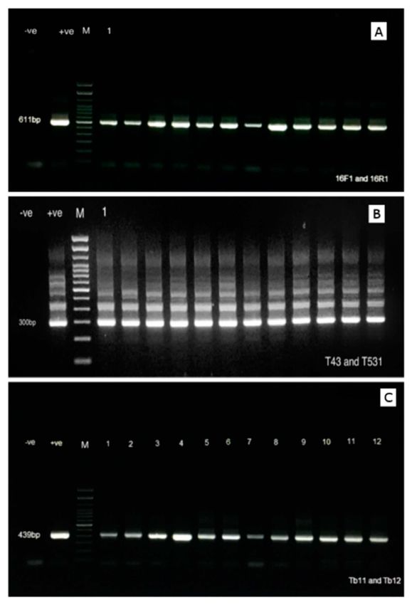
Microorganisms 2020, 8, 1305                                                                         2 of 11

methods have been utilized for the identification of this genus, in particular molecular diagnostic
methods based on the DNA or RNA [8].
      NTM are found in a wide range of geographical locations and they have been isolated from a variety
of samples originating from many different environmental conditions, including low organic matter
concentrations and low oxygen level concentrations [9]. These mycobacteria have been reported from
water, biofilm and soil and have been found in association with infections in a wide range of hosts, such as
mammals, including humans, and birds as well as aquatic animals [3,10–13]. The most common species
identified in water samples include M. marinum, M. gordonae, M. flavescens, M. fortuitum, and M. chelonae
(isolated from aquariums and swimming pools). In addition, other species can cause disease,
especially in ornamental fish where M. triviale, M. avium, M. abscessus, and M. peregrinum have been
regularly associated with diseases [14–16], while M. flavescens has been more infrequently reported [5].
NTM have a predilection for aquatic environments and it is likely that water plays a significant role as a
vector in the transmission of Mycobacterium sp. [4]: all of these species have been isolated from several
fish species [10,17], and zoonotic cases are often associated with exposure through water or aerosols
or handling of contaminated seafood or ornamental fish [18–21]. Consequently, mycobacteriosis is
often linked to the professional occupation of the patients, and people whose occupation involves
contact with water and fish are more likely to be exposed to the infection [17,22]. Additionally, due to
being frequently reported as a disease from swimmers, the infection has occasionally been referred
to as a “Swimming pool granulomas” [17], although this form of the disease is much less common
nowadays because of the systematic application of disinfectants. On the other hand, the prevalence of
M. marinum in natural waters has been estimated at a low level, and it seems that the risk of infection
in human is also low [5]. Because of the bacteria’s slow growth and low thermal preferences, infections
in humans are often limited to superficial infections with nodules and to the skin and extremities,
although deeper infections have also been reported, including deep bursitis, tenosynovitis, arthritis,
and osteomyelitis. Moreover, more systemic forms of mycobacteriosis can also occur, including those
which involve the respiratory system particularly in immunocompromised patients [5,23]. Additionally,
other NTM such as M. chelonae, M. fortuitum, M. flavescens, and M. gordonae have also been associated
with granulomatous lesions, hepatitis, endocarditis, and meningitis, and infections have been observed
in the ocular, bone, joint, and skeletal system [19,21,24].
      In fish, M. avium has been isolated from Cockatoo Dwarf Cichlid (Apistogramma cacatuodes)
in the Czech Republic [25]. M. gordonae has similarly been reported from several fish species
including Gold fish (Carassius auratus), Guppy (Poecilia reticulate), Angel fish (Pterophyllum scalare),
and Common carp (Cyprinus carpio) [26]. Additionally, M. fortuitum and M. chelonae have
both been reported in the ornamental and wild fish, including Neon tetra (Paracheirodon innesi),
Goldfish (Carassius auratus auratus), Three-spot gourami (Trichogaster trichopterus), Cichlid fish
(Microgeophagus altispinosus), Sterlet (Acipenser ruthenus), Siamese fighting fish (Betta splendens),
Dwarf gourami (Colisa lalia), Sailfin molly (Poecilia latipinna), Giant sailfin molly (Poecilia velifera),
Discus fish (Symphysodon discus), Green swordtail (Xiphophorus helleri), Australian lungfish
(Neoceratodus fosteri), Silver mullet (Mugil curema), Atlantic salmon (Salmo salar), and juvenile Pacific
salmon (Oncorhynchus tshawytscha) [16,24,27–31]. M. marinum is an important bacterial agent causing
fish tuberculosis, and transmission to humans can be observed via contaminated water in aquarium
and fish breeding. It has been associated with salt and fresh water exposure [5]. Furthermore, this
species has also been isolated from decorative and farm fish.
      Piscine mycobacteriosis is a slow developing chronic disease, although a more acute form of this
disease has also been reported, and the disease may not always be associated with obvious clinical signs.
Asymptomatic mycobacteriosis has also been reported and is associated with reduced fish growth in
aquaculture [17]. When present, clinical signs of mycobacteriosis include nonspecific signs, such as the
ones commonly associated with systemic disease in fish such as a swollen abdomen, red lesions on the
lateral line, exophthalmia, and pile gills. Additionally, internal signs including organomegaly of the
liver, kidney, and spleen have also been reported [6]. A more characteristic sign is the development of
Microorganisms 2020, 8, 1305 3 of 11
granulomatous lesions on the internal organs, which is an uncommon feature in fish. Several virulence
factors have been identified in Mycobacterium spp. pathogenesis including secretion system 1 (ESX-1)
to 5 (ESX-5), PE_PGRs family and PPE proteins (that are considered the most important factor for the
replication of Mycobacterium sp. in macrophage), and PknG (protein kinase G). The most important
virulence factor is the Esx secretion system that is important for both M. marinum and M. tuberculosis
pathogenesis [22,32]. In Austria, the Federal Ministry of Agriculture, Forestry, Environment and Water
Management recently announced the objective of increasing national fish production to raise the degree
of self-supply from the current 34% to 60%, corresponding to an increase in production from 2400 to
5500 tons annually [33]. This aquaculture production is mostly composed of carp as well as rainbow
trout (Oncorhynchus mykiss) introduced for farming purposes. Moreover, the endemic brown trout
(Salmo trutta fario) populates several rivers and waterways. However, these populations are considered
at risk as the reported numbers of fish are considered in decline, despite several reintroduction efforts.
Because Mycobacterium sp. are known pathogens of wild fish, notably isolated in 2018 from brown
trout originating in the Kamp river in Austria [34], we decided to estimate the prevalence of these
organisms in the Kamp and the adjacent Wulka river.
2. Materials and Methods
2.1. Origin of the Water Samples
In total, 70 natural water samples were taken from the Kamp and Wulka rivers on two different
sampling dates in 2018 and 2019 as a part of the project ClimateTrout. In total, 45 samples originated
from the Kamp and 25 samples originated from the Wulka. The aim of the ClimateTrout project was to
investigate the prevalence of the myxozoan T. bryosalmonae in wild brown trout and water samples
by using PCR, notably in order to determine the role of this parasite in the decline of wild brown
trout populations in Austria. The results from this screening were published by Waldner et al. in
2019 [35], and it was decided to use the remaining samples to further investigate additional organisms
of interest. Briefly, a 4 L water sample of the Kamp and Wulka rivers was collected and brought to the
University of Veterinary Medicine of Vienna. Samples were vacuum filtered with Whatman 1.5 µm
GF/F filters (Whatman, Maidstone, United Kingdom) according to Hutchins et al. [36] to concentrate
microorganisms. Afterwards, environmental DNA (eDNA) was extracted using the DNeasy Power
Soil kit (Qiagen Inc., Hilden, Germany) according to the manufacturer’s instructions. Unfortunately,
the water samples did not allow for bacterial isolation by cultures on media.
The samplings took place in June and July 2018 as well as in January 2019 (Table 1), and the water
temperature in the rivers at the time ranged from 16 to 22 ◦ C in the summer to 0–2 ◦ C in January.
Table 1. Mycobacterium sp. identified in water sample in Austrian rivers.
Sampling Number of Positive/Prevalence Rate of
River Sites
Date Number Mycobacterium sp.
June 2018 25 25/25 (100%)
Kamp
January 2019 20 20/20 (100%)
July 2018 10 10/10 (100%)
Wulka
January 2019 15 11/15 (73.33%)
Total for 2018 35 35/35 (100%)
Totals Total for 2019 35 31/35 (88.57%)
Both years 70 66/70 (94.28%)Microorganisms 2020, 8, 1305 4 of 11
2.2. PCR Assay for the 16S rRNA and hsp65 Genes
Three different PCR procedures and primers sets were used to detect Mycobacterium sp. in water
samples in order to maximize our confidence in the results. Initially, a PCR assay was performed
according to the protocol developed at the University of Veterinary Medicine and published by
Delghandi et al. in 2020 [34], using Myco 16F1 (50 -AGCTCGTAGGTGGTTTGTCG-30 ) and Myco 16R1
(50 -CCACCTTCCTCCGAGTTGAC-30 ) for the detection of the 16S rRNA gene [34]. The total volume
of amplification was 25 µL, comprising 12.5 µL Dream Taq Green PCR Master Mix, 1 µL of each primer
(10 pmol) and 4 µL eDNA solution. The amplification program consisted of 95 ◦ C for 5 min and
35 cycles of 95 ◦ C for 1 min, 54 ◦ C for 1 min, and 72 ◦ C for 1 min. The resulting PCR amplicon was
611 bp in size.
A second confirmatory nested PCR (nPCR) assay was conducted as previously described by
Talaat et al. in 1997 [37] to identify the 16S rRNA gene in members of the Mycobacterium genus.
Briefly, two different primers were used for the first round and second round PCR (T39 , T13 and
T43, T531, respectively). The primers used in the first round amplification were the T39 outer F
(50 -GCGAACGGGTGAGTAACACG-30 ) and T13 outer R (50 -TGCACACAGGCCACAAGGGA-30 )
primers. Afterward, 2 µL of this product was used in a second round of amplification using the T43
inner F (50 -AATGGGCGCCAAGCCTGATG-30 ) and T531 inner R (50 -ACCGCTACACCAGGAAT-30 )
primers. The amplification conditions for both rounds were one cycle of 95 ◦ C for 5 min and 30 cycles
of 94 ◦ C for 1 min, 50 ◦ C for 1 min, and 72 ◦ C for 1 min. The nPCR assay produced a 300 bp
amplification product.
Finally, we also utilized Tb11 (50 -ACCAACGATGGTGTGTCCAT-30 ) and Tb12 (50 -CTTGTCGAA
CCGCATACCCT-30 ) to identify a 65 kDa heat shock protein (hsp65) gene of Mycobacterium according to
the procedure described by Telenti et al. [38]. The amplification for these primer pairs was carried out
as follow: one cycle of 95 ◦ C for min, followed by 45 cycles of denaturing at 94 ◦ C for 1 min, annealing
at 60 ◦ C for 1 min, and extension at 72 ◦ C for 1 min. The resulting amplicon was 439 bp in size.
Each set of samples for the round of amplification included a negative control (using genomic
DNA from the Gram-positive aquatic bacterium R. salmoninarum) as well as a positive control in the
form of DNA extracted from a pure culture of M. marinum on Middlebrook 7H10 agar extracted using
a DNeasy kit (Qiagen) according to the manufacturer’s instructions. Eight microliters of each PCR
product were analyzed by gel electrophoresis on 1% agarose gels and examined under UV illumination.
All samples were screened two times with all three PCR protocols in order to confirm the
results. The PCR amplicons were cut from the agarose gels, and DNA were extracted utilizing the
MinElute Gel Extraction kit (Qiagen Inc.). Nine positive samples were randomly selected from both
rivers; four microliters of each primer (T531, Myco 16F1, and Tb11) in 5 pmol concentration was
added to purified samples and sent for sequencing to LGC Genomics Company (Berlin, Germany)
by Sanger sequencing to confirm that these samples were homologous to sequences from known
members of the Mycobacterium genus; sequencing results were analyzed for homology using BLAST
(Basic Local Alignment Sequence Tool; National Center for Biotechnology Information; USA).
Afterwards, a ClustalW analysis was conducted on the 16s RNA sequences using the software Clustal
Omega from the European Bioinformatics Institute of the European Molecular Biology Laboratory
(EMBL-EBI). In addition, we added the corresponding sequences from Mycobacterium sp. isolated from
fish in our previous survey as well as three sequences from known strains of Mycobacterium from the
NCBI database.
3. Results
In total, 45 water samples were collected from the Kamp and 25 samples from the Wulka River.
Genomic DNAs were extracted from these samples, and PCRs were performed for each sample in
order to detect the presence of NTM based on three different PCR protocols by Delghandi et al. [34],
Talaat et al. [37], and Telenti et al. [38] (Figure 1). Notably, the results were identical for all three PCR
protocols, and Mycobacterium sp. were detected in all samples originating from the Kamp River, at bothMicroorganisms 2020, 8, 1305 5 of 11
sampling time points (June and January, Table 1). In the Wulka, the prevalence was also high: all
samples collected in the summer 2018 were positive, while only 11 out of 15 samples collected in the
winter 2019 were positive (prevalence of 73.33%). There were no significant effect of month or place of
sampling (p > 0.5). When comparing with previous results regarding the screening of Mycobacterium
sp. in wild brown trout, all fish that had been found infected with Mycobacterium sp. originated from
the Kamp River, which had the highest prevalence in the present study [34].
Figure 1. Agarose gel electrophoresis showing the amplicons generated by the various PCR conducted
on the positive samples through the 3 different procedures: (A) 611 bp amplicon product generated
using the PCR procedure described by Delghandi et al. [34]; (B) 300 bp amplicon generated using
primers targeting the 16S rRNA according to the procedure described by Talaat et al. [37]; (C) 439 bp
amplicon generated using the primers targeting the hsp65 gene of Mycobacterium sp. according to the
procedure described by Telenti et al. [38]. For each gel, 5 µL of the amplicons was loaded in each well.
Analysis of the 16S rRNA and hsp65 sequences confirmed that the bacteria detected most likely
belonged to the genus Mycobacterium, and the sequences were between 98.5% and 99.2% and 94.2%
and 95.0% identical to other sequences from Mycobacterium species when sequencing the amplicons
generated using the Talaat and Telenti primers, respectively. Similarly, the primers Myco 16 F1 andMicroorganisms 2020, 8, 1305 6 of 11
Myco 16 R1 produced amplicons with 92.8–99.22% identity with the sequences from other mycobacterial
species. Notably, none of the three primer-pairs were specific for a single species of Mycobacterium,
and sequencing always matched more than one species (see Supplementary Table S1). The sequences
were deposited in the GenBank database under accession number PRJNA647541.
4. Discussion
This survey aimed to investigate the prevalence of Mycobacterium sp. in water. Mycobacterium
spp. are important organisms associated with both aquaculture and human diseases. While members
of the genus Mycobacterium are considered common inhabitants of aquatic environments, including
rivers, lakes, ponds, and streams, there has been no previous study regarding the prevalence of
Mycobacterium sp. in natural Austrian waters. However, members of the Mycobacterium genus have
been frequently isolated from water samples as an environmental bacterium. For example, M. fortuitum
and M. chelonae represent the species most frequently isolated from tap water and reservoirs [5].
M. avium subsp. paratuberculosis has similarly been isolated and identified from the Taff river in
Southern Wales using both PCR and culture on Herrold’s egg yolk medium (HEYM) [39] and reported
a geographical correlation between the presence of these bacteria and the prevalence of Crohn’s
Disease in the population. Notably, culture attempts using M. avium have shown that the bacterium
was unable to grow when exposed to high NaCl concentrations; on the other hand, its growth rate
was enhanced under low concentrations of dissolved oxygen [40]. M. gordonae has been frequently
isolated from contaminated water [5]; more importantly, this pathogen has been isolated from tap
water from hospitals and homes in Germany by Peters et al., using isolation and culture methods [41],
which has important public health implications. Moreover, Le Dantec et al. isolated these organisms
from membrane filtered water samples originating from the Paris water distribution system on
Lowenstein–Jensen medium followed by sequencing of the 16S rDNA gene [42]. NTMs were more
common in this study with 78% of the samples being positive for Mycobacterium sp. and about 15%
contaminated with mycobacteria with pathogenic potential [42]. Moreover, Chilima et al. detected
Mycobacterium sp. using both Ziehl–Nielsen staining and PCR amplification of the 16S rRNA gene
in both water and soil samples from Northern Malawi [43]. Notably, these two approaches resulted
in very different results with 75% of the samples appearing positive using the staining method,
while Mycobacterium DNA was only detected in 54% of them [43]. However, the investigators were
unable to identify the bacterial isolates at the species level. Concerning fish farms and aquaculture,
mycobacteriosis-causing M. marinum was observed in rainbow trout and brown trout fish farm
population in Italy [44]. Other Mycobacterium spp. that were frequently reported in water included
M. kansasii and M. xenopi. While, M. kansasii has been rarely reported in aquaculture. This species
was isolated from zebrafish (Danio rerio) by Kusar et al. in 2017 [45]. However, there is no report of
isolation of M. xenopi in aquaculture. Additionally, Mycobacterium sp. were present in two Finnish
lake water samples, and this organism was detected by Niva et al. in 2006 using PCR procedures [46].
Interestingly, M. pseudoshotsii has been detected in water in the Chesapeake Bay. This species was
isolated frequently in striped bass (Morone saxatilis) in this region [47]. In addition, M. fortuitum and
M. chelonae were identified in water samples collected from freshwater rivers, ponds, and brooks
in Iran by Rahbar et al. in 2010 using isolation on Lowenstein–Jensen (LJ) medium [48]. Notably,
these species have a potential to infect fish (farmed and wild fish) [27,28] and humans [24]. Likewise,
Mycobacterium spp. have been isolated from tank water and aquariums, and M. marinum was reported
from aquariums causing infection in humans [49].
Environmental mycobacteria can survive under a wide range of environmental conditions.
They have been classified as atypical mycobacteria and are considered opportunistic [50]. Remarkably,
all samples were positive with the exception of four samples that had been collected from the river
Wulka during the winter. This high level of prevalence of Mycobacterium sp. in our samples was
consistent with our previous findings, published in 2020 [34], where screening of kidney samples for
DNA sequences from Mycobacterium sp. in wild brown trout discovered a high prevalence in the KampMicroorganisms 2020, 8, 1305 7 of 11
river in June 2018. Interestingly, all of the positive fish samples in this study originated from the same
sampling location and time, which could suggest that an outbreak of NTM was taking place in the
population at the time of the sampling [34].
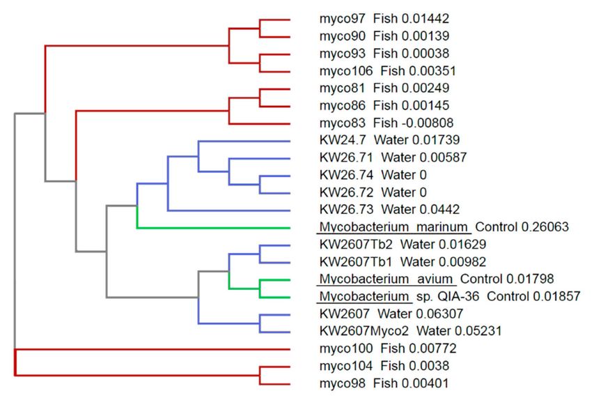
When compared with the results from our previous survey performed in wild brown trout [34],
the species detected in the present survey appeared more diverse (Figure 2). This could be explained
by the fact that the fish sampled in the previous survey originated from a single outbreak, and so it
would be plausible that all bacteria involved originated from the clonal expansion of a single bacterial
cell, while the present survey involves bacteria from a comparatively large geographic and temporal
area. Our samples also bracketed several known mycobacterial species, suggesting that more than one
species was detected here; although, because of the lack of specificity at the species level of the primers
used in the study, such conclusions are difficult to make.
Figure 2. Phylogenetic tree constructed by ClustalW analysis of the 16S rRNA sequences from the
amplicons from the present study. Sequences of these amplicons (water, indicated by blue branches)
were compared to sequences from fish samples obtained during our previous samples of wild fish in
the same rivers (fish, indicated by red branches, see Delghandi et al. [38]) and that of three control
Mycobacterium sp. from the NCBI dataset (control, indicated by green branches).
It is indeed important to note that none of the three sets of protocols and primers were found to be
specific at the species level and did not allow for the specific identification of pathogenic mycobacteria.
Moreover, molecular methods are also able to detect bacterial DNA even in the absence of biologically
active pathogens [51] and as a result do not discriminate between live and dead organisms. Therefore,
the actual risk for public health and fish farm associated with this high prevalence is difficult to assess.
On the other hand, other investigation projects have made use of isolation and cultures on specific agars.
While this approach has the advantage of increased specificity, because it only detects live bacteria and
allows for further tests to identify the bacteria at the species level, it is not considered as sensitive, as
Mycobacterium spp. are difficult to cultivate and are easily outgrown by other environmental bacteria.
Another more recently developed technique is immunomagnetic separation polymerase chain reaction
(IMS-PCR) where samples are incubated with antibody-coated immunomagnetic beads, to allow the
purification of samples. Whan et al. have developed an IMS-PCR method for the detection of M. avium
subsp. paratuberculosis [52] and screened 192 samples of untreated water from Northern Ireland,
detecting the bacterium in 15 (8%) of these samples [53].
In the future, it would be beneficial to perform a more thorough investigation, for example, using
more diverse sampling, including other rivers and bodies of water as well as various sampling times
covering other months and seasons. It would also be beneficial to use a combination of techniques
and approaches, in particular, decontamination, for example using NaOH or antibiotics followed byMicroorganisms 2020, 8, 1305 8 of 11
isolation on specific agar and identification of the isolates at the species level, for example using mass
spectrophotometry, to maximize the quality of our results. This would allow for a better understanding
of the public health risks associated with the presence of Mycobacterium sp. in Austrian waters.
Supplementary Materials: The following are available online at http://www.mdpi.com/2076-2607/8/9/1305/s1.
Table S1: Most similar sequences for the amplicons in the database of the National Center for Biotechnology
Information, based on the results from a search using the Basic Local Alignment Tool.
Author Contributions: K.W. performed the initial sampling and the genomic DNA isolation. M.R.D. performed
the rest of the experimental work. M.R.D. and S.M.-L. wrote the manuscript, while S.M.-L. and M.E.-M. designed
and supervised the study. All authors have read and agreed to the published version of the manuscript.
Funding: This work was supported in part by the Austrian Science Funds (Fonds zur Förderung der
wissenschaftlichen Forschung), project P28837-B22. The funding body did not contribute to the study’s design or
analysis of the data.
Acknowledgments: Open Access Funding by the Austrian Science Fund (FWF).
Conflicts of Interest: The authors declare no conflict of interest.
Abbreviations
NTM Nontuberculous mycobacteria
eDNA Environmental DNA
IMS-PCR Immunomagnetic separation polymerase chain reaction
nPCR Nested PCR
BLAST Basic Local Alignment Sequence Tool
HEYM Herrold’s egg yolk medium
IMS-PCR Immunomagnetic separation polymerase chain reaction
PPE proteins Proline-Proline-Glutamic Acid proteins
References
1. Chinabut, S. Fish Disease and Disorders: Viral, Bacterial, and Fungal Infections; CAB International: Wallingford,
UK, 1999; pp. 319–340.
2. Akbari, S.; Mosavari, N.; Tadayon, K.; Rahmati-Holasoo, H. Isolation of Mycobacterium fortuitum from fish
tanks in Alborz, Iran. Iran. J. Microbiol. 2014, 6, 234–239. [PubMed]
3. Johansen, M.D.; Herrmann, J.-L.; Kremer, L. Non-tuberculous mycobacteria and the rise of Mycobacterium
abscessus. Nat. Rev. Genet. 2020, 18, 392–407. [CrossRef] [PubMed]
4. Neumann, M.; Schulze-Robbecke, R.; Hagenau, C.; Behringer, K. Comparison of methods for isolation of
mycobacteria from water. Appl. Environ. Microbiol. 1997, 63, 547–552. [CrossRef] [PubMed]
5. Dailloux, M.; Laurain, C.; Weber, M.; Hartemann, P. Water and nontuberculous mycobacteria. Water Res.
1999, 33, 2219–2228. [CrossRef]
6. Elliot, D.G. Fish Viruses and Bacteria: Pathobiology and Protection; CABI Publishing: Wallingford, UK, 2017;
pp. 286–297.
7. Goodfellow, M.; Magee, J.G. Taxonomy of Mycobacteria. In Mycobacteria; Springer Science and Business
Media LLC: Berlin/Heidelberg, Germany, 1998; pp. 1–71.
8. Hruska, K.; Kaevska, M. Mycobacteria in water, soil, plants and air: A review. Veterinární Medicína 2013, 57,
623–679. [CrossRef]
9. Falkinham, J.O. Impact of human activities on the ecology of nontuberculous mycobacteria. Futur. Microbiol.
2010, 5, 951–960. [CrossRef]
10. Beran, V.; Matlova, L.; Dvorska, L.; Svastova, P.; Pavlik, I. Distribution of mycobacteria in clinically healthy
ornamental fish and their aquarium environment. J. Fish Dis. 2006, 29, 383–393. [CrossRef]
11. Slany, M.; Makovcova, J.; Jezek, P.; Bodnarova, M.; Pavlik, I. Relative prevalence of Mycobacterium marinum
in fish collected from aquaria and natural freshwaters in central Europe. J. Fish Dis. 2014, 37, 527–533.
[CrossRef]Microorganisms 2020, 8, 1305 9 of 11
12. Dowdell, K.; Haig, S.J.; Caverly, L.J.; Shen, Y.; Lipuma, J.J.; Raskin, L. Nontuberculous mycobacteria in
drinking water systems—The challenges of characterization and risk mitigation. Curr. Opin. Biotechnol. 2019,
57, 127–136. [CrossRef]
13. Ghazesaeed, K.; Mohammadi, M. Study of different types of mycobacteria in sediments of fish breeding
pools of north of Iran. Tehran Univ. Med. J. TUMS Publ. 1997, 55, 45–49.
14. Francis-Floyd, R. Mycobacterial Infections of Fish; Southern Regional Aquaculture Center: Stoneville, MS, USA,
2011; pp. 1–12.
15. Novotny, L.; Halouzka, R.; Matlova, L.; Vavra, O.; Bartosova, L.; Slany, M.; Pavlik, I. Morphology and
distribution of granulomatous inflammation in freshwater ornamental fish infected with mycobacteria.
J. Fish Dis. 2010, 33, 947–955. [CrossRef] [PubMed]
16. Puk, K.; Banach, T.; Wawrzyniak, A.; Zi˛etek, J.A.; Winiarczyk, S. Detection of Mycobacterium marinum, M.
peregrinum, M. fortuitum and M. abscessus in aquarium fish. J. Fish Dis. 2018, 41, 153–156. [CrossRef]
[PubMed]
17. Gauthier, D.T.; Rhodes, M.W. Mycobacteriosis in fishes: A review. Veter J. 2009, 180, 33–47. [CrossRef]
18. Dousa, K.M.; Babiker, A.; Van Aartsen, D.; Shah, N.; A Bonomo, R.; Johnson, J.L.; Skalweit, M.J. Ibrutinib
Therapy and Mycobacterium chelonae Skin and Soft Tissue Infection. Open Forum Infect. Dis. 2018, 5, 168.
[CrossRef]
19. Rudra, P.; Rajesh, S.; Pritam, N. Pulmonary Infection with Mycobacterium Gordonae in an Immunocompetent
Patient: A Case Report. Am. J. Infect. Dis. Microbiol. 2013, 2, 1–3. [CrossRef]
20. Rotman, D.A.; Blauvelt, A.; Kerdel, F.A. Widespread primary cutaneous infection with mycobacterium
fortuitum. Int. J. Dermatol. 1993, 32, 512–514. [CrossRef] [PubMed]
21. Allen, D.M.; Chng, H.H. Disseminated Mycobacterium flavescens in a probable case of chronic granulomatous
disease. J. Infect. 1993, 26, 83–86. [CrossRef]
22. Aubry, A.; Mougari, F.; Reibel, F.; Cambau, E. Mycobacterium Marinum. Tuberc. Nontuberculous Mycobact.
Infect. 2017, 5, 735–752. [CrossRef]
23. Hashish, E.A.; Merwad, A.-R.M.; Elgaml, S.; Amer, A.; Kamal, H.; Elsadek, A.; Marei, A.; Sitohy, M.
Mycobacterium marinum infection in fish and man: Epidemiology, pathophysiology and management; a
review. Vet. Q. 2018, 38, 35–46. [CrossRef]
24. Gonzalez-Diaz, E.; Morfin-Otero, R.; Perez-Gomez, H.R.; Esparza-Ahumada, S.; Rodriguez-Noriega, E.
Rapidly Growing Mycobacterial Infections of the Skin and Soft Tissues Caused by M. fortuitum and M.
chelonae. Curr. Trop. Med. Rep. 2018, 5, 162–169. [CrossRef]
25. Lescenko, P.; Mátlová, L.; Dvorská, L.; Bartos, M.; Vavra, O.; Navratil, S.; Novotny, L.; Pavlik, I. Mycobacterial
infection in aquarium fish. Veterinární Medicína 2012, 48, 71–78. [CrossRef]
26. Mrlik, V.; Slany, M.; Kubečka, J.; Seda, J.; Necas, A.; Babák, V.; Slana, I.; Kriz, P.; Pavlik, I. A low prevalence
of mycobacteria in freshwater fish from water reservoirs, ponds and farms. J. Fish Dis. 2012, 35, 497–504.
[CrossRef] [PubMed]
27. Zanoni, R.G.; Florio, D.; Fioravanti, M.L.; Rossi, M.; Prearo, M. Occurrence of Mycobacterium spp. in
ornamental fish in Italy. J. Fish Dis. 2008, 31, 433–441. [CrossRef] [PubMed]
28. Bruno, D.; Griffiths, J.; Mitchell, C.; Wood, B.; Fletcher, Z.; Drobniewski, F.; Hastings, T. Pathology attributed
to Mycobacterium chelonae infection among farmed and laboratory-infected Atlantic salmon Salmo salar.
Dis. Aquat. Org. 1998, 33, 101–109. [CrossRef]
29. Strike, T.B.; Feltrer, Y.; Flach, E.; MacGregor, S.K.; Guillaume, S. Investigation and management of an outbreak
of multispecies mycobacteriosis in Australian lungfish (Neoceratodus fosteri) including the use of triple
antibiotic treatment. J. Fish Dis. 2016, 40, 557–570. [CrossRef]
30. Pate, M.; Jencic, V.; Zolnir-Dovc, M.; Ocepek, M. Detection of mycobacteria in aquarium fish in Slovenia by
culture and molecular methods. Dis. Aquat. Org. 2005, 64, 29–35. [CrossRef]
31. Jacobs, J.; Stine, C.B.; Baya, A.M.; Kent, M.L. A review of mycobacteriosis in marine fish. J. Fish Dis. 2009, 32,
119–130. [CrossRef]
32. Pradhan, G.; Shrivastva, R.; Mukhopadhyay, S. Mycobacterial PknG Targets the Rab7l1 Signaling Pathway
To Inhibit Phagosome–Lysosome Fusion. J. Immunol. 2018, 201, 1421–1433. [CrossRef]Microorganisms 2020, 8, 1305 10 of 11
33. Blaas, K. Aquaculture 2020—Austrian Strategy to Increase the National Fish Production. 2012.
Available online: https://www.google.com/url?sa=t&rct=j&q=&esrc=s&source=web&cd=1&cad=rja&uact=
8&ved=2ahUKEwiujP_TmobfAhVyh4sKHeP6CboQFjAAegQICRAC&url=https%253A%252F%252Fwww.
bmnt.gv.at%252Fdam%252Fjcr%253A19848c67-c1f8-406e-a3d5-186a977ece5f%252FAquaculture%
2525202020%252520-%252520Austrian%25 (accessed on 26 August 2020).
34. Delghandi, M.R.; Menanteau-Ledouble, S.; Waldner, K.; El-Matbouli, M. Renibacterium salmoninarum and
Mycobacterium spp.: Two bacterial pathogens present at low levels in wild brown trout (Salmo trutta fario)
populations in Austrian rivers. BMC Vet. Res. 2020, 16, 1–12. [CrossRef]
35. Waldner, K.; Bechter, T.; Auer, S.; Borgwardt, F.; El-Matbouli, M.; Unfer, G. A brown trout (Salmo trutta)
population faces devastating consequences due to proliferative kidney disease and temperature increase:
A case study from Austria. Ecol. Freshw. Fish 2019, 29, 465–476. [CrossRef]
36. Hutchins, P.R.; Sepulveda, A.J.; Martin, R.M.; Hopper, L.R. A probe-based quantitative PCR assay for detecting
Tetracapsuloides bryosalmonae in fish tissue and environmental DNA water samples. Conserv. Genet. Resour.
2017, 10, 317–319. [CrossRef]
37. Talaat, A.M.; Reimschuessel, R.; Trucksis, M. Identification of mycobacteria infecting fish to the species
level using polymerase chain reaction and restriction enzyme analysis. Vet. Microbiol. 1997, 58, 229–237.
[CrossRef]
38. Telenti, A.; Marchesi, F.; Balz, M.; Bally, F.; Böttger, E.C.; Bodmer, T. Rapid identification of mycobacteria to
the species level by polymerase chain reaction and restriction enzyme analysis. J. Clin. Microbiol. 1993, 31,
175–178. [CrossRef] [PubMed]
39. Pickup, R.W.; Rhodes, G.; Arnott, S.; Sidi-Boumedine, K.; Bull, T.J.; Weightman, A.J.; Hurley, M.;
Hermon-Taylor, J. Mycobacterium avium subsp. paratuberculosis in the Catchment Area and Water
of the River Taff in South Wales, United Kingdom, and Its Potential Relationship to Clustering of Crohn’s
Disease Cases in the City of Cardiff. Appl. Environ. Microbiol. 2005, 71, 2130–2139. [CrossRef] [PubMed]
40. Bartram, J.; Dufour, A. Pathogenic Mycobacteria in Water: A Guide to Public Health Consequences, Monitoring and
Management; World Health Organization: Geneva, Switzerland; IWA Publishing: London, UK, 2004.
41. Peters, M.; Muller, C.; Rüsch-Gerdes, S.; Seidel, C.; Gobel, U.; Pohle, H.; Ruf, B. Isolation of atypical
mycobacteria from tap water in hospitals and homes: Is this a possible source of disseminated MAC infection
in AIDS patients? J. Infect. 1995, 31, 39–44. [CrossRef]
42. Le Dantec, C.; Duguet, J.-P.; Montiel, A.; Dumoutier, N.; Dubrou, S.; Vincent, V. Occurrence of Mycobacteria
in Water Treatment Lines and in Water Distribution Systems. Appl. Environ. Microbiol. 2002, 68, 5318–5325.
[CrossRef]
43. Chilima, B.Z.; Clark, I.M.; Floyd, S.; Fine, P.E.M.; Hirsch, P. Distribution of Environmental Mycobacteria in
Karonga District, Northern Malawi. Appl. Environ. Microbiol. 2006, 72, 2343–2350. [CrossRef]
44. Salogni, C.; Zanoni, M.; Covi, M.; Pacciarini, M.L.; Alborali, G.L. Infezione da Mycobacterium marinum:
Descrizione di un focolaio di malattia in trota iridea (Oncorhynchus mykiss) e trota fario (Salmo trutta)
d’allevamento. Ittiopatologia 2007, 4, 227–237.
45. Kušar, D.; Zajc, U.; Jenčič, V.; Ocepek, M.; Higgins, J.; Žolnir-Dovč, M.; Pate, M. Mycobacteria in aquarium
fish: Results of a 3-year survey indicate caution required in handling pet-shop fish. J. Fish Dis. 2016, 40,
773–784. [CrossRef]
46. Niva, M.; Hernesmaa, A.; Haahtela, K.S.S.M.; Sivonen, K.H.K. Actinobacterial communities of boreal forest
soil and lake water are rich in mycobacteria. Boreal. Environ. Res. 2006, 11, 45–53.
47. Gauthier, D.T.; Reece, K.S.; Xiao, J.; Rhodes, M.W.; Kator, H.I.; Latour, R.J.; Bonzek, C.F.; Hoenig, J.M.;
Vogelbein, W.K. Quantitative PCR Assay for Mycobacterium pseudoshottsii and Mycobacterium shottsii and
Application to Environmental Samples and Fishes from the Chesapeake Bay. Appl. Environ. Microbiol. 2010,
76, 6171–6179. [CrossRef] [PubMed]
48. Rahbar, M.; Lamei, A.; Babazadeh, H.; Yavari, S.A. Isolation of rapid growing mycobacteria from soil and
water in Iran. Afr. J. Biotechnol. 2010, 9, 3618–3621.
49. Slany, M.; Jezek, P.; Fiserova, V.; Bodnarova, M.; Štork, J.; Havelkova, M.; Kalat, F.; Pavlik, I. Mycobacterium
marinum infections in humans and tracing of its possible environmental sources. Can. J. Microbiol. 2012, 58,
39–44. [CrossRef] [PubMed]
50. Cardinal, J.L. Mycobacteriosis in Striped Bass, Morone Saxatilis, from Virginia Waters of Chesapeake Bay.
Master’s Thesis, The Collage of William and Mary, Williamsburg, VA, USA, 2001.Microorganisms 2020, 8, 1305 11 of 11
51. Josephson, K.L.; Gerba, C.P.; Pepper, I.L. Polymerase chain reaction detection of nonviable bacterial pathogens.
Appl. Environ. Microbiol. 1993, 59, 3513–3515. [CrossRef]
52. Whan, L.; Ball, H.J.; Grant, I.R.; Rowe, M.T. Development of an IMS–PCR assay for the detection of
Mycobacterium avium subsp. paratuberculosis in water. Lett. Appl. Microbiol. 2005, 40, 269–273. [CrossRef]
53. Whan, L.; Ball, H.J.; Grant, I.R.; Rowe, M.T. Occurrence of Mycobacterium avium subsp. paratuberculosis in
Untreated Water in Northern Ireland. Appl. Environ. Microbiol. 2005, 71, 7107–7112. [CrossRef]
© 2020 by the authors. Licensee MDPI, Basel, Switzerland. This article is an open access
article distributed under the terms and conditions of the Creative Commons Attribution
(CC BY) license (http://creativecommons.org/licenses/by/4.0/).You can also read