Manganese systemic distribution is modulated in vivo during tumor progression and affects tumor cell migration and invasion in vitro - Nature
←
→
Page content transcription
If your browser does not render page correctly, please read the page content below
www.nature.com/scientificreports
OPEN Manganese systemic distribution
is modulated in vivo during tumor
progression and affects tumor cell
migration and invasion in vitro
Mariana Paranhos Stelling1,2,8, Mariana Alves Soares2,8,
Simone Coutinho Cardoso3,8, Juliana Maria Motta2, Joice Côrtes de Abreu2,
Maria Júlia Mansur Antunes2, Vitória Gonçalves de Freitas1,2, João Alfredo Moraes4,
Morgana Teixeira Lima Castelo‑Branco5, Carlos Alberto Pérez6 &
Mauro Sérgio Gonçalves Pavão2,7*
Metastatic disease remains the leading cause of death in cancer and understanding the mechanisms
involved in tumor progression continues to be challenging. This work investigates the role of
manganese in tumor progression in an in vivo model of tumor growth. Our data revealed that
manganese accumulates within primary tumors and secondary organs as manganese-rich niches.
Consequences of such phenomenon were investigated, and we verified that short-term changes in
manganese alter cell surface molecules syndecan-1 and β1-integrin, enhance collective cell migration
and invasive behavior. Long-term increased levels of manganese do not affect cell growth and
viability but enhance cell migration. We also observed that manganese is secreted from tumor cells in
extracellular vesicles, rather than in soluble form. Finally, we describe exogenous glycosaminoglycans
that counteract manganese effects on tumor cell behavior. In conclusion, our analyses describe
manganese as a central element in tumor progression by accumulating in Mn-rich niches in vivo,
as well as in vitro, affecting migration and extracellular vesicle secretion in vitro. Manganese
accumulation in specific regions of the organism may not be a common ground for all cancers,
nevertheless, it represents a new aspect of tumor progression that deserves special attention.
Cell migration is a relevant aspect of cancer1, it participates in tumor progression since the very early steps
of tumor microenvironment formation2 with the recruitment of local cells and arrival of distant cells. Integ-
rins are central molecules in migration connecting the extracellular matrix with the cytoskeleton. They medi-
ate tumor microenvironment formation and arrival of inflammatory and metastatic cells into the healthy
microenvironment3. Therefore, understanding the mechanisms of integrin activation is essential for the study
of tumor progression. Integrins are modulated by divalent cations that bind to distinct sites and regulate its
function4. Divalent cations are important for stabilizing integrin structure and modulating integrin-binding to its
ligand, either enhancing or suppressing said b inding5. Specific concentrations of C
a2+ usually present inhibitory
1
Instituto Federal de Educação, Ciência e Tecnologia do Rio de Janeiro, Rua Senador Furtado, 121, Rio de
Janeiro 20270, Brazil. 2Instituto de Bioquímica Médica Leopoldo de Meis, Universidade Federal do Rio de Janeiro,
Rua Professor Rodolpho Paulo Rocco, 255 – Hospital Universitário Clementino Fraga Filho, Rio de Janeiro 21941,
Brazil. 3Instituto de Física, Universidade Federal do Rio de Janeiro, Avenida Athos da Silveira Ramos, 149 ‑ Centro
de Tecnologia, bloco A, Rio de Janeiro 21941, Brazil. 4Redox Biology Laboratory, Instituto de Ciências Biomédicas,
Universidade Federal do Rio de Janeiro, Avenida Carlos Chagas Filho, 373, Rio de Janeiro 21941, Brazil. 5Instituto
de Ciências Biomédicas and Hospital Universitário Clementino Fraga Filho, Universidade Federal do Rio de Janeiro,
Rua Professor Rodolpho Paulo Rocco, 255, Rio de Janeiro 21941, Brazil. 6Brazilian Synchrotron Light Laboratory
(LNLS), Brazilian Center for Research in Energy and Materials (CNPEM), Campinas, SP 13083, Brazil. 7Laboratório
de Bioquímica e Biologia Celular de Glicoconjugados, Hospital Universitário Clementino Fraga Filho, Rua Professor
Rodolpho Paulo Rocco, 255, 4º andar, sala 4A‑08, Cidade Universitária, Rio de Janeiro, Rio de Janeiro CEP
21941‑913, Brazil. 8These authors contributed equally: Mariana Paranhos Stelling, Mariana Alves Soares and
Simone Coutinho Cardoso. *email: mpavao@hucff.ufrj.br
Scientific Reports | (2021) 11:15833 | https://doi.org/10.1038/s41598-021-95190-5 1
Vol.:(0123456789)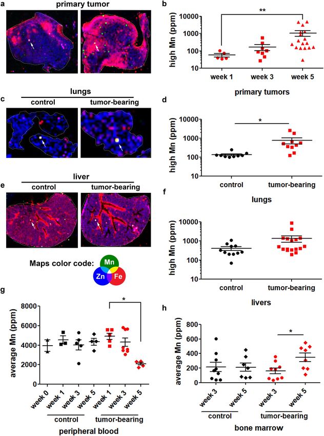
www.nature.com/scientificreports/

          Scientific Reports |   (2021) 11:15833 |   https://doi.org/10.1038/s41598-021-95190-5   2

Vol:.(1234567890)
www.nature.com/scientificreports/
◂Figure 1. Manganese distribution is affected during tumor progression. Control and tumor-bearing mice were
analyzed regarding elemental tissue content and distribution by X-ray fluorescence. Elemental distribution
maps from (a) primary tumors—examples from two tumor-bearing mice; (c) lungs—from one control and one
tumor-bearing mice; and (e) livers—from one control and one tumor-bearing mice were built from the acquired
spectra; map color code: green (Mn—manganese), red (Fe—iron) and blue (Zn—zinc). Maps’ dimensions for
primary tumor: 7.6 mm × 6.0 mm (left panel) and 6.9 mm × 7.7 mm (right panel); lungs: 8.6 mm × 6.2 mm
(control) and 3.2 mm × 4.5 mm (tumor-bearing); livers: 11.0 mm × 14.0 mm (control) and 9.3 mm × 13.7 mm
(tumor-bearing). White arrows indicate regions where manganese was found to be in highest concentration
(high-Mn niches). Elemental concentration of manganese from these regions were plotted for all (b) primary
tumors, (d) lungs, (f) livers, (g) peripheral blood and (h) bone marrow. Primary tumors were analyzed
from week 1 to week 5, lungs and livers were analyzed at week 5 of tumor progression; peripheral blood was
analyzed from week 0 to week 5 of tumor progression and bone marrows were analyzed at weeks 3 and 5 of
tumor progression. Control mice samples are represented in black symbols, tumor-bearing mice samples are
represented in red symbols. Units are expressed in concentration as ppm (parts per million). *p < 0.05; **p < 0.01,
Kruskal–Wallis test and Dunn’s multiple comparison post-test. Primary tumors (week 1) N = 5, (week 3) N = 9,
(week 5) N = 17; Lungs (control) N = 9, (tumor-bearing) N = 10; Livers (control) N = 11, (tumor-bearing) N = 17;
Peripheral blood (week 0) N = 3, (week 1/control) N = 3, (week 1/tumor-bearing) N = 5, (week 3/control) N = 5,
(week 3/tumor-bearing) N = 8, (week 5/control) N = 4, (week 5/tumor-bearing) N = 5; Bone marrow (week 3/
control) N = 9, (week 3/tumor-bearing) N = 9, (week 5/control) N = 7, (week 5/tumor-bearing) N = 8.
effect, while Mn2+ enhances integrin-ligand binding by shifting integrins into high-affinity conformation5. The
role of metals in cancer progression remains to be further investigated.
Primary tumors secrete molecules that promote microenvironment modulation, producing suitable niches
for metastasis6, 7. Recent findings have shown that extracellular vesicles (EVs)8 released by primary tumors
present a relevant role in tumorigenesis and pre-metastatic niche f ormation6, 9, a tumoral modulation of distant
sites10, 11. Melanoma-derived EVs, for instance, were designated relevant in the promotion of metastatic niche
formation12. Another important aspect of premetastatic niche formation is the array of cell surface and extracel-
lular matrix molecules involved in tumor cell adhesion and migration. Integrins, cadherins, fibronectins and
laminins are central molecules in this process, however, the cytoskeleton and cytoskeleton-associated proteins
are also essential for directional activity towards a chemokine s ource13, 14.
In this work we investigate tumor progression from an underexplored point of view: metals modulation by
the primary tumor and its systemic influence. Our findings highlight manganese as a relevant character in tumor
progression, participating in enhanced tumor cell migration and forming manganese-rich niches in primary
tumors and distant organs.
Results
Tumor‑bearing mice present altered manganese systemic distribution. Multi-elemental quanti-
fication and mapping of tissue sections from healthy and tumor-bearing mice were achieved by X-ray fluores-
cence (XRF). Animals were analyzed from 0 to 5 weeks of tumor progression, a time frame that allows obser-
vation of early to late stages of malignancy. We observed that average Mn concentration (ppm) is not affected
by tumor presence in lungs and livers, (SI Appendix, Fig. S1, and Table S1), however, primary tumors present
increased Mn average concentration from 3 to 5 weeks of tumor progression (SI Appendix, Fig. S1), confirming
that this structure is accumulating Mn in a time-dependent manner.
Next, we investigated if Mn accumulation occurs heterogeneously within primary tumors. We screened sam-
ples for regions of highest Mn concentration (Fig. 1). We observed that primary tumors present regions rich in
Mn (Fig. 1a), stated as ‘Mn-rich niches’, which present increased Mn concentration in a time-dependent manner
(Fig. 1b and SI Appendix, Table S2). Interestingly, when analyzing distant organs, we noted that they also present
Mn-rich niches (Fig. 1c, e) in both control and tumor-bearing mice. Lungs of tumor-bearing animals presented
significantly increased Mn concentration within Mn-rich niches (Fig. 1d and SI Appendix, Table S2). Liver
sections from control and tumor-bearing mice also presented regions of Mn accumulation (Fig. 1e) however,
statistical significance was not reached (Fig. 1f—p=0.16, unpaired Student’s T-test—and SI Appendix, Table S2).
A time-dependent detailed multi-elemental analysis of these Mn-rich niches was performed, and we found
that niches found within primary tumors present other altered elements, while lungs and livers present only
altered Mn. Primary tumor Mn-rich niches presented a decrease in P from week 1 to 5, while the elements K, Fe
and Cu presented an increase, and were characterized to be positively correlated with Mn, while P was negatively
correlated (Table 1).
Peripheral blood and bone marrow were also analyzed, revealing that peripheral blood remained stable in
control mice along the 5 weeks of the experiment, while tumor-bearing mice showed a decrease in Mn content
at week 5 (Fig. 1g and SI Appendix, Table S1). At this advanced tumoral stage, we also observed decreased blood
iron (Fe) levels (2.06 ratio control/tumor-bearing mice at week 5), suggesting that decreased Mn levels may not
be due to a direct regulation by the tumor, rather than indirectly affected processes such as Mn/Fe absorption
and transport. Bone marrow, on the other hand, presented a discrete, however significant, increase in Mn content
from week 3 to week 5 in tumor-bearing mice (Fig. 1h and SI Appendix, Table S1).
Observed alterations indicate that the presence of a primary tumor induced by the inoculation of LLC cells
into C57BL/6 mice systemically affects Mn distribution within the organism in a time-dependent manner. Mn-
rich niches are found within primary tumor-specific regions, lungs and bone marrow.
Scientific Reports | (2021) 11:15833 | https://doi.org/10.1038/s41598-021-95190-5 3
Vol.:(0123456789)www.nature.com/scientificreports/
Primary tumors Lungs Livers
Control and tumor-bearing groups at Control and tumor-bearing groups at
Comparison parameters Weeks 1–5
week 5 weeks 1, 3 and 5
P Decreased, p=0.028* Unaltered Unaltered
S Unaltered Unaltered Unaltered
K Increased, p=0.004* Unaltered Unaltered
Ca Unaltered Unaltered Unaltered
Fe Increased, p=0.001* Unaltered Unaltered
Cu Increased, p=0.006* Unaltered Unaltered
Zn Unaltered Unaltered Unaltered
Table 1. Analysis of elemental composition within Mn-rich niches. * Kruskal–Wallis test.
Figure 2. Manganese affects tumor cell survival. LLC cell survival was evaluated after 24 h of incubation with
different MnCl2 concentrations. Cell number represents adhered live cells only. *p < 0.05; **p < 0.01; ***pwww.nature.com/scientificreports/
Figure 3. Manganese promotes tumor cell migration in vitro and heparin counteracts its effects at a non-
anticoagulant concentration. LLC cells migration was evaluated in matrigel-covered transwell chambers.
Transmigration was analyzed after 3 h of incubation in control and Mn-treated ( MnCl2 5 µM) conditions.
Transwell inserts were (a) stained and imaged for (b) cell quantification. N = 3. UFH (bovine unfractionated
heparin—0.1 ng/mL). Scale bars 50 µm. *p < 0.05, one-way ANOVA and Bonferroni’s multiple comparison post-
test.
UFH-treated cells—26 ± 2 µm/h). The fact that Mn-pretreated cells present pronounced collective migration
behavior without alterations in cell speed reveals that Mn enhances cell invasiveness by stimulating collective
migration rather than affecting cell speed.
Other tumor cell lines were evaluated regarding their migration potential in wound healing assays after brief
exposure to M nCl2-supplemented medium (SI Appendix, S2a–c). Data revealed that HeLa cells present similar
behavior compared to LLC cells. On the other hand, B16 and MDA-MB-231 cell lines do not respond significantly
to Mn, an indication that underlining mechanisms regulate cell migration.
We have also tested tumor cell behavior in the presence of other divalent metals known to affect integrin
function, magnesium (Mg) and zinc (Zn). We could observe that MgCl2 does not affect tumor cell survival
or migration (SI Appendix, S3a, b), while ZnCl2 is cytotoxic at 500 µM, but does not affect cell migration (SI
Appendix, S3c, d) under tested conditions.
To further investigate Mn influence on LLC cell migration, modified culture media were prepared with fetal
bovine serum (FBS) reduced to 0.7× (Mn-low) or increased in 9.5× (Mn-high) standard Mn concentration (SI
Appendix, Table S3). LLC cells growth and viability were assessed in Mn-low and Mn-high media for 48 h. After
this period, viable LLC cells were quantified (Fig. 5a) and cell viability was assessed by MTT assay (Fig. 5b).
Mn-low and Mn-high media do not affect LLC cell growth and viability in 48 h of culture. Next, LLC cells were
cultured in Mn-low or Mn-high media for 24 h and migration was assessed by wound healing assays (Fig. 5c).
Interestingly, migration (Fig. 5d) in Mn-low conditions is similar to control, however, when pretreated with Mn
(pre-Mn), migration is slightly, but significantly affected (pre-Mn 56 ± 1.22% vs. Mn-low + pre-Mn 52.2 ± 1.82%;
average ± s.e.m.). On the other hand, LLC cells cultured in Mn-high conditions migrate more, even in the absence
of a Mn pretreatment (control 45.65 ± 1.11% vs. Mn-high 56.93 ± 1.5% vs Mn-high + pre-Mn 55.4 ± 1.12%; aver-
age ± s.e.m.). These data confirm that LLC cell migration is, to some extent, directly affected by Mn availability.
Manganese is preferentially secreted from tumor cells in extracellular vesicles. In vivo data
revealed the presence of Mn-rich niches within primary tumors and distant organs, however, only niches found
within primary tumors presented alterations in other elements alongside Mn, while niches found within lungs
and livers only presented alterations regarding Mn content (Table 1). Therefore, we sought to further under-
stand the formation of local (primary tumor) and distant Mn-rich niches by investigating Mn mechanisms of
distribution, such as extracellular vesicles (EVs) or secreted in soluble form. First, we incubated LLC cells with
MnCl2-supplemented medium and after 4h of conditioning, cells were collected and analyzed for Mn content. A
similar protocol was performed; however, medium conditioning took place for 24 h in basal FBS-free medium,
which was submitted to an EV isolation protocol. Both control and Mn-pre-exposed cells secreted EVs in a
large size-range, compatible with the secretion of mostly exosomes, but also microvesicles (Fig. 6a). In order
to confirm EVs identity, an immunoblotting assay was performed for the detection of EV markers CD63 and
syntenin-1 (Fig. 6b). Next, all samples were prepared for Mn quantification by XRF (Fig. 6c), whereas LLC cells
exposed to Mn-pretreatment presented increased levels of retained Mn.
Interestingly, other tumor cell lines were tested (SI Appendix, S2d-f) and we observed that similar to LLC cells,
HeLa cells retain Mn after brief exposure and show increased migration (SI Appendix, S2a and d), whereas B16
Scientific Reports | (2021) 11:15833 | https://doi.org/10.1038/s41598-021-95190-5 5
Vol.:(0123456789)www.nature.com/scientificreports/
Figure 4. Manganese modulates tumor cell migration pattern. LLC cells migration pattern was evaluated in
wound healing assays by intermittent (a, b) and continuous (c–e) monitoring. Cells were (a) imaged and (b)
total migrated distance was quantified. N = 9. (c) Time lapse images—example from a Mn-pretreated cells
migration video—were also acquired and compilated into videos for (d) collective migration quantification. (e)
Single-cell speed was calculated from videos. N = 3. UFH (bovine unfractionated heparin—0.1 ng/mL); pre-Mn
(MnCl2 5 µM 1 h pretreatment prior to migration). Scale bars 50 µm. *p < 0.05, one-way ANOVA test and
Bonferroni’s multiple comparison post-test.
Scientific Reports | (2021) 11:15833 | https://doi.org/10.1038/s41598-021-95190-5 6
Vol:.(1234567890)www.nature.com/scientificreports/
Figure 5. Tumor cells cultured in manganese-low and manganese-high conditions present different migration
patterns. LLC cells were cultured in Mn-low and Mn-high conditions for 48 h and (a) cell growth was evaluated by
counting live, adhered cells using trypan blue. (b) Cell viability was evaluated by the MTT assay. LLC cell migration
pattern was evaluated in wound healing assays by intermittent monitoring at 0 h and 12 h. Cells were (c) imaged and
migrated distance was (d) quantified. Mn-low (standard high glucose DMEM + Mn-low FBS); Mn-high (standard high
glucose DMEM + Mn-high FBS). Scale bars 200 µm. Cell growth assay N = 4; MTT assay N = 6; wound healing assay
N = 6. *pwww.nature.com/scientificreports/
Figure 6. Tumor cell-derived extracellular vesicles are enriched in manganese and affect tumor cell migration.
LLC cells-derived extracellular vesicles were concentrated by ultracentrifugation. (a) EVs quantification and size
determination. N = 6. (b) CD63 and syntenin-1 detection by Western blotting. Ponceau was used as total protein
control. (c) Tumor cells, EV-enriched fraction, EV-free conditioned medium and basal medium were analyzed
by X-Ray Fluorescence. N = 6. Units are expressed in concentration as ppm (parts per million). Control cells—
white bars; Mn-pretreated cells (Mn) – black bars. *p < 0.05, Student’s T test. (d) Wound healing assays of LLC
cells incubated with extracellular vesicle-enriched medium from control and Mn-pretreated LLC cells. N = 12.
*p < 0.05; **p < 0.01, one-way ANOVA and Bonferroni’s Multiple Comparison Test.
cells retain Mn after brief exposure, but do not respond with accentuated migration (SI Appendix, S2b and e).
Finally, MDA-MB-231 cells do not retain Mn and do not migrate more compared to control cells (SI Appendix,
S2c, f).
Next, we were able to verify that Mn-pretreated LLC cells secreted EVs with elevated Mn concentration
(1.42 ratio Mn-pretreated/control cells), whereas the remaining EV-free medium did not present detectable dif-
ferences between conditions (41 ± 8; 38 ± 2; 43 ± 8 nM Mn for basal medium, control conditioned, and pre-Mn
Scientific Reports | (2021) 11:15833 | https://doi.org/10.1038/s41598-021-95190-5 8
Vol:.(1234567890)www.nature.com/scientificreports/
treated conditioned medium, respectively, mean ± s.e.m.—Fig. 6c). Additionally, all the other detected elements
(P, S, K, Ca, Fe, Cu and Zn) did not present differences in concentration between control EVs and pre-Mn EVs.
Finally, to evaluate the contribution of Mn-enriched EVs to tumor cell migration, we performed wound heal-
ing assays in the presence of LLC-derived EVs. The amount of EVs used for migrations were 2.5*108 ± 6.2*107
for control EVs and 2.9*108 ± 1.5*108 for pre-Mn EVs, which corresponds to 4 times the number of vesicles LLC
cells would naturally secrete in culture. We could observe that naive LLC cells exposed to EVs derived from
Mn-pretreated cells migrate significantly more compared to control cells (Fig. 6d).
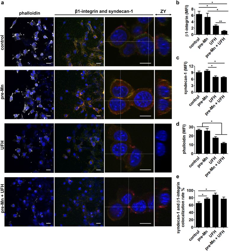
Manganese and heparin affect β1‑Integrin and the heparan sulfate proteoglycan syndecan‑1
during tumor cell migration. To unveil possible mechanisms related to Mn effect on tumor cell migra-
tion, we analyzed the expression pattern of the migration-related molecules β1-integrin and syndecan-1, as well
as F-actin, in wound healing assays. PCR analyses were performed in order to confirm the presence of ITGB1
and SDC1 mRNA in LLC cells (SI Appendix, S4a). Next, we analyzed syndecan-1 expression in LLC cells by flow
cytometry (SI Appendix, S4b, c). We have followed the wound healing assay protocol and collected cells at 8 h of
migration. Syndecan-1 expression did not present significant changes between conditions when analyzing total
cell population.
Next, under similar conditions portrayed in Fig. 4a, LLC cells were let to migrate for 8 h, then cells were fixed
and stained for the specific molecules (Fig. 7a). Interestingly, we observed that Mn-pretreated cells presented
similar levels of β1-integrin and syndecan-1 staining compared to control cells (Fig. 7b, c), however, the colo-
calization rate of these molecules was significantly higher (Fig. 7e). On the other hand, heparin (UFH) treatment
led to a decrease in β1-integrin and syndecan-1 staining (Fig. 7b, c) and, despite the fact that the colocalization
rate in this condition is higher (Fig. 7e), we found a lower signal from these molecules in our analysis. Finally,
phalloidin staining revealed that Mn does not alter F-actin, however, heparin (UFH) treatment significantly
decreases F-actin staining (Fig. 7d).
Analysis of total cell population (SI Appendix, S4) has not shown differences between conditions; however,
we were able to detect differences in β1-integrin and syndecan-1 expression near the migration edge. Higher
magnification images revealed that these molecules are located on the cell surface, as well as within cells (Fig. 7a
ZY projections and SI Appendix, S5 XZ projections), which could explain the differences in molecules’ expression
found between total cell population and cells in the migration edge.
Primary tumors and livers were also analyzed for syndecan-1 expression. We were able to find high syn-
decan-1 cell clusters within primary tumors (N = 4) and tumor-bearing livers (N = 6), while analyzed control
livers (N = 4) did not present similar clusters (SI Appendix, S6a–h). Additionally, a side-by-side comparison was
made for sequential sections of a tumor-bearing mouse liver immunostained for syndecan-1 and irradiated for
multi-elemental analyses. These twin images show the occurrence of 5 regions of high syndecan-1 cell clusters
nearby high-Mn niches, 1 region of high syndecan-1 cell cluster distant from any high-Mn niche and 1 region
of high-Mn niche distant from any high syndecan-1 cell cluster (SI Appendix, S6i and j). These data indicate a
possible relationship between high-Mn niches and syndecan-1 in our in vivo mouse model of tumor growth.
A dermatan sulfate found in the marine invertebrate Styela plicata presents similar antitu‑
moral effect in vitro compared to bovine heparin. Ascidians are tunicates that produce sulfated
glycosaminoglycans in their viscera. Ascidian-derived glycosaminoglycans may be considered as mammalian
heparin analogs and antitumoral molecules16. We tested a dermatan sulfate derived from the ascidian Styela pli-
cata regarding its ability to inhibit Mn-stimulated tumor cell migration in vitro. We observed that this molecule
inhibits LLC cells exacerbated migration after brief Mn exposure (Fig. 8).
To investigate if UFH and the ascidian dermatan sulfate bind Mn, we performed XRF analyses on glycans that
were incubated with a MnCl2 solution, followed by extensive dialysis. We used the structures published by Pavão
et al.17 and R
abenstein18 to estimate how many Mn atoms would bind to the ascidian dermatan and to UFH.
Our calculations estimated that both glycosaminoglycans efficiently retained Mn at the ratio of, approximately,
one Mn atom per disaccharide, according to mass fraction estimations (SI Appendix, Table S4) that took into
consideration the heterogeneous nature of glycosaminoglycans modifications, such as sulfate group additions.
Sample contamination by free Mn was evaluated by analyzing water collected from the last dialysis day. We could
observe that background levels are low and not responsible for the significant amounts detected in the glycans
dialyzed solutions.
Finally, we tested if UFH would, indeed, affect Mn content and distribution in vivo. To this end, we treated
tumor-bearing mice with UFH at 2 and 4 weeks of tumor development with local subcutaneous injections of
UFH at a non-anticoagulant concentration. After 5 weeks of tumor development, primary tumors were collected
for XRF analyses and Mn quantification and mapping. We were able to verify that both experimental groups,
tumor-bearing mice (control) and UFH-treated tumor-bearing mice, presented unchanged average Mn and
Mn-rich regions (SI Appendix, S7a; (11 ± 4)*102 (untreated) vs. (28 ± 6)*10 (UFH-treated), average ± std error
Mn (ppm); p = 0.23, T test); S7b; 29 ± 2 (untreated) vs. 35 ± 1 (UFH-treated), average ± std error Mn (ppm);
p = 0.13, Student’s t test).
Discussion
Trace elements, especially metals, are regulators of cellular homeostasis and present key roles on pathological
processes, such as cancer19. They function as cofactors, electron donors and are central for bioelectrogenesis and
electrophysiology. Although first reports date from many decades ago20, metals have recently received especial
attention and, nowadays, their multiple roles are highlighted in metallomic s tudies21, 22.
Scientific Reports | (2021) 11:15833 | https://doi.org/10.1038/s41598-021-95190-5 9
Vol.:(0123456789)www.nature.com/scientificreports/
Figure 7. Manganese affects β1-integrin and syndecan-1 expression in tumor cells. Confocal microscopy
images of (a) immunostainings for β1-integrin, syndecan-1 and F-actin (phalloidin). DAPI (blue), β1-integrin
(red), syndecan-1 (green) and phalloidin (white). ZY axis projections were generated from higher magnification
images of β1-integrin and syndecan-1 merged stainings. Scale bars are 25 µm (lower magnification) and 10 µm
(higher magnification). N = 6. Images were quantified regarding fluorescence intensity for (b) β1-integrin, (c)
syndecan-1 and (d) phalloidin-Alexa 488. (e) β1-integrin and syndecan-1 colocalization rates. MFI, mean
fluorescence intensity. One-way ANOVA with Bonferroni’s post test. *p < 0.05; **p < 0.01.
The present work brings to light the relevance of manganese (Mn) modulation during tumor progression. Mn
is an important cofactor for many systems, especially for integrin outside-in a ctivation23–26 and Mn-dependent
superoxide dismutase (SOD2) a ctivity27–29. Consequently, Mn is particularly relevant in cell migration and sur-
vival, two central aspects of tumor progression and metastasis.
Interestingly, Mn has been widely studied and applied as a contrast agent for tumoral magnetic resonance
imaging30–32. In this work we have focused our investigations on Mn distribution and accumulation in primary
Scientific Reports | (2021) 11:15833 | https://doi.org/10.1038/s41598-021-95190-5 10
Vol:.(1234567890)www.nature.com/scientificreports/
Figure 8. Ascidian dermatan sulfate counteracts migration-promoting effects of manganese on tumor cells. LLC cell
migration was evaluated by wound healing assays after brief exposure to manganese followed by treatment with an ascidian
dermatan sulfate (DS). Cells were (a) imaged at 0 h and 12 h and migrated distance was (b) quantified. N = 9. Scale bars 50 µm.
*p < 0.05, one-way ANOVA test and Bonferroni’s multiple comparison post-test.
Scientific Reports | (2021) 11:15833 | https://doi.org/10.1038/s41598-021-95190-5 11
Vol.:(0123456789)www.nature.com/scientificreports/
tumors and its effects on tumor cell behavior. We aimed to observe trace elements’ changes imposed by tumor
progression and their contribution to microenvironment modulation.
In depth XRF analyses of tumor-bearing mice revealed that LLC-induced ectopic primary tumors accumu-
late Mn in a time-dependent manner. Accumulation occurs not only when average Mn was measured in the
tissue, but much more intensely in specific regions of approximately one to four pixels (360–1440 µm2) in XRF
imaging. Interestingly, we did not detect average Mn changes in livers and lungs. Nevertheless, when analyzing
XRF elemental maps, we were able to depict Mn-rich niches, as found in primary tumors, an indication that Mn
distribution is affected by tumor progression. Due to the fact that tumor-bearing mice bone marrows presented
elevated average Mn levels, while peripheral blood presented a decrease at the same stage of tumor progression,
we conclude that occurrence of Mn-rich niches in primary tumors and lungs, as well as elevated Mn levels in
bone marrow are regulated processes strongly associated with tumor progression. Also, the fact that different
tissues presented varied profiles of Mn distribution indicates that tissue Mn levels are not simply a result of Mn
availability, rather, they may be the result of multiple regulation mechanisms, such as Mn transporters, Mn-
binding molecules alone or associated with Mn-related ions, such as Fe, Ca and Zn.
Mn absorption and excretion are well controlled physiological p rocesses33. Diet intake and gastrointestinal
absorption are the main source of systemic Mn, while liver Mn processing and excretion through the gut is
the main pathway for Mn losses34. Overall, great systemic changes in Mn are rarely observed, unless in severe
intoxication cases and disease development, causing mostly neural impairment s ymptoms35. Our data revealed
an interesting aspect of Mn imbalance during disease progression: occurrence of Mn-rich niches in the absence
of obvious Mn systemic changes.
Mn-rich niches could be found in clusters at different locations, including near blood vessels, indicating that
this element is probably originating from the circulation36. In addition, detailed analysis of the niches revealed
that primary tumors present Mn alterations before distant organs. Also, Mn-rich niches from primary tumors
present correlated alterations in P, K, Fe and Cu, while Mn-rich niches from lungs and livers only present altera-
tions in Mn. These data indicate that Mn alterations initiate in primary tumors and may influence the occurrence
of Mn-rich niches in healthy tissues.
Mn absorption, distribution and excretion are well regulated systemic processes, which points towards the
formation of Mn-rich niches as a hidden process, undetectable to widely used metal detection techniques and,
more importantly, to biological systemic sensing mechanisms. Our in vitro data show that even brief changes
in Mn levels in culture medium allow LLC cells to significantly accumulate Mn and secrete it in extracellular
vesicles (EVs), indicating a Mn route in LLC cells responsible for Mn retention, internalization, and secretion in
EVs. The relevance of these data is highlighted in different works, such as by Harischandra and c olleagues37. The
authors show that Mn exposure promotes α-synuclein secretion in exosomal vesicles, which subsequently evokes
proinflammatory and neurodegenerative responses. They report that, in dopaminergic neurons, Mn stimulates
exosome release37. Additionally, exosome-associated integrins present important functions in premetastatic niche
formation and metastasis. As reviewed by Hoshino et al.38, some works showed that exosome-associated integrin
expression profiles underlie tumor cell organotropism. For instance, exosomal integrins α6β4 and α6β1 were
linked to lung metastasis, while exosomal integrin αvβ5 was related to liver metastasis. Different pairs of bone
marrow associated integrins, such as α4β1, were shown to be essential for hematopoietic cell migration within
bone marrow11, therefore the detected elevated levels of Mn within tumor-bearing mice bone marrow may be
due to the arrival of extracellular vesicles expressing Mn-activated integrins released by cancer cells from primary
tumor. We hypothesize that LLC cells continuously retain Mn from circulation, resulting in average elevated
Mn levels in primary tumors, as well as formation of Mn-rich niches through extracellular vesicle delivery.
Multi-elemental analyses of Mn-pretreated cells-derived EVs revealed that Mn is the only detected element at
significantly different levels compared to control EVs. This information points toward EVs as one of the likely
routes of Mn distribution from the primary tumor to distant organs during tumor progression. Finally, we have
analyzed different tumor cell lines and found that Mn accumulation does not occur in all cells, for instance HeLa
and B16 cell lines retain Mn after brief in vitro exposure, while MDA-MB-231 cells do not. These additional data
confirm the specificity of Mn retention, internalization and secretion in some types of tumors.
There are a number of pathways related with Mn distribution. The Mn transporter DMT1 is reported to be
involved in tumor progression, however, most reports focus on iron, rather than Mn, as a DMT1-associated
tumor-inducing agent39–41. ZIP (Zrt- and Irt-like protein) transporters, on the other hand, have been associ-
ated with zinc and Mn transport, and being expressed by endothelial c ells42, are stronger candidates for Mn
retention in primary tumors. Alterations in the expression levels of Mn transporters are also described to lead
to altered Mn absorption. The work by Mercadante and colleagues shows that Slc30a10-deficient mice present
severe Mn excess accumulation, and this phenomenon was linked to Slc30a10 deficiency in the liver and small
intestines43. Mutated SLC39A14 has also been described to cause severe Mn accumulation in humans, leading
to the early onset of parkinsonism-dystonia44. Our data shows that, even in a regular diet, free from Mn intoxi-
cation sources, tumor-bearing animals present a unique feature, which is the formation of Mn-rich niches. We
show that these niches are formed in a time-dependent manner after Mn accumulation in the primary tumor
and reach impressive levels in advanced stages of tumor progression. Further investigations of alterations in Mn
transport mechanisms in tumor cells are an interesting and relevant perspective for the field.
Charge interactions are a fast and less specific pathway for Mn retention and subsequent internalization. In the
present work we show that LLC cells express the heparan sulfate proteoglycan syndecan-1 and confocal analyses
revealed that upon Mn treatment, syndecan-1 and β1-integrin present higher colocalization rates. Proteoglycans
have been described as relevant co-receptors and their sulfated glycosaminoglycans chains offer many binding
sites for Mn on the cell s urface45, 46. Mn could be solely responsible for affecting syndecan-1 cell surface distribu-
tion pattern if proteoglycan internalization or shedding were promoted by binding to the cation. On the other
hand, these processes could also be mediated by proteoglycan-mediated Mn presentation to its receptor on the
Scientific Reports | (2021) 11:15833 | https://doi.org/10.1038/s41598-021-95190-5 12
Vol:.(1234567890)www.nature.com/scientificreports/
cell surface. High magnification confocal analyses of syndecan-1 and β1-integrin immunostainings revealed that
these molecules can be found on the cell surface, as well as intracellularly, confirming that both processes are
equally possible. Finally, syndecan-1 immunostainings of primary tumors and livers from control and tumor-
bearing mice indicate that this proteoglycan may be involved in Mn distribution in vitro, as well as in vivo.
Mn-rich niches in distant organs are discrete in comparison to primary tumors, nevertheless they can also be
found clustered near vessels (Fig. 1e). Following the time-associated evolution of this pattern, we hypothesize that
distant organs may receive Mn from primary tumors, forming Mn-rich niches later in tumor progression. Mn
distribution from primary tumors can occur by secreting soluble factors or vesicles into the circulation. Our data
shows that, in vitro, if briefly exposed to excess Mn, LLC cells release Mn-rich EVs. We have also detected that
the remaining vesicle-free medium contains the same levels of Mn concentration as basal medium, confirming
that EVs are the preferential route for Mn secretion in these cells. The release of Mn-rich EVs in the circulation
would explain the observed pattern of Mn-rich niches without impacting Mn average tissue levels. Taking into
consideration Mn-dependent processes of cell migration and survival, we hypothesize that occurrence of Mn-
rich niches in distant organs may be a characteristic of microenvironment modulation.
Cell survival and migration is a crucial step in metastasis, and Mn is a well-established integrin a ctivator23,
25, 47
, an essential factor to both processes. Our data shows that LLC cells present enhanced migration when
exposed to Mn-rich culture medium. Indeed, we have observed that this behavior is not repeated by incubating
cells with divalent cations, magnesium and zinc, supporting the specific relevance of Mn modulation during
tumor progression. We hypothesize that tumor cells able to retain Mn in vivo migrate more efficiently due to a
more efficient integrin activation and collective migration behavior. Supporting evidence of this phenomenon
has been described in an in vitro model of tumor cell migration, whereas MDA-MB-231 cells are exposed to
MnCl2 and monitored for migration and proliferation a ctivities48. Findings show that M nCl2 5 µM mainly
stimulates transmigration through type I collagen (chemotaxis assay), while transmigration through matrigel
(chemoinvasion assay) is more pronounced at M nCl2 100 µM. Our group has found that MDA-MB-231 cells do
not retain Mn and do not present enhanced migration in the wound healing assay at M nCl2 5 µM, in consonance
with the results described above by Luparello (2019). Nevertheless, differences in cell behavior according to Mn
concentration and ECM setup may arise from variations in Mn transport mechanisms and in the expression
profile of ECM-binding cell surface molecules. LLC cells were also cultured in Mn-modified media (Mn-low and
Mn-high) and we could see that cell viability was not affected by these culture conditions. Next, we verified that
basal migration is not affected in Mn-low culture conditions, however, when cells are pretreated with a pulse of
high Mn, Mn-low cells migrate slightly less than control cells. On the hand, LLC cells cultured in Mn-high condi-
tions migrate similarly to pre-Mn cells (control cells + Mn pretreatment), and, interestingly, pre-Mn treatment in
Mn-high cells does not further enhance migration. These data reveal that tumor cell migration is affected by both
short-term and long-term Mn exposure. This model of in vitro tumor cell migration indicates that tumor cells
in vivo may experience similar conditions and present enhanced migration when near or within Mn-rich niches.
Other tumor cell lines were analyzed regarding migration behavior after brief exposure to Mn-rich culture
medium, interestingly, cell lines able to retain Mn may (HeLa cells) or may not (B16 cells) respond to Mn,
however, among the tested cell lines the one that does not retain Mn, also does not change its migration pattern
upon Mn stimulation (MDA-MB-231 cells).
Finally, we tested if exogenous sulfated polysaccharides: heparin and ascidian dermatan sulfate would act
as Mn-chelating agents. These molecules are great candidates to interfere with tumor progression because they
counteract Mn effects at exceptionally low concentrations, below their anticoagulant activity, in addition to
being physiological metal m odulators49. Both glycosaminoglycans bind to Mn, regulate Mn-stimulated tumor
cell migration and, in the case of heparin, decrease F-actin staining during migration. However, different gly-
cosaminoglycans may promote or inhibit tumor p rogression45. Marine-derived molecules have been studied as
antitumoral agents16. Groult and colleagues have tested red seaweed carrageenan regarding its anti-heparanase
effect on MDA-MB-231 breast cancer c ells50. Styela plicata is an ascidian that produces a dermatan sulfate with
structural similarities with mammalian heparin but does not share the same potent anticoagulant e ffect51.
In summary, our analyses point to a time-dependent process that promotes the formation of Mn-rich niches.
These niches are prominent within primary tumors; however, they can also be observed in distant organs. Our
results show changes in LLC cells migration even after a short-term exposure to Mn from invasive to highly
invasive. Interestingly, β1-integrin and syndecan-1 are found colocalized after manganese short-term exposure
in migrating cells. EVs also change from presenting low manganese content to higher manganese content.
In conclusion, the overall results revealed an unexplored role of Mn in tumor progression and possible
mechanisms involving its effect on tumor cell motility. Mn accumulation in specific regions of the organism
may not be a common ground for all cancers, nevertheless it represents a new aspect of tumor progression that
deserves special attention.
Materials and methods
Cell lines and standard cell culture conditions. Cells were cultured under standard conditions at 37 °C
and CO2 5% atmosphere. Cell culture medium was composed of DMEM (Sigma-Aldrich) supplemented with
fetal bovine serum (FBS) 10% (v/v) (Vitrocell) and glucose 4500 mg/L (Sigma-Aldrich). Cells were passaged
with a trypsin-EDTA solution (trypsin 0.25% and EDTA 1 mM). Protease-free cell detachment was achieved
by incubating cells in a Ca2+ and M
g2+-free PBS solution with EDTA 1 mM, followed by detachment with a cell
scraper and further pipetting. Cell viability was assessed by counting detached cells using a Neubauer chamber
in trypan blue 0.2% solution. Each quantification was performed at least three times.
Cell lines used in this work were LLC (mouse Lewis lung carcinoma, ATCC), MDA-MB-231 (human breast
adenocarcinoma, ATCC), HeLa (human cervix adenocarcinoma, ATCC) and B16 (mouse melanoma, ATCC).
Scientific Reports | (2021) 11:15833 | https://doi.org/10.1038/s41598-021-95190-5 13
Vol.:(0123456789)www.nature.com/scientificreports/
Manganese cell toxicity assays. LCC cells were seeded in 24-well plates (6 × 10⁴ cells/well) and after
cultures reached 80% confluence, cells were incubated for 24 h in increasing concentrations of M
nCl2 (5, 10, 25,
50, 100 and 500 μM). Cells were harvested by enzymatic treatment and viable cells were quantified in a Neubauer
chamber using trypan blue.
Transmigration assays. Semi-confluent LLC cells were detached using the protease-free method and
quantified as described above. Next, transwell inserts 8 µm pore-size (Corning) were placed on 24-well culture
plates and the top chamber was incubated with 100 µL matrigel (BD Biosciences) diluted in FBS-free medium
to 1 mg/mL. Inserts were incubated at room temperature for 2 h. Next, excess medium was removed by gentle
aspiration and inserts were prepared for transmigration. Bottom chamber was filled with 650 µL culture medium
supplemented with FBS 10%, while top chamber received 100 µL FBS-free medium with a total of 105 cells. Plates
were incubated in standard culture conditions for 3 h, when inserts were collected for processing. Quantification
of transmigrated cells was achieved by removing inserts from culture plates, followed by gentle scraping of non-
transmigrated cells from the top chamber using a cotton swab. Next, inserts were rinsed in PBS, fixed and stained
in a crystal violet 0.5% (m/v) solution in methanol 20%. Stained inserts were imaged, and cell quantification was
performed using ImageJ software 1.52a.
Cell migration wound healing assays. Cells were cultured in 6-well plates until 80% confluence, then
culture medium was changed to control (regular culture medium) or pre-Mn (regular culture medium with the
addition of manganese chloride ( MnCl2—Sigma-Aldrich) 5 µM. Cells were incubated in these conditions for
1 h. Next, medium (control or Mn-added) was removed, cells were rinsed and mechanically removed from the
plate in a cross-shaped pattern using a sterile P1000 tip. Cells were rinsed again for removal of cell debris and
incubated in control (regular culture medium) or polysaccharide-treated (regular culture medium with the addi-
tion of unfractionated bovine heparin—UFH or ascidian-derived dermatan sulfate—ascDS at 0.1 ng/mL) for 12
h. In order to evaluate cell migration and wound closure, cells were imaged, using the cross center as a reference
at 0 h and 12 h. Migration was quantified using ImageJ software and expressed as the percentage of migrated
distance relative to the original wound width.
An automatic microphotography system was used to monitor migrating cells under standard culture condi-
tions and generate time lapse videos. Cells were imaged every 2 min for 24 h. Images were compiled into videos
that were analyzed regarding migrating pattern (collective cell migration) and single-cell speed (ImageJ software).
Extracellular vesicles concentration from tumor cell conditioned medium. LLC cells were cul-
tured until 70% confluence, next, control culture medium was changed to pre-Mn culture medium and cells
were incubated for 4 h. Afterwards, cells were rinsed and incubated in serum-free basal medium (high glucose
DMEM) for 24 h for medium conditioning. Extracellular vesicles’ concentration was achieved by an ultracen-
trifugation protocol52 as follows: conditioned medium was centrifuged at 300g for 10 min, 2000g for 10 min,
and 10,000g for 30 min. Pellets were discarded after each centrifugation round. Next, the remaining supernatant
was ultracentrifuged (Beckman 70Ti rotor) at 100,000g for 70 min. The extracellular vesicle-enriched pellet was
washed in PBS to eliminate contaminating proteins and centrifuged one last time at 100,000g for 70 min. The
final pellet was resuspended in 100 µl of PBS. A Nanoparticle Tracking Analysis – NTA Zetaview - was used to
monitor separation.
Extracellular vesicles markers characterization by immunoblotting. For extracts, exosomes pre-
viously purified were lysed in 50 mM HEPES, pH 6.4, 1 mM MgCl2, 10 mM EDTA, 1% Triton X-100, 1 µg/mL
DNase, 0.5 µg/mL Rnase, 1 mM PMSF, 1 mM benzamidine, 1 µg/mL leupeptin and 1 µg/mL soybean trypsin
inhibitor. Total protein content in the exosomes extracts was determined by BCA method. Lysates were dena-
tured in sample buffer (50 mM Tris·HCl, pH 6.8, 1% SDS, 5% 2-ME, 10% glycerol, and 0.001% bromophenol
blue) and heated in boiling water for 3 min. Samples (10 µg total protein) were resolved by 15% SDS-PAGE and
proteins transferred to polyvinylidine difluoride membranes. Molecular weight standards were run in parallel.
Membranes were blocked with Tween-TBS (TBS, 0.01% Tween 20; T-TBS) containing 5% BSA and probed with
primary antibody (1:500) overnight at 4 °C. Primary antibodies used in western analysis were anti-CD63 cat:
10628D and anti-Syntenin-1 cat: PA528813 (Invitrogen, Carlsbad, CA, USA). The membranes were rinsed with
T-TBS and incubated for 1h at room temperature with HRP-conjugated secondary antibody (1:5000). Immuno-
reactive proteins were visualized by the ECL detection (GE, Chicago, IL, USA).
Extracellular vesicles in cell migration wound healing assays. LLC cells were submitted to wound
healing assays incubated with extracellular vesicles (EVs) enriched medium containing 4 times more EVs than
a naturally conditioned medium. Cells received EVs from naïve LLC cells (control EVs) and from M nCl2 pre-
exposed cells (pre-Mn EVs). In order to evaluate cell migration and wound closure, cells were imaged at 0 h
and 12 h. Migration was quantified using ImageJ software and expressed as the percentage of migrated distance
relative to the original wound width.
Animal model of spontaneous metastasis. C57BL/6 male and female mice between 8 and 12 weeks
of age were anesthetized and inoculated subcutaneously in the posterior dorsolateral region with 5 × 10⁵ LLC
cells in 60 µL serum-free DMEM as vehicle (tumor-bearing group), whereas control animals were inoculated
with vehicle only. Animals were randomly assigned to experimental groups, comprising a minimum of five
specimens per experimental group in order to accurately assess variations between specimens. All experimental
Scientific Reports | (2021) 11:15833 | https://doi.org/10.1038/s41598-021-95190-5 14
Vol:.(1234567890)www.nature.com/scientificreports/
groups were kept under standard conditions and daily monitored for vital signs, behavior and tumor size from
0 to 5 weeks of tumor development. Animals that presented signs of possible discomfort were anesthetized with
a combination of xylazine and ketamine and submitted to a cervical dislocation procedure according to the
Brazilian animal experimentation guidelines. Death was confirmed by observing all of the following: absence of
breathing movements, absence of pulse and absence of heartbeat. These animals were excluded from the experi-
ments.
Tissue samples were collected in order to investigate tumor progression in a time-dependent manner. Spe-
cifically, primary tumors, liver and lungs, in addition to peripheral blood and bone marrow were harvested at
weeks 0–5 and processed for analyses. Solid tissues were rinsed in saline, briefly and superficially fixed in 4%
paraformaldehyde for 5 min and cryopreserved in OCT (Optimal Cutting Temperature compound – Tissue-Tek)
for further sectioning. Liquid tissues were collected fresh and immediately processed for further analyses. All
procedures involving animal experimentation were approved by the Federal University of Rio de Janeiro Animal
Experimentation Committee (protocol number: 015/18) and were performed in accordance with the Brazilian
guidelines for scientific use of animals. This work is in compliance with the ‘Animal Research: Reporting of
In Vivo Experiments’ (ARRIVE) essential 10 guidelines.
X‑ray microfluorescence analyses. X-ray microfluorescence analyses were performed at the UVX Light
Source at the Brazilian Synchrotron Light Source Laboratory (LNLS). Analyzed samples comprised of homoge-
neous liquid samples, cultured cells or tissue sections placed on Ultralene thin film (SPEX SamplePrep). Homo-
geneous liquids and resuspended cells were placed on Ultralene film in drops and let to air dry. Tissue samples
were cryosectioned into 20 µm-thick slices and placed on Ultralene film. Cultured cell samples were quantified,
scraped from the plate, placed on Ultralene film and let air dry. Samples were analyzed at D09B X-ray Fluores-
cence beamline at room temperature and ambient pressure according to Sartore et al.53.
Immunostaining imaging. LLC cells were cultured on RS glass slides (Nunc) covered in growth factor-
reduced matrigel (BD Biosciences). When 80–90% confluence was reached, cells were submitted to the migra-
tion wound healing assay for 8 h. Cells were fixed in 4% paraformaldehyde for 10 min. Next, cells were rinsed
with PBS and incubated in blocking solution (2% bovine serum albumin in PBS) for 30 min, followed by incu-
bation with Triton 0.1% for 5 min. Primary and secondary antibodies were incubated separately for 90 min
each in the following order: rat anti-syndecan-1, anti-rat-Cy5, goat anti-β1-integrin, anti-goat-Cy3, phalloidin-
Alexa 488. Cells were rinsed with PBS between steps. Then, slides were mounted in anti-fade mounting medium
Vectashield (Vector Laboratories) with DAPI. Antibodies used for immunostaining were diluted in 0.5% albu-
min in PBS and are anti-CD138 (1:100 - RD Systems) and anti-β1 integrin (1:100 - Santa Cruz Biotechnology).
Images were acquired on the confocal microscope system TCS SPE (Leica) and processed using LAS X
software (Leica). Cells were imaged using a 10X objective lens and a 40X oil immersion objective lens (with
additional 3X zoom). Lasers used were 488, 583 and 633 nm wavelength. Images were acquired in stacks of 500
nm thickness and the number of stacks varied according to sample height.
Traditional widefield immunofluorescence images were acquired using an Axio Imager A1 microscope and
Axiovision System software (both Zeiss; Oberkochen, Germany).
Mn‑low and Mn‑high fetal bovine serum. LLC cells were cultured in modified cell media and evalu-
ated for cell viability and migration pattern. Cell media were prepared with standard basal high-glucose DMEM
medium and Mn-depleted (Mn-low) and Mn-supplemented (Mn-high) fetal bovine serum (FBS). Modified FBS
solutions were prepared from standard FBS by treatment with activated Dowex 50Wx4 cationic exchange resin
(Dow) for removal of c ations54. Next, a mix of 80% dowex-treated FBS and 20% standard FBS was made in order
to partially replenish FBS of Dowex-removed cations. Next, to recover FBS standards of cation composition, the
following salts and respective concentrations were added to the mix: CaCl2 3.85 µM, FeCl2 21 µM, KCl 7 mM
and NaCl 101.5 mM. Mn-low FBS did not receive any additional supplementation, while Mn-high received
additional MnCl2 in order to meet average FBS Mn levels55. Finally, Mn-low and Mn-high FBSs were filtered
using a 0.22 µm syringe filter (Millipore) in order to ensure all solutions were sterile. Mn-low and Mn-high
manipulated FBS were both prepared from the same initial solution in order keep the same overall composition
for both conditions, with the exception of M nCl2. ICP-OES analyses were performed in order to define elemental
composition of modified fetal bovine sera (SI appendix).
Cell survival and viability in Mn‑low and Mn‑high medium. LLC cells were cultured in standard
conditions until experiments were performed. LLC cell survival was evaluated by seeding 2.6*104 cells/cm2 into
6-well plates in standard cell medium. After 3 h, medium was carefully exchanged to experimental media as:
control medium, Mn-low medium and Mn-high medium. Cells were cultured for 48 h and quantified in a Neu-
bauer chamber using trypan blue.
Cell viability was evaluated using the MTT assay. LLC cells were cultured in standard and respective experi-
mental conditions for 48 h in 96-well plates. Cell viability was assessed by incubation with tetrazolium salt
(MTT, Roche) 0.5 mg/mL in cell medium for 2 h. After incubation, cell medium was discarded, and formazan
crystals were dissolved in 200 µL dimethyl sulfoxide (DMSO, Sigma-Aldrich) per well. Absorbance at 560 nm
was acquired using SpectraMax Plus microplate reader (Molecular Devices) and analyzed using Softmax Pro
software (Molecular Devices).
Cell migration wound healing assays in Mn‑low and Mn‑high medium. LLC cell migration in
Mn-low and Mn-high conditions was evaluated following the same protocol described for cell migration wound
Scientific Reports | (2021) 11:15833 | https://doi.org/10.1038/s41598-021-95190-5 15
Vol.:(0123456789)www.nature.com/scientificreports/
healing assay. Cells were seeded onto 6-well plates in standard culture conditions and, after 24 h of incubation,
medium was exchanged to control, Mn-low and Mn-high medium accordingly. After 12 h of incubation in
the modified media, cells were briefly exposed to a Mn pulse by incubation in their respective medium with
added MnCl2 5 µM. After this brief exposure, cells were rinsed and the scratch was done. Cells were imaged at
0 h and 12 h of migration in their respective modified medium and were named: control, pre-Mn, Mn-low (no
Mn pulse before migration), Mn-low + pre-Mn (Mn pulse before migration), Mn-high and Mn-high + pre-Mn,
respectively.
Ascidian dermatan sulfate isolation from Styela plicata. Styela plicata specimens were collected at
Praia da Urca, Rio de Janeiro, Brazil. Ascidian dermatan sulfate (ascDS) was isolated from Styela plicata viscera
by proteolytic digestion followed by anion exchange chromatography, as previously d escribed17. Collection of
ascidian specimens was authorized by the Brazilian regulatory agency ICMBio, under the protocol number
66457-1 (SISBIO).
Sulfated glycosaminoglycans and manganese interaction assays. Unfractionated bovine heparin
(UFH) and ascidian dermatan sulfate (ascDS) were tested for manganese binding. A total mass of 1 mg of each
polysaccharide was incubated in M nCl2 1 M for 24 h at room temperature, followed by 5 days of dialysis. X-ray
fluorescence was used to assess polysaccharide-bound manganese. Water from the last dialysis day (control
water) was used as a control for residual free manganese.
Statistical analyses. Statistical analyses were performed using GraphPad Prism 5 for Windows, version
5.03 (2009). Kruskal–Wallis test with Dunn’s post test was applied to all in vivo experiments, while one-way
ANOVA with Bonferroni’s post test was applied to in vitro experiments where applicable, otherwise, Student’s T
test was applied for comparisons when there were only two experimental groups to be analyzed.
Received: 20 December 2019; Accepted: 22 July 2021
References
1. Friedl, P. & Wolf, K. Tumour-cell invasion and migration: Diversity and escape mechanisms. Nat. Rev. Cancer 3, 362–374 (2003).
2. Salvatore, V. et al. The tumor microenvironment promotes cancer progression and cell migration. Oncotarget 8, 9608–9616 (2017).
3. Maziveyi, M. & Alahari, S. K. Cell matrix adhesions in cancer: The proteins that form the glue. Oncotarget 8, 48471–48487 (2017).
4. Xiong, J.-P. et al. Crystal structure of the extracellular segment of integrin alpha Vbeta3 in complex with an Arg-Gly-Asp ligand.
Science 296, 151–155 (2002).
5. Zhang, K. & Chen, J. The regulation of integrin function by divalent cations. Cell Adhes. Migr. 6, 20–29 (2012).
6. Peinado, H., Lavotshkin, S. & Lyden, D. The secreted factors responsible for pre-metastatic niche formation: Old sayings and new
thoughts. Semin. Cancer Biol. 21, 139–146 (2011).
7. Psaila, B. & Lyden, D. The metastatic niche: Adapting the foreign soil. Nat. Rev. Cancer 9, 285–293 (2009).
8. Colombo, M., Raposo, G. & Théry, C. Biogenesis, secretion, and intercellular interactions of exosomes and other extracellular
vesicles. Annu. Rev. Cell Dev. Biol. 30, 255–289 (2014).
9. Ratajczak, J., Wysoczynski, M., Hayek, F., Janowska-Wieczorek, A. & Ratajczak, M. Z. Membrane-derived microvesicles: Important
and underappreciated mediators of cell-to-cell communication. Leukemia 20, 1487–1495 (2006).
10. Sleeman, J. P. The metastatic niche and stromal progression. Cancer Metastasis Rev. 31, 429–440 (2012).
11. Kaplan, R. N., Psaila, B. & Lyden, D. Bone marrow cells in the ‘pre-metastatic niche’: Within bone and beyond. Cancer Metastasis
Rev. 25, 521–529 (2006).
12. Peinado, H. et al. Melanoma exosomes educate bone marrow progenitor cells toward a pro-metastatic phenotype through MET.
Nat. Med. 18, 883–891 (2012).
13. Yamaguchi, H., Wyckoff, J. & Condeelis, J. Cell migration in tumors. Curr. Opin. Cell Biol. 17, 559–564 (2005).
14. Bozzuto, G., Ruggieri, P. & Molinari, A. Molecular aspects of tumor cell migration and invasion. Ann. Ist. Super Sanita 46, 66–80
(2010).
15. Roper, J. A., Williamson, R. C. & Bass, M. D. Syndecan and integrin interactomes: Large complexes in small spaces. Curr. Opin.
Struct. Biol. 22, 583–590 (2012).
16. Kozlowski, E. O. & Pavao, M. S. G. Effect of sulfated glycosaminoglycans on tumor invasion and metastasis. Front. Biosci. (Schol.
Ed.) 3, 1541–1551 (2011).
17. Pavão, M. S. et al. Highly sulfated dermatan sulfates from Ascidians. Structure versus anticoagulant activity of these glycosami-
noglycans. J. Biol. Chem. 273, 27848–27857 (1998).
18. Rabenstein, D. L. Heparin and heparan sulfate: Structure and function. Nat. Prod. Rep. 19, 312–331 (2002).
19. Callejón-Leblic, B., Arias-Borrego, A., Pereira-Vega, A., Gómez-Ariza, J. L. & García-Barrera, T. The metallome of lung cancer
and its potential use as biomarker. Int. J. Mol. Sci. 20, 778 (2019).
20. Schwartz, M. K. Role of trace elements in cancer. Cancer Res. 35, 3481–3487 (1975).
21. Kellett, A., Molphy, Z., Slator, C., McKee, V. & Farrell, N. P. Molecular methods for assessment of non-covalent metallodrug-DNA
interactions. Chem. Soc. Rev. 48, 971–988 (2019).
22. Stelling, M. P. et al. Metal ions and the extracellular matrix in tumor migration. FEBS J. 286, 2950–2964 (2019).
23. Dormond, O., Ponsonnet, L., Hasmim, M., Foletti, A. & Rüegg, C. Manganese-induced integrin affinity maturation promotes
recruitment of alpha V beta 3 integrin to focal adhesions in endothelial cells: Evidence for a role of phosphatidylinositol 3-kinase
and Src. Thromb. Haemost. 92, 151–161 (2004).
24. Lein, P., Gallagher, P. J., Amodeo, J., Howie, H. & Roth, J. A. Manganese induces neurite outgrowth in PC12 cells via upregulation
of alpha(v) integrins. Brain Res. 885, 220–230 (2000).
25. Tenaud, I., Leroy, S., Chebassier, N. & Dreno, B. Zinc, copper and manganese enhanced keratinocyte migration through a functional
modulation of keratinocyte integrins. Exp. Dermatol. 9, 407–416 (2000).
26. Walowitz, J. L. & Roth, J. A. Activation of ERK1 and ERK2 is required for manganese-induced neurite outgrowth in rat pheochro-
mocytoma (PC12) cells. J. Neurosci. Res. 57, 847–854 (1999).
Scientific Reports | (2021) 11:15833 | https://doi.org/10.1038/s41598-021-95190-5 16
Vol:.(1234567890)You can also read


