MANUAL SLIDE PREPARATION - GEOMX DSP - NANOSTRING
←
→
Page content transcription
If your browser does not render page correctly, please read the page content below

GeoMx® DSP
Manual Slide Preparation
User Manual

FOR RESEARCH USE ONLY. Not for use in diagnostic procedures.
© 2022 NanoString Technologies, Inc. All rights reserved.

MAN-10150-01 | April 2022
NanoString, Inc.
              530 Fairview Ave N
          Seattle, Washington 98109

             www.nanostring.com

              T: 888.358.6266
              F: 206.378.6288
      E: geomxsupport@nanostring.com

                  Sales Contacts
United States: us.sales@nanostring.com EMEA:
          europe.sales@nanostring.com
Asia Pacific & Japan: apac.sales@nanostring.com
      Other Regions: info@nanostring.com

         EU Authorized Representative
           NanoString Technologies
               Germany Gmbh
                 Birketweg 31
                80639 Munich
                   Germany

         UK Authorized Representative
           NanoString Technologies
               Europe Limited
            11th Floor Whitefriars
                Lewins Mead
               Bristol BS1 2NT
               United Kingdom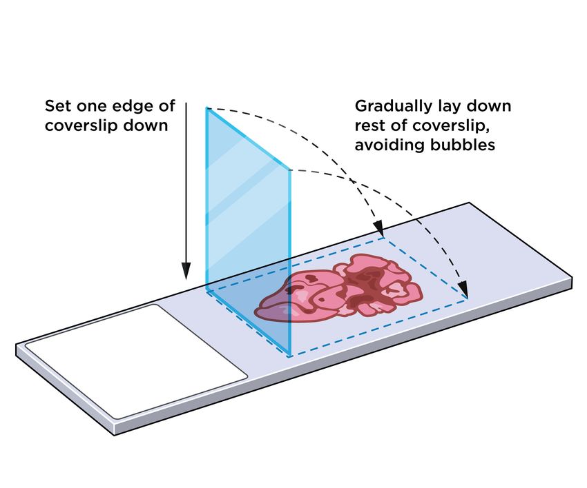
MAN-10150-01                                                   GeoMx DSP Manual Slide Preparation
Rights, License, & Trademarks

Rights, License, & Trademarks
Use
For research use only. Not for use in diagnostic procedures.
Intellectual Property Rights
This GeoMx ® Digital Spatial Profiler (DSP) User Manual and its contents are the property of
NanoString Technologies, Inc. (“NanoString”), and are intended solely for use by NanoString
customers, for the purpose of operating the GeoMx DSP System. The GeoMx DSP System
(including both its software and hardware components) and this User Guide and any other
documentation provided to you by NanoString in connection therewith, are subject to patents,
copyright, trade secret rights and other intellectual property rights owned by, or licensed to,
NanoString. No part of the software or hardware may be reproduced, transmitted, transcribed,
stored in a retrieval system, or translated into other languages without the prior written consent of
NanoString. For a list of patents, see www.nanostring.com/company/patents.
Limited License
Subject to the terms and conditions of the GeoMx DSP System contained in the product quotation,
NanoString grants you a limited, non-exclusive, non-transferable, non-sublicensable, research use
only license to use the proprietary GeoMx DSP System only in accordance with the manual and
other written instructions provided by NanoString. Except as expressly set forth in the terms and
conditions, no right or license, whether express, implied or statutory, is granted by NanoString
under any intellectual property right owned by, or licensed to, NanoString by virtue of the supply of
the proprietary GeoMx DSP System. Without limiting the foregoing, no right or license, whether
express, implied or statutory, is granted by NanoString to use the GeoMx DSP System with any
third party product not supplied or licensed to you by NanoString or recommended for use by
NanoString in a manual or other written instruction provided by NanoString.
Trademarks
NanoString, NanoString Technologies, the NanoString logo, GeoMx, and nCounter are trademarks
or registered trademarks of NanoString Technologies, Inc., in the United States and/or other
countries. All other trademarks and/or service marks not owned by NanoString that appear in this
document are the property of their respective owners.
Copyright
©2022 NanoString Technologies, Inc. All rights reserved.

FOR RESEARCH USE ONLY. Not for use in diagnostic procedures.

                                                                                                   3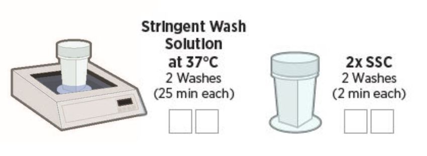
GeoMx DSP Manual Slide Preparation                                   MAN-10150-01
Table of Contents

Table of Contents

    GeoMx DSP Manual Slide Preparation User Manual                              1
    Contacts                                                                    2
    Rights, License, & Trademarks                                               3
    Changes in This Revision                                                    6
    Conventions                                                                 7
    GeoMx DSP Workflow                                                          8
     User Manuals and Resources                                                10
    Slide Prep Introduction                                                    11
    Protein Slide Preparation Protocol (FFPE)                                  12
     Equipment, Materials, and Reagents                                        12
     Prepare reagents                                                          16
     Prepare tissue samples                                                    17
     Deparaffinize and rehydrate FFPE tissue sections (45 minutes)             18
     Perform antigen retrieval (1 hour)                                        19
     Blocking (1 hour)                                                         20
     Primary antibody incubation (overnight)                                   21
     Postfix (70 minutes)                                                      23
     Nuclei staining (20 minutes)                                              24
     Safe storage guidelines for protein slides                                25
     Slide unmounting procedure                                                25
     Stripping and re-probing procedure for protein slides                     26
    RNA Slide Preparation Protocol (FFPE)                                      27
     Equipment, Materials, and Reagents                                        27
     Prepare reagents                                                          32
     Prepare tissue samples                                                    33
     Deparaffinize and rehydrate FFPE tissue sections (35 minutes)             34
     Perform target retrieval (25 minutes)                                     35
     Expose RNA targets (10–30 minutes)                                        37

FOR RESEARCH USE ONLY. Not for use in diagnostic procedures.

4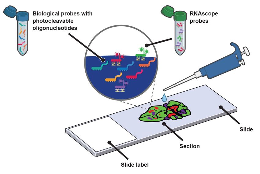
MAN-10150-01                                               GeoMx DSP Manual Slide Preparation
Table of Contents

    Postfix: Preserve tissue morphology for soft tissues (20 minutes)                      38
    In situ hybridization (overnight)                                                      39
    Perform stringent washes to remove off-target probes (90 minutes)                      42
    Add morphology markers (100 minutes)                                                   43
    Safe storage guidelines for RNA slides                                                 44
    Stripping and re-probing procedure for RNA slides                                      45
  Appendix I: Selecting and Sectioning FFPE Samples                                        46
  Appendix II: Modifications to Protocol for Fresh Frozen Samples                          48
    Selecting fresh frozen blocks                                                          48
    Sectioning fresh frozen blocks                                                         48
    Fresh frozen sample preparation for protein assays                                     49
    Fresh frozen sample preparation for RNA assays                                         50
    Cryosectioning technique                                                               51
  Appendix III: Modifications to Protocol for Fixed Frozen Samples                         52
    Preparing fixed frozen tissue block from harvested tissue                              52
    Sectioning fixed frozen blocks                                                         54
    Required fixed frozen sample preprocessing steps                                       55
  Appendix IV: Substitute Probe R Guidance                                                 56
  Appendix V: Adding Custom Barcoded Antibodies                                            57
  Appendix VI: RNAscope® and GeoMx RNA Assays                                              58
  Appendix VII: Secondary Antibody Immunofluorescence Staining for RNA Assays              60
  Appendix VIII: Tyramide Signal Amplification (TSA) of Morphology Markers                 62
  Troubleshooting                                                                          64
  Final page                                                                               66

FOR RESEARCH USE ONLY. Not for use in diagnostic procedures.

                                                                                           5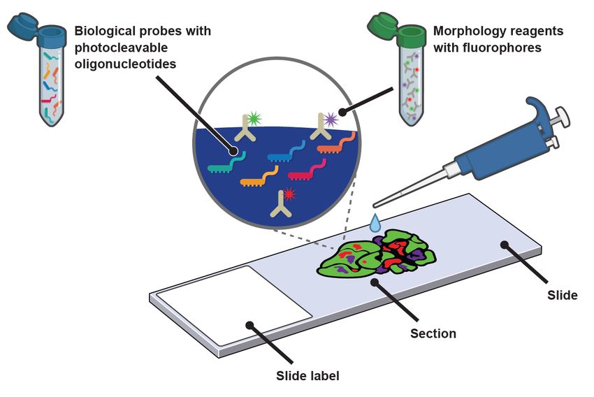
GeoMx DSP Manual Slide Preparation                                               MAN-10150-01
Changes in This Revision

Changes in This Revision
The GeoMx DSP Slide Preparation User Manuals were restructured for clarity and simplicity. This
GeoMx DSP Manual Slide Preparation User Manual (MAN-10150) covers protein and RNA
sample preparation using manual (non-automated) methods, for assays with NGS or nCounter
readout. The GeoMx DSP Automated Slide Preparation User Manual (MAN-10151) covers
sample preparation using semi- and fully- automated methods on the BOND RX/RX m Fully
Automated IHC/ISH Stainer from Leica Biosystems ® . These manuals replace the GeoMx-NGS
Slide Preparation User Manual (SEV-00153-05) and GeoMx-nCounter Slide Preparation User
Manual (SEV-00172-08).
NGS-specific information is separated from nCounter-specific information with colored text boxes,
as described in Conventions on page 7.

Other changes in this manual revision include:

l   Updated items and links in Equipment, Materials, and Reagents lists on page 12 and on
    page 27
l   Updated slide dimensions diagrams (example on page 17) to clarify GeoMx DSP scan area
l   Added guidance for epitope retrieval and Proteinase K conditions for mouse tissue on page 36
    and on page 37, and updated cell pellet Proteinase K digestion conditions on page 37
l   Extended safe storage guidelines to 3 months for protein slides on page 25 and 7 days for
    RNA slides on page 44
l   Revised storage and handling guidance for RNA probe mixes in step 1 on page 39
l   Updated fresh frozen sample preparation guidance on page 48
l   Updated fixed frozen sample preparation guidance on page 52
l   Improved guidance for including custom barcoded antibodies in GeoMx protein assays on
    page 57
l   Added guidance to use secondary antibodies in morphology marker labeling on page 60
l   Improved guidance for tyramide signal amplification (TSA) in morphology marker labeling on
    page 62
l   Added a section on Troubleshooting on page 64

FOR RESEARCH USE ONLY. Not for use in diagnostic procedures.

6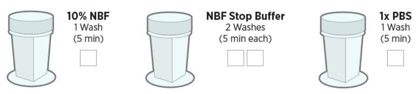
MAN-10150-01                                                       GeoMx DSP Manual Slide Preparation
Conventions

Conventions
The following conventions are used in the GeoMx DSP user manuals and are described for your
reference.
Bold text is typically used to highlight a specific button, keystroke, or menu option. It may also be
used to highlight important text or terms.
Blue underlined text is typically used to highlight links and/or references to other sections of the
manual. It may also be used to highlight references to other manuals or instructional material.

 A gray box indicates general information that may be useful for improving assay performance. These
 notes aim to clarify other instructions or provide guidance to improve the efficiency of the assay workflow.

       IMPORTANT: This symbol indicates important information that is critical to ensuring a successful
      assay. Following these instructions may help improve the quality of your data.

       WARNING: This symbol indicates the potential for bodily injury or damage to the instrument if the
      instructions are not followed correctly. Always carefully read and follow the instructions accompanied
      by this symbol to avoid potential hazards.

  For NGS readout: Content in blue boxes             For nCounter readout: Content in green boxes
  denotes steps or information specific to           denotes steps or information specific to nCounter
  NGS readout of GeoMx DSP. Follow                   readout of GeoMx DSP. Follow these instructions
  these instructions if using Illumina® NGS          if using nCounter® MAX/FLEX, Pro, or SPRINT to
  to read out GeoMx DSP counts.                      read out GeoMx DSP counts.

FOR RESEARCH USE ONLY. Not for use in diagnostic procedures.

                                                                                                                7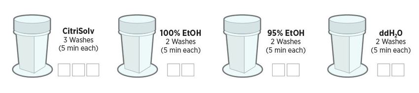
GeoMx DSP Manual Slide Preparation MAN-10150-01
GeoMx DSP Workflow
GeoMx DSP Workflow
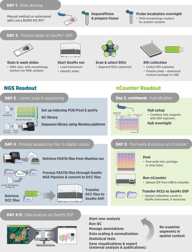
The GeoMx Digital Spatial Profiler (DSP) is a novel platform developed by NanoString. This
product relies on antibody or nucleic acid probes coupled to photocleavable oligonucleotide tags.
After probes hybridize to targets in slide-mounted tissue sections, the oligonucleotide tags are
released from discrete regions of the tissue via UV exposure. Released tags are quantitated by
nCounter technology or Illumina Next Generation Sequencing (NGS). Counts are mapped back to
tissue location, yielding a spatially resolved digital profile of analyte abundance (see Figure 1).
l Day 1: Slide Staining. Prepare slides and incubate biological targets with UV- cleavable
probes. Prepare manually or using the BOND RX/RX m Fully Automated IHC/ISH Stainer from
Leica Biosystems®.
l Day 2: Process Slides on GeoMx DSP. Load prepared slides into the GeoMx DSP instrument
and enter slide/study information. Slides are scanned to capture fluorescent images used to
select regions of interest (ROIs). The instrument collects UV-cleaved oligos from the ROIs into
the wells of a collection plate.
For NGS readout: For nCounter readout:
Day 3: Transfer the collected aspirates to a Day 2, continued : Transfer the collected
PCR plate and perform Library Prep with aspirates to a hybridization plate along with
Seq Code primers. Pool and purify the GeoMx Hyb Code reagents. Hybridization
products, then Sequence on an Illumina occurs overnight.
NGS instrument. Day 3 : Pool wells and Process on an
Day 4: Process FASTQ sequencing files into nCounter MAX/FLEX or Pro Analysis
digital count conversion (DCC) files using System or SPRINT Profiler. Upload
NanoString's GeoMx NGS Pipeline on reporter count conversion (RCC) files to the
Illumina DRAGEN™ accessed via GeoMx DSP.
BaseSpace™ Sequence Hub, or using
GeoMx NGS Pipeline standalone software.
Upload DCC files on to the GeoMx DSP.
l Day 4 or 5: Create a Data Analysis study in the Data Analysis suite and perform quality-control
checks and data analysis, and generate analysis plots.
FOR RESEARCH USE ONLY. Not for use in diagnostic procedures.
8
MAN-10150-01                                              GeoMx DSP Manual Slide Preparation
GeoMx DSP Workflow

                             Figure 1: GeoMx DSP workflow summary

FOR RESEARCH USE ONLY. Not for use in diagnostic procedures.

                                                                                          9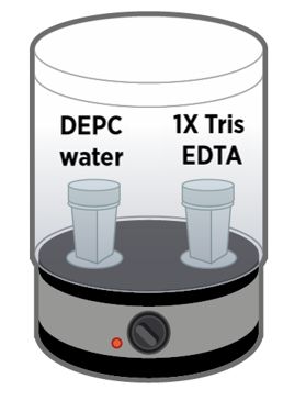
GeoMx DSP Manual Slide Preparation                                                 MAN-10150-01
GeoMx DSP Workflow

User Manuals and Resources
The GeoMx DSP workflow is divided into the following user manuals:

                              GeoMx DSP Manual Slide Preparation User Manual
                                             MAN-10150
       Workflow
        Step 1
                            GeoMx DSP Automated Slide Preparation User Manual
                                             MAN-10151

       Workflow                      GeoMx DSP Instrument User Manual
        Step 2                                MAN-10152

                           For NGS readout:                  For nCounter readout:
       Workflow         GeoMx DSP NGS Readout             GeoMx DSP nCounter Readout
        Step 3               User Manual                          User Manual
                              MAN-10153                           MAN-10089

       Workflow                     GeoMx DSP Data Analysis User Manual
        Step 4                                 MAN-10154

User manuals and other documents can be found online in the NanoString University Document
Library at https://university.nanostring.com.
Instrument and workflow training courses are available in NanoString University.

  For NGS readout:                             For nCounter readout:
  For documentation specific to the Illumina   For documentation specific to the nCounter Pro,
  platform, see                                MAX/FLEX, and SPRINT instruments, see
  https://support.illumina.com.                https://www.nanostring.com/support/support-
                                               documentation/ or the NanoString University
                                               Document Library at
                                               https://university.nanostring.com.

FOR RESEARCH USE ONLY. Not for use in diagnostic procedures.

10
MAN-10150-01 GeoMx DSP Manual Slide Preparation
Slide Prep Introduction
Slide Prep Introduction
Slide preparation is the first step of the GeoMx DSP workflow. Tissue sections are processed for
staining, followed by the addition of morphology reagents and biological probes (see Figure 2).
l Morphology reagents are antibody-fluorophore complexes that bind to specific targets on the
tissue. Tissue structure and cell components most important to your analysis are illuminated
using the fluorescence imaging on the DSP system. Alternative labeling techniques are
described in the Appendices.
l Biological probes are either antibodies or in situ hybridization (ISH) probes that bind to protein
or RNA targets, respectively. Each probe is coupled to a photocleavable oligonucleotide. These
oligos, when exposed to the GeoMx DSP instrument's UV light, will be released into solution and
aspirated into a collection plate well for downstream processing.
Figure 2: Slide preparation with ISH probes
Slide Prep Equipment, Materials, and Reagents
Required equipment, materials, and reagents are listed at the start of each slide preparation
process in the manual. Individual Equipment, Materials, and Reagents lists for every application
of the GeoMx DSP are available in the NanoString University Document Library
(https://university.nanostring.com).
FOR RESEARCH USE ONLY. Not for use in diagnostic procedures.
11GeoMx DSP Manual Slide Preparation MAN-10150-01
Protein FFPE Equipment, Materials, Reagents
Protein Slide Preparation Protocol (FFPE)
IMPORTANT: For any given study, NanoString recommends using only one sample type
preparation method (e.g., FFPE or fresh frozen samples, but not a combination of sample types).
Equipment, Materials, and Reagents
The following tables list equipment, materials, and reagents that are required for this protocol but
are not supplied by NanoString.
Table 1: Equipment for protein slide prep not provided by NanoString.
Equipment Source Part No.
Quincy Lab, Inc. (or Various GC
Baking oven
comparable) models
TintoRetriever pressure cooker (rated for 110V; requires transformer to
Bio SB BSB 7008
operate on 220V)*
*A TintoRetriever pressure cooker is recommended for this protocol. These alternatives are acceptable and may be
more easily purchased outside the U.S., but have not been validated by NanoString : Tefal CY505E 6 Liter ;
AmazonBasics multipurpose pressure cooker 5.5L.
Table 2: Materials for protein slide prep not provided by NanoString.
Materials Source Part No.
Pipettes for 5–1,000 μL Various Various
12-channel P20 multi-channel pipetter Various Various
Filter tips (DNase/RNase free) Various Various
Microcentrifuge tubes (DNase/RNase free) Various Various
Superfrost Plus microscope slides or Fisher Scientific or 12-550-15
Leica BOND Plus microscope slides Leica Biosystems S21.2113.A
Slide staining jars (Coplin jars) (recommended number: 12) and slide 25608-904, 25608-
VWR (or comparable)
holder inserts 868
M920-2 (select black
Humidity chamber Simport (or comparable)
lid)
Vector Labs (or
Hydrophobic barrier pen H-4000
comparable)
RNase AWAY® or 10% Bleach (RNaseZap® is not a substitute) Thermo Fisher 7003PK
Razor blades Various Various
Coverslips (optional) Various Various
FOR RESEARCH USE ONLY. Not for use in diagnostic procedures.
12MAN-10150-01 GeoMx DSP Manual Slide Preparation
Protein FFPE Equipment, Materials, Reagents
Materials Source Part No.
Heat/cold protectant handling glove Various Various
SanDisk (or SDCZ800-128G-
USB drive v3.0, 64 GB or higher (able to be NTFS formatted)
comparable) G46
Table 3: Reagents for protein slide prep not provided by NanoString. RT = room temperature
Reagents Source, Part No. Storage
Thermo Fisher, AM9922 (or comparable)
DEPC-treated water NOTE: As an alternative to commercial DEPC-treated water, prepare RT
your own following standard protocols.
10X tris-buffered saline
Cell Signaling Technologies, 12498S RT
(TBS)
10X TBS with Tween- 20
Cell Signaling Technologies, 9997S RT
(TBS-T)
4 or 16%
Thermo Scientific, 4% concentration, FB002, R37814 or
paraformaldehyde (PFA) 4°C (or manufacturer
16% concentration (must be diluted to 4% with PBS), 28906, 28908
instructions)
(or comparable)
1X phosphate buffered
saline pH 7.4 (PBS)
Thermo Fisher, 10010031KU (or comparable) RT
NOTE: Used only to dilute
16% PFA
Fluoromount-G mounting
SouthernBiotech, 0100-01 RT
media (optional)
CitriSolv or
Xylene or D-Limonene
Fisher Scientific, 04-355-121
((R)-(+)-Limonene) RT
Sigma Aldrich, 183164-100ML or 183164-500ML (or comparable)
100% ethanol (EtOH): ACS
grade or better
Various RT
10X citrate buffer pH 6 Sigma-Aldrich, C9999-100ML or C9999-1000ML (or comparable) 4°C
FOR RESEARCH USE ONLY. Not for use in diagnostic procedures.
13GeoMx DSP Manual Slide Preparation MAN-10150-01 Protein FFPE Equipment, Materials, Reagents NanoString Reagents The following reagents are supplied by NanoString. Contact your NanoString Sales Representative to use our reagent planning tools to calculate required quantities. GeoMx Protein Slide Prep Kit GeoMx Morphology Kit — Human or Mouse Protein compatible (various available) Example morphology kit (Solid Tumor TME) GeoMx Nuclear Stain Morphology Kit FOR RESEARCH USE ONLY. Not for use in diagnostic procedures. 14
MAN-10150-01                                                       GeoMx DSP Manual Slide Preparation
Protein FFPE Equipment, Materials, Reagents

 For NGS readout:                                      For nCounter readout:

 GeoMx Protein Cores and optional                      GeoMx Protein Cores and optional Modules for
 Modules for NGS readout (various                      nCounter readout (various available, including
 available, including custom options)                  custom options)

*Each Protein Core and Module for nCounter readout includes a Probe R reagent, from Probe R_1
to Probe R_9. DO NOT combine two modules with a common Probe R_number in the same
experiment run. Substitute Probe R reagents are available for assays with overlapping Probe R_
numbers. Refer to Appendix IV: Substitute Probe R Guidance on page 56 for more information
and to plan Probe R usage in your experiment.

 Each tube of detection antibody (Ab) mix contains sufficient reagent for 12 slides (112 µL). If you are using
 the entire Ab mix in one week, then store at 4°C. If not, aliquot the Ab mix into 4-slide aliquots (37 µL +
 37 µL + 38 µL) and freeze unused aliquots at -80°C. Do not exceed more than 2 freeze/thaw cycles and
 do not freeze diluted antibody.

 The morphology marker antibodies are stored at 4°C. Aliquoting is not required.

FOR RESEARCH USE ONLY. Not for use in diagnostic procedures.

                                                                                                            15
GeoMx DSP Manual Slide Preparation MAN-10150-01
Protein FFPE Equipment, Materials, Reagents
Prepare reagents
Prepare your reagents using the dilution instructions (see Table 4).
Table 4: Reagent prep for protein slide preparation
Reagent Dilution Storage
Dilute 10X citrate buffer (pH 6) in DEPC- treated water.
1X citrate buffer (pH 6) Must be prepared on the day of slide prep. Do not prepare 4°C
ahead of time.
Prepare 500 mL of 95% ethanol by adding 25 mL of
95% ethanol (EtOH) RT
DEPC-treated water to 475 mL of 100% ethanol.
NOTE: Use only for post-fixation step 4°C (or
4% paraformaldehyde (PFA) If using 16% stock, dilute from 16% to 4% in 1X PBS, manufacturer
aliquot, and store. instructions)
Prepare 1 L by diluting 100 mL of 10X TBS-T in 900 mL
1X tris-buffered saline with Tween-20 (TBS-
DEPC-treated water. Total volume needed for slide prep RT
T)
depends on volume on staining jars to be used.
Dilute 500 µL of 10X TBS in 4.5 mL DEPC-treated water in
1X TBS RT
order to prepare a total of 5 mL 1X TBS.
FOR RESEARCH USE ONLY. Not for use in diagnostic procedures.
16MAN-10150-01 GeoMx DSP Manual Slide Preparation
Protein FFPE Slide Prep Protocol
Prepare tissue samples
Appendix I: Selecting and Sectioning FFPE Samples on page 46 covers FFPE block selection
and sectioning in detail. Review it prior to beginning the Protein Slide Preparation protocol.
GeoMx has been validated for sample blocks up to 3 years old prepared from tissues with a cold ischemic
time of less than 1 hour using 10% NBF or similar fixative. For best results, do not use FFPE blocks
greater than 10 years old. Assay performance will be influenced by tissue block age and treatment
conditions such as cold/warm ischemic time, fixative, and storage.
Slide Preparation
1. Tissue sections should be 5 µm thick and
mounted on Superfrost Plus or
BOND Plus slides. Tissue sections must
be placed in the Scan Area (shown in
green) (see Figure 3) in the center of the
slide and be no larger than 35.3 mm
long by 14.1 mm wide. Mounted material
should not overlap the slide gasket (shown
in blue) or the Tip Calibration area (shown
in red). If mounting multiple sections per Figure 3: Slide dimensions
slide, ensure that tissues are at least 2–3
mm apart and still fit within the Scan Area.
If sections are too large and/or placed off-center, continue with slide preparation as usual. Just before
loading the slide in the instrument slide tray, scrape off the parts of the tissue exceeding the scan area,
making sure the slide gasket and tip calibration area are free of tissue. Scraping off tissue before slide
preparation could generate tissue folds that may result in staining or binding artifacts.
IMPORTANT: The GeoMx DSP instrument will only image the area inside the Scan Area. Tissue
outside of the Scan Area will not be imaged and may cause problems in tissue detection by
the optical system.
2. Bake slides with mounted sections in a 60°C drying oven for 30 minutes to 3 hours prior to
deparaffinization. Stand slides vertically during baking to allow excess paraffin to flow off. Longer
baking times may be necessary for some tissues to sufficiently adhere to the slide; this should be
empirically tested.
FOR RESEARCH USE ONLY. Not for use in diagnostic procedures.
17GeoMx DSP Manual Slide Preparation MAN-10150-01
Protein FFPE Slide Prep Protocol
Deparaffinize and rehydrate FFPE tissue sections (45 minutes)
You will need the following items and reagents for this step: Staining jars, Citrisolv (or
acceptable substitute), 100% EtOH, 95% EtOH, and DEPC-treated water. See the Equipment,
Materials, and Reagents lists on page 12 for more details.
1. Prepare the pressure cooker by adding water to the correct level per the manufacturer's
instructions (for the TintoRetriever, above 4 cups) and preheating to 80°C. Do not preheat the
Citrate Buffer.
2. Deparaffinize and rehydrate FFPE tissue sections. Place slides in a rack and perform the
following washes in staining jars (see Figure 4). Ensure you have sufficient buffer volume to
cover all slides. Slides should be dipped up and down gently several times when placing in and
before removing from staining jars.
Figure 4: Wash steps
WARNING: Dispose of CitriSolv or its substitute in accordance with your lab's safety procedures.
FOR RESEARCH USE ONLY. Not for use in diagnostic procedures.
18MAN-10150-01 GeoMx DSP Manual Slide Preparation
Protein FFPE Slide Prep Protocol
Perform antigen retrieval (1 hour)
You will need the following items and reagents for this step: staining jars, pressure cooker, 1X
Citrate Buffer and 1X TBS-T. See the Equipment, Materials, and Reagents lists on page 12 and
Prepare Reagents steps on page 16.
1. Place FFPE slides in a staining jar containing 1X freshly prepared
Citrate Buffer pH 6 at room temperature. Place a lid on the staining jar
to prevent evaporation. To prevent pressurization, DO NOT tighten or seal
the jar lid.
2. Place the staining jar containing the slides and lid into the preheated pressure cooker.
3. Secure the pressure cooker lid and run on high pressure and high temperature for 15
minutes.
4. When the timer reaches zero, carefully release the pressure and
transfer the staining jar with slides to a lab bench (room temperature),
remove the staining jar lid, and let stand for at least 25 minutes
(maximum one hour).
5. Wash the slides in 1X TBS-T in a staining jar for 5 minutes.
FOR RESEARCH USE ONLY. Not for use in diagnostic procedures.
19GeoMx DSP Manual Slide Preparation MAN-10150-01
Protein FFPE Slide Prep Protocol
Blocking (1 hour)
You will need the following items and reagents for this step: hydrophobic pen, humidity
chamber, and Buffer W. See the Equipment, Materials, and Reagents lists on page 12 for more
details.
IMPORTANT: When creating a hydrophobic barrier around the tissue on a slide, prepare slides
one at a time, leaving the others in the TBS-T solution. DO NOT allow the tissue sections to dry out
during slide preparation.
1. Fill a humidity chamber with enough water to cover the bottom. Avoid overfilling to prevent
splashing while moving the chamber. If your chamber is light- permeable, minimize light
exposure (e.g., wrap the lid in aluminum foil).
2. Remove one slide from the 1X TBS-T, tap on an absorbent, clean
surface such as a paper towel, then use an absorbent wipe to carefully
remove excess buffer from the slide, without touching the tissue.
3. Make a closed hydrophobic barrier around each tissue section with a
hydrophobic pen. Ensure that a complete barrier is made while
minimizing the size of the area within the barrier.
4. Place the slide in the humidity chamber in a horizontal position and
add enough Buffer W to completely fill the hydrophobic barrier (up to 200
µL per slide, depending on the size of the tissue). Repeat steps 2-4 for
any additional slides.
5. Incubate slides with Buffer W for 1 hour at room temperature in the closed humidity
chamber.
6. Thaw detection probe antibody (Ab) mix (cores and modules) on ice. Keep tube protected
from light.
FOR RESEARCH USE ONLY. Not for use in diagnostic procedures.
20MAN-10150-01 GeoMx DSP Manual Slide Preparation
Protein FFPE Slide Prep Protocol
Primary antibody incubation (overnight)
You will need the following materials and reagents for this step: prepared humidity chamber,
thawed detection antibody mix (core and optional modules), morphology markers, and Buffer
W. See the Equipment, Materials, and Reagents lists on page 12 for more details.
IMPORTANT: Probe mixes should be assembled in an area separate from nCounter work, NGS
library prep, or other GeoMx workflows. GeoMx detection reagents can cross-contaminate probe
mixes and give misleading or incorrect results. Areas should be cleaned thoroughly with RNase
AWAY after probe mix formulation. Alternatively, probe mixes can be made in PCR workstations that
are decontaminated with UV light. Gloves should be changed after handling probe mixes to avoid
cross-contamination.
Due to the high sensitivity of this assay, it is recommended that you change pipette filter tips for every
step, change gloves frequently, and use fresh wipes to remove excess liquids.
For nCounter readout: Not all protein modules are compatible with one another. Some
combinations require Substitute Probe R for nCounter readout. Plan your modules by
referring to Appendix IV: Substitute Probe R Guidance on page 56.
1. Mix the detection Ab mix by flicking then spin down. Do not vortex.
Each tube of detection antibody (Ab) mix contains sufficient reagent for 12 slides (112 µL). If you are
using the entire Ab mix in one week, then store at 4°C. If not, aliquot the Ab mix into 4-slide aliquots
(37 µL + 37 µL + 38 µL) and freeze unused aliquots at -80°C. Do not exceed more than 2 freeze/thaw
cycles and do not freeze diluted antibody.
The morphology marker antibodies are stored at 4°C. Aliquoting is not required.
2. Make a working Ab solution by diluting detection antibodies and morphology markers into
Buffer W (n = number of slides) (see Table 5). Adjust to reflect the number of core, module, and
morphology reagents, and the number of slides to be prepared (up to a total volume of 200 µL
per slide). Table 5 addresses both NGS assays and nCounter assays.
FOR RESEARCH USE ONLY. Not for use in diagnostic procedures.
21GeoMx DSP Manual Slide Preparation MAN-10150-01
Protein FFPE Slide Prep Protocol
Table 5: Working antibody mix equation for protein slide prep for NGS or nCounter assays (n = number of slides)
Other Morph Morph Other Final
Core Mix Module 1 Module 2 Buffer W***
Modules* Marker1** Marker2 Markers Volume
(up to
8 μL x n 8 μL x n 8 μL x n ... 5 μL x n 5 μL x n ... 200 μL x n
200 μL) x n
* If adding a custom-barcoded detection antibody, follow instructions in Appendix V: Adding Custom
Barcoded Antibodies on page 57.
** If using non-NanoString morphology markers, optimal concentration in the working antibody mix must be
determined by user testing.
*** If using a different number of detection or morphology reagents, Buffer W amount must be adjusted to
bring total volume up to 200 µL/slide.
3. Remove slide from humidity chamber and remove Buffer W by tapping the slide on a
clean, absorbent surface, such as a paper towel, then using an absorbent wipe to carefully
remove excess buffer from the slide, without touching the tissue.
4. Place the slide back into the humidity chamber in a horizontal
position. Cover the tissue with 200 μl of the diluted antibody
solution . Make sure the entire tissue is covered and no bubbles are
present.
IMPORTANT: From this point on, minimize the slides' exposure to light to preserve the integrity
of the photocleavable barcodes.
5. Transfer the humidity chamber to a 4°C refrigerator and incubate overnight. Ensure the
humidity chamber stays level to avoid losing antibody solution.
FOR RESEARCH USE ONLY. Not for use in diagnostic procedures.
22MAN-10150-01 GeoMx DSP Manual Slide Preparation
Protein FFPE Slide Prep Protocol
Postfix (70 minutes)
You will need the following materials and reagents for this step: staining jars, 1X TBS-T, and 4%
PFA. See the Equipment, Materials, and Reagents lists on page 12 and Prepare Reagents steps
on page 16.
IMPORTANT: Everything that comes into contact with the antibody solution, such as containers for
TBS-T, must be dedicated to this protocol and thoroughly cleaned with RNase AWAY, as probes
may contaminate subsequent runs. Use separate staining jars for different probe mixes. Staining
jars should be cleaned with RNase AWAY before each use.
1. Removing one slide at a time from the humidity chamber, carefully tap off the antibody
solution from each slide on a clean, absorbent surface, such as several paper towels.
2. Wash the slides in 3 washes of 1X TBS-T for 10 minutes each.
IMPORTANT: Washes are critical for best quality data. Do not shorten or skip washes.
3. Removing one slide at a time, carefully tap off each slide on a clean, absorbent surface to
remove excess wash solution.
4. Ensure that the hydrophobic barrier is still intact or draw a fresh barrier over the old one using the
hydrophobic pen.
5. Cover the sample with up to 200 µL 4% PFA and incubate for 30
minutes in the humidity chamber at room temperature.
6. (Optional) Remove SYTO 13 nuclear stain from -20°C and allow it to warm to room temperature
for use in the next step.
7. Carefully tap each slide on clean, absorbent surface to remove excess 4%
PFA. Wash slides in two washes of 1X TBS-T for 5 minutes each.
FOR RESEARCH USE ONLY. Not for use in diagnostic procedures.
23GeoMx DSP Manual Slide Preparation MAN-10150-01
Protein FFPE Slide Prep Protocol
Nuclei staining (20 minutes)
You will need the following materials and reagents for this step: humidity chamber, staining
jars, razor, SYTO 13 nuclear stain, 1X TBS, and 1X TBS-T. See the Equipment, Materials, and
Reagents lists on page 12 and Prepare Reagents steps on page 16.
IMPORTANT: Before using the humidity chamber in the following steps, clean it with RNase
AWAY. Prep the humidity chamber by lining with Kimwipes and adding just enough water to cover
the bottom of the chamber.
1. Allow SYTO 13 to warm to room temperature.
2. Once thawed, vortex then picofuge SYTO 13 for at least 1 minute to bring the solution and
insoluble particles to the bottom of the vial. When pipetting SYTO 13, pipette from the top of the
vial to avoid insoluble particles.
3. Dilute SYTO 13 1:10 in 1X TBS. Prepare a sufficient volume per slide to completely cover
tissue (~200 µL per slide). Mix by pipetting up and down. Close SYTO 13 stock vial tightly and
store at -20°C.
4. Remove one slide at a time from the 1X TBS-T, remove excess liquid
by tapping on a clean, absorbent surface, then place slides in humidity
chamber in a horizontal position and cover the tissue with diluted
SYTO 13.
5. Stain for 15 minutes at room temperature in the humidity chamber.
6. Wash slides by dipping in a staining jar with 1X TBS-T.
7. Transfer to another staining jar with fresh 1X TBS-T.
8. Working with one slide at a time, and dipping back into 1X TBS-T to avoid
drying out, carefully scrape off the hydrophobic pen with a razor blade.
Be sure to remove all of the wax without damaging or removing any of the
tissue.
9. Store stained slides in 1X TBS-T. If it is necessary to re-stain with SYTO 13, re-draw the
hydrophobic barrier, then repeat steps 1-8.
Once the slides are prepared, load them onto the GeoMx DSP (see the GeoMx DSP Instrument
User Manual (MAN-10152)) or store slides according to guidelines on next page. DO NOT let
slides dry out.
FOR RESEARCH USE ONLY. Not for use in diagnostic procedures.
24MAN-10150-01 GeoMx DSP Manual Slide Preparation
Protein FFPE Slide Prep Protocol
Safe storage guidelines for protein slides
l Storage for up to 1 day: submerge in 1X TBS-T and store at 4°C, protected from light to
maintain the integrity of the photocleavable barcodes.
l Storage for 1 day to 3 months:
1. Rinse slide to be mounted with TBS-T or PBS-T. Touch the slide edge to a paper
towel to remove excess liquid. Place slide on a flat surface.
2. Using a pipette tip (200 µL tip works well), add one drop (~50 µL) of Fluoromount-
G to the slide; add more as necessary to ensure the slide does not dry out and tissue
is adequately covered.
3. Mount coverslip by aligning one edge of the coverslip then slowly lowering from
one side to the other. Remove excess mounting medium.
4. Allow slide to dry at room temperature overnight, protected from light (e.g. in a
bench drawer).
5. Store slide at 4°C, protected from light, for up to 3 months.
Slide unmounting procedure
1. Submerge mounted slide in 1X TBS-T or PBS-T until coverslip is loose or has fallen off. With
gentle agitation, the coverslip typically falls off within 15 minutes.
2. If coverslip has fallen off, slide is ready for use. If coverslip is still attached but loose, gently
remove it using tweezers.
3. Wash slide with fresh 1X TBS-T for 5 minutes to ensure removal of mounting media.
FOR RESEARCH USE ONLY. Not for use in diagnostic procedures.
25GeoMx DSP Manual Slide Preparation MAN-10150-01 Protein FFPE Slide Prep Protocol Stripping and re-probing procedure for protein slides GeoMx Protein assay slides are reusable and can be restained with a different commercial or custom panel following this protocol. This protocol requires a UV light box or transilluminator capable of emitting 302/312 nm UV light (example) and 1X TBS-T. 1. Place the slide flat on the surface of a UV transilluminator. 2. Apply enough 1X TBS-T to completely cover the tissue (50–200 μL depending on the size of the tissue). 3. Expose to UV light for 3 minutes to cleave tags from bound antibodies. 4. Carefully tap each slide on a clean, absorbent surface (e.g. paper towel) to remove liquid and avoid oligo contamination. 5. Wash slides by dipping in a staining jar with 1X TBS-T. 6. Transfer to another staining jar with fresh 1X TBS-T. 7. To apply new probes, proceed to Perform antigen retrieval (1 hour) on page 19. FOR RESEARCH USE ONLY. Not for use in diagnostic procedures. 26
MAN-10150-01                                                                GeoMx DSP Manual Slide Preparation
RNA FFPE Equipment, Materials, Reagents

RNA Slide Preparation Protocol (FFPE)

         IMPORTANT: For any given study, NanoString recommends using only one sample type
        preparation method (e.g., FFPE or fresh frozen samples, but not a combination of sample types).

Equipment, Materials, and Reagents
The following tables list equipment, materials, and reagents that are required for this protocol but
are not supplied by NanoString.
                              Table 6: Equipment for RNA slide prep not provided by NanoString.
                           Equipment                                                Source              Part No.
                                                                        Quincy Lab, Inc. (or        Various GC
Baking oven
                                                                        comparable)                 models
Hybridization oven including hybridization chamber*:

        HybEZ II Hybridization System or                                ACDBio                      321710/321720

        RapidFISH Slide Hybridizer                                      Boekel Scientific           240200 for 120V
Water bath (programmable to at least 37°C)                              Various                     Various
                                                                        Hamilton Beach              37530Z
5-quart steamer**
                                                                        Nesco                       ST-25F
Hot plate programmable up to 85°C (only needed for preparation of       Various                     Various
cell pellet tissue type)
                                                                        ThermoPro                   TP01A
Digital thermometer
                                                                        1EasyLife                   1EasyLife

*NanoString recommends the listed hybridization ovens for this protocol. The following alternatives are acceptable, but
have not been validated by NanoString. Test to ensure slides remain hydrated overnight. Abbott ThermoBrite, Leica
ThermoBrite

**NanoString recommends the listed steamers for this protocol. These alternatives are acceptable, but have not been
validated by NanoString: Philips HD9140.

FOR RESEARCH USE ONLY. Not for use in diagnostic procedures.

                                                                                                                   27
GeoMx DSP Manual Slide Preparation MAN-10150-01
RNA FFPE Equipment, Materials, Reagents
Table 7: Materials for RNA slide prep not provided by NanoString.
Materials Source Part No.
Pipettes for 5–1,000 μL Various Various
12-channel P20 multi-channel pipette Various Various
Filter tips (DNase/RNase free) Various Various
Microcentrifuge tubes (DNase/RNase free) Various Various
Superfrost Plus microscope slides or Fisher Scientific or 12-550-15
Leica BOND Plus microscope slides Leica Biosystems S21.2113.A
Slide staining jars (Coplin jars) (recommended number: 16) 25608-904, 25608-
VWR (or comparable)
and slide holder inserts 868
Humidity chamber Simport M920-2
Benchtop protector sheet (fits inside the hybridization oven, Fisher Scientific (or
14-206-62
optional) comparable)
HybriSlip hybridization covers (22 mm x 40 mm x 0.25 mm)
Grace Bio-Labs 714022
Note: Other products have not been validated by NanoString.
RNase AWAY® or 10% Bleach (RNaseZap® is not a substitute) Thermo Fisher 7003PK
Heat/cold protectant handling glove Various Various
Forceps (for slide handling) Various Various
Aluminum foil Various Various
SDCZ800-128G-
USB drive v3.0, 64 GB or higher (able to be NTFS formatted) SanDisk (or comparable)
G46
FOR RESEARCH USE ONLY. Not for use in diagnostic procedures.
28MAN-10150-01 GeoMx DSP Manual Slide Preparation
RNA FFPE Equipment, Materials, Reagents
Table 8: Reagents for RNA slide prep not provided by NanoString. RT = room temperature
Reagents Source, Part No. Storage
Thermo Fisher, AM9922 (or comparable)
DEPC-treated water NOTE: As an alternative to commercial DEPC-treated water, prepare RT
your own following standard protocols.
10X phosphate buffered Sigma-Aldrich, P5368-10PAK, P5368-5X10PAK
RT
saline pH 7.4 (PBS) (or comparable)
10% neutral buffered
formalin (NBF)
EMS Diasum, 15740-04 (or comparable) RT
100% deionized formamide Thermo Fisher , AM9342 or VWR, VWRV0606 (or comparable) 4°C
NOTE: If deionized formamide is unavailable, molecular grade (bring to RT
formamide may be substituted. before opening)
20X SSC (DNase/RNase
Sigma-Aldrich, S6639 RT
free)
Ambion, 2546
Proteinase K See manu-
Thermo Fisher, AM2548 or 25530049
facturer's
Note: Use of Proteinase K from any other vendor will require instructions
optimization of incubation times and concentration.
Antigen Retrieval Solution
eBioscience™ IHC Antigen Retrieval Solution - High pH, 00-4956-58 4°C
(10X Tris-EDTA pH 9.0 )
Tris base Sigma-Aldrich, 10708976001 (or comparable) RT
Glycine Sigma-Aldrich, G7126 (or comparable) RT
CitriSolv
or Xylene or D-Limonene
Fisher Scientific, 04-355-121
((R)-(+)-Limonene) RT
Sigma Aldrich, 183164-100ML or 183164-500ML (or comparable)
100% ethanol (ACS grade or
better)
Various RT
10% Tween- 20 Teknova, T0710 (or comparable) RT
FOR RESEARCH USE ONLY. Not for use in diagnostic procedures.
29GeoMx DSP Manual Slide Preparation MAN-10150-01 RNA FFPE Equipment, Materials, Reagents NanoString Reagents The following reagents are supplied by NanoString. Contact your NanoString Sales Representative to use our reagent planning tools to calculate required quantities. GeoMx RNA Slide Prep Kit GeoMx Morphology Kit - Human or Mouse RNA compatible (various available) Example morphology kit (Solid Tumor TME) GeoMx Nuclear Stain Morphology Kit FOR RESEARCH USE ONLY. Not for use in diagnostic procedures. 30
MAN-10150-01                                              GeoMx DSP Manual Slide Preparation
RNA FFPE Equipment, Materials, Reagents

 For NGS readout:                                For nCounter readout:

                                                 GeoMx Probe Mix for nCounter readout
 GeoMx Probe Mix for NGS readout (various
 available; green or white cap)

                                                 Custom RNA-nCounter Probe Mix (optional)

 Custom RNA-NGS Probe Mix (optional)

Be sure to use probe mixes for manual (non-automated) RNA slide preparation. Probe mixes for
fully automated slide preparation on BOND RX/RXm (yellow label, red cap) are not compatible with
this manual RNA slide preparation protocol. Refer to GeoMx DSP Automated Slide Preparation
User Manual (MAN-10151) for information about automated protocols.

FOR RESEARCH USE ONLY. Not for use in diagnostic procedures.

                                                                                             31
GeoMx DSP Manual Slide Preparation MAN-10150-01
RNA FFPE Equipment, Materials, Reagents
Prepare reagents
Prepare the reagents using the dilution instructions (see Table 9). Use DEPC-treated water for
all dilutions.
IMPORTANT: Take care to maintain nuclease-free conditions. The greatest risk of contamination
comes from GeoMx probes and other oligos. We recommend the use of RNase AWAY (Thermo
Fisher 7002) for cleaning of all surfaces and equipment, as it will limit contamination from oligos,
GeoMx probes and nucleases. After using RNase AWAY, allow area/items to air dry completely, or
rinse with DEPC-treated water. See manufacturer's instructions for details.
Table 9: Reagent prep for RNA slide preparation
Reagent Dilution Storage
Prepare 500 mL of 95% ethanol by adding 25 mL of DEPC-treated water to
95% EtOH 475 mL of 100% ethanol. NanoString recommends to prepare fresh each RT
week.
Prepare 1 L of 1X PBS by combining 100 mL of 10X PBS and 900 mL of
1X PBS pH 7.4 RT
DEPC-treated water. Don't reuse.
Default concentration (1 µg/mL) is made by adding 10 μL of 20 mg/mL
Proteinase K to 200 mL of 1X PBS made with DEPC-treated water. See Table
Proteinase K 11 for alternative concentrations by tissue type. n/a
Note: Prepare fresh and do not reuse. Take care to pipette accurately.
Inaccurate concentration of Proteinase K will affect assay performance.
Add 24.5 g Tris base and 15 g Glycine to 2 L DEPC-treated water.
NBF stop buffer RT
Do not reuse. Solution will lose efficacy with repeated use.
Prepare 1 L of 2X SSC by combining 100 mL of 20X SSC and 900 mL of
2X SSC RT
DEPC-treated water. Do not reuse.
Prepare 250 mL of 2X SSC-T by combining 25 mL of 20X SSC, 2.5 mL of
2X SSC-T (optional) RT
10% Tween-20, and 222.5 mL of DEPC-treated water. Do not reuse.
Prepare 1 L of 4X SSC by combining 200 mL of 20X SSC and 800 mL of
4X SSC RT
DEPC-treated water. Do not reuse.
1X Tris-EDTA pH 9.0 Prepare 1 L of 1X Tris-EDTA pH 9.0 by combining 100 mL of 10X Tris-EDTA
RT
(antigen retrieval solution) pH 9.0 and 900 mL of DEPC-treated water.
FOR RESEARCH USE ONLY. Not for use in diagnostic procedures.
32MAN-10150-01 GeoMx DSP Manual Slide Preparation
RNA FFPE Slide Prep Protocol
Prepare tissue samples
Appendix I: Selecting and Sectioning FFPE Samples on page 46 covers FFPE block selection
and sectioning in detail. Review it prior to beginning the RNA Slide Preparation protocol.
GeoMx has been validated for sample blocks up to 3 years old prepared from tissues with a cold ischemic
time of less than 1 hour using 10% NBF or similar fixative. For best results, do not use FFPE blocks
greater than 10 years old. Assay performance will be influenced by tissue block age and treatment
conditions such as cold/warm ischemic time, fixative, and storage.
Slide Preparation
1. Tissue sections should be 5 µm thick and
mounted on Superfrost Plus or
BOND Plus slides. Tissue sections must
be placed in the Scan Area (shown in
green) (see Figure 5) in the center of the
slide and be no larger than 35.3 mm
long by 14.1 mm wide. Mounted material
should not overlap the slide gasket (shown
in blue) or the Tip Calibration area (shown
in red). If mounting multiple sections per Figure 5: Slide dimensions
slide, ensure that tissues are at least 2–3
mm apart and still fit within the Scan Area.
If sections are too large and/or placed off-center, continue with slide preparation as usual. Just before
loading the slide in the instrument slide tray, scrape off the parts of the tissue exceeding the scan area,
making sure the slide gasket and tip calibration area are free of tissue. Scraping off tissue before slide
preparation could generate tissue folds that may result in staining or binding artifacts.
IMPORTANT: The GeoMx DSP instrument will only image the area inside the Scan Area. Tissue
outside of the Scan Area will not be imaged and may cause problems in tissue detection by
the optical system.
2. Bake slides with mounted sections in a 60°C drying oven for 30 minutes to 3 hours prior to
deparaffinization. Stand slides vertically during baking to allow excess paraffin to flow off. Longer
baking times may be necessary for some tissues to sufficiently adhere to the slide; this should be
empirically tested.
FOR RESEARCH USE ONLY. Not for use in diagnostic procedures.
33GeoMx DSP Manual Slide Preparation MAN-10150-01
RNA FFPE Slide Prep Protocol
Deparaffinize and rehydrate FFPE tissue sections (35 minutes)
You will need the following materials and reagents for this step: Citrisolv (or acceptable
substitute), 100% EtOH, 95% EtOH, and 1X PBS.
The steamer, staining jars, 1X Tris-EDTA (pH 9.0) (antigen retrieval solution), DEPC-treated
water, water bath and Proteinase K solution are preheated here for their use in a later step. (To
prepare cell pellet samples, use a hot plate set to 85°C, rather than the steamer, as described on
next page). See the Equipment, Materials, and Reagents lists on page 27 for more details.
1. Fill the steamer reservoir up to the fill line with water. Place two staining jars inside, one
containing DEPC-treated water and one containing 1X Tris-EDTA (pH 9.0) (Antigen Retrieval
Solution). Ensure sufficient reagent volume to cover slides up to the label. Loosely cover each
jar with aluminum foil instead of the jar lid to allow for a thermometer reading in a later step.
Preheat the steamer to 100°C. More water may need to be added to the steamer during
preheating.
The Nesco steamer takes 1 hour to heat the liquid in the jars to a stable maximum temperature near
100°C. Final temperature can be checked by inserting a digital thermometer through the hole in the lid
of the steamer into the staining jars.
2. Deparaffinize and rehydrate FFPE tissue sections. Place slides in a rack and perform the
following washes in staining jars (see Figure 6). Ensure you have sufficient buffer volume to
cover all slides. Slides should be dipped up and down gently several times when placing in and
before removing from staining jars. After the last wash, slides can be stored in the 1X PBS for up
to one hour.
Figure 6: Wash steps
WARNING: Dispose of CitriSolv or its substitute in accordance with your lab's safety procedures.
3. During wash steps, preheat the water bath to 37°C. Prepare the Proteinase K dilution, if not
yet done, and add the diluted Proteinase K solution to a staining jar and place in the
water bath to preheat to 37°C . Refer to Table 11 for the recommended Proteinase K
concentration for your tissue type.
FOR RESEARCH USE ONLY. Not for use in diagnostic procedures.
34MAN-10150-01 GeoMx DSP Manual Slide Preparation
RNA FFPE Slide Prep Protocol
Perform target retrieval (25 minutes)
You will need the following materials and reagents for this step: Steamer, Staining jars, 1X Tris-
EDTA (pH 9.0) (antigen retrieval solution), DEPC-treated water (all preheated in the previous
step) and 1X PBS. See the Equipment, Materials, and Reagents lists on page 27 and Prepare
Reagents steps on page 32.
(To prepare cell pellet samples, use a hot plate set to 85°C, rather than the steamer, to preheat
buffers and for target retrieval (see Table 10). Follow the steps as described below to move slides
between buffers).
1. Without removing the lid, place an instant-read digital thermometer through the vents in the
steamer lid and pierce the aluminum foil covering the 1X Tris- EDTA.
Ensure the 1X Tris-EDTA has reached ~99°C. Reset the steamer's timer
to ensure consistent heating during incubation and add more water as
needed.
WARNING: Removing the steamer lid releases high- temperature steam. Use a thermal
protection glove with full hand coverage and transfer slides using forceps or rack.
2. Once the 1X Tris-EDTA has reached 99°C, carefully remove the steamer lid and jar covers.
Dip the slides into the DEPC-treated water for 10 seconds to bring the slide temperature up to
~99°C. Quickly transfer the slides to the 1X Tris-EDTA. Replace jar cover, then replace steamer
lid.
IMPORTANT: The steamer temperature will plateau at ~100°C. Once the lid is removed, the
temperature of the buffers will start to fall rapidly. Try to limit the time the steamer is uncovered to
30 seconds(maximum uncovered time is 2 minutes). Reproducible results rely upon minimizing
this variation in temperature.
3. Incubate the slides. Incubation times and temperatures may differ by tissue and may need to
be empirically determined. Conditions for certain tissues are listed below (see Table 10). If the
tissue type you wish to use is not listed, start with 15 minutes.
FOR RESEARCH USE ONLY. Not for use in diagnostic procedures.
35GeoMx DSP Manual Slide Preparation MAN-10150-01
RNA FFPE Slide Prep Protocol
Epitope retrieval times were determined based on FFPE tissue blocks meeting the constraints
outlined in the sample guidance section with minimal normal adjacent tissue. These conditions may
vary by sample, the amount of normal adjacent tissue, and other factors. These conditions were
optimized for large tumor sections and may not apply to arrayed tissues, cored tissues, and needle
biopsies.
If preparing fresh frozen or fixed frozen samples instead of FFPE, target retrieval time is 15 min. See
Appendix II: Modifications to Protocol for Fresh Frozen Samples on page 48 or Appendix III:
Modifications to Protocol for Fixed Frozen Samples on page 52 for more information.
Table 10: Target retrieval times by tissue type
Tissue Type (FFPE) Target Retrieval in Tris-EDTA
Breast 20 min
Cell pellets 15 min at 85°C (use hot plate instead of steamer)
Colorectal 20 min
Melanoma 20 min
Mouse tissue 20 min
NSCLC 20 min
Prostate tumor 20 min
Tonsil 15 min
4. When target retrieval time is up, move slides to room temperature 1X
PBS immediately.
5. Wash in room temperature 1X PBS for 5 minutes . Slides can be
stored up to 1 hour in 1X PBS.
FOR RESEARCH USE ONLY. Not for use in diagnostic procedures.
36MAN-10150-01 GeoMx DSP Manual Slide Preparation
RNA FFPE Slide Prep Protocol
Expose RNA targets (10–30 minutes)
You will need the following materials and reagents for this step: preheated water bath, preheated
Proteinase K dilution, and 1X PBS . See the Equipment, Materials, and Reagents lists on
page 27 for more details.
1. Incubate slides in Proteinase K solution at 37°C for the time
specified for the tissue type (see Table 11). Proteinase K
concentration and incubation times may need to be
empirically determined. If the tissue type you wish to use is not
listed, start with a concentration of 1 µg/mL for 15 minutes.
2. Wash slides in 1X PBS for 5 minutes. During the wash,
ensure that the 10% NBF and NBF Stop Buffer needed in
the next step are ready. Proceed to the next step
immediately.
Table 11: Proteinase K digest concentrations and times by tissue type
Tissue Type Proteinase K Digest
Breast 0.1 µg/mL for 15 min
Cell pellets 1 µg/mL for 5 min
Colorectal 1 µg/mL for 15 min
Melanoma 1 µg/mL for 15 min
Mouse tissue 1 µg/mL for 15 min
NSCLC 1 µg/mL for 15 min
Prostate tumor 1 µg/mL for 15 min
Tonsil 1 µg/mL for 15 min
Proteinase K digestion times and concentrations were determined based on FFPE tissue blocks meeting
the constraints outlined in the sample guidance section with minimal normal adjacent tissue. The
conditions were optimized for large tumor sections and may not apply to arrayed tissues, cored tissues,
and needle biopsies. Theconditions required may vary by sample, the amount of normal adjacent tissue,
and other factors. Use of Proteinase K from any other vendor will require optimization of incubation times
and concentration.
If preparing fresh frozen or fixed frozen samples instead of FFPE, digest with Proteinase K at 1 µg/mL for
15 min. See Appendix II: Modifications to Protocol for Fresh Frozen Samples on page 48 or
Appendix III: Modifications to Protocol for Fixed Frozen Samples on page 52 for more information.
FOR RESEARCH USE ONLY. Not for use in diagnostic procedures.
37GeoMx DSP Manual Slide Preparation MAN-10150-01
RNA FFPE Slide Prep Protocol
Postfix: Preserve tissue morphology for soft tissues (20 minutes)
If preparing fixed frozen samples instead of FFPE, DO NOT perform this Postfix step. Proceed to In situ
hybridization (overnight) on page 39 . See Appendix III: Modifications to Protocol for Fixed
Frozen Samples on page 52 for more information.
You will need the following materials and reagents for this step: Staining jars, 10% NBF, NBF
Stop Buffer and 1X PBS. See the Equipment, Materials, and Reagents lists on page 27 and
Prepare Reagents steps on page 32.
WARNING: Use of appropriate personal protective equipment is advised. Used NBF Stop Buffer
contains NBF and must be disposed of in the same manner as the NBF.
Post-fix the tissue by performing these washes (see Figure 7):
Figure 7: Post-fix wash steps
Slides can be stored in the final 1X PBS wash up to 1 hour at room temperature or 6 hours at 4°C.
FOR RESEARCH USE ONLY. Not for use in diagnostic procedures.
38MAN-10150-01 GeoMx DSP Manual Slide Preparation
RNA FFPE Slide Prep Protocol
In situ hybridization (overnight)
You will need the following materials and reagents for this step: hybridization chamber,
hybridization oven, Buffer R, RNA Probe Mix, and 2X SSC or DEPC-treated water. See the
Equipment, Materials, and Reagents lists on page 27 for more details.
IMPORTANT: Probe mixes should be handled in an area separate from nCounter work, NGS
library prep, or other GeoMx workflows. GeoMx detection reagents can cross-contaminate probe
mixes and give misleading or incorrect results. Areas should be cleaned thoroughly with RNase
AWAY after probe mix formulation. Alternatively, handle probe mixes in PCR workstations that are
decontaminated with UV light. Gloves should be changed after handling any probe mixes to avoid
cross-contamination.
Due to the high sensitivity of this assay, it is recommended that you change pipette filter tips for every
step, change gloves frequently, and use fresh wipes to remove excess liquids.
1. Prepare reagents : Warm Buffer R and RNA detection probes to room temperature before
opening. (Warming RNA detection probes reduces viscosity, improving pipetting accuracy.)
Before use, briefly vortex, then spin down. Store unused RNA detection probes at 4°C for up to 6
months or re-freeze.
2. Clean the hybridization chamber and other equipment with RNase AWAY and allow to dry,
or rinse with DEPC- treated water. The hybridization chamber can be a key source of
contamination by oligos. Arrange fresh Kimwipes on the bottom of the chamber and wet with 2X
SSC or DEPC-treated water. Kimwipes should be thoroughly damp, but liquid should not pool. If
your chamber is light-permeable, wrap the lid in aluminum foil to minimize light exposure.
If your hybridization chamber does not seal with a gasket, place the chamber in a zip-lock bag to
simulate a sealed chamber. Chambers sealed in this manner should be tested prior to use to ensure
they maintain humidity (i.e. that slides do not dry out) for 24 hours. Unsealed chambers can result in
evaporation of the hybridization solution.
3. Make hybridization solution following Table 12 for NGS assays or Table 13 for nCounter
assays. Confirm that you are using probe mix for manual/semi- automated slide preparation
(white label, green/white/amber cap) and not probe mix for fully automated slide prep (yellow
label, red cap).
FOR RESEARCH USE ONLY. Not for use in diagnostic procedures.
39You can also read