Morphological and molecular identification of a new Kudoa thyrsites isolate in Mediterranean silver scabbardfish Lepidopus caudatus
←
→
Page content transcription
If your browser does not render page correctly, please read the page content below
Vol. 132: 125–134, 2019                  DISEASES OF AQUATIC ORGANISMS
                                                                                                                 Published January 10
 https://doi.org/10.3354/dao03316                        Dis Aquat Org

 Morphological and molecular identification of a
new Kudoa thyrsites isolate in Mediterranean silver
       scabbardfish Lepidopus caudatus
  Lucilla Giulietti1,*, Simonetta Mattiucci2, Michela Paoletti3, Didrik H. Grevskott1,
                       Miguel Bao1, Paolo Cipriani1, Arne Levsen1
                          1
                           Institute of Marine Research (IMR), Nordnes, Bergen 5009, Norway
  2
   Department of Public Health and Infectious Diseases, Section of Parasitology, Sapienza-University of Rome 00185, Italy
                3
                 Department of Ecological and Biological Sciences, Tuscia University, Viterbo 01100, Italy

        ABSTRACT: Myxozoans of the genus Kudoa (Myxosporea, Multivalvulida) infect marine and
        estuarine fish species worldwide. Some Kudoa species are of concern to the seafood industry since
        they may generate macroscopic cysts in the fish host’s musculature, or cause post mortem myo-
        liquefaction, commonly known as ‘soft flesh’. One of the economically most important species is
        K. thyrsites, a myoliquefactive myxosporean parasite that occurs in many wild and cultured mar-
        ine fish species worldwide. Here we identified a K. thyrsites isolate as the causative agent of
        myoliquefaction in silver scabbardfish Lepidopus caudatus from the Alboran Sea (western Medi-
        terranean Sea). For comparative and validation purposes, the morphological and molecular char-
        acteristics of the isolate were compared with fresh spores of a K. thyrsites isolate infecting Atlantic
        mackerel Scomber scombrus from the Norwegian Sea. Myxospores of both isolates shared a stel-
        late appearance and contained 4 unequal pyriform polar capsules (1 large, 1 small and 2 interme-
        diate). These morphological traits were consistent with all other previously described K. thyrsites
        isolates. Moreover, the small subunit rDNA sequences of the Mediterranean and Norwegian Sea
        isolates revealed 100% similarity, and matched 100% with K. thyrsites isolates previously re-
        corded in myoliquefactive Atlantic mackerel from the North Sea and off southern England. The
        findings suggest that K. thyrsites is the primary cause of myoliquefaction in silver scabbardfish
        from the Alboran Sea. This report represents the first morphological and molecular characteriza-
        tion of K. thyrsites in the Mediterranean Sea. A set of new allometric characters is proposed as
        additional descriptors for more accurate and specific description of kudoid myxospores.

        KEY WORDS: Kudoa thyrsites · Mediterranean Sea · ‘Soft flesh’ · Myoliquefaction · Lepidopus
        caudatus · Molecular identification · New morphological characters · Fish parasite
                                Resale or republication not permitted without written consent of the publisher

                  1. INTRODUCTION                                         oligochaetes and polychaetes) as invertebrate defini-
                                                                          tive hosts and fish as vertebrate intermediate hosts
  Species of the genus Kudoa Meglitsch, 1947                              (Wolf & Markiw 1984, Shaw et al. 1997, Young 2002,
(Myxozoa, Multivalvulida) are parasites of marine                         Lom & Dyková 2006). Actinospores released from the
and estuarine fishes with a worldwide geographical                        annelid host infect fish through various entry sites,
distribution (Moran et al. 1999, Whipps & Kent 2006,                      such as the skin, fins or gills (Yokoyama et al. 2012).
Eiras et al. 2014, MacKenzie & Kalavati 2014). The                        However, the transmission pathways of Kudoa spe-
typical life cycle of myxozoans involves annelids (e.g.                   cies, as well as the life cycle putatively involving an

*Corresponding author: lucilla.giulietti@gmail.com                        © Inter-Research 2019 · www.int-res.com
126 Dis Aquat Org 132: 125–134, 2019 alternate invertebrate host and a fish infective stage, range of most Kudoa species, and many myxozoans are still unresolved. The kudoids are defined by their in general (Moran et al. 1999, Whipps & Kent 2006, myxospore life cycle stage that occurs in the verte- Burger et al. 2008, Yokoyama et al. 2012), K. thyrsites brate host (i.e. fish) (Lom 1987). The myxospores are has been recorded from more than 30 phylogeneti- characterized by 4 or more shell valves, each con- cally comparatively distant teleost fish species. taining a single polar capsule (Lom & Dyková 2006). Moreover, infections with K. thyrsites have been In early studies, the identification of kudoids to the reported from commercially important wild fish species level was based on morphology and morpho- stocks such as Northeast Atlantic mackerel (Levsen metry of myxospores, i.e. shape and dimensions of et al. 2008), mahi-mahi Coryphaena hippurus off the spore, polar capsules and polar filaments (Lom & western Australia (Langdon 1991, Whipps et al. 2003) Arthur 1989), and more recently on molecular ana- and snoek Thyrsites atun off South Africa (Henning lyses of the small (SSU) and large (LSU) ribosomal et al. 2013), the latter being the type host species of DNA (rDNA) subunits (Whipps & Kent 2006, Burger K. thyrsites (Gilchrist 1924). K. thyrsites has also been & Adlard 2010, 2011). A more accurate identification documented in several important sea-reared fish of Kudoa species can be achieved by integrating both species such as olive flounder Paralichthys olivaceus morphological and molecular data, together with from Japanese waters (Yokoyama et al. 2004), coho ecological and biological aspects such as tissue tro- salmon Oncorhynchus kisutch and Atlantic salmon pism, host specificity, pathogenicity and geographi- Salmo salar in British Columbia, Canada (Whitaker & cal distribution (Whipps et al. 2004, Adlard et al. Kent 1991, St-Hilaire et al. 1998, Moran et al. 1999, 2005, Shin et al. 2016). Marshall et al. 2016), as well as other areas including To date, more than 100 nominal Kudoa species Ireland, Chile and Australia (Palmer 1994, Munday have been described from many phylogenetically et al. 1998, Lopez & Navarro 2000). For instance, distant fish host species (Moran et al. 1999, Lom & K. thyrsites infections caused significant problems for Dyková 2006, Eiras et al. 2014, Sato & Kasai 2016). the Atlantic salmon aquaculture industry in British Most of these are histozoic parasites infecting the Columbia, where the loss of revenue due to skeletal muscles of fish (Moran et al. 1999, Lom & degraded fish products reached 50 million Canadian Dyková 2006, Eiras et al. 2014), while others occur in dollars in 2002 (Funk et al. 2007, Marshall et al. the brain, heart, gills, kidney, ovary or intestines 2016). (Eiras et al. 2014). Despite its almost cosmopolitan distribution (Moran Kudoa spp. are considered non-pathogenic para- et al. 1999, Whipps & Kent 2006, Henning et al. 2013, sites of fish. However, several species can generate MacKenzie & Kalavati 2014), K. thyrsites has not macroscopic cysts in the muscle and may induce post been previously reported in fish from the Mediter- mortem myoliquefaction (Alvarez-Pellitero & Sitjà- ranean Sea. Only a few reports of Kudoa sp. infec- Bobadilla 1993, Moran et al. 1999, Eiras et al. 2014). tions in fish from the Mediterranean Sea exist so far. The muscle degeneration, commonly known as ‘soft A case of ‘soft flesh’ was observed in 1 swordfish flesh’, ‘milky flesh’ or ‘jelly flesh’, may irreversibly Xiphias gladius caught off Sicily, caused by an reduce the quality of the fish fillet and the mar- unidentified Kudoa sp. (Gaglio et al. 2010). Pam- ketability of the fish product, resulting in economic poulie et al. (1999) described K. camarguensis as the losses to the seafood industry, as well as loss of con- causative agent of myoliquefaction in 2 gobiid fish sumer confidence (Moran et al. 1999, Lom & Dyková caught in the lagoon waters of the River Rhone delta 2006, Levsen 2015). Furthermore, some human in southern France. Moreover, in the same geograph- health issues (e.g. gastrointestinal disorders) associ- ical area, macroscopic pseudocysts of K. lunata were ated with the consumption of raw fish products from found in the muscle of 3 species of scaldfish (Arno- wild and farmed fish infected by Kudoa spp. (e.g. K. glossus spp.) (Lom et al. 1983). Along the Mediter- septempunctata) have recently been reported (Kawai ranean North African coast, spores of K. nova were et al. 2012, Iwashita et al. 2013, Suzuki et al. 2015, detected in whitish macroscopic pseudocysts embed- Yahata et al. 2015). ded in the muscle of Atlantic horse mackerel Trachu- One of the most conspicuous ‘soft flesh’-inducing rus trachurus (Campbell 2005). Some other species species is K. thyrsites (Gilchrist 1924), which infects infecting the ovaries (K. azevedoi, Mansour et al. the somatic and cardiac musculature of several wild 2013), spleen, intestine (K. trifolia, Holzer et al. 2006) and cultured marine fish species throughout temper- and mesenteries (K. unicapsula, Yurakhno et al. 2007) ate seas of the world (Moran et al. 1999, Whipps & of several different Mediterranean fish species have Kent 2006). In contrast to the rather narrow fish host also been reported. However, none of the ‘soft flesh’-
Giulietti et al.: Kudoa thyrsites in Mediterranean silver scabbardfish                  127

inducing Kudoa species so far reported from the                   Two subsamples of soft muscle tissue were taken
Mediterranean Sea were genetically identified to               from each fillet of the affected fish. Following St-
species level.                                                 Hilaire et al. (1997), the samples were placed on
   The silver scabbardfish Lepidopus caudatus is a             glass slides, moistened with saline water and then
benthopelagic trichiurid, widely distributed through-          minced with a scalpel blade. The resulting minced
out temperate oceans of the world (Parin 1986, De-             tissue was collected into plastic tubes and allowed to
mestre et al. 1993). It generally inhabits the continen-       rest for 30 min, and then centrifuged. A small aliquot
tal shelf and the upper slope, seasonally migrating in         was pipetted from the bottom of each tube and used
the water column (Demestre et al. 1993, Nakamura &             to prepare 2 wet smears of each sample. The slides
Parin 1993, D’Onghia et al. 2000). In the Mediter-             were then examined using bright-light microscopy at
ranean region, especially Spain, Italy, Albania and            400× magnification. Whenever kudoid spores were
Tunisia, L. caudatus is one of the commercially most           detected, the wet smears were embedded and moun-
important fish species intended for human consump-             ted in glycerine jelly (Crookham & Dapson 1991). For
tion (Rosa et al. 2006, Iwamoto 2015). To date, little         further morphological and molecular analyses, 10
is known about the epidemiology of Kudoa sp. in                Eppendorf tubes (1.5 ml) were filled with ‘soft’ mus-
L. caudatus, except of a single report of myolique-            cle tissue of the 2 Kudoa-positive silver scabbardfish,
factive K. thyrsites in this fish species, caught off          and stored frozen at −20°C.
South Africa (Henning et al. 2013).                               For comparative and validation purposes, the same
   The aim of the present study was to identify and            inspection and analytical procedures were applied to
characterize, by morphological and molecular means,            8 specimens of Atlantic mackerel showing muscle
a ‘soft flesh’-inducing Kudoa species in silver scab-          liquefaction after manual texture testing and visual
bardfish caught in the Alboran Sea (western Medi-              inspection. The mackerel examined belonged to a
terranean Sea). For comparative and validation pur-            batch of 916 specimens (mean total length and
poses, the spore characteristics of this new isolate           weight: 36 ± 2 cm, 434 ± 65 g) caught in September
were compared with those of freshly obtained spores            2017 during a research cruise in the southern Norwe-
of a K. thyrsites isolate from Atlantic mackerel Scom-         gian Sea (approx. 64° 04’ N, 00° 40’ E).
ber scombrus caught in the Norwegian Sea.

                                                                       2.2. Morphological analysis of spores
          2. MATERIALS AND METHODS
                                                                 The measurements and the overall morphological
               2.1. Sample collection                          analysis of the spores were performed using Nikon
                                                               Digital Sight DS-L1 measuring tool software on mul-
   A total of 35 silver scabbardfish, caught in October        tiple digital images obtained in an Olympus BX51
2014 off the coast of Motril, Alboran Sea (36° 34’ N,          microscope at 1000× magnification. In total, 60
3° 30’ W), were parasitologically examined for ani-            spores (30 in lateral and apical view, respectively)
sakid nematodes as part of the EU Project PARASITE             from each of the silver scabbardfish and Atlantic
(GA no. 312068). The fish (n = 35; mean ± SD total             mackerel samples were measured in accordance
length and weight: 122 ± 7.8 cm and 1468 ± 309 g),             with the morphometric characters commonly used to
were obtained during regular commercial fishing                identify Kudoa species (Lom & Arthur 1989, Levsen
operations. The samples were cool-stored on board              et al. 2008). Morphometric characters include spore
and subsequently transported in a refrigerated truck           length, width and thickness, and length and width of
(2°C) to the laboratory of the Parasitology Section at         the large, intermediate and small polar capsules
the Department of Public Health and Infectious Dis-            (PCs) (Lom & Arthur 1989, Levsen et al. 2008) (Fig. 1).
eases of the Sapienza-University of Rome, Italy.               All measurements are given in µm, while the overall
   During inspection for parasites (approximately 48 h         measuring uncertainty of the morphometric spore
after the fish were caught), 2 of the silver scabbard-         characters was set to 0.5 µm.
fish showed abnormally soft and mushy texture of the             Various allometric relationships were assessed
muscle tissue comprising the fillets. The extent of            with respect to their usefulness as additional specific
muscle liquefaction in each fish was assessed by               morphological descriptors of kudoid myxospores (see
manual muscle texture testing and visual inspection            Table 1). Thus, the ratios between spore width 1 (W1)
of the muscle appearance, i.e. whether the basic seg-          and width 2 (W2), thickness 1 (T1) and thickness 2
mental myomere structure was intact or not.                    (T2), length of the large PC (LPCL) and length of the
128 Dis Aquat Org 132: 125–134, 2019
Fig. 1. Schematic drawings for the morphological characterization of kudoid spores. (A) Apical view of spore. W1 (W2): width
1 (2), T1 (T2): thickness 1 (2). (B) Measurements of the polar capsules (PC). LPCL: length of the large PC; IPCL: length of the
intermediate PC; SPCL: length of the small PC. (C) Lateral view of spore. L: length
intermediate PC (IPCL), as well as the ratio between muscle tissue, using the DNeasy® Blood and Tissue
the length of the large PC (LPCL) and the length of Kit (Qiagen). The SSU rDNA (18S rDNA) was ampli-
the small PC (SPCL) were calculated (see Table 1). fied using the primer pair K.thyr18Sfor_3 (5’-GGT CAT
Measurements of morphological characteristics of ATG CTC GTC TCA AAG-3’) and K.thyr18Srev_3
Kudoa sp. spores obtained from silver scabbardfish (5’-TCG GTC AAG ACA ATT TAA CCG-3’) (Levsen
and Atlantic mackerel were statistically compared 2015). Polymerase chain reaction (PCR) was carried
using Student’s t-test. A Shapiro-Wilk test was per- out in a 50 µl volume containing 100 ng template
formed to assess if the data were normally distributed DNA, 1 µl of each primer (10 mM), 2 µl of MgSO4
(95% confidence level). Equal variances were as- (50 mM) (Invitrogen), 5 µl of 10× high-fidelity buffer
sessed graphically. Subsequently, measurement data (Invitrogen), 4 µl of dNTPs (10 mM) (Invitrogen),
were transformed into base-10 logarithmic scale to 0.2 µl Platinum Taq DNA polymerase (Invitrogen)
achieve normality. All statistical analyses were per- and MilliQ water. The PCR reaction was run with the
formed using Statistica v13.1. The level of statistical following conditions: 95°C for 5 min (initial denatura-
significance was set to 95% (p < 0.05). tion), then 35 cycles of 95°C for 30 s (denaturation),
Finally, the morphological appearance and mor- 55°C for 30 s (annealing) and 72°C for 1 min (exten-
phometric data of the present Kudoa sp. myxospores sion), followed by a final post-amplification at 72°C
were compared with the published data of K. thyr- for 7 min.
sites isolates described from various fish host species All PCR products were purified using the QIA-
and localities (see Table S1 in the Supplement at quick® PCR purification kit (Qiagen) and sequenced
www.int-res.com/articles/suppl/d132p125_supp.pdf). with an Automated Sanger DNA Sequencer using
BigDye™ Terminator v3.1 mix (Thermo Fisher Scien-
tific) at the DNA Sequencing Laboratories of the Uni-
2.3. Molecular analysis versity of Bergen, Norway. Sequencing was per-
formed in both directions to ensure reading accuracy.
Molecular identification of Kudoa sp. was carried All SSU rDNA sequences obtained were aligned in
out on samples of liquefied muscle collected from 2 Clustal X version 2.0 software (Larkin et al. 2007) and
silver scabbardfish caught in the Alboran Sea and analysed in GenBank (BLAST, www.ncbi.nlm.nih.
from 2 Atlantic mackerel fished in the Norwegian gov/BLAST), in comparison with other kudoid se-
Sea. Kudoa sp. DNA was extracted from 40 mg of quences previously deposited.Giulietti et al.: Kudoa thyrsites in Mediterranean silver scabbardfish 129
3. RESULTS
3.1. Epidemiological data
Two out of 35 silver scabbardfish caught in the Alb-
oran Sea showed signs of abnormally soft and mushy
texture upon arrival at the laboratory, approximately
48 h post catch and stored at 2°C during transport.
Total length and total weight of the 2 affected fish
was 135 and 121 cm, and 1778 and 1500 g, respec-
tively. Microscopic analysis of liquefied muscles of
the affected specimens revealed the presence of
numerous myxospores throughout the fish flesh, sub-
sequently identified as Kudoa sp. spores. The Atlan-
tic mackerel samples consisted of 916 fish, of which 8
showed clear signs of muscle liquefaction. Subse-
quent microscopy of muscle samples from each of the
affected mackerel revealed the presence of numer-
ous Kudoa sp. spores. Thus, the relative occurrence
of post mortem myoliquefactive kudoosis (‘soft flesh’)
was 5.7% in silver scabbardfish and 0.9% in Atlantic
mackerel.
3.2. Morphological and morphometric
characteristics of spores
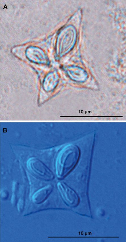
The spores obtained from silver scabbardfish
(Fig. 2A) and Atlantic mackerel (Fig. 2B) showed
similar morphological appearance. In apical view,
mature spores were stellate, with 4 unequal pyriform
PCs, each within thin-walled valves (Fig. 2). The
Kudoa sp. spores from both fish species were charac-
terized by 1 large, 1 small and 2 intermediate PCs,
directed apically to one another (Fig. 2). Single-
coiled polar filaments were sometimes visible within
Fig. 2. Comparison between mature spores of 2 isolates of
each PC (Fig. 2). In lateral view, spores were subcon- Kudoa thyrsites in apical view. (A) K. thyrsites in Lepidopus
ical in shape, with posterior apices of valves forming caudatus from the Alboran Sea. (B) K. thyrsites in Scomber
lanceolate processes. scombrus from the Norwegian Sea
In accordance with the morphological characteris-
tics described by Egusa (1986), Kent et al. (1994),
Canning & Okamura (2004) and Lom & Dyková 3.3. Molecular identification
(2006), all present spore samples were assigned to
the genus Kudoa. The morphometric and allometric SSU rDNA sequences (1200 bp in length) were
data (mean ± SD, range) of spores isolated from silver obtained from Kudoa spores sampled from liquefied
scabbardfish and Atlantic mackerel, are given in muscle of the examined fish specimens. The align-
Table 1. ment by Clustal X of the SSU rDNA sequences from
Statistical analysis confirmed that there was no sig- silver scabbardfish (GenBank accession no. MH-
nificant morphometric variation between the 2 host 899080), showed 100% match with the correspon-
isolates of Kudoa sp. The overall morphology, to- ding sequence of the Kudoa sp. isolate from Atlantic
gether with the morphometric spore measurements, mackerel (GenBank accession no. MH899081).
showed close similarity with isolates of K. thyrsites Moreover, the latter sequences matched 100%
described in the literature (Table S1). (over 1200 bp) with the SSU rDNA sequence of130 Dis Aquat Org 132: 125–134, 2019
Table 1. Comparative spore morphometry of Kudoa thyrsites isolates primarily associated with Kudoa spp. infec-
from silver scabbardfish Lepidopus caudatus and Atlantic mackerel tions, as well as some other species of mul-
Scomber scombrus from the Alboran Sea and the Norwegian Sea, re-
tivalvulid myxosporeans (Moran et al. 1999).
spectively. Sixty spores were measured for each parameter and fish
host species. Measurements (mean, SD and range) are given in µm. To date, only a few cases of ‘soft flesh’-
Morphological character definitions are given in Fig. 1. TL (TW): total inducing Kudoa species have been reported
length (width) of myoliquefactive fish from fish caught in the Mediterranean Sea
(Pampoulie et al. 1999, Gaglio et al. 2010).
Morphometrical Locality and host species However, none of the Kudoa species was
characteristic Alboran Sea Norwegian Sea identified or described molecularly.
or ratio Lepidopus caudatus Scomber scombrus In the present study, a case of post
N=2 N=8
mortem myoliquefactive kudoosis in 2 silver
(TL: 135 and 121.0 cm; (mean TL: 37 cm;
TW: 1778 and 1500 g) mean TW: 436.5 g) scabbardfish from the Alboran Sea is re-
Mean SD (Range) Mean SD (Range) ported. Based on overall spore morphology
and molecular analysis, the causative spe-
W1 15.5 1.0 (13.0−17.5) 15.5 1.5 (13.0−18.5) cies was identified as K. thyrsites (Gilchrist
W2 14.5 1.0 (12.0−17.0) 14.5 1.5 (11.5−17.5)
1924). The finding represents the first
T1 12.0 1.0 (9.5−14.5) 12.0 1.5 (9.5−17.5)
T2 10.0 1.0 (8.5−12.5) 9.5 1.0 (7.0−11.5) molecular and morphological identification
L 7.5 1.0 (6.5−11.0) 7.0 1.0 (5.5−8.0) of K. thyrsites in fish from the Mediterran-
LPCL 5.0 0.5 (4.0−6.5) 5.5 1.0 (4.5−7.5) ean Sea. For comparative and validation
IPCLa 3.5 1.0 (2.5−6.0) 4.0 1.0 (2.5−5.5) purposes, the morphological and molecular
SPCL 3.0 0.5 (1.5−4.5) 3.0 0.5 (2.0−4.0) characteristics of the present K. thyrsites
W1:W2 1.1 0.1 (1.0−1.2) 1.1 0.1 (0.9−1.2)
isolate from the Mediterranean Sea were
T1:T2 1.2 0.2 (0.9−1.6) 1.2 0.2 (1.0−1.6)
LPCL:IPCL 1.4 0.3 (0.7−1.8) 1.4 0.2 (1.2−2.0) compared with equally processed and ana-
LPCL:SPCL 1.8 0.3 (1.1−3) 1.9 0.3 (1.5−2.7) lysed fresh spores of a K. thyrsites isolate
a infecting Atlantic mackerel caught in the
Combined measurements (mean) of the 2 intermediate polar cap-
sules of K. thyrsites spore Norwegian Sea.
The overall morphological characteristics
of the spores from liquefied muscle tissue of
the previously identified K. thyrsites isolate from the silver scabbardfish samples were in close agree-
Atlantic mackerel caught in the North Sea (Gen- ment with those of the K. thyrsites isolate from
Bank accession no. EU154349, Levsen et al. 2008), Atlantic mackerel (Fig. 2), as well as with other K.
as well as the SSU rDNA sequence of another thyrsites isolates previously described from different
K. thyrsites isolate from Atlantic mackerel caught fish host species and geographical areas (Table S1).
off southern England (GenBank accession no. Despite the similarities in overall shape and
AY542482, Whipps & Kent 2006). On the other appearance, it appears from the literature that vari-
hand, the present K. thyrsites SSU rDNA sequences ous spore measurements, as well as some other spe-
obtained from silver scabbardfish and Atlantic cific characteristics, differ considerably between the
mackerel shared 99% similarity with K. thyrsites K. thyrsites isolates described to date (Table S1). For
sequences of the same gene recorded from other instance, the presence of unequally sized PCs
geographical areas and deposited in GenBank, i.e. (1 large, 1 small and 2 intermediate) in both K. thyr-
South Africa (accession nos. AY542481, AY941819, sites isolates presently analysed (Fig. 2), has only
Whipps & Kent 2006; and AY078430, Whipps et al. been reported in a few other studies of this species,
2003), eastern Australia (accession no. AY152747, including the original description (Gilchrist 1924,
Whipps et al. 2003), British Columbia (accession no. Langdon 1991, Munday et al. 1998, Levsen et al.
AF031412, Hervio et al. 1997) and Japan (accession 2008). Additionally, the number of coils of the polar
nos. LC128644, LC128645, Kasai et al. 2016; and filament varies considerably between different re-
AY382607, Yokoyama & Itoh 2005). ports (i.e. 1−3.5 coils) (Gilchrist 1924, Kabata &
Whitaker 1981, Langdon 1991, Whipps et al. 2003,
Levsen et al. 2008), thus rendering it a less reliable
4. DISCUSSION taxonomic character (Whipps et al. 2003). Spore
width is another important morphological character
Post mortem liquefaction of fish muscle tissue, of K. thyrsites that seems to vary widely among iso-
commonly referred to as ‘soft flesh’, is a phenomenon lates, ranging from 12.0 µm in spores isolated fromGiulietti et al.: Kudoa thyrsites in Mediterranean silver scabbardfish 131
South African snoek Thyrsites atun to 16.7 µm in the K. thyrsites samples from silver scabbardfish
spores obtained from Pacific hake Merluccius pro- (GenBank accession no. MH899080) were 100%
ductus caught off Pacific North America (Whipps & identical to SSU rDNA sequences of K. thyrsites sam-
Kent 2006). ples from Atlantic mackerel from the Norwegian Sea
The intraspecific variability in spore dimensions (GenBank accession no. MH899081). A complete
appears to be relatively high in Myxosporea (Dia- match (100%) was also observed when the se-
mant et al. 2005). For example, observations in sev- quences of both isolates were aligned with published
eral other Kudoa species (e.g. K. gunterae, K. hypo- sequences of K. thyrsites isolates from Atlantic mack-
epicardialis, K. islandica, K. iwatai, K. shiomitsui, erel caught in the North Sea (GenBank accession no.
K. whippsi), as well as some other Myxozoa (e.g. En- EU154349, Levsen et al. 2008) and off the southern
teromyxum leei) indicate that spore size apparently coast of England (GenBank accession no: AY542482,
depends on developmental stage, geographical lo- Whipps & Kent 2006). The results indicate that the
cality or fish host species (Padrós et al. 2001, Blaylock 4 K. thyrsites isolates identified from geographically
et al. 2004, Diamant et al. 2005, Burger & Adlard different waters in Europe (i.e. Alboran Sea, Norwe-
2010, Kristmundsson & Freeman 2014). Other techni- gian Sea, North Sea and southern coast of England)
cal factors (e.g. heterogeneity of measuring tech- are genetically indistinguishable at the locus exam-
niques adopted by different authors, 3-dimensional- ined (i.e. SSU rDNA).
ity of the spores), may introduce uncertainty as to The present finding suggests that the geographical
measurement accuracy. Measuring a curved 3-dimen- distribution of K. thyrsites includes the western
sional spore with a standard microscope has obvious Mediterranean Sea. Furthermore, the Alboran Sea is
limitations since only 1 focal plane can be imaged at considered an oceanographic transition zone be-
a given point. Being a result of a projection, the tween the Atlantic Ocean and the Mediterranean
image of the spore could therefore become consider- Sea, retaining some oceanographic characteristics
ably distorted, increasing the uncertainty associated more similar to the Atlantic basin than to the Medi-
with a measurement (Prabhat et al. 2004). The meas- terranean Sea (Tintore et al. 1988). From this area,
uring uncertainty may increase even more when several cases of fish and marine mammals infected
focussing on the smallest characters of a spore. Thus, with parasites of oceanic waters (e.g. the nematode
although the width of the PCs is commonly reported Anisakis simplex s.s. and the monogenean Heteraxi-
as a specific character in Kudoa spp. spore descrip- noides atlanticus) of the Northeast Atlantic Ocean
tions, the accuracy of this measurement obtained have been reported (MacKenzie et al. 2008, Mat-
here was considered not informative, and is thus not tiucci et al. 2018).
reported. The apparently low host specificity of K. thyrsites
In light of these issues, we propose a set of addi- and its wide geographical distribution render this
tional morphometric characters, based on calculated parasite a potential loss factor in wild-catch fisheries
allometric ratios of various spore dimensions, which and marine fish cultures in most temperate and sub-
can be useful when used together with conventional tropical seas and coastal areas. However, due to the
measurements. Thus, the presently considered ratios lack of knowledge about the ecology, life cycle and
(W1:W2, T1:T2, LPCL:IPCL and LPCL:SPCL) were transmission pathways of K. thyrsites, it is difficult to
virtually identical in both K. thyrsites isolates exam- evaluate the boundaries of its geographical range
ined here, i.e. they did not differ significantly. This and its actual distribution in the Mediterranean Sea.
observation suggests that these allometric ratios are Thus, further research is required to assess the possi-
consistent between the 2 isolates and can therefore ble economic consequences for the fishing industry
be used as supplementary descriptors in species or inflicted by myoliquefactive K. thyrsites infections in
isolates (e.g. K. thyrsites) in which spore size varies L. caudatus, as well as other K. thyrsites-susceptible
widely. In fact, since allometric ratios are derived rel- and commercially exploited Mediterranean fish
ative values, they are unaffected by the variability of species.
morphometrical spore dimensions.
The molecular identification of the present Kudoa
isolates infecting silver scabbardfish and Atlantic Acknowledgements. We thank Hui-Shan Tung for her assis-
mackerel was consistent with the morphological tance and help with the molecular analyses. Stefania
Billera’s contribution in drawing the images was also highly
identification, thus confirming K. thyrsites as the pri- appreciated. The study was partly supported by the Euro-
mary cause of post mortem myoliquefaction in both pean Union’s Seventh Framework Program for Research,
fish species. The SSU rDNA sequences (1200 bp) of Technological Development and Demonstration, project132 Dis Aquat Org 132: 125–134, 2019
‘Parasite risk assessment with integrated tools in EU fish muscolari da Kudoa sp. (Myxosporea: Multivalvulida) in
production value chains (PARASITE)’, Grant agreement un esemplare di pesce spada (Xiphias gladius) pescato
(GA) no. 312068. nel Mediterraneo. Large Anim Rev 16:1−3
Gilchrist JDF (1924) A protozoal parasite Chloromyxum
thyrsites sp. n. of the Cape sea-fish, the ‘snoek’ (Thyrsites
LITERATURE CITED atun Euphr.). Trans R Soc S Afr 11:263−273
Henning SS, Hoffman LC, Manley M (2013) A review of
Adlard RD, Bryant MS, Whipps CM, Kent ML (2005) Multi- Kudoa-induced myoliquefaction of marine fish species in
valvulid myxozoans from eastern Australia: three new South Africa and other countries. S Afr J Sci 109:1−5
species of Kudoa from scombrid and labrid fishes of the Hervio DML, Khattra J, Devlin RH, Kent ML, Sakanari J,
Great Barrier Reef, Queensland, Australia. J Parasitol 91: Yokoyama H (1997) Taxonomy of Kudoa species (Myxo-
1138−1142 sporea), using a small-subunit ribosomal DNA sequence.
Alvarez-Pellitero P, Sitjà-Bobadilla A (1993) Pathology of Can J Zool 75:2112−2119
Myxosporea in marine fish culture. Dis Aquat Org 17: Holzer AS, Blasco-Costa I, Sarabeev VL, Ovcharenko MO,
229−238 Balbuena JA (2006) Kudoa trifolia sp. n.: Molecular phy-
Blaylock RB, Bullard SA, Whipps CM (2004) Kudoa hypoepi- logeny suggests a new spore morphology and unusual tis-
cardialis n. sp. (Myxozoa: Kudoidae) and associated sue location for a well-known genus. J Fish Dis 29:743−755
lesions from the heart of seven perciform fishes in the Iwamoto T (2015) Lepidopus caudatus. In: IUCN Red List of
northern Gulf of Mexico. J Parasitol 90:584−593 Threatened Species 2015:e.T198721A42691759. http://
Burger MAA, Adlard RD (2010) Phenotypic variation in a dx.doi.org/10.2305/IUCN.UK.2015-4.RLTS.T198721A42
significant spore character in Kudoa (Myxosporea: 691759.en
Multivalvulida) species infecting brain tissue. Parasitol- Iwashita Y, Kamijo Y, Nakahashi S, Shindo A and others
ogy 137:1759−1772 (2013) Food poisoning associated with Kudoa septem-
Burger MAA, Adlard RD (2011) Low host specificity in the punctata. J Emerg Med 44:943−945
Kudoidae (Myxosporea:Multivalvulida) including seven- Kabata Z, Whitaker DJ (1981) Two species of Kudoa (Myxo-
teen new host records for Kudoa thalassomi. Folia Para- sporea: Multivalvulida) parasitic in the flesh of Merluc-
sitol 58:1−16 cius productus (Pisces: Teleostei) in the Canadian Pacific.
Burger MAA, Barnes AC, Adlard RD (2008) Wildlife as Can J Zool 59:2085−2091
reservoirs for parasites infecting commercial species: Kasai A, Li YC, Mafie E, Sato H (2016) New host records of
host specificity and a redescription of Kudoa amamiensis monacanthid fish for three Kudoa spp. (K. septempunc-
from teleost fish in Australia. J Fish Dis 31:835−844 tata, K. thyrsites and K. shiomitsui) prevalent in the olive
Campbell N (2005) The myxosporean parasitofauna of the flounder (Paralichthys olivaceus), with the description of
Atlantic horse mackerel, Trachurus trachurus (L.) in the K. parathyrsites n. sp. from a black scraper (Thamna-
North-East Atlantic Ocean and Mediterranean Sea. Acta conus modestus). Parasitol Res 115:2741−2755
Parasitol 50:97−101 Kawai T, Sekizuka T, Yahata Y, Kuroda M and others (2012)
Canning EU, Okamura B (2004) Biodiversity and evolution Identification of Kudoa septempunctata as the causative
of the Myxozoa. Adv Parasitol 56:43−131 agent of novel food poisoning outbreaks in Japan by con-
Crookham J, Dapson R (1991) Hazardous chemicals in the sumption of Paralichthys olivaceus in raw fish. Clin Infect
histopathology laboratory, 2nd edn. Anatech, Battle Dis 54:1046−1052
Creek, MI Kent ML, Margolis L, Whitaker DJ, Hoshs GE, McDonald TE
D’Onghia G, Mastrotaro F, Maiorano P (2000) Biology of sil- (1994) Review of Myxosporea of importance in salmonid
ver scabbard fish, Lepidopus caudatus (Trichiuridae), fisheries and aquaculture in British Columbia. Folia
from the Ionian Sea (Eastern-Central Mediterranean). Parasitol 41:27−37
Cybium 24:249−262 Kristmundsson Á, Freeman MA (2014) Negative effects of
Demestre M, Moli B, Recases L, Sanches M (1993) Life his- Kudoa islandica n. sp. (Myxosporea: Kudoidae) on aqua-
tory and fishery of Lepidopus caudatus (Pisces: Trichiuri- culture and wild fisheries in Iceland. Int J Parasitol Para-
dae) in the Catalan Sea (Northwestern Mediterranean). sites Wildl 3:135−146
Mar Biol 115:23−31 Langdon JS (1991) Myoliquefaction post mortem (‘milky
Diamant A, Ucko M, Paperna I, Colorni A, Lipshitz A (2005) flesh’) due to Kudoa thyrsites (Gilchrist) (Myxosporea:
Kudoa iwatai (Myxosporea: Multivalvulida) in wild and Multivalvulida) in mahi mahi, Coryphaena hippurus L.
cultured fish in the Red Sea: redescription and molecular J Fish Dis 14:45−54
phylogeny. J Parasitol 91:1175−1189 Larkin MA, Blackshields G, Brown NP, Chenna R and others
Egusa S (1986) The order Multivalvulida Shulman, 1959 (2007) Clustal W and Clustal X version 2.0. Bioinforma-
(Myxozoa; Myxosporea): a review. Fish Pathol 21: tics 23:2947−2948
261−274 (in Japanese) Levsen A (2015) A review of Kudoa species (Myxozoa, Mul-
Eiras JC, Saraiva A, Cruz C (2014) Synopsis of the species of tivalvulida) affecting seafood quality, with emphasis on
Kudoa Meglitsch, 1947 (Myxozoa: Myxosporea: Multi- K. thyrsites in Atlantic mackerel (Scomber scombrus).
valvulida). Syst Parasitol 87:153−180 J Fish Sci Technol Spec Issue 2015:3−9
Funk VA, Raap M, Sojonky K, Jones S, Robinson J, Levsen A, Jørgensen A, Mo TA (2008) Occurrence of post-
Falkenberg C, Miller KM (2007) Development and val- mortem myoliquefactive kudoosis in Atlantic mackerel,
idation of an RNA- and DNA-based quantitative PCR Scomber scombrus L., from the North Sea. J Fish Dis 31:
assay for determination of Kudoa thyrsites infection 601−611
levels in Atlantic salmon Salmo salar. Dis Aquat Org Lom J (1987) Myxosporea: a new look at long-known para-
75:239−249 sites of fish. Parasitol Today 3:327−332
Gaglio G, Marino F, Monaco S, Giannetto S (2010) Lesioni Lom J, Arthur JR (1989) A guideline for the preparationGiulietti et al.: Kudoa thyrsites in Mediterranean silver scabbardfish 133 of species descriptions in Myxosporea. J Fish Dis 12: Paris, p 976−980 151−156 Prabhat P, Ram S, Ward ES, Ober RJ (2004) Simultaneous Lom J, Dyková I (2006) Myxozoan genera: definition and imaging of different focal planes in fluorescence notes on taxonomy, life cycle terminology and patho- microscopy for the study of cellular dynamics in three genic species. Folia Parasitol 53:1−36 dimensions. IEEE Trans Nanobioscience 3:237−242 Lom J, Dyková I, Lhotáková S (1983) Kudoa lunata n. sp. Rosa A, Menezes G, Melo O, Pinho MR (2006) Weight- (Myxozoa, Myxosporea) and notes on the nature of length relationships of 33 demersal fish species from muscular ‘cysts’ of the genus. Arch Protistenk 127: Azores archipelago. Fish Res 80:329−332 387−397 Sato H, Kasai A (2016) Kudoa species (Myxozoa: Myxo- Lopez JC, Navarro J (2000) Descripcion de casos clinicos sporea: Multivalvulida) recorded in Japan or its sur- producidos por nuevos agentes patogenos de importan- rounding natural waters (1930-2016). Jpn J Vet Parasitol cia en salmones de cultivo en Chile. XI Congreso de 15:111−138 (in Japanese with English summary) Medicina Verterinaria Universidad de Chile. Jornadas Shaw RW, Hervio DML, Devlin RH, Adamson ML (1997) de Salmonicultura, 25-27 October, Puerto Varas, Chile, Infection of Aulorhynchus flavidus (Gill) (Osteichthyes: p 106 Gasterosteiformes) by Kudoa thyrsites (Gilchrist) (Myxo- MacKenzie K, Kalavati C (2014) Myxosporean parasites of sporea: Multivalvulida). J Parasitol 83:810−814 marine fishes: their distribution in the world’s oceans. Shin SP, Shirakashi S, Hamano S, Kato K, Lasso LT, Yoko- Parasitology 141:1709−1717 yama H (2016) Phylogenetic study of the genus Kudoa MacKenzie K, Campbell N, Mattiucci S, Ramos P, Pinto AL, (Myxozoa: Multivalvulida) with a description of Kudoa Abaunza P (2008) Parasites as biological tags for stock rayformis sp. nov. from the trunk muscle of Pacific identification of Atlantic horse mackerel Trachurus tra- sierra Scomberomorus sierra. Mol Phylogenet Evol 98: churus L. Fish Res 89:136−145 337−345 Mansour L, Thabet A, Chourabi K, Harrath AH, Gtari M, Al St-Hilaire S, Ribble C, Whitaker DJ, Kent ML (1997) Evalu- Omar SY, Hassine OKB (2013) Kudoa azevedoi n. sp. ation of a nondestructive diagnostic test for Kudoa thyr- (Myxozoa, Multivalvulida) from the oocytes of the At- sites in farmed Atlantic salmon (Salmo salar). Aquacul- lantic horse mackerel Trachurus trachurus (Perciformes, ture 156:139−144 Carangidae) in Tunisian coasts. Parasitol Res 112: St-Hilaire S, Ribble C, Whitaker DJ, Kent M (1998) Preva- 1737−1747 lence of Kudoa thyrsites in sexually mature and imma- Marshall WL, Sitjà-Bobadilla A, Brown HM, MacWilliam T ture pen-reared Atlantic salmon (Salmo salar) in British and others (2016) Long-term epidemiological survey of Columbia, Canada. Aquaculture 162:69−77 Kudoa thyrsites (Myxozoa) in Atlantic salmon (Salmo Suzuki J, Murata R, Yokoyama H, Sadamasu K, Kai A (2015) salar L.) from commercial aquaculture farms. J Fish Dis Detection rate of diarrhoea-causing Kudoa hexapunctata 39:929−946 in Pacific bluefin tuna Thunnus orientalis from Japanese Mattiucci S, Cipriani P, Levsen A, Paoletti M, Nascetti G waters. Int J Food Microbiol 194:1−6 (2018) Molecular epidemiology of Anisakis and anisakia- Tintore J, La Violette PE, Blade I, Cruzado A (1988) A study sis: an ecological and evolutionary road map. In: of an intense density front in the eastern Alboran Sea: Rollinson D, Stothard JR (eds) Advances in parasitology. the Almeria-Oran front. J Phys Oceanogr 18:1384−1397 Academic Press, London, p 93−263 Whipps CM, Kent ML (2006) Phylogeography of the cos- Moran JDW, Whitaker DJ, Kent ML (1999) A review of the mopolitan marine parasite Kudoa thyrsites (Myxozoa: myxosporean genus Kudoa Meglitsch, 1947, and its Myxosporea). J Eukaryot Microbiol 53:364−373 impact on the international aquaculture industry and Whipps CM, Adlard RD, Bryant MS, Lester RJG, Findlay V, commercial fisheries. Aquaculture 172:163−196 Kent ML (2003) First report of three Kudoa species from Munday BL, Su XQ, Harshbarger JC (1998) A survey of Eastern Australia: Kudoa thyrsites from mahi mahi product defects in Tasmanian Atlantic salmon (Salmo (Coryphaena hippurus), Kudoa amamiensis and Kudoa salar). Aquaculture 169:297−302 minithyrsites n. sp. from sweeper (Pempheris ypsilych- Nakamura I, Parin NV (1993) Snake mackerels and cutlass- nus). J Eukaryot Microbiol 50:215−219 fishes of the world. FAO Species Catalogue. FAO Fish- Whipps CM, Grossel G, Adlard RD, Yokoyama H, Bryant eries Synopsis No. 125, Vol 15. FAO, Rome MS, Munday BL, Kent ML (2004) Phylogeny of the Mul- Padrós F, Palenzuela O, Hispano C, Tosas O, Zarza C, tivalvulidae (Myxozoa:Myxosporea) based on compara- Crespo S, Alvarez-Pellitero P (2001) Myxidium leei tive ribosomal DNA sequence analysis. J Parasitol 90: (Myxozoa) infections in aquarium-reared Mediterranean 618−622 fish species. Dis Aquat Org 47:57−62 Whitaker DJ, Kent ML (1991) Myxosporean Kudoa thyrsites: Palmer R (1994) Kudoa — the Irish experience. In: Conley a cause of soft flesh in farm-reared Atlantic salmon. DC, Nanaimo BC (eds) Kudoa Workshop Proceedings, 17 J Aquat Anim Health 3:291−294 and 18 February 1994. Aquacult Ind Dev Rep 94. Wolf K, Markiw ME (1984) Biology contravenes taxonomy in Province of British Columbia, Ministry of Agriculture, the Myxozoa: New discoveries show alternation of inver- Fisheries and Food, Victoria, p 18−21 tebrate and vertebrate hosts. Science 225:1449−1452 Pampoulie C, Marques A, Rosecchi E, Crivelli AJ, Bochereau Yahata Y, Sugita-Konishi Y, Ohnishi T, Toyokawa T, Naka- JL (1999) A new myxosporean parasite, Kudoa camar- mura N, Taniguchi K, Okabe N (2015) Kudoa septem- guensis n. sp., recorded on two goby species (Teleostei: punctata-induced gastroenteritis in humans after floun- Pisces) in the Rhone Delta (Mediterranean Sea, France). der consumption in Japan: a case-controlled study. Jpn J J Eukaryot Microbiol 46:304−310 Infect Dis 68:119−123 Parin NV (1986) Trichiuridae. In: Whitehead PJP, Bauchot Yokoyama H, Itoh N (2005) Two multivalvulid myxozoans ML, Hureau JC, Nielsen J, Tortonese E (eds) Fishes of causing postmortem myoliquefaction: Kudoa megacap- the Northeastern Atlantic and Mediterranean. UNESCO, sula n. sp. from red barracuda (Sphyraena pinguis) and
134                                            Dis Aquat Org 132: 125–134, 2019

   Kudoa thyrsites from splendid alfonso (Beryx splendens).         Rijeka, p 1–44
   J Parasitol 91:1132−1137                                      Young CA (2002) An immunolocalization study of the life
Yokoyama H, Whipps CM, Kent ML, Mizuno K, Kawakami                  stages of Kudoa thyrsites in Atlantic salmon (Salmo
   H (2004) Kudoa thyrsites from Japanese flounder and              salar). Research report. Malaspina University-College,
   Kudoa lateolabracis n. sp. from Chinese sea bass: cau-           Nanaimo
   sative myxozoans of post-mortem myoliquefaction. Fish         Yurakhno VM, Ovcharenko AS, Holzer AS, Sarabeev VL,
   Pathol 39:79−85                                                  Balbuena JA (2007) Kudoa unicapsula n. sp. (Myxo-
Yokoyama H, Grabner D, Shirakashi S (2012) Transmission             sporea: Kudoidae) a parasite of the Mediterranean mul-
   biology of the Myxozoa. In: Carvalho E, David GS, Silva          lets Liza ramada and L. aurata (Teleostei: Mugilidae).
   RJ (eds) Health and environment in aquaculture. InTech,          Parasitol Res 101:1671−1680

Editorial responsibility: Dieter Steinhagen,                     Submitted: May 17, 2018; Accepted: November 2, 2018
Hannover, Germany                                                Proofs received from author(s): December 23, 2018
You can also read