Morphological features of large layer V pyramidal neurons in cortical motor related areas of macaque monkeys: analysis of basal dendrites
←
→
Page content transcription
If your browser does not render page correctly, please read the page content below
www.nature.com/scientificreports
OPEN Morphological features of large
layer V pyramidal neurons
in cortical motor‑related areas
of macaque monkeys: analysis
of basal dendrites
Yu Takata1, Hiroshi Nakagawa1,2, Taihei Ninomiya3,4, Hajime Yamanaka1 &
Masahiko Takada1*
In primates, large layer V pyramidal neurons located in the frontal motor-related areas send a variety
of motor commands to the spinal cord, giving rise to the corticospinal tract, for execution of skilled
motor behavior. However, little is known about the morphological diversity of such pyramidal neurons
among the areas. Here we show that the structure of basal dendrites of the large layer V pyramidal
neurons in the dorsal premotor cortex (PMd) is different from those in the other areas, including the
primary motor cortex, the supplementary motor area, and the ventral premotor cortex. In the PMd,
not only the complexity (arborization) of basal dendrites, i.e., total dendritic length and branching
number, was poorly developed, but also the density of dendritic spines was so low, as compared to the
other motor-related areas. Regarding the distribution of the three dendritic spine types identified, we
found that thin-type (more immature) spines were prominent in the PMd in comparison with stubby-
and mushroom-type (more mature) spines, while both thin- and stubby-type spines were in the other
areas. The differential morphological features of basal dendrites might reflect distinct patterns of
motor information processing within the large layer V pyramidal neurons in individual motor-related
areas.
Pyramidal neurons are the main projection neurons in the cerebral cortex. Thus, various lines of information
processed in a given cortical area are conveyed to other cortical areas or subcortical regions through axonal
branches of the pyramidal neurons. Transmission of such information from neuron to neuron takes place at
synapses, and postsynaptic neurons receive it through their dendrites and dendritic spines.
Layer V is a major output layer of the cerebral cortex. In primates, large layer V pyramidal neurons in the
motor-related areas of the frontal lobe, including the primary motor cortex (M1), the supplementary motor area
(SMA), and the dorsal and ventral divisions of the premotor cortex (PMd, PMv), send their axons extensively
to the brainstem and the spinal cord for control of voluntary m ovements1–5. These layer V pyramidal neurons
have many dendritic spines, which are distributed more frequently on their basal than apical d endrites6. It has
been reported in rodents that the basal dendrites of large layer V pyramidal neurons receive inputs from their
neighboring neurons7 and layer II/III pyramidal neurons8. Such inputs through the basal dendrites may exert a
strong impact on activity of the layer V pyramidal neurons. To reveal the structural basis for control of large layer
V pyramidal neuron activity, it is essential to define the morphology of their basal dendrites and dendritic spines.
In general, wide variations in pyramidal neuron structure depend on the areal and laminar specificity of the
cortex. For example, the morphological features of basal dendrites of pyramidal neurons in layer III, i.e., their
complexity and spine number, vary among the visual cortical areas of primates9, thus reflecting a certain func-
tional diversity of individual areas. Likewise, the frontal motor-related areas are involved in different aspects of
1
Systems Neuroscience Section, Primate Research Institute, Kyoto University, Inuyama, Aichi 484‑8506,
Japan. 2Department of Molecular Neuroscience, World Premier International Immunology Frontier Research
Center, Osaka University, 3‑1 Yamadaoka, Suita, Osaka 565‑0871, Japan. 3Department of Developmental
Physiology, National Institute for Physiological Sciences, Okazaki 444‑8585, Japan. 4Department of Physiological
Sciences, School of Life Science, The Graduate University for Advanced Studies (SOKENDAI), Hayama,
Kanagawa 240‑0193, Japan. *email: takada.masahiko.7x@kyoto‑u.ac.jp
Scientific Reports | (2021) 11:4171 | https://doi.org/10.1038/s41598-021-83680-5 1
Vol.:(0123456789)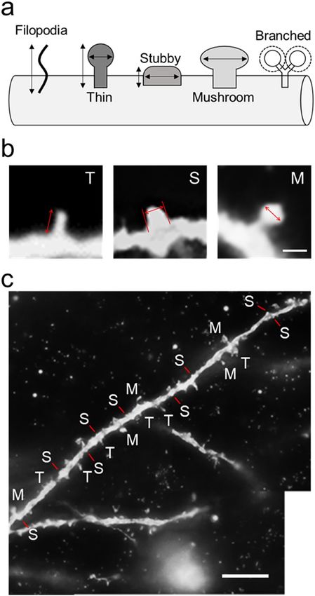
www.nature.com/scientificreports/
motor control10–13. In addition, each area receives somewhat distinct inputs from other cortical a reas14,15. These
facts lead us to hypothesize that the structure of large pyramidal neurons in layer V may be different among the
areas. However, little is known about the differential morphological characteristics of large layer V pyramidal
neurons in the motor-related areas. To understand how the morphological features of these pyramidal neurons
represent the functional role(s) specific to each area, we investigated the possible differences in pyramidal neuron
structure, with particular reference to the basal dendrites, among the M1, SMA, PMd, and PMv of macaque
monkeys. In the present study, the morphology of basal dendrites of the large layer V pyramidal neurons was
analyzed and compared in individual motor-related areas, especially in their digit regions.
Results
Before analyzing the morphological features of large layer V pyramidal neurons giving rise to the corticospinal
tract (CST), especially for digit movement, we performed two preparatory experiments: retrograde labeling of
CST neurons and intracortical microstimulation (ICMS) mapping in the motor-related areas of the frontal lobe.
Retrograde labeling of CST neurons. In the present study, it is critical to sample CST neurons out of
pyramidal neurons in layer V of the frontal motor-related areas. To solve this issue, we employed retrograde
transport of rabies virus to examine the largeness of CST neurons in individual motor-related areas projecting
to the cervical enlargement. The use of rabies virus for labeling CST neurons was meritorious in that this virus is
taken up specifically from axon terminals, but not from passing fibers, and provides the explicit Golgi-like mor-
phology of labeled neurons with the somal size unchanged16–18. After rabies injections into the cervical enlarge-
ment, especially into the C6–T1 levels for digit innervation, retrogradely labeled CST neurons were observed
in the motor-related areas (Fig. 1a–l). All labeled neurons were confined to layer V across the areas, indicating
that only monosynaptically-connected neurons were traced in this monkey. We sampled 112, 54, 66, and 44
neurons from the M1, SMA, PMd, and PMv, respectively, and measured their somal size using Neurolucida
explorer. The same number of unlabeled neurons was sampled from layer V of each area. As shown in Fig. 1m–p,
the somal size of the labeled CST neurons was 416.54 ± 23.42 μm2 for the M1, 331.78 ± 20.67 μm2 for the SMA,
235.55 ± 5.81 μm2 for the PMd, and 236.69 ± 10.13 μm2 for the PMv (for the somal size distribution, see also
Fig. 1q), while that of the unlabeled neurons was 268.05 ± 21.55 μm2, 175.4 ± 7.64 μm2, 203.06 ± 6.47 μm2, and
154.47 ± 6.70 μm2 for individual areas, respectively. The somal size of the labeled CST neurons was significantly
larger than that of the unlabeled neurons in each motor-related area (TukeyHSD; p < 0.01). Based on these data,
putative CST neurons, the somal size of which was larger than the first quartile for the labeled CST neurons
(238.29 μm2, 221.49 μm2, 199.78 μm2, and 179.6 μm2 for individual areas), were selected for their morphological
analyses.
ICMS mapping. In the present study, it is prerequisite to dissociate the digit region in each of the motor-
related areas as accurately as possible. To achieve this purpose, we carried out ICMS to identify the digit regions
in individual motor-related areas. In a representative case shown in Fig. 2, movements of the digits were evoked
from five loci within the M1, three loci within the SMA, three loci within the PMd, and one locus within the
PMv. According to the results of ICMS mapping, we determined the border between the digit region and other
body-part regions in each area and dissected out a tissue block containing its digit representation alone for mor-
phological analyses of putative CST neurons (Fig. 2).
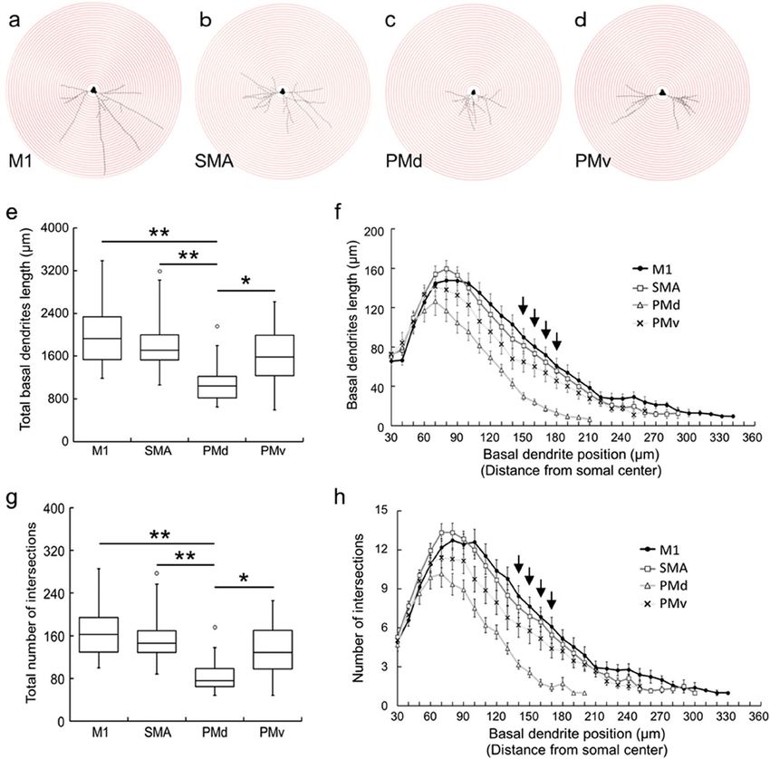
Complexity of basal dendrites. We selected 20 putative CST neurons from each motor-related area
based on the somal size: 464.75 ± 30.22 μm2 for the M1; 460.89 ± 37.68 μm2 for the SMA; 287.74 ± 8.02 μm2 for
the PMd; and 319.47 ± 15.76 μm2 for the PMv. We traced the basal dendrites of putative CST neurons within the
digit regions of individual motor-related areas, as the full length of each dendrite appeared to be followed suc-
cessfully in single sections. We then surveyed the complexity of basal dendrites, i.e., total dendritic length and
intersection number, by means of Sholl analysis (for details, see Methods; Fig. 3a–d). Data obtained were as fol-
lows: (1) The total length of basal dendrites in the PMd was significantly shorter than in the M1, SMA, and PMv
(TukeyHSD; p < 0.05; Fig. 3e). There were no significant differences in the total basal dendrites length among
the M1, SMA, and PMv (TukeyHSD; Fig. 3e); (2) The basal dendrites length around 150–180 µm apart from the
somal center was significantly shorter in the PMd than in the other motor-related areas (TukeyHSD; p < 0.05;
Fig. 3f); (3) The total number of intersections in the PMd was significantly smaller than in the M1, SMA, and
PMv (TukeyHSD; p < 0.05; Fig. 3g). There were no significant differences in the total intersection number among
the M1, SMA, and PMv (TukeyHSD; Fig. 3g); (4) The intersection number around 140–170 µm apart from the
somal center was significantly smaller in the PMd than in the other motor-related areas (TukeyHSD; p < 0.05;
Fig. 3h); and (5) In close proximity of the soma (~ 30 µm from the somal center), there were no significant dif-
ferences in the intersection number among the motor related areas (TukeyHSD; Fig. 3h).
Density of dendritic spines. The density of dendritic spines was analyzed for the basal dendrites of CST
neurons within the digit regions of individual motor-related areas using Neurolucida explorer (Fig. 4a–d). In
the present experiments, we counted the number of spines on two dendrites in each area and converted it to the
value per 10-µm segment. It was found that the density of dendritic spines in the PMd was significantly lower
than in the other motor-related areas (TukeyHSD; p < 0.01; Fig. 4e). To confirm whether such a distribution
pattern of dendritic spines might depend on the basal dendrite position, we examined the density of spines on
every 20- or 50-µm segment. In each motor-related area, there was no significant difference in the spine density
between the two dendrites (data not shown). Depending on the basal dendrite position, on the other hand, there
were some differences in the spine density in the M1, SMA, and PMv (TukeyHSD; p < 0.05; Fig. 4f–i). Moreover,
Scientific Reports | (2021) 11:4171 | https://doi.org/10.1038/s41598-021-83680-5 2
Vol:.(1234567890)www.nature.com/scientificreports/
Figure 1. Retrograde labeling of CST neurons in frontal motor-related areas and their somal size measurement.
(a–d) Low-magnification photos of retrograde labeling of CST neurons in the frontal motor-related areas after
rabies injections into the C6–T1 levels of the spinal cord. (a) M1, (b) SMA, (c) PMd, (d) PMv. Scale bar, 5 mm.
cs, central sulcus; sps, superior precentral sulcus; spur, spur of the arcuate sulcus. (e–h) Higher-magnification
photos in the M1 (e), SMA (f), PMd (g), and PMv (h). Scale bar, 500 µm. II, layer II; III, layer III; V, layer V. (i–l)
Representative CST neurons (i.e., large layer V pyramidal neurons) taken respectively from dotted squares of
panels (a–d). Scale bar, 20 µm. (m–p) Measurement of the somal size of labeled CST neurons in the motor-related
areas. As control, the same number of unlabeled layer V pyramidal neurons was sampled from each area. Data
were obtained in 112, 54, 66, and 44 neurons sampled from 14 sections through the M1 (m), SMA (n), PMd (o),
and PMv (p), respectively. Dotted line indicates the first quartile for the labeled CST neurons in each area. (q)
Distribution of the somal size of labeled CST neurons in each area (left, box plot; right, dot plot).
Scientific Reports | (2021) 11:4171 | https://doi.org/10.1038/s41598-021-83680-5 3
Vol.:(0123456789)www.nature.com/scientificreports/
Figure 2. ICMS mapping of frontal motor-related areas. (a) Dorsal view of the monkey brain showing the
frontal regions examined for ICMS mapping. (b) Results of ICMS mapping for identification of the digit
representation of the SMA. (c) Results of ICMS mapping for identification of digit representations of the M1,
PMd, and PMv. The identified digit regions (in gray) are taken for morphological analyses of the large layer
V pyramidal neurons. The body parts of which movements were evoked by ICMS are indicated as follows: D,
digits; E, elbow; F, face; L, leg; S, shoulder; W, wrist; X, no response. ias, inferior limb of the arcuate sulcus; ml,
midline; sas, superior limb of the arcuate sulcus. Other abbreviations are as in Fig. 1.
we analyzed the correlation between the spine number/density and the dendrite length for single basal dendrites
of putative CST neurons. A strong positive correlation between the spine number and the dendrite length was
detected in each of the motor-related areas (Pearson correlation coefficient; Fig. 5a–d). By contrast, no clear
correlation between the spine density and the dendrite length was found in any area (Pearson correlation coef-
ficient; Fig. 5e–h).
Distribution of dendritic spine types in motor‑related areas. Finally, the distribution of the five
dendritic spine types, i.e., the filopodia, thin, stubby, mushroom, and branched types (Fig. 6), was investigated
in the motor-related areas. For each type, the spine density was expressed as the number per 10-µm segment of
single basal dendrites. Both the filopodia and the branched types were only a few or almost none in each of the
motor-related areas (TukeyHSD; p < 0.01; Fig. 7). The other three types of spines were consistently observed in all
motor-related areas (Figs. 7a and 8). In the M1 and SMA, the density of thin- and stubby-type spines was com-
parable to each other, and these types of spines were much more abundant than mushroom-type spines (Tuk-
eyHSD; p < 0.01; Fig. 7a–c). On the other hand, the patterns of spine type distribution in the PMd and PMv were
somewhat different from those in the M1 and SMA. The density of each of thin-, stubby-, and mushroom-type
spines was relatively low in the PMd as compared to the other areas (Figs. 7a and 8). Particularly, the density of
stubby-type spines was far lower in the PMd than in all of the M1, SMA, and PMv (TukeyHSD; p < 0.01; Fig. 8b).
Within the PMd and PMv, the density of thin-type spines was significantly higher than those of stubby- and
mushroom-type spines (TukeyHSD; p < 0.01; Fig. 7a,d,e). Also, there was no significant difference in the PMd
between the density of stubby- and mushroom-type spines (Fig. 7a,d).
Regarding the spine morphology, both the length and the width of thin-type spines were significantly larger
in the M1 than in the other motor-related areas (TukeyHSD; p < 0.01; Fig. 8d–g). Also, there were no significant
differences in the morphology of thin-type spines among the SMA, PMd, and PMv (Fig. 8d–g). On the other
hand, the width of stubby-type spines was significantly larger in the M1 than in the other areas (TukeyHSD;
p < 0.05; Fig. 8d–g). With respect to the correlation between the distribution of spine types and the distance from
the dendritic origin, the number of thin- and stubby-type spines at the distal segment was much smaller in all
motor-related areas (TukeyHSD; p < 0.05; Fig. 8h–j). On the other hand, the number of mushroom-type spines
at the middle segment was significantly larger in all areas (TukeyHSD; p < 0.05; Fig. 8h–j).
Discussion
In the present study, we morphologically analyzed the large layer V pyramidal neurons by quantitatively compar-
ing the structure of their basal dendrites, i.e., dendritic arbors and spines, in the motor-related areas of macaque
monkeys. We selected representative neurons for analysis as accurately as possible by measuring the somal area
Scientific Reports | (2021) 11:4171 | https://doi.org/10.1038/s41598-021-83680-5 4
Vol:.(1234567890)www.nature.com/scientificreports/
Figure 3. Complexity of basal dendrites of large layer V pyramidal neurons in frontal motor-related areas.
(a–d) Plots of Sholl analysis of basal dendrites of large layer V pyramidal neurons. (a) M1, (b) SMA, (c) PMd,
(d) PMv. For Sholl analysis, concentric circles were utilized starting at 30 µm away from the center of soma and
increasing radii by 10 µm. (e) Total length of basal dendrites in the motor-related areas. Taken from 20 neurons
in each area. Tukey–Kramer method. *p < 0.05, **p < 0.01. (f) Basal dendrites length at every 10-µm position.
Expressed as the summation of the length of single basal dendrites measured per 10 µm. Error bars denote SEM.
Arrows indicate the positions where the basal dendrites length is significantly shorter in the PMd than in the
other three motor-related areas (post hoc pairwise Tukey–Kramer method comparison of the basal dendrites
length; p < 0.05). At most of the other positions, the basal dendrites length is significantly lower in the PMd than
in at least one motor-related area. (g) Total number of intersections of basal dendrites in the motor-related areas.
Taken from 20 neurons in each area. Tukey–Kramer method. *p < 0.05, **p < 0.01. (h) Intersection number at
every 10-µm position. Expressed as the summation of the number of intersections on the concentric circles.
Error bars denote SEM. Arrows indicate the positions where the intersection number is significantly smaller in
the PMd than in the other three motor-related areas (post hoc pairwise Tukey–Kramer method comparison of
the intersection number; p < 0.05). At most of the other positions, the intersection number is significantly lower
in the PMd than in at least one motor-related area.
of CST neurons retrogradely labeled from the C6–Th1 segments of the spinal cord and by identifying the digit
region of each motor-related area with ICMS mapping. We have found that the complexity (arborization) of
basal dendrites, i.e., the total dendritic length and intersection number, in the large layer V pyramidal neurons
seems poorly developed in the PMd as compared to the other motor-related areas, including the M1, SMA, and
PMv. Interestingly, it has been reported that the dendritic arborization of layer III pyramidal neurons in the
PMd is more complex than in the M119. These data suggest that the dendritic arborization of pyramidal neurons
differs in a layer-dependent manner. We have further demonstrated that the spine density of basal dendrites in
Scientific Reports | (2021) 11:4171 | https://doi.org/10.1038/s41598-021-83680-5 5
Vol.:(0123456789)www.nature.com/scientificreports/
Figure 4. Spine density of basal dendrites of large layer V pyramidal neurons in frontal motor-related areas.
(a–d) Representative morphology of basal dendrite spines of Golgi-impregnated large layer V pyramidal
neurons in the M1 (a), SMA (b), PMd (c), and PMv (d). Scale bar, 2 µm. (e) Spine density of basal dendrites in
the motor-related areas. Data on two dendrites taken from each of 20 neurons in individual areas. Expressed
as the mean number of spines per 10-µm segment of single dendrites. Tukey–Kramer method. **p < 0.01. (f–i)
Spine density of two basal dendrites on every 20- or 50-µm segment. Taken from 20 neurons in the M1 (f), SMA
(g), PMd (h), and PMv (i). Error bars denote SEM. *p < 0.05, **p < 0.01.
the large layer V pyramidal neurons is lower in the PMd than in the other motor-related areas. By contrast, it
has been shown that the number of dendritic spines of layer III pyramidal neurons is larger in the PMd than in
the M119. A similar layer-specific diversity has also been observed in the cortical areas of macaque m onkeys20.
In our statistical analysis at the single dendrite level, we could detect a strong positive correlation between the
Scientific Reports | (2021) 11:4171 | https://doi.org/10.1038/s41598-021-83680-5 6
Vol:.(1234567890)www.nature.com/scientificreports/
Figure 5. Correlations between basal dendrite length and spine number/density of large layer V pyramidal
neurons. (a–d) Correlations between the basal dendrite length and the spine number in the M1 (a), SMA (b),
PMd (c), and PMv (d). Taken from 20 neurons of one dendrite (Dendrite 1 in Fig. 4f–i) in each area. (e–h)
Correlations between the basal dendrite length and the spine density in M1 (e), SMA (f), PMd (g), and PMv (h).
Taken from 20 neurons of one dendrite (Dendrite 1 in Fig. 4f–i) in each area.
Scientific Reports | (2021) 11:4171 | https://doi.org/10.1038/s41598-021-83680-5 7
Vol.:(0123456789)www.nature.com/scientificreports/
Figure 6. Five types of dendritic spines. (a) Schematic drawings of five spine types. (b) Typical examples of
thin (T)-, stubby (S)-, and mushroom (M)-type spines. Arrows represent the length of a thin-type spine, and
the width of a stubby- and a mushroom-type spine. Scale bar, 1 µm. (c) Thin (T)-, stubby (S)-, and mushroom
(M)-type spines distributed on a single basal dendrite. Taken from large layer V pyramidal neuron in the M1.
Scale bar, 10 µm. For the precise criteria on spine type classification, see the Methods.
dendritic length and the spine number of basal dendrites in each of the motor-related areas. However, no posi-
tive correlation between the dendritic length and the spine density was found in any area. This indicates that the
density of dendritic spines does not depend on the dendritic length.
Manual dexterity, represented when manipulating a small object, is most developed in higher primates,
including monkeys and humans. Accumulated evidence using monkeys implies that skilled motor behavior with
the digits is achieved by neuronal activity in the frontal motor-related areas21–25. Functional imaging studies in
humans have further reported that these motor-related areas are co-activated during fine digit m ovements26,27.
However, it has been shown that the number of CST neurons projecting to the cervical enlargement is smaller
in the PMd and PMv than in the M1 and S MA28. In favor of this, Morecraft et al. have demonstrated that CST
terminals from the PMd and PMv are less dense in the cervical enlargement, compared with the M1 and SMA3,4.
Thus, the PMd as well as the PMv might make a smaller contribution to dexterous movements of the digits.
In addition, we classified dendritic spines into five types according to the prior studies29,30 to explore the
distribution pattern of these spine types on the basal dendrites. It should be noted here, however, that the pre-
sent spine type classification was not performed three dimensionally by high-resolution fluorescent microscopy
Scientific Reports | (2021) 11:4171 | https://doi.org/10.1038/s41598-021-83680-5 8
Vol:.(1234567890)www.nature.com/scientificreports/
Figure 7. Distribution of dendritic spine types. (a) Density of five spine types in the basal dendrites of large
layer V pyramidal neurons in the motor-related areas. Data on two dendrites taken from each of 20 neurons
in individual areas. Expressed as the mean number of spines per 10-µm segment of single dendrites. Error bars
denote SEM. (b–e) Cross tables showing the results of post hoc pairwise Tukey–Kramer method comparison of
the distribution of the five spine types (F, filopodia; T, thin; S, stubby; M, mushroom; B, branched) in the M1 (b),
SMA (c), PMd (d), and PMv (e). In these cross tables, asterisks indicate that the value for one spine type on rows
is significantly higher than for other spine type(s) on columns. *p < 0.05, **p < 0.01. Note that both the filopodia
and the branched types are quite a few or almost none in each of the motor-related areas, and that in the M1,
SMA, and PMv, the density of thin- and stubby-type spines is comparable to each other and much higher than
that of mushroom-type spines, but that in the PMd, the density of thin-type spines is significantly higher than
those of stubby- and mushroom-type spines.
or electron microscopy, but was carried out under a two-dimensional light microscope. A recent work31 has
described that there are two limitations in spine type classification using light microscopy. The first limitation
is the resolution level, because the xy-plane resolution of a light microscope is limited up to 200 nm and the
z-axis resolution is much lower. This may make it quite difficult to determine the spine size, especially filopo-
dia- and thin-type spines. The second limitation is the direction of observation, because observation from the
only one direction can lead to incorrect classification of spine types. Therefore, the same weakness is inherent
in our analysis of the spine morphology. It has been well documented that the filopodia and thin types of spines
contribute to the learning process, while the stubby, mushroom, and branched types of spines are involved in
the memory f ormation32,33. Moreover, the mushroom-type spines have been implicated in long-term memory
because they are more mature and stable. These overall results indicate that filopodia- and thin-type spines are
more immature, whereas mushroom- and branched-type spines are more mature. In our analysis, all motor-
related areas were commonly devoid of the filopodia and branched types, and conversely, rich in the thin, stubby,
and mushroom types. We have further found that the density of thin-type spines is higher in the PMd compared
with stubby- and mushroom-type spines, and that not only thin-type spines, but also stubby-type spines exhibit
higher density than mushroom-type spines in the other motor-related areas. The present data suggest that the
large layer V pyramidal neurons in the PMd may have a higher neuroplastic capability.
It has been shown in mice that the basal dendrites of large layer V pyramidal neurons receive presumably
synchronized inputs from their neighboring n eurons7, and diverse inputs from other cortical areas via layer II/
III pyramidal n eurons8. According to a previous electrophysiological work on motor learning, recurrent inputs to
layer V pyramidal neurons could play an important role in their synchronized a ctivity34. Therefore, the morpho-
logical diversity of basal dendrites and their spines might reflect distinct patterns of motor information process-
ing within the large layer V pyramidal neurons, i.e., CST neurons, in the frontal motor-related areas and, also,
a variety of motor commands to be issued from the CST neurons. In fact, previous studies have demonstrated
that the motor-related areas other than the M1 modulate outputs of the M1 through interareal connections35,36,
and that these areas make relatively small contributions to direct innervation over the cervical enlargement
Scientific Reports | (2021) 11:4171 | https://doi.org/10.1038/s41598-021-83680-5 9
Vol.:(0123456789)www.nature.com/scientificreports/
Figure 8. Density comparison of thin-, stubby-, and mushroom-type spines among frontal motor-related areas.
(a–c) Density of thin-type (a), stubby-type (b), and mushroom-type (c) spines in the basal dendrites of large
layer V pyramidal neurons in the motor-related areas. Expressed as in Fig. 7a. Error bars denote SEM. Tukey–
Kramer method. *p < 0.05, **p < 0.01. Note that the density of stubby-type spines is much lower in the PMd than
in the other areas. (d–g) Scatter plots of thin- and stubby-type spine morphology (length vs. width) for the M1
(d), SMA (e), PMd (f), and PMv (g). (h–j) Number of thin-type (h), stubby-type (i), and mushroom-type (j)
spines on the proximal, middle, and distal segments of a single basal dendrite. Error bars denote SEM. *p < 0.05,
**p < 0.01.
based on the number of CST neurons and the density of CST terminals3,4. However, the relationship between
the structure and the function of basal dendrites of CST neurons is poorly understood. Of particular interest
is that dendrites and dendritic spines of monkey cortical neurons have the capacity to change their morphol-
ogy, number, density, and motility not only during development, but also in a dulthood37. Moreover, it has been
reported that the plastic change of dendritic spine morphology of mouse CST neurons occurs during motor
recovery from spinal cord injury38. Thus, in-depth studies are needed to understand the correlation between
CST neuron-related neuroplastic events and motor outputs.
Methods
Animals. Three adult macaque monkeys, one rhesus monkey (Macaca mulatta; male, 6.0 kg) and two Japa-
nese monkeys (Macaca fuscata; one male, 7.0 kg; one female, 6.0 kg), were used for this study. The rhesus mon-
key was to specify the large layer V pyramidal neurons by evaluating the largeness of CST neurons arising from
the frontal motor-related areas, and the Japanese monkeys were to analyze the morphology of basal dendrites
of the large layer V pyramidal neurons. The experimental protocols were approved by the Animal Welfare and
Animal Care Committee of Primate Research Institute, Kyoto University, and all experiments were conducted
in accordance with the Guidelines for the Care and Use of Laboratory Primate (Ver. 3, 2010) set by the Primate
Research Institute of Kyoto University, Japan.
Retrograde labeling of CST neurons. To label retrogradely CST neurons in individual motor-related
areas, the challenge-virus-standard (CVS)-11 strain of rabies virus was injected unilaterally into the cervical
enlargement in a rhesus monkey. The virus was originally derived from the Center for Disease Control and Pre-
vention (Atlanta, GA, USA) and was donated by Dr. Satoshi Inoue (The National Institute of Infectious Diseases,
Tokyo, Japan). It has been demonstrated that the rabies strain CVS-11 is transsynaptically transported in the
retrograde direction16,18. When the rate of retrograde transport for the viral batch used in the present study was
Scientific Reports | (2021) 11:4171 | https://doi.org/10.1038/s41598-021-83680-5 10
Vol:.(1234567890)www.nature.com/scientificreports/
calibrated by evaluating transneuronal labeling in the cortico-basal ganglia loop circuit in our previous work39,
we concluded that the two-day survival period was appropriate to label monosynaptically-connected neurons.
The titer of a viral suspension was 1.4 × 10⁸ focus-forming units (FFU)/ml. The monkey was sedated with a
combination of ketamine hydrochloride (10 mg/kg, i.m.) and xylazine hydrochloride (1 mg/kg, i.m.), and then
anesthetized with sodium pentobarbital (20 mg/kg, i.v.). Under aseptic conditions, the spinal cord between the
C4 and the Th2 segment was exposed by laminectomy with the monkey fixed in a stereotaxic frame. By using
a 10-μl Hamilton microsyringe, a total of eight penetrations were made just medial to the lateral funiculus of
the C6–Th1 segments. In each penetration, a 0.5-μl viral suspension was infused at the depth of 4 mm and then
2 mm from the dorsal surface, and the microsyringe was kept in place for a few min. After the rabies injections,
the back muscles and skin were sutured.
After a survival of two days, the monkey was anesthetized deeply with an overdose of sodium pentobarbital
(50 mg/kg, i.v.) and perfused transcardially with 0.1 M phosphate-buffered saline (PBS; pH 7.4), followed by
10% formalin in 0.1 M phosphate buffer (pH 7.4). The brain was removed from the skull, post-fixed in the same
fresh fixative overnight at 4 °C, and then saturated with 30% sucrose at 4 °C. The histochemical procedures for
rabies visualization were as described elsewhere40. Briefly, the cerebral hemisphere contralateral to the rabies
injections was serially cut into 60-μm-thick coronal sections on a freezing microtome. Every sixth section was
first immersed in 0.3% H 2O2 for 30 min. After several washes in PBS, the sections were immersed in 1% skim
milk for 1 h and incubated overnight at 4 °C with rabbit anti-rabies virus antibody41 (diluted at 1:10,000) in
PBS containing 0.1% Triton X-100 and 1% normal goat serum. The sections were then incubated for 2 h in the
same fresh medium containing biotinylated goat anti-rabbit IgG antibody (diluted at 1:200; Vector Laborato-
ries, Burlingame, CA, USA) and treated with the ABC Elite kit (Vector Laboratories, Burlingame, CA, USA)
for 1.5 h. The sections were reacted in 0.05 M Tris-HCl buffer containing 0.04% 3,3′-diaminobenzidine, 0.04%
nickel chloride, and 0.002% hydrogen peroxide to visualize rabies-labeled CST neurons. Finally, these sections
were counterstained with 0.1% Neutral red. The adjacent series of the sections were Nissl-stained with 1% Cresyl
violet to determine the areal and laminar boundaries of the motor-related areas.
Measurement of somal area of CST neurons. The somal area of rabies-labeled CST neurons were
measured in the M1, SMA, PMd, and PMv of the hemisphere opposite to the tracer injections. In 14 coronal
sections, images of the CST neurons were traced and analyzed by using Neurolucida and Neurolucida explorer
(MBF Bioscience, Williston, VT, USA). For the present measurement, a total of 112, 54, 66, and 44 labeled neu-
rons were sampled from the M1, SMA, PMd, and PMv, respectively, and their somal size was measured with
Neurolucida explorer. The same number of unlabeled neurons were sampled from layer V of each area. Somata
of the CST neurons were circumscribed as previously reported42. All images were acquired with microscopes
(for lower-power images, Biorevo BZ-9000, Keyence, Japan; for higher-power images, Axio Imager Z1, Carl
Zeiss, Germany).
ICMS mapping. In two Japanese monkeys, ICMS was performed to identify the digit regions of the motor-
related areas electrophysiologically, as previously described43. Briefly, after sedation with ketamine hydrochlo-
ride (10 mg/kg, i.m.) and xylazine hydrochloride (1 mg/kg, i.m.), the monkeys were anesthetized with 2–3%
sevoflurane. Two head holders were mounted in parallel over the skull for head fixation, and several screws were
implanted into the skull as anchor. A skull portion corresponding to the frontal lobe was removed, and a plastic
chamber (67 mm long × 32 mm wide × 15 mm deep) was attached onto the exposed skull. One week later, the
head of each monkey who was seated in a primate chair was fixed to a stereotaxic frame attached to the chair. A
glass-coated tungsten microelectrode (0.5–1.5 MΩ at 1 kHz; Alpha Omega, USA) was inserted perpendicularly
into the M1, SMA, PMd, and PMv to identify their digit representations. Parameters of stimulation currents were
as follows: lower than 70 μA, 200-μs duration at 333 Hz, and trains of 11 or 44 cathodal pulses. Evoked move-
ments were carefully monitored by muscle palpation and visual inspection, thereby preparing an ICMS map of
the motor-related areas.
Golgi‑Cox staining. Following ICMS mapping, the monkeys were perfused transcardially with PBS under
deep anesthesia with an overdose of sodium pentobarbital (30 mg/kg, i.v.). The identified digit region in each
of the frontal motor-related areas was rapidly dissected out and processed for Golgi-Cox staining (i.e., Golgi
impregnation) according to the manufacturer’s protocol (FD Rapid GolgiStain Kit, FD NeuroTechnologies, Bal-
timore, MD, USA). In brief, blocks containing the motor-related areas were placed in a mixture of solutions A
and B (1:1) for two weeks at room temperature in the dark, and the mixed solution was replaced after 24 h. The
blocks were then immersed in solution C for cryoprotection for three days at 4 °C in the dark, and the solution
was replaced after 24 h. Subsequently, each block was sectioned coronally at 200-μm thickness on a vibratome
(Neo-LinearSlicer MT, Dosaka EM, Japan). The sections were mounted onto gelatin-coated glass slides and
reacted with a mixture of solutions D and E and distilled water (1:1:2) for ten min at room temperature to visual-
ize pyramidal neurons. After several washes in distilled water, the sections were dehydrated in graded alcohols,
defatted in xylene, coverslipped, and then observed under a light microscope (Axio imager Z1) with an objec-
tive lens (63 × oil, N.A 1.4, working distance 0.19 mm, ZEISS). Dendritic spine images were taken with Axio
Imager Z1 at a resolution of 150 dpi and 2D-reconstructed by Neurolucida. Black and white reversal was done
to emphasize the spine shape.
Analyses of complexity and spine density of basal dendrites in Golgi‑impregnated pyramidal
neurons. Based on the somal size of Golgi-impregnated pyramidal neurons and the basal dendrite structure,
large layer V pyramidal neurons (i.e., putative CST neurons) in each of the motor-related areas were selected
Scientific Reports | (2021) 11:4171 | https://doi.org/10.1038/s41598-021-83680-5 11
Vol.:(0123456789)www.nature.com/scientificreports/
for analyses of the complexity of basal dendrites and the density of dendritic spines (for complexity analysis, 20
neurons; for spine analysis, 20 neurons, two dendrites each). Morphological analyses of these pyramidal neu-
rons were carried out as reported elsewhere44. A previous work was adopted in terms of the layered structure
of the motor-related a reas45, and the criteria of basal dendrites of pyramidal neurons were in accordance with
prior studies46. In our experiments, we selected the large layer V pyramidal neurons which had at least two
basal dendrite arbors with multiple branching. The basal dendrites of such large layer V pyramidal neurons
were traced using Neurolucida, and all data were incorporated into Neurolucida explorer. The complexity of the
basal dendrites, comprising their total length and the number of intersections, was assessed by Sholl analysis47
which serves to analyze the whole structure of dendritic arbors. Using this analysis, we counted the number of
intersections of single basal dendrites on concentric circles which start at 30 µm away from the center of soma
and gradually increase radii by 10 µm, and then measured the length of single basal dendrites per 10 µm. For the
two dendrites selected from each neuron, the density of dendritic spines was analyzed as the number per 10-µm
dendritic segment. The total number of dendritic spines was counted by summing up all spines on every 10-µm
segment of a single dendrite. Also, the density of spines was examined on every 20- or 50-µm segment to confirm
whether the spine distribution might depend on the position of the basal dendrite. Since it has been described
that there are only a few spines in the close vicinity of a s oma48, we precluded the proximal segment within 30 µm
of the dendritic origin from analysis. Moreover, the dendritic spines were classified into the following five types
by their shapes29,30: filopodia type, length ≥ 2 μm, no head or head width < 0.7 µm; thin type, length:width > 1;
stubby type, length:width < 1; mushroom type, head width ≥ 0.7 μm; branched type, spine head > 1 (Fig. 6). For
each type, the density of dendritic spines was analyzed as described above. Furthermore, we randomly chose
four neurons (two neurons from each monkey) in each of the motor-related areas and measured the spine
length and width of thin- and stubby-type spines (Fig. 8d–g). In our analysis, spines with longer than 0.2-µm
length/width were collected on account of spatial resolution. We counted the number of thin-, stubby-, and
mushroom-type spines in the proximal, middle, and distal segments of a single dendrite (Fig. 8h–j). For the M1,
SMA, and PMv, the proximal, middle, and distal segments were 30–50, 120–140, and 210–230 µm apart from
the dendritic origin, respectively. For the PMd, the proximal, middle, and distal segments were 30–50, 90–110,
and 150–170 µm apart from the dendritic origin, respectively.
Statistical analyses. All morphological data about the basal dendrites of large layer V pyramidal neurons
were statistically analyzed by using R software. The somal size of retrogradely labeled CST neurons and unla-
beled neurons was analyzed by using Student’s t-test. The total basal dendrites length, total intersection number,
and spine density were compared among the motor-related areas by using one-way ANOVA with the Tukey–
Kramer method. For Sholl analysis, the basal dendrite length and intersection number per 10-µm segment were
compared among the motor-related areas by using two-way ANOVA with the Tukey–Kramer method. The spine
density on 20- or 50-µm segment in each area was compared among the segments by using two-way ANOVA
with the Tukey–Kramer method. Correlation analysis was performed by using Pearson correlation coefficient.
All data were expressed as mean ± SEM, and the statistical significance was accepted at p < 0.05.
Received: 28 August 2020; Accepted: 8 February 2021
References
1. Borra, E., Belmalih, A., Gerbella, M., Rozzi, S. & Luppino, G. Projections of the hand field of the macaque ventral premotor area
F5 to the brainstem and spinal cord. J. Comp. Neurol. 518, 2570–2591 (2010).
2. Fregosi, M., Contestabile, A., Hamadjida, A. & Rouiller, E. M. Corticobulbar projections from distinct motor cortical areas to the
reticular formation in macaque monkeys. Eur. J. Neurosci. 45, 1379–1395 (2017).
3. Morecraft, R. J. et al. Terminal distribution of the corticospinal projection from the hand/arm region of the primary motor cortex
to the cervical enlargement in rhesus monkey. J. Comp. Neurol. 521, 4205–4235 (2013).
4. Morecraft, R. J. et al. Terminal organization of the corticospinal projection from the lateral premotor cortex to the cervical enlarge-
ment (C5–T1) in rhesus monkey. J. Comp. Neurol. 527, 2761–2789 (2019).
5. Rouiller, E. M., Moret, V., Tanne, J. & Boussaoud, D. Evidence for direct connections between the hand region of the supplementary
motor area and cervical motoneurons in the macaque monkey. Eur. J. Neurosci. 8, 1055–1059 (1996).
6. Larkman AU (1991) Dendritic morphology of pyramidal neurones of the visual cortex of the rat: III. Spine distributions. J. Comp.
Neurol. 306, 332–343
7. Markram, H., Lübke, J., Frotscher, M., Roth, A. & Sakmann, B. Physiology and anatomy of synaptic connections between thick
tufted pyramidal neurones in the developing rat neocortex. J. Physiol. 500, 409–440 (1997).
8. Mao, T. et al. Long-range neuronal circuits underlying the interaction between sensory and motor cortex. Neuron 72, 111–123
(2011).
9. Elston, G. N., Tweedale, R. & Rosa, M. G. Cortical integration in the visual system of the macaque monkey: large-scale morpho-
logical differences in the pyramidal neurons in the occipital, parietal and temporal lobes. Proc. Biol. Sci. 266, 1367–1374 (1999).
10. Kurata, K. & Hoffman, D. S. Differential effects of muscimol microinjection into dorsal and ventral aspects of the premotor cortex
of monkeys. J. Neurophysiol. 71, 1151–1164 (1994).
11. Penfield, W. & Boldrey, E. Somatic motor and sensory representation in man. Brain 60, 389–443 (1937).
12. Spinks, R. L., Kraskov, A., Brochier, T., Umilta, M. A. & Lemon, R. N. Selectivity for grasp in local field potential and single neuron
activity recorded simultaneously from M1 and F5 in the awake macaque monkey. J. Neurosci. 28, 10961–10971 (2008).
13. Shima, K. & Tanji, J. Neuronal activity in the supplementary and presupplementary motor areas for temporal organization of
multiple movements. J. Neurophysiol. 84, 2148–2160 (2000).
14. Kaas, J. H. Evolution of somatosensory and motor cortex in primates. Anat. Rec. A Discov. Mol. Cell. Evol. Biol. 281, 1148–1156
(2004).
15. Takada, M. et al. Organization of prefrontal outflow toward frontal motor-related areas in macaque monkeys. Eur. J. Neurosci. 19,
3328–3342 (2004).
16. Kelly, R. M. & Strick, P. L. Rabies as a transneuronal tracer of circuits in the central nervous system. J. Neurosci. Methods 103,
63–71 (2000).
Scientific Reports | (2021) 11:4171 | https://doi.org/10.1038/s41598-021-83680-5 12
Vol:.(1234567890)www.nature.com/scientificreports/
17. Nassi, J. J., Lyon, D. C. & Callaway, E. M. The parvocellular LGN provides a robust disynaptic input to the visual motion area MT.
Neuron 50, 319–327 (2006).
18. Ugolini, G. Specificity of rabies virus as a transneuronal tracer of motor networks: transfer from hypoglossal motoneurons to
connected second-order and higher order central nervous system cell groups. J. Comp. Neurol. 356, 457–480 (1995).
19. Elston, G. N., Benavides-Piccione, R., Elston, A., Defelipe, J. & Manger, P. R. Specialization in pyramidal cell structure in the
sensory-motor cortex of the vervet monkey (Cercopethicus pygerythrus). Neuroscience 134, 1057–1068 (2005).
20. Elston, G. N. Interlaminar differences in the pyramidal cell phenotype in cortical areas 7m and STP (the superior temporal poly-
sensory area) of the macaque monkey. Exp. Brain Res. 138, 141–152 (2001).
21. Dum, R. P. & Strick, P. L. Motor areas in the frontal lobe of the primate. Physiol. Behav. 77, 677–682 (2002).
22. Lemon, R. N. & Griffiths, J. Comparing the function of the corticospinal system in different species: organizational differences for
motor specialization?. Muscle Nerve 32, 261–279 (2005).
23. Quallo, M. M., Kraskov, A. & Lemon, R. N. The activity of primary motor cortex corticospinal neurons during tool use by macaque
monkeys. J. Neurosci. 32, 17351–17364 (2012).
24. Rizzolatti, G. & Luppino, G. The cortical motor system. Neuron 31, 889–901 (2001).
25. Umilta, M. A., Brochier, T., Spinks, R. L. & Lemon, R. N. Simultaneous recording of macaque premotor and primary motor cortex
neuronal populations reveals different functional contributions to visuomotor grasp. J. Neurophysiol. 98, 488–501 (2007).
26. Ehrsson, H. H., Fagergren, A. & Forssberg, H. Different cortical activity when fine or large grip force is applied in a precision grip
task: An fMRI study. Neuroimage 11, 852 (2000).
27. Ehrsson, H. H., Fagergren, A. & Forssberg, H. Differential fronto-parietal activation depending on force used in a precision grip
task: an fMRI study. J. Neurophysiol. 85, 2613–2623 (2001).
28. Dum, R. P. & Strick, P. L. The origin of corticospinal projections from the premotor areas in the frontal lobe. J. Neurosci. 11, 667–689
(1991).
29. Harris, K. M., Jensen, F. E. & Tsao, B. Erratum: Three-dimensional structure of dendritic spines and synapses in rat hippocampus
(CA1) at postnatal day 15 and adult ages: implications for the maturation of synaptic physiology and long-term potentiation. J.
Neurosci. 12, 2685–2705 (1992).
30. McKinstry, S. U. et al. Huntingtin is required for normal excitatory synapse development in cortical and striatal circuits. J. Neurosci.
34, 9455–9472 (2014).
31. Festa, L. K. et al. CXCL12-induced rescue of cortical dendritic spines and cognitive flexibility. Elife 9, 1–28 (2020).
32. Kasai, H., Matsuzaki, M., Noguchi, J., Yasumatsu, N. & Nakahara, H. Structure-stability-function relationships of dendritic spines.
Trends Neurosci. 26, 360–368 (2003).
33. Hayashi-Takagi, A. et al. Labelling and optical erasure of synaptic memory traces in the motor cortex. Nature 525, 333–338 (2015).
34. Troncoso, J., Múnera, A. & Delgado-García, J. M. Learning-dependent potentiation in the vibrissal motor cortex is closely related
to the acquisition of conditioned whisker responses in behaving mice. Learn. Mem. 14, 84–93 (2007).
35. Ninomiya, T., Inoue, K. ichi, Hoshi, E. & Takada, M. Layer specificity of inputs from supplementary motor area and dorsal premo-
tor cortex to primary motor cortex in macaque monkeys. Sci. Rep. 9, 1–11 (2019).
36. Dum, R. P. & Strick, P. L. Frontal lobe inputs to the digit representations of the motor areas on the lateral surface of the hemisphere.
J. Neurosci. 25, 1375–1386 (2005).
37. Elston, G. N. & Fujita, I. Pyramidal cell development: postnatal spinogenesis, dendritic growth, axon growth, and electrophysiol-
ogy. Front. Neuroanat. 8, 78 (2014).
38. Ghosh, A. et al. Heterogeneous spine loss in layer 5 cortical neurons after spinal cord injury. Cereb. Cortex 22, 1309–1317 (2012).
39. Miyachi, S. et al. Organization of multisynaptic inputs from prefrontal cortex to primary motor cortex as revealed by retrograde
transneuronal transport of rabies virus. J. Neurosci. 25, 2547–2556 (2005).
40. Ninomiya, T., Sawamura, H., Inoue, K. I. & Takada, M. Differential architecture of multisynaptic geniculo-cortical pathways to
V4 and MT. Cereb. Cortex 21, 2797–2808 (2011).
41. Inoue, S. et al. Cross-reactive antigenicity of nucleoproteins of lyssaviruses recognized by a monospecific antirabies virus nucleo-
protein antiserum on paraffin sections of formalin-fixed tissues. Pathol. Int. 53, 525–533 (2003).
42. Miller, M. Maturation of rat visual cortex. I. A quantitative study of Golgi-impregnated pyramidal neurons. J. Neurocytol. 10,
859–878 (1981).
43. Nakagawa, H., Ninomiya, T., Yamashita, T. & Takada, M. Treatment with the neutralizing antibody against repulsive guidance
molecule-a promotes recovery from impaired manual dexterity in a primate model of spinal cord injury. Cereb. Cortex 29, 561–572
(2019).
44. Zemmar, A. et al. Oligodendrocyte- and neuron-specific Nogo-A restrict dendritic branching and spine density in the adult mouse
motor cortex. Cereb. Cortex 28, 2109–2117 (2018).
45. Matelli, M., Luppino, G. & Rizzolatti, G. Patterns of cytochrome oxidase activity in the frontal agranular cortex of the macaque
monkey. Behav. Brain Res. 18, 125–136 (1985).
46. Bianchi, S. et al. Dendritic morphology of pyramidal neurons in the chimpanzee neocortex: regional specializations and comparison
to humans. Cereb. Cortex 23, 2429–2436 (2013).
47. Sholl, D. A. Dendritic organization in the neurons of the visual and motor cortices of the cat. J. Anat. 87, 387-406.1 (1953).
48. Kim, B. G., Dai, H.-N., McAtee, M., Vicini, S. & Bregman, B. S. Remodeling of synaptic structures in the motor cortex following
spinal cord injury. Exp. Neurol. 198, 401–415 (2006).
Acknowledgements
This work was supported by Grants-in-Aid for Scientific Research on Innovative Areas ‘Adaptive Circuit Shift’
(to M.T., 17H05567) and Grant-in-Aid for Japan Society for the Promotion of Science (JSPS) Fellows (to Y.T.,
19J14692).
Author contributions
Y.T., H.N., T.N., and H.Y. performed experiments. H.N. and M.T. conceived the experiments. Y.T. analyzed
the data. Y.T., H.N. and M.T. wrote the manuscript. All authors discussed the data and commented on the
manuscript.
Competing interests
The authors declare no competing interests.
Additional information
Correspondence and requests for materials should be addressed to M.T.
Reprints and permissions information is available at www.nature.com/reprints.
Scientific Reports | (2021) 11:4171 | https://doi.org/10.1038/s41598-021-83680-5 13
Vol.:(0123456789)www.nature.com/scientificreports/
Publisher’s note Springer Nature remains neutral with regard to jurisdictional claims in published maps and
institutional affiliations.
Open Access This article is licensed under a Creative Commons Attribution 4.0 International
License, which permits use, sharing, adaptation, distribution and reproduction in any medium or
format, as long as you give appropriate credit to the original author(s) and the source, provide a link to the
Creative Commons licence, and indicate if changes were made. The images or other third party material in this
article are included in the article’s Creative Commons licence, unless indicated otherwise in a credit line to the
material. If material is not included in the article’s Creative Commons licence and your intended use is not
permitted by statutory regulation or exceeds the permitted use, you will need to obtain permission directly from
the copyright holder. To view a copy of this licence, visit http://creativecommons.org/licenses/by/4.0/.
© The Author(s) 2021
Scientific Reports | (2021) 11:4171 | https://doi.org/10.1038/s41598-021-83680-5 14
Vol:.(1234567890)You can also read