MORPHOLOGY AND MINERAL COMPOSITION OF PINEAL GLAND CONCRETIONS IN VULPES LAGOPUS L., 1758 (MAMMALIA: CARNIVORA)
←
→
Page content transcription
If your browser does not render page correctly, please read the page content below
Transactions of the Karelian Research Centre of the           Труды Карельского научного центра РАН
Russian Academy of Sciences                                   № 7. 2021. С. 89–99
No. 7. 2021. P. 89–99
DOI: 10.17076/them1413

УДК 591.481.3:599.742.1

MORPHOLOGY AND MINERAL COMPOSITION OF PINEAL GLAND
CONCRETIONS IN VULPES LAGOPUS L., 1758
(MAMMALIA: CARNIVORA)
S. N. Kalinina1, S. Yu. Chazhengina2, V. A. Ilyukha1, S. A. Svetov2,
E. A. Khizhkin1
1
    Institute of Biology, Karelian Research Centre, Russian Academy of Sciences, Petrozavodsk, Russia
2
    Institute of Geology, Karelian Research Centre, Russian Academy of Sciences, Petrozavodsk, Russia

Mammalian pineal gland is known to often contain calcified concretions (brain sand, cor-
pora arenacea, acervuli, concrements) with understudied biological significance, mineral
and chemical compositions. Previous studies reported about the chemistry, shape, size
and structure of these biominerals from human and rodent pineal gland. This study ad-
dresses the morphology, mineral and chemical composition of calcified concretions in pi-
neal gland of blue fox Vulpes lagopus L. (Mammalia: Carnivora). We used routine histolo-
gical methods as well as scanning electron microscopy coupled with energy-dispersive
detector and Raman spectroscopy. The results suggest that the process of pineal gland
mineralization is most likely not age-related. Our data concerning the location and mineral
composition of calcium concretions in blue fox pineal gland are in agreement with those
obtained by other researchers for rodent and human pineal glands. Calcified concretions
were located in pineal gland capsule, protruding septae, and parenchyma. Two morpho-
logical types of concrements were distinguished, including mulberry-like and irregular
elongated structures. The acervuli of mulberry-like structure contained hydroxylapatite
and calcite, and the irregular elongated aggregates were composed of hydroxylapatite
only. The latter has not been previously recorded from calcified concretions in mammals.
These findings give the first insight into the morphology, mineral and chemical composi-
tion of calcium concrements in pineal gland of blue fox.

K e y w o r d s: calcified concretions; calcite; hydroxylapatite; pineal gland; Vulpes lago-
pus L.

С. Н. Калинина,  С. Ю. Чаженгина, В. А. Илюха, С. А. Светов,
Е. А. Хижкин. МОРФОЛОГИЯ И МИНЕРАЛЬНЫЙ СОСТАВ КОНКРЕЦИЙ
ШИШКОВИДНОЙ ЖЕЛЕЗЫ У ПЕСЦА VULPES LAGOPUS L., 1758
(MAMMALIA: CARNIVORA)

Известно, что шишковидная железа (эпифиз мозга) млекопитающих часто содер-
жит кальцинированные конкременты (мозговой песок, corpora arenacea, acervuli,
конкременты), биологическое значение, минеральный и химический состав кото-
рых не полностью изучены. Ранее сообщалось о химическом составе, форме, раз-
мере и структуре этих биоминералов эпифиза человека и грызунов. Настоящее
исследование посвящено морфологии, минеральному и химическому составу
кальцинированных конкрементов в шишковидной железе песца Vulpes lagopus L.

                                                                                        89
(Mammalia: Carnivora). Использовали стандартные гистологические методы, а так-
же сканирующую электронную микроскопию в сочетании с энергодисперсионным
детектором и рамановской спектроскопией. Результаты свидетельствуют о том,
что процесс минерализации шишковидной железы, скорее всего, не связан с воз-
растом. Наши данные о расположении и минеральном составе конкреций кальция
в шишковидной железе песца согласуются с данными, полученными другими ис-
следователями на шишковидной железе грызунов и человека. Кальцинированные
конкременты располагались в капсуле, перегородках и паренхиме шишковидной
железы. Выделили два морфологических типа конкрементов: виноградоподобные
и неправильной удлиненной формы. Конкременты виноградоподобной структуры
включали гидроксилапатит и кальцит, а агрегаты неправильной вытянутой формы
состояли только из гидроксилапатита. Последний ранее не был обнаружен в каль-
цинированных конкрециях млекопитающих. Результаты дают первое представ-
ление о морфологии, минеральном и химическом составе конкрементов кальция
в шишковидной железе песца.
К л ю ч е в ы е с л о в а: кальцинированные конкреции; кальцит; гидроксилапатит;
шишковидная железа; Vulpes lagopus L.
Introduction mammalian pineal gland. Chemical methods show
the presence of a large amount of Ca and P [Boc-
Biogenic minerals, or biominerals, are the com- chi, Valdre, 1993] and traces of S, Mg, N, Fe, Zn,
posite materials containing an organic matrix and Cu in concrements [Kodaka et al., 1994; Na-
and nano- or micro-scale amorphous or crystal- kamura et al., 1995].
line minerals [Gilbert et al., 2005]. In mammali- Pineal concretions have long been studied
an organism, the biomineral composite materials using histochemical methods [Humbert, Pévet,
include bone, dentine, enamel, otoliths, pineal 1992; Bulc et al., 2010], transmission and scanning
concrements, etc. The latter are also called ‘brain electron microscopy in combination with X-ray mi-
sand’ (corpora arenacea, calcified concretions, croanalysis [Kodaka et al., 1994; Nakamura et al.,
acervuli), which is often detected in pineal glands 1995; Baconnier, Lang, 2004; Kim et al., 2012] as
of humans [Bocchi, Valdre, 1993; Maślińska et al., well as a number of label-free surface characte-
2010; Kim et al., 2012] and many mammalian spe- rization techniques such as X-ray Photoelectron
cies [Lewinski et al., 1983; Vígh et al., 1998; Bulc Spectroscopy (XPS) and Energy Dispersive X-ray
et al., 2010]. Spectroscopy (EDX) [Tofail et al., 2019]. However,
Two morphological types of concrements are these studies are few and usually based on the ap-
distinguished under light microscope. The first plication of one or two analytical methods, which
type consists of single concretions with a concen- cannot provide a comprehensive description
tric laminar structure marked by light and dark lay- of the morphology, mineral and chemical composi-
ers, while the second one is represented by a mul- tion of the solid-phase pineal concretions. Hence, it
berry-like structure consisting of a large number is of critical importance to use a complex and mul-
of interconnected nodules [Kim et al., 2012]. It is tidisciplinary approach to studying the properties
noteworthy that both types often coexist within of pineal concrements. We employed routine histo-
a pineal gland [Kim et al., 2012]. In mammals, pi- logical methods, as well as scanning electron mic-
neal concretions reach a size of 2–3 μm, forming roscopy coupled with energy-dispersive detector
conglomerates of up to 1 mm or more [Vigh et al., and Raman spectroscopy to study brain sand.
1998; Bulc et al., 2010; Kim et al., 2012]. Currently, pineal calcification is the one
The composition of concrements is heteroge- of the most intriguing phenomena, since the ques-
neous and includes inorganic and organic compo- tions of its physiological relevance for the or-
nents [Krstić, 1976; Kodaka et al., 1994]. The for- ganism and the sources of its formation are yet
mer generally comprises hydroxyapatite [Bocchi, unresolved. The formation of concrements has
Valdre, 1993], calcite [Bacconier, Lang, 2004], been associated with aging, reproductive status,
fluorite [Luke, 2001] and aragonite [Tofail et al., ethnicity, geographic location, gender, and envi-
2019], while the latter includes glucosaminogly- ronmental factors such as altitude and exposure
cans and their complexes with proteins, pineal to sunlight [Zimmerman, Bilaniuk, 1982; Lewinski
hormones, structures of membranes, and the cy- et al., 1983; Schmid, 1993; Mori et al., 2003; Tur-
toplasmic matrix of pinealocytes [Vígh et al., 1998]. gut et al., 2008; Admassie, Mekonnen, 2009; Bulc
Hydroxylapatite has not so far been reported from et al., 2010]. The pineal gland has one of the high-
90est calcification rates in the human body [White- in ascending series of alcohol grades, cleared
head et al., 2015]. In a study of 12,000 healthy sub- in xylene, then embedded in paraffin wax and sec-
jects, it was observed that 71.6% of them had pine- tioned with a thickness of 5 μm in the coronal or
al gland calcifications. Large amounts of evidence sagittal planes. The sections were then stained
suggest that the pineal calcification is associated with Ehrlich’s haematoxylin and counterstained
with human pathological disorders (Alzheimer’s with eosin (H&E) and Masson-Goldner to visualize
disease, schizophrenia, etc.) and aging [review by connective tissue. The stained sections were then
Tan et al., 2018]. Hence, the study of the morpho- mounted in distyrene plasticizer and observed un-
genesis of pineal biominerals is of high relevance. der light microscope AxioScope. A1 (Zeiss, Ger-
The mineralization of mammalian pineal gland may many). The images were made using video came-
be species-specific. For example, pineal concre- ra AxioCam MRc 5 (Zeiss, Germany) connected
ments are more often revealed in species such as to the microscope, and image-processing system
Mongolian gerbil (Meriones unguiculatus, Muri- AxioVision (Zeiss, Germany).
dae, Rodentia) [Lewinski et al., 1983] or in humans
[Turgut et al., 2008; Admassie, Mekonnen, 2009]. Concrement isolation from pineal gland
To date, blue fox (Vulpes lagopus) is the only Cani-
dae species (Mammalia: Carnivora) whose pineal Calcified concretions were extracted from
gland has been found to contain the concretions the pineal glands of 5 blue fox adults using
[Bulc et al., 2010]. a procedure described in a study by Baconnier
It is thus of high interest to investigate the mor- and Lang [2004]. Five pineal glands with the total
phology, mineral and chemical properties of pine- mass of 0.5 g were placed in a microcentrifuge
al gland concretions to understand in the near fu- tube. 1.5 ml of 2.5 % sodium hypochlorite solution
ture the reasons for their formation as well as their was added to the brain substance and sonicated
possible biological significance. So, the aim of this for 10 min. The sample was allowed to settle for
study was to analyze the morphology, mineral 1 min and then the supernatant liquid was removed
and chemical composition of pineal calcium con- to a second microcentrifuge tube and centrifuged
cretions in blue fox. at approximately 9000 g for 1 min. Then the sample
was immediately washed twice with 95 % etha-
Material and methods nol and then resuspended in approximately 50 ml
of 100 % ethanol. From the ethanol solution,
The study was carried out using the equipment the specimens were deposited on glass plate. It is
of the Core Facility of the Karelian Research Cen- noteworthy that the samples did not come in con-
tre of the Russian Academy of Sciences and ac- tact with solutions containing calcium ions.
cording to EU Directive 2010/63/EU for animal ex-
periments with special permission from the Local Scanning electron microscopy (SEM)
Ethics Committee of the Institute of Biology.
Scanning electron microscopy was applied
Animals and material collection to study the morphology and composition of cal-
cified concretions. The experiments were carried
Juveniles (n=4) and adults (n=6) of blue fox out using unstained sections and extracts of pine-
(V. lagopus L., 1758) (Mammalia: Carnivora) were al glands. The analysis was facilitated by a VEGA II
used. The animals were reared in a fur farm in in- LSH scanning electron microscope (Tescan, Czech
dividual, outdoor cages. They were fed in accor- Republic) with an energy dispersive detector
dance with the nutritional regime for fur-bearing INCA Energy 350 (Oxford Instruments). The setup
animals with free access to water. The animals of the SEM study was the following: W-cathode,
were sacrificed between 8 a. m. and 9 a. m. in De- a voltage of 20 kW, and a spectrum setting time
cember (period of sexual rest; photoperiod 07 h at analytical points of 90 sec in a standard expe-
light : 17 h dark) in line with the approved proce- riment. For the SEM observations, the specimens
dure for fur animal farms. Pineal glands were re- were covered with carbon or beryllium film.
moved immediately after skinning.
Raman spectroscopy
Histology and light microscopic examination
Raman analysis of the calcified concretions was
After removal, the pineal glands were imme- carried out using a dispersive Nicolet Almega XR
diately fixed by immersion in 10 % neutral buffe- Raman spectrometer with a green laser (532 nm,
red formalin at room temperature for histological Nd-YAG). The spectra were collected on unstained
preparations. The fixed glands were dehydrated sections and extracts of pineal glands at 2‑cm-1
91spectral resolution. The spectrometer was calibra- Micromorphological features and chemical
ted before each analytical session by ‘zero-point’ composition of pineal concretions
centering and by analyzing an Si-standard with
a characteristic Si Raman band at 520.4 cm‑1. SEM observations are the most important
A confocal microscope with a 50× objective lens in this study permitting to describe the morphology
was used to focus the excitation laser beam of calcified concretions in detail. Among concen-
on the sample and to collect the Raman signal trates from pineal gland, two types of concrements
from a 2‑μm diameter area. The final laser power were distinguished according to their morphology.
was about 5–10 mW at the sample surface. Raman The first type is characterized by ellipsoidal to ap-
spectra were acquired in the 200–4000 cm‑1 spec- proximately spherical shape and size in a range
tral region, with 30 s exposition time. Raman spec- of 7–10 μm (Fig. 2, a, b). This morphology is si-
tral data of calcified concretions, such as band po- milar to the so-called “mulberry-like” structure
sition and full width at half maximum (FWHM), were reported for human pineal glands [Krstić, 1976;
determined using OMNIC software with a Gauss- Kodaka et al., 1994; Baconnier, Lang, 2004; Kim
ian/Lorentzian function. et al., 2012]. The second type of concrements was
elongate irregular-shaped particles with 1:2 ratio
Results between sides in two directions (Fig. 2, c, d). They
were larger, up to 25 μm. This type of concretions
Pineal morphology in blue fox was the most abundant in the observed samples. It
should be emphasized that no single crystals were
Pineal gland of blue fox is classified into A or AB revealed in the studied pineal glands, and both va-
types according to Vollrath’s classification [Voll- rieties of calcified concretions were represented by
rath, 1981] and displays large individual variability aggregates of irregular or elongated particles with-
in shape and size. Generally speaking, the pineal is in < 1–6 mm in size.
a conical organ (up to 5–6 mm long and 3–4 mm EDS analysis was carried out to determine
wide) with or without invagination on the surface the composition of the concrements. The SEM
and sometimes divided into two parts by connec- images of acervuli and the corresponding EDS
tive tissue fibers. The pineal gland is surrounded by results are shown in Figure 3. The principal ele-
a pial capsule. Pial cells are flattened connective ments identified were calcium, phosphorous,
tissue cells derived from the mesoderm and with carbon, and oxygen. We were unable to quanti-
some cells from the neural crest. Glandular paren- fy carbon and oxygen because of organic mat-
chyma comprises more numerous pinealocytes, ter presence in the sample. Moreover, additional
cells with large nuclei of oval or round shape, less oxygen content might come from the glass base
abundant glial cells, probably astrocytes, with of the sample. The Na, Mg and Si impurities were
highly heterochromatic (strongly stained) small also due to the glass base. Microprobe analysis re-
nuclei, fibroblasts, blood vessels, reticular and col- vealed two types of calcified concretions. The first
lagen fibers. group was apatite (Fig. 3, 1a, 2a). The Ca/P ratio
Pineal acervuli were round, oval or irregular was similar in the two morphological types of con-
in shape, and located in capsule, septae and pa- crements, and varied within 1.25–1.76 with an ave-
renchyma (Fig. 1). At the light microscopic level, rage of 1.36. The chemical composition of the se-
they were observed in both juveniles and adults, cond group of calcified concretions (Fig. 3, 1b, 2b)
but not in all individuals. Some glands contained included carbon and oxygen, which can be inter-
numerous deposits of different sizes, whereas preted as calcium carbonate or oxalate.
others had only few acervuli.
In the sections stained with H&E, concrements Spectroscopic features of pineal concretions
were colored purple (Fig. 1, a–d). Some of them
were dark without clear evident laminar structure, Raman spectroscopy is a widely-recognized
others were light-stained and seemed to be hollow, technique for the identification and crystallo-
and the third group had a marked laminar structure chemical interpretation of biominerals, including
with alternative layers of light- and dark-stained apatite from bone [Timlin et al., 1999; Carden,
rings. In samples stained according to the Mas- Morris, 2000; Thomas et al., 2011; Pasteris et al.,
son-Goldner method, acervuli were colored pink, 2014] and pineal gland [Baconnier, Lang, 2004].
light green or both (Fig. 1, e, f). In some concre- The Raman spectrum of hydroxylapatite is de-
ments several layers or the point of calcification fined by the occurrence of narrow bands at 588,
initiation were black. In the immediate vicinity 960 and 1044 cm‑1, which come from the symmet-
of acervuli denegerated cells, fibroblasts and col- ric P-O stretch for PO4 tetrahedra [Pasteris et al.,
lagen fibers were often observed. 2014]. Additionally, the Raman spectrum of hydro-
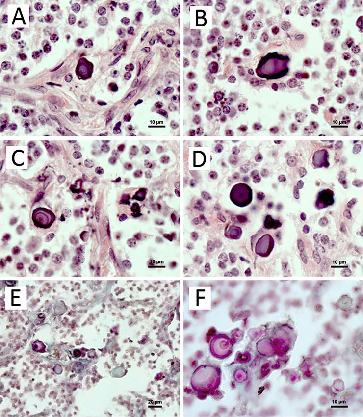
92Fig 1. Calcified concretions in the pineal gland of adult blue fox.
Structure: (a), (c), (f) are laminar concretions with alternative layers of light- and dark-stained rings; (b) are
hollow-like ones; (d), (e), (f) are acervuli without clear laminar structure.
(a–d) H&E, (e–f) Masson-Goldner staining. Scale bar is 10 μm
xylapatite displayed a narrow band at ~ 3572 cm‑1, mens was in the 958 cm-1 position and had a band
which corresponded to the O-H stretch for hydroxyl width of FWHM = 18 cm-1. The low-intensity broad
in the channel site of apatite structure and broad band was detected at 1056 cm-1. It should probably
band centered at about 3400 cm‑1, indicative be attributed to the combination of apatite band
of molecular water [Pasteris et al., 2014]. The Ra- at 1044 cm-1 and carbonate band at 1075 cm-1
man spectra of calcium concretions were con- [Karampas, Kontoyannis, 2013]. Additionally, Ra-
sistent with hydroxylapatite, although the spec- man spectra of calcified concretions displayed
tra were “poor” compared to the synthetic one two broad bands centered at 3476 and 3700 cm‑1,
(Fig. 4). Only two peaks were detected in the Ra- mainly corresponding to adsorbed and crystallo-
man spectra of studied specimens, which cor- graphically incorporated water (Fig. 4). No bands
responded to the symmetric P-O stretch for PO4 indicative of carbonate substitution for phosphate
tetrahedra. The most intensive peak of apatite de- in apatite were recorded in the Raman spectra
tected in the Raman spectra of the studied speci- of the calcified concretions. However, it has been
93Fig. 2. Secondary electron (SE) images of mulberry-like (a, b) and irregular elongate (c, d) pineal gland
concrements. Scale bar is 5 μm
established [Thomas et al., 2011] that the posi- recorded total water content. Therefore, the Ra-
tion and width (FWHM) of the band correspond man data suggest that the studied calcified con-
to the symmetric stretching mode ν1 – PO4 cor- cretions are composed of the carbonated hyd-
related with the hydroxylapatite composition, in- roxylapatite. Hydroxylapatite was recognized
cluding the carbonate content. Synthetic hydro- in a majority of the studied calcified concretions.
xylapatite without impurities was characterized It was represented by both “mulberry-like” struc-
by Raman parameters including the Raman fre- tures and elongate irregular-shaped particles with
quencies of 960 cm−1 and FWHM = 9.2 cm−1 [Tho- the size ca. 3–25 mm.
mas et al., 2011]. Hydroxylapatite from the con- Raman spectroscopy was used to distinguish
crements was characterized by a slightly lower between calcium carbonate or oxcalate minerals
band position (958 cm-1) and a significantly higher in the calcified concretions of blue fox pineal gland.
band width FWHM = 18 cm-1. These spectroscopic Raman spectroscopic data revealed that the calci-
features are specific to apatite with the carbona- fied concretions composed of calcium and carbon
te substitution in PO4 tetrahedra [Thomas et al., were represented only by calcite. It was identified
2011]. Another evidence of phosphate substitution by the characteristic bands at 712 and 1088 cm-1
by carbonates comes from the relatively high total (Fig. 5). Calcite was found in all the studied cal-
water content marked by broad bands at around cified concretions. In the calcified concretions
3500 cm-1 detected for the calcified concretions. composed of both calcite and hydroxylapatite,
According to Pasteris et al. [2014], the increase the number of calcite grains appeared to be higher
in carbonate concentration within the apatite is than the number of hydroxylapatite grains. In con-
correlated with an increase in spectroscopically trast to hydroxylapatite, calcite was represented
94Fig. 3. Secondary electron (SE) images (1) and elemental energy dispersive spectra (2) of pineal gland concre-
ments composed of apatite (a) and calcite (b). Dots indicate EDS measurement location. Scale bar is 5 μm
only by the “mulberry-like” structure with the size Admassie, Mekonnen, 2009; Bulc et al., 2010] de-
ca. 2–13 mm. spite several irrelevant cases [Tapp, Huxley, 1972;
Maślińska et al., 2010].
Discussion The calcified concrements were located
in the parenchyma of distal part of the blue fox
The reported findings give a first insight into gland, in the capsule surrounding it and in the pro-
the morphology, mineral and chemical properties truding septae. Our results are in agreement with
of pineal calcified concretions in blue fox. data of Bulc et al. [2010], who also observed
The brain sand is the one of the most intrigu- the concretions in the pineal capsule and pa-
ing structure in the pineal gland, which is able renchyma of blue fox. Other authors revealed
to form the brain sand due to the high calcium con- the differences in structure of capsular concre-
tent and the high phosphate turnover compared ments and of parenchymal ones. The first show
to other tissues [Borell, Örström, 1947; Vígh et al., clear concentric lamination, but the second have
1989]. However, the biological significance of pi- the globular structure. As the capsule and septae
neal calcium concrements is still unknown. of the pineal are formed by the arachnoid and pia
We detected the presence of pineal acervuli mater sheets of the meninges, the concre-
in both juveniles and adults blue fox. However, pre- ments of the capsule and septae are the example
viously Bulc et al. [2010] revealed the brain sand of meningeal calcification [Vígh et al., 1998].
in the pineal of blue foxes aged 3 years, but not Similarly, acervuli from the capsule in our study
in those aged 7–8 months. The relevance of con- had laminated structure, but most of the concre-
cretions to aging is still arguable; in general views ments formed in the pineal parenchyma also had
their incidence and amount are believed to in- alternative layers of light- and dark-stained rings.
crease with age [Schmid, 1993; Mori et al., 2003; Others were strongly dark-stained, and the third
95Fig. 4. Raman spectra of commercial hydroxylapatite (a) [modified after Pasteris et al.,
2014] and hydroxylapatite from pineal gland concrements (b)
Fig. 5. Raman spectra of standard calcite sample (a) [RRUFF…] and calcite from pineal
gland concrements (b)
group were light-stained and seemed to have of pineal calcified concretions have been predo-
hollow structure (in sections stained with H&E). minately revealed in humans [Krstić, 1976; Bocchi,
However, acervuli in samples stained according Valdre, 1993; Kodaka et al., 1994; Nakamura et al.,
to the Masson-Goldner method were colored pink, 1995; Bacconier, Lang, 2004]. Such data for other
light green or both, some of them had black rings, mammalian species are scarce [Krstić, Golaz, 1977;
probably indicating differences in the composition Humbert, Pévet, 1995; Tofail et al., 2019]. Identifi-
of the concrements. cation of the mineral composition of pineal calcified
Although studies of the pineal gland have a long concretions is often challenging due to the small size
history, the mineral and chemical compositions and low bioavailability of these minerals. The SEM-
96EDS and Raman spectroscopy data obtained for Overviewing the data available about pineal cal-
the blue fox pineal indicate that pineal calcified con- cification, one can conclude that a multifactorial
cretions are made up of hydroxylapatite and calcite. mechanism may be responsible for its formation
Hydroxylapatite is characterized by the average [Vígh et al., 1998]. Moreover, the nature and crys-
Ca/P ratio of 1.36, which is similar to the compo- tallinity of the inorganic tissue of pineal concretions
sition variations in bioapatites [Frank-Kamenets- give reason to assume that the corpora arenacea
kaya et al., 2011; Combes et al., 2016]. The iden- is of a physiological rather than pathological ossi-
tification of apatite in pineal concretions from blue fication type, with characteristics between enamel
fox is in agreement with the data reported by other and dentine, but with more marked analogies to-
researchers, who revealed that acervuli are mainly wards the latter [Bocchi, Valdre, 1993].
built up from hydroxyapatite [Bocchi, Valdre, 1993;
Kodaka et al., 1994]. It should be emphasized that Conclusions
the morphology of the mulberry-like structure is si-
milar for both blue fox pineal gland, as it is shown The reported findings give a first insight into
in the present study, and for human pineal gland the morphology, mineral and chemical composition
[Kodaka et al., 1994]. However, hydroxylapati- of pineal calcium concrements in blue fox. The re-
te in the calcified concretions from blue fox pineal sults suggest that the process of pineal gland mi-
gland occurred not only as mulberry-like structure, neralization is most likely not age-related. Our data
but also as elongate irregular-shaped particles with concerning the location and mineral composition
larger size of up to 25 mm (Fig. 2). of calcium concretions in blue fox pineal gland
The SEM-EDS and Raman spectroscopy stu- are in agreement with those obtained by other re-
dies of calcified concretions from blue fox pineal searchers on rodent and human pineal glands.
gland showed that the additional mineral phase Calcified concretions were located in the capsule,
in the pineal gland was calcite. The data are protruding septae and parenchyma of pineal gland.
in agreement with those obtained for the pineal Two morphological types of concrements were dis-
gland of humans [Bacconier, Lang, 2004] and rats tinguished, including the mulberry-like and the ir-
[Tofail et al., 2019]. However, other polymorphs regular elongate structures. The acervuli of mulber-
of calcium carbonate, namely vaterite and arago- ry-like structure were made up of hydroxylapatite
nite, have also been recognized in the pineal gland and calcite, and the irregular elongate aggregates
of rats [Tofail et al., 2019]. Calcite in calcified con- were composed of hydroxylapatite only. The lat-
cretions from blue fox pineal gland appeared as ter had not been previously recorded from calci-
mulberry-like structure smaller that the elongate fied concretions from mammals. The data report-
irregular-shaped hydroxyapatite particles. ed in this study contribute to the understanding
The data concerning the mineral composi- of the calcification mechanism in the pineal gland.
tion of calcium concretions in blue fox pineal are
in agreement with those obtained by other re- The authors are grateful to Director of FSUE
searchers on rodent and human gland [Kodaka “Russian Sable” V. L. Shevyrkov, to Z. S. Ruchki-
et al., 1994; Bacconier, Lang, 2004; Tofail et al., na and to the Head of the Laboratory of Ecologi-
2019]. The presence of two different mineral cal Biochemistry of IB KarRC RAS S. A. Murzina
phases in the same organ is considered to be bio- for help in conducting the research. We also thank
logically significant [Bacconier, Lang, 2004; Tofail G. L. Kuranov for the methodology discussion.
et al., 2019], but the mechanism of biomineraliza- The authors have no conflicts of interest that are
tion remains unknown. directly relevant to the contents of this manuscript.
To date, there is no clear understanding of how The study was carried out under state order
and why calcium concretions form, but some (project № 0218‑2019‑0073).
mechanisms for their formation are suggested.
Among them the impairment of Ca2+-dependent References
ATPase, changes in calcium channels, or a con-
stant depolarization of pinealocytes calcium pomp Admassie D., Mekonnen A. Incidence of normal
leading to elimination of calcium out of the cell pineal and choroids plexus calcification on Brain CT
[Krstić, 1986]; death or degeneration of pine- (computerized tomography) at Tikur Anbessa Teaching
Hospital Addis Ababa, Ethiopia. Ethiopian Med. J. 2009.
alocytes, resulting in an overall decrease in pine-
Vol. 47. P. 55–60.
al activity [Schmid, 1993; Humbert, Pévet, 1995], Baconnier S., Lang S. B. Calcite microcrystals in the
and active participation of tryptase mast cells pineal gland of the human brain: Second harmonic ge-
in the pineal calcification process as sites where it nerators and possible piezoelectric transducers. IEEE T.
starts [Maślińska et al., 2010]. Dielect. El. In. 2004. Vol. 11. P. 203–209.
97Bocchi G., Valdre G. Physical, chemical, and mine- culi and age in human adult males. Anat. Sci Int. 2003.
ralogical characterization of carbonate-hydroxyapatite Vol. 78. P. 181–184.
concretions of the human pineal gland. J. Inorganic Bio- Nakamura K. T., Nakahara H., Nakamura M., Tokio-
chem. 1993. Vol. 49. P. 209–220. ka T., Kiyomura H. Ultrastructure and x-ray microanalyti-
Borell U., Örström Å. The turnover of phosphate cal study of human pineal concretions. Ann. Anat. 1995.
in the pineal body compared with that in other parts Vol. 177. P. 413–419.
of the brain. Biochem. J. 1947. Vol. 41. P. 398–403. Pasteris J. D., Yoder C. H., Wopenka B. Molecular
Bulc M., Lewczuk B., Prusik M., Gugolek A., Przy- water in nominally unhydrated carbonated hydroxylapa-
bylska-Gornowicz B. Calcium concrements in the pineal tite: The key to a better understanding of bone mineral.
gland of the Arctic fox (Vulpes lagopus) and their rela- Am. Miner. 2014. Vol. 99. P. 16–27.
tionship to pinealocytes, glial cells and type I and III col- RRUFF database of Raman spectroscopy, X-ray dif-
lagen fibers. Polish J. Vet. Sci. 2010. Vol. 13. P. 269–278. fraction and chemistry of minerals. URL: https://rruff.info
Carden A., Morris M. D. Application of vibrational (accessed: 20.02.2021).
spectroscopy to the study of mineralized tissues (re- Schmid H. A. Decreased melatonin biosynthesis, cal-
view). J. Biomed. Optics. 2000. Vol. 5. P. 259–268. cium flux, pineal gland calcification and aging: a hypo-
Combes C., Cazalbou S., Rey C. Apatite biominerals. thetical framework. Gerontol. 1993. Vol. 39. P. 189–199.
Minerals. 2016. Vol. 6, no. 2. P. 34. Tan D. X., Xu B., Zhou X., Reiter R. J. Pineal cal-
Frank-Kamenetskaya O., Kol’tsov A., Kuz’mina M., cification, melatonin production, aging, assocated
Zorina M., Poritskaya L. Ion substitutions and non-stoi- health consequences and rejuvenation of the pineal
chiometry of carbonated apatite- (CaOH) synthesised by gland. Molecules. 2018. Vol. 23. Art. 301. doi: 10.3390/
precipitation and hydrothermal methods. J. Mol. Struc- molecules23020301
ture. 2011. Vol. 992. P. 9–18. Tapp E., Huxley M. The histological appearance
Gilbert P., Abrecht M., Frazer B. H. The organic-mi- of the human pineal gland from puberty to old age.
neral interface in biominerals. Rev. Mineral. Geochem. J. Pathol. 1972. Vol. 108. P. 137–144.
2005. Vol. 59. P. 157–185. Thomas D. B., McGoverin C. M., Fordyce R. E.,
Humbert W., Pévet P. Permeability of the pineal Frew R. D., Gordon K. C. Raman spectroscopy of fossil
gland of the rat to lanthanum: Significance of dark pine- bioapatite – A proxy for diagenetic alteration of the oxy-
alocytes. J. Pineal Res. 1992. Vol. 12. P. 84–88. gen isotope composition. Palaeogeogr. Palaeoclimatol.
Humbert W., Pévet P. Calcium concretions in the pi- Palaeoecol. 2011. Vol. 310, no. 1–2. P. 62–70.
neal gland of aged rats: an ultrastructural and microana- Timlin J. A., Carden A., Morris M. D. Chemical micro-
lytical study of their biogenesis. Cell Tissue Res. 1995. structure of cortical bone probed by Raman transects.
Vol. 279. P. 565–573. App. Spectr. 1999. Vol. 53. P. 1429–1435.
Karampas I. A., Kontoyannis C. G. Characterization Tofail S. A. M., Mouras R., McNamara K., Patyk-
of calcium phosphates mixtures. Vib. Spectrosc. 2013. Kazmierczak E., Geaney H., Zaworotko M., Ryan K. M.,
Vol. 64. P. 126–133. Soulimane T., Silien C., Kopáni M. Multimodal surface
Kim J., Kim H. W., Chang S., Kim J. W., Je J. H. analyses of chemistry and structure of biominerals in ro-
Growth patterns for acervuli in human pineal gland. Sci. dent pineal gland concretions. Appl Surf. Sci. 2019.
Rep. 2012. Vol. 2. P. 984–988. Vol. 469. P. 378–386.
Kodaka T., Mori R., Debari K., Yamada M. Scanning Turgut A. T., Karakaş H. M., Özsunar Y., Altın L.,
electron microscopy and electron probe microanalysis Çeken K., Alıcıoğlu B., Sönmez İ., Alparslan A., Yürü-
studies of human pineal concretions. Microscopy. 1994. mez B., Çelik T., Kazak E., Geyik P. Ö., Koşar U. Age-re-
Vol. 43. P. 307–317. lated changes in the incidence of pineal gland calcifi-
Krstić R. A combined scanning and transmission cation in Turkey: A prospective multicenter CT study.
electron microscopic study and electron probe micro- Pathophysiol. 2008. Vol. 15. P. 41–48.
analysis of human pineal acervuli. Cell Tissue Res. 1976. Vígh B., Vígh-Teichmann I., Heinzeller T., Tutter I.
Vol. 174. P. 129–137. Meningeal calcification of the rat pineal organ. Histo-
Krstić R. Pineal calcification: its mechanism and signifi- chem. 1989. Vol. 91. P. 161–168.
cance. J. Neural Transmis. Supp. 1986. Vol. 21. P. 415–432. Vígh B., Szél A., Debreceni K., Fejér Z., e Silva M. M.,
Krstić R., Golaz J. Ultrastructural and X-ray micro- Vígh-Teichmann I. V. Comparative histology of pineal cal-
probe comparison of gerbil and human pineal acervuli. cification. Histol. Histopathol. 1998. Vol. 13. P. 851–870.
Experientia. 1977. Vol. 33. P. 507–508. Vollrath L. The pineal organ. Oksche A. and Voll-
Lewinski A., Vaughan M. K., Champney T. H. Dark- rath L. (eds) Handbuch Der Mikroskopischen Anatomie
exposure increases the number of pineal concretions Des Menschen vol 7. Springer, Berlin, Heidelberg, New
in male gerbils (Meriones unguiculatus). IRCS Med. Sci. York, 1981.
1983. Vol. 11. P. 977–978. Whitehead M. T., Oh C., Raju A., Choudhri A. F.
Luke J. Fluoride deposition in the aged human pineal Physiologic pineal region, choroid plexus, and dural cal-
gland. Caries Res. 2001. Vol. 35. P. 125–128. cifications in the first decade of life. Am. J. Neuroradiol.
Maślińska D., Laure-Kamionowska M., Deręgow- 2015. Vol. 36 (3). P. 575–580. doi: 10.3174/ajnr. A4153
ski K., Maśliński S. Association of mast cells with calci- Zimmerman R. A., Bilaniuk L. T. Age-related inci-
fication in the human pineal gland. Folia Neuropathol. dence of pineal calcification detected by computed to-
2010. Vol. 48. P. 276–282. mography. Radiol. 1982. Vol. 142. P. 659–662.
Mori R., Kodaka T., Sano T. Preliminary report on the
correlations among pineal concretions, prostatic cal- Received March 20, 2021
98СВЕДЕНИЯ ОБ АВТОРАХ: CONTRIBUTORS: Калинина Светлана Николаевна Kalinina, Svetlana заведующая лаб. экологической физиологии животных, Institute of Biology, Karelian Research Centre, к. б. н. Russian Academy of Sciences Институт биологии КарНЦ РАН, 11 Pushkinskaya St., 185910 Petrozavodsk, Karelia, Russia Федеральный исследовательский центр e-mail: cvetnick@yandex.ru «Карельский научный центр РАН» tel.: +79114241881 ул. Пушкинская, 11, Петрозаводск, Республика Карелия, Россия, 185910 эл. почта: cvetnick@yandex.ru тел.: +79114241881 Чаженгина Светлана Юрьевна Chazhengina, Svetlana старший научный сотрудник лаб. геохимии, четвертичной Institute of Geology, Karelian Research Centre, геологии и геоэкологии, к. г.‑м. н. Russian Academy of Sciences Институт геологии КарНЦ РАН, 11 Pushkinskaya St., 185910 Petrozavodsk, Karelia, Russia Федеральный исследовательский центр e-mail: chazhengina@mail.ru «Карельский научный центр РАН» tel.: +79116601533 ул. Пушкинская, 11, Петрозаводск, Республика Карелия, Россия, 185910 эл. почта: chazhengina@mail.ru тел.: +79116601533 Илюха Виктор Александрович Ilyukha, Victor директор ИБ КарНЦ РАН, главный научный сотрудник Institute of Biology, Karelian Research Centre, лаб. экологической физиологии животных, д. б. н. Russian Academy of Sciences Институт биологии КарНЦ РАН, 11 Pushkinskaya St., 185910 Petrozavodsk, Karelia, Russia Федеральный исследовательский центр e-mail: ilyukha@bio.krc.karelia.ru «Карельский научный центр РАН» ул. Пушкинская, 11, Петрозаводск, Республика Карелия, Россия, 185910 эл. почта: ilyukha@bio.krc.karelia.ru Светов Сергей Анатольевич Svetov, Sergey директор ИГ КарНЦ РАН, руководитель и главный Institute of Geology, Karelian Research Centre, научный сотрудник лаб. геохимии, четвертичной Russian Academy of Sciences геологии и геоэкологии, д. г.‑м. н., старший научный 11 Pushkinskaya St., 185910 Petrozavodsk, Karelia, Russia сотрудник, проф. e-mail: ssvetov@krc.karelia.ru Институт геологии КарНЦ РАН, Федеральный исследовательский центр «Карельский научный центр РАН» ул. Пушкинская, 11, Петрозаводск, Республика Карелия, Россия, 185910 эл. почта: ssvetov@krc.karelia.ru Хижкин Евгений Александрович Khizhkin, Evgeny старший научный сотрудник лаб. экологической Institute of Biology, Karelian Research Centre, физиологии животных, к. б. н. Russian Academy of Sciences Институт биологии КарНЦ РАН, 11 Pushkinskaya St., 185910 Petrozavodsk, Karelia, Russia Федеральный исследовательский центр e-mail: hizhkin84@mail.ru «Карельский научный центр РАН» ул. Пушкинская, 11, Петрозаводск, Республика Карелия, Россия, 185910 эл. почта: hizhkin84@mail.ru
You can also read