Multi-scale Regional Attention Deeplab3+: Multiple Myeloma Plasma Cells Segmentation in Microscopic Images
←
→
Page content transcription
If your browser does not render page correctly, please read the page content below
Multi-scale Regional Attention Deeplab3+:
                                           Multiple Myeloma Plasma Cells Segmentation in
                                                         Microscopic Images

                                                  Afshin Bozorgpour1,2∗ , Reza Azad1,2∗ , Eman Showkatian2 , and Alaa
                                                                              Sulaiman2†
arXiv:2105.06238v1 [eess.IV] 13 May 2021

                                                               1
                                                                   Sharif University of Technology, Tehran, Iran
                                                                    2
                                                                      BmDeep Group, contact@bmdeep.com

                                                   Abstract. Multiple myeloma cancer is a type of blood cancer that hap-
                                                   pens when the growth of abnormal plasma cells becomes out of control in
                                                   the bone marrow. There are various ways to diagnose multiple myeloma
                                                   in bone marrow such as complete blood count test (CBC) or counting
                                                   myeloma plasma cell in aspirate slide images using manual visualization
                                                   or through image processing technique. In this work, an automatic deep
                                                   learning method for the detection and segmentation of multiple myeloma
                                                   plasma cell have been explored. To this end, a two-stage deep learning
                                                   method is designed. In the first stage, the nucleus detection network is
                                                   utilized to extract each instance of a cell of interest. The extracted in-
                                                   stance is then fed to the multi-scale function to generate a multi-scale
                                                   representation. The objective of the multi-scale function is to capture
                                                   the shape variation and reduce the effect of object scale on the cyto-
                                                   plasm segmentation network. The generated scales are then fed into a
                                                   pyramid of cytoplasm networks to learn the segmentation map in vari-
                                                   ous scales. On top of the cytoplasm segmentation network, we included
                                                   a scale aggregation function to refine and generate a final prediction.
                                                   The proposed approach has been evaluated on the SegPC2021 grand-
                                                   challenge and ranked second on the final test phase among all teams.

                                                   Keywords: Myeloma Plasma Cell · Segmentation · Attention Deeplabv3+
                                                   · Deep Learning · SegPC2021 · Grand Challenge.

                                           1     Introduction
                                           Cancer happens when the cells start to grow out of control and spread to healthy
                                           surrounding tissue. Myeloma, also known as multiple myeloma, is a type of
                                           blood cancer that arises from plasma cells in the bone marrow [12, 19]. More
                                           specifically, bone marrow is a kind of soft tissue found inside some part of larger
                                           bones in human body. Different types of blood cells such as red blood cells,
                                                 Support: This work is fully supported by BmDeep Group.
                                               † To whom correspondence should be addressed: E-mail: alaa@bmdeep.com.
                                               ∗ These authors contributed equally to this work.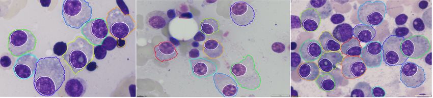
2 A. Bozorgpour et al.
white blood cells, and platelets are made in the bone marrow [13]. Plasma cells
developed by the B lymphocytes (type of white blood cells) form part of the
body’s immune system. In order to fight the infections, antibodies, also known
as immunoglobulin, are produced by normal plasma cells. In myeloma cancer
plasma cells crow in the bone marrow in a way there is no space for normal
red cells, white cells, and platelets. Myeloma begins to develop when the DNA is
damaged or changed during the production of new plasma cells. These abnormal
plasma cells (myeloma cells) will spread in a different part of bone marrow
and produce more abnormal cells. Myeloma cells will produce a large number
of paraproteins (type of antibody) which are useless and unable to fight the
infections [6]. Unlike other cancers, myeloma will not form a tumor or lump but
it will lead to the accumulation of abnormal plasma cells in the bone marrow
and paraproteins in the body. Multiple myeloma is referred to the situation when
myeloma cancers affect multiple parts of the body [1]. Since myeloma cell can
be differentiated from normal plasma cells based on histology and morphological
features, it is a common method to diagnose multiple myeloma cancer through
the aspirate slide images [15, 17, 18]. In this method, at first, blood samples
will be extracted from bone marrow by using the injection of the needle onto the
bone. The extracted blood sample will be transferred to a slide and stained using
hematoxylin and eosin. Abnormal plasma cells will be detected and marked using
manual microscopic visualization (sample is shown in figure 1). Finally, based
on the estimation of the normal plasma cells in bone marrow, the presence or
absence of myeloma cancer will be concluded [16].
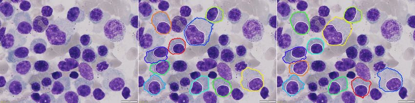
Fig. 1. Some samples of myeloma plasma cells microscopy images [10], where the my-
loma cancer cells are detected and highlighted with colored boundary.
Although manual inspection of stained slide images is a gold standard of
diagnosis of myeloma cancer, it is time-consuming and prone to inter and in-
traobserver variation. These limitations could be compensated by the use of
advanced digital image processing techniques such as object detection and seg-
mentation. Automation of the abnormal plasma cell detection alongside expert
pathologist decision could lead to the reduction of diagnosis time and workload
of the pathologist. To this end, in this paper, we propose a deep network that
utilizes a pyramid of Attention Deeplabv3+ model in a regional-based manner to
segment each instance of a myeloma cancer cell. We utilize our approach on the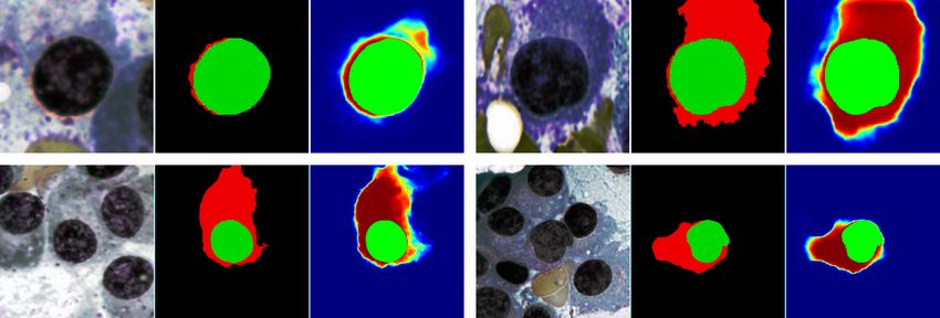
Multi-scale Regional Attention Deeplab3+        3

multiple mylomia plasma cell segmentation challenge which provided by [8–11].
Our contribution is summarized as follows:

 – Second ranking on the SegPC2021 challenge for multiple myeloma cancer
   cell segmentation.
 – Regional base instance segmentation approach.
 – guiding cytoplasm segmentation network with additional nucleus mask as a
   supervisory signal.

2   Related Work

Advanced image processing and machine learning methods such as image clas-
sification, object detection, and segmentation could have promising applications
in various medical domain [2, 3, 7]. Detection and segmentation of abnormal
cells in microscopic images have been proposed by several researchers in recent
works. For instance, Vyshnav et al. used a deep learning-based approach for
multiple myeloma cancer detection in stained microscopic images. They com-
pared the performance of Mask R-CNN and U-Net in segmentation and inferred
that Mask R-CNN has a very better performance than U-Net in myeloma cell
segmentation [23]. Authors of [21] used convolutional neural networks to classify
the normal and abnormal plasma cells in stained microscopic images. For the
classification task, they used AlexNet [14] to extract features from microscopic
images and then used Support Vector Machine (SVM) for the classifier. Their
novelty was the preprocessing stage where they used a median filter for each
R, G, and B color channel (on the importance of color space [5]) individually
and linear contrast stretching for the color enhancement. Then they compared
their result for classification with state of art techniques. They reported that
their networks reached 89% sensitivity for the cell classification. Vuola et al.
used Mask-RCNN and U-Net ensembled for nuclei segmentation in microscopic
images. They inferred that Mask-RCNN and U-Net have similar results on the
nucleus segmentation task. They reported that U-Net has better performance in
nucleus segmentation than Mask R-CNN in the term of similarity index. On the
other hand, the Mask R-CNN has better performance in the term of precision as-
sessment. Finally, they concluded that an ensembled model improves the model
performance in nucleus segmentation [22]. Saeedizadeh et al. used a bottleneck
algorithm, modified watershed, and SVM for myeloma cell detection in micro-
scopic images. At first, they separated the white blood cell from red blood cells
by use of the color normalization. After that the used thresholding technique to
separate the nucleus from the cytoplasm and by the use of watershed and bot-
tleneck algorithm they separated the connected cells. Finally, by using the series
of decision rules and the use of an SVM classifier they achieved the sensitivity of
96.52% and precision of 95.28% in recognition of myeloma cells [20]. Even though
the literature work gained promising results, their applicability in the multiple
myeloma cancer cell segmentation is limited due to the challenges such as over-
lap between cytoplasm’s of instances, the fuzzy boundary of the cytoplasm, and
4       A. Bozorgpour et al.

overlaying of one nucleus on another cytoplasm in microscopic images. To miti-
gate these limitations, we propose to regional attention deep model to segment
each cell with precise attention.

3     Methodology
A general diagram of the proposed structure is depicted in figure 2. The proposed
methods consist of two stages: in the first stage, the nucleus segmentation net-
work extracts all the nucleus instances. Then each instance fed into a multiscale
cytoplasm segmentation network. This network utilizes the Attention Deeplab3+
model to segment the cytoplasm area. In the next subsections, we will elaborate
on each part in more detail.

                                                                 3.0
                                                                        …       …
                                Multi-scale Instance Selection

                                                                                    Aggregation Function
                                                                 2.4
                                                                        …       …

                                                                                                           …
                                                                 1.75
                                                                        …       …

                                                                                …
                                                                            …
                                                                        …

                                                                 0.5    …       …

Fig. 2. Proposed regional Attention Deeplabv3+ model for multiple myeloma plasma
cells segmentation. The proposed method applies a U-net structure to learn the segmen-
tation map for each nucleus instance then it utilizes multi-scale attention deeplabv3+
model to generate the segmentation mask for cytoplasm.

3.1   Nucleus Segmentation
In the proposed architecture instead of jointly learning the segmentation of the
nucleus and cytoplasm mask, we utilize a two-stage strategy. Our main moti-
vation is to use the detected nucleus instance as a supervisory signal for the
cytoplasm instance segmentation to deal with overlapped areas. In other words,
the man objective of the first stage is to extract all the possible nucleus in-
stances from the input image. Then each extracted nucleus instance alongside
the cropped image patch is fed to the multi-scale instance selection function.
The instance selection function is simply an image cropping function with a pre-
defined scale. We use multi-scale to deal with varying cytoplasm scales. In figure
2, a sample of cropped nucleus instances with varying scale sizes (0.5 to 3.0) is
demonstrated. To learn the nucleus segmentation map we train a U-net model
using a nucleus annotation mask. It is worthwhile to mention that we include
the predicted nucleus instance alongside the cropped image as an input for the
Multi-scale Regional Attention Deeplab3+          5

cytoplasm segmentation network. The goal of this extra input is to guide the
network for the object of interest.

3.2         Cytoplasm Segmentation

In a regular auto-encoder decoder structure the encoder network consists of sev-
eral convolutions blocks followed by pooling operations to encode the object of
interest in high-level representation space. In this structure, due to the consec-
utive pooling operation, the spatial dimension of the network may considerably
decrease which can result in less discriminated representation power for objects
with varying scale. To mitigate this problem, the Deeplabv3+ model utilized an
atrous convolution structure. The atrous convolution applies a set of upsampled
convolutional kernels to describe the object of interest in higher receptive filed
size. To further improve the representation power of the Deeplab model, Azad
et al. [4] proposed a two-level add-on attention mechanism to extract more in-
formative features from the atrous convolutions. Where the first level attention
mechanism scales the representation space to highlight the more informative
channels then the second attention mechanism utilizes a 3D convolution kernel
between each atrous scale to learn a robust non-learn feature set. In this section,
we use the Attention Deeplabv3+ model to tackle the cytoplasm segmentation
problem. Cytoplasm boundary has a high overlap with the background area
and it requires careful attention to discriminate the cytoplasm boundary from
the background area. In our implementation, we fed the extracted nucleus area
alongside the image batch to the model to learn the instance segmentation mask.
We also apply the image histogram equalization method to normalize samples.
Sample of the estimated masks for the given nucleus instance is depicted in figure
3.

            Input   GT Mask   Estimation       Input       GT Mask     Estimation
Scale 1.0
Scale 2.6

Fig. 3. Segmentation results of the proposed method for both nucleus and cytoplasm
area. The cropped image alongside the predicted nucleus mask is fed to the cytoplasm
network to generate the instance cytoplasm segmentation.
6 A. Bozorgpour et al. 3.3 Aggregation function Learning objects of interest in multi-scale fashion can produce a robust segmen- tation mask. In this work, we apply the multi-scale technique on the input level. Consequently, the model generates a multi-scale segmentation mask. The main objective of this multi-scale technique is to tackle the problem of cytoplasm boundary. More specifically, the cytoplasm boundary has a non-rigid shape. If the boundary area is less then the model requires precise attention around the nucleus to separate it from the background. On the other hand, if the boundary is wast then the model needs to consider the big area to separate its boundary from other instances. We solve this limitation by defining several scales. Since the ultimate objective of the model is to produce a single segmentation mask for each instance, we propose to use an aggregation function to combine and select a single segmentation mask. The aggregation function can use the out- put of all scales to generate a single prediction (like non-maxima suppression), however, our experiment selection approach produced better performance. To perform this operation, we simply start from the lowest scale and calculate the relation between the detected cytoplasm area and the nucleus area. If the ratio is higher than a threshold value then the next scale is evaluated. The process goes through the next scales until finding the appropriate condition. 3.4 Training Procedure Our training procedure consists of two stages. In the first stage, we train the U- net model to segment the nucleus from the input images. The training process takes into account the training and validation set and learns the nucleus mask. We train the model for 100 epochs using the Adam optimization with a learning rate of 1e−4. In the second phase, we trained each Attention Deeplabv3+ model using the patches extracted from the input image alongside the nucleus mask (resulted from the nucleus segmentation network). For each scale, we train the model for 100 epochs using the Adam optimization with a learning rate of 1e−5. All training is done using cross-entropy loss on a single GTX 1080 GPU. 3.5 Inference Procedure The inference stage uses the trained models to generate the segmentation mask for both nucleus and cytoplasm instances. In our inference, we use a fixed number of scales (4 scales) for the test phase. 4 Results The proposed method is evaluated on multiple myeloma cell segmentation grand challenges which are provided by the SegPC 2021. The challenge data set consists of a training set with 290 samples, validation and test sets with 200 and 277 sam- ples respectively. All the samples are annotated by the pathologist and instance
Multi-scale Regional Attention Deeplab3+ 7
base segmentation masks are provided for the object of interest (myeloma plasma
cells). We trained our model using the training and validation set. During the
competition time, we generated the segmentation mask for each instance. The
challenge leader-board compared each team using the MIOU metric, where our
method ranked second among all teams. Table 1 shows the comparison results
for the top five winning teams.
Table 1. Performance comparison on the final test phase for SegPC2021 challenge.
Teams Score (mIoU)
XLAB Insights 0.9389
bmdeep 0.9385
DSC-IITISM 0.9382
507 0.9366
AIVIS 0.9276
As shown in table 1, the proposed method outperformed most of the com-
petitors and achieved the second-best place with a small gap (0.0004) from the
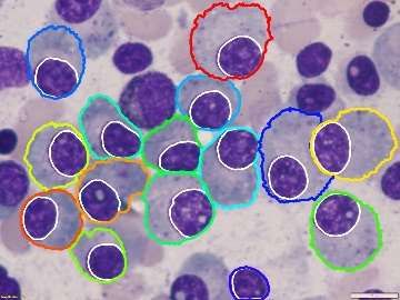
first team. Figure 4 demonstrates some prediction results where the proposed
method estimated both nucleus and cytoplasm masks with high performance.
Input Ground-Truth Estimation
Fig. 4. A sample of prediction results on SegPC2021 grand challenge.
As explained before the proposed method uses a multi-scale strategy to gen-
erate a precise segmentation mask for the cytoplasm instances. In this section,
we will elaborate on the scale selection strategy and its effect on the final per-
formance. To this end, we have extracted the statistical scale information from
the training set. The histogram information is depicted in figure 5.
According to figure 5, it is crystal clear that the ratio of cytoplasm area to
nucleus area is distributed in almost four different peaks. Thus, we select four
scales to generate a precise segmentation mask. It is worthwhile to mention that
we select the scale value a bit higher than the histogram peaks (shown with red
line on figure 5) to generate an image patch to cover the appropriate receptive
field size. In our experiment for the final test phase, we selected 4 different scales
as depicted in figure 2.8 A. Bozorgpour et al.
Fig. 5. Histogram of the area of cytoplasm to nucleus on the validation set.
5 Conclusion
In this paper, we proposed a multi-scale regional Attention Deeplabv3+ model
for myeloma plasma cell segmentation. The proposed method utilized a U-net
model for nucleus instance segmentation. The segmented nucleus instance is
extracted from the input image and alongside the predicted nucleus mask fed
into a multi-scale cytoplasm detector network. The cytoplasm detector took
into account the strength of the Attention Deeplabv3+ model to segment each
cytoplasm instance. We further proposed an aggregation function to select the
more related scale to fulfill the prediction score. Evaluation results on the final
challenge phase demonstrated outstanding results.
Acknowledgements. This work was fully supported by the BmDeep group. All
the implementation code is available: https://github.com/bmdeep/SegPC2021
References
1. Alexanian, R., Dimopoulos, M.: The treatment of multiple myeloma. New England
Journal of Medicine 330(7), 484–489 (1994)
2. Asadi-Aghbolaghi, M., Azad, R., Fathy, M., Escalera, S.: Multi-level context gating
of embedded collective knowledge for medical image segmentation. arXiv preprint
arXiv:2003.05056 (2020)
3. Azad, R., Asadi-Aghbolaghi, M., Fathy, M., Escalera, S.: Bi-directional convlstm
u-net with densley connected convolutions. In: Proceedings of the IEEE/CVF In-
ternational Conference on Computer Vision Workshops. pp. 0–0 (2019)
4. Azad, R., Asadi-Aghbolaghi, M., Fathy, M., Escalera, S.: Attention deeplabv3+:
Multi-level context attention mechanism for skin lesion segmentation. In: European
Conference on Computer Vision. pp. 251–266. Springer (2020)
5. Azad, R., Shayegh, H.R.: Novel and tuneable method for skin detection based on
hybrid color space and color statistical features. arXiv preprint arXiv:1407.6506
(2014)Multi-scale Regional Attention Deeplab3+ 9
6. Bird, J.M., Owen, R.G., D’Sa, S., Snowden, J.A., Pratt, G., Ashcroft, J., Yong, K.,
Cook, G., Feyler, S., Davies, F., et al.: Guidelines for the diagnosis and management
of multiple myeloma 2011. British journal of haematology 154(1), 32–75 (2011)
7. Feyjie, A.R., Azad, R., Pedersoli, M., Kauffman, C., Ayed, I.B., Dolz, J.: Semi-
supervised few-shot learning for medical image segmentation. arXiv preprint
arXiv:2003.08462 (2020)
8. Gehlot, S., Gupta, A., Gupta, R.: Ednfc-net: Convolutional neural network with
nested feature concatenation for nuclei-instance segmentation. In: ICASSP 2020-
2020 IEEE International Conference on Acoustics, Speech and Signal Processing
(ICASSP). pp. 1389–1393. IEEE (2020)
9. Gupta, A., Duggal, R., Gehlot, S., Gupta, R., Mangal, A., Kumar, L., Thakkar,
N., Satpathy, D.: Gcti-sn: Geometry-inspired chemical and tissue invariant stain
normalization of microscopic medical images. Medical Image Analysis 65, 101788
(2020)
10. Gupta, A., Gupta, R., Gehlot, S., Goswami, S.: Segpc-2021: Segmentation of mul-
tiple myeloma plasma cells in microscopic images. IEEE Dataport 1(1), 1 (2021)
11. Gupta, A., Mallick, P., Sharma, O., Gupta, R., Duggal, R.: Pcseg: Color model
driven probabilistic multiphase level set based tool for plasma cell segmentation in
multiple myeloma. PloS one 13(12), e0207908 (2018)
12. Guyton, A.C., Hall, J.E.: of medical (2006)
13. Hideshima, T., Mitsiades, C., Tonon, G., Richardson, P.G., Anderson, K.C.: Un-
derstanding multiple myeloma pathogenesis in the bone marrow to identify new
therapeutic targets. Nature Reviews Cancer 7(8), 585–598 (2007)
14. Krizhevsky, A., Sutskever, I., Hinton, G.E.: Imagenet classification with deep con-
volutional neural networks. Advances in neural information processing systems 25,
1097–1105 (2012)
15. Kyle, R.A., Remstein, E.D., Therneau, T.M., Dispenzieri, A., Kurtin, P.J., Hodne-
field, J.M., Larson, D.R., Plevak, M.F., Jelinek, D.F., Fonseca, R., et al.: Clinical
course and prognosis of smoldering (asymptomatic) multiple myeloma. New Eng-
land Journal of Medicine 356(25), 2582–2590 (2007)
16. Minges Wols, H.A.: Plasma cells. e LS (2001)
17. Nau, K.C., Lewis, W.D.: Multiple myeloma: diagnosis and treatment. American
family physician 78(7), 853–859 (2008)
18. Palumbo, A., Sezer, O., Kyle, R., Miguel, J., Orlowski, R., Moreau, P., Niesvizky,
R., Morgan, G., Comenzo, R., Sonneveld, P., et al.: International myeloma work-
ing group guidelines for the management of multiple myeloma patients ineligible
for standard high-dose chemotherapy with autologous stem cell transplantation.
Leukemia 23(10), 1716–1730 (2009)
19. Rajkumar, S.V., Dimopoulos, M.A., Palumbo, A., Blade, J., Merlini, G., Mateos,
M.V., Kumar, S., Hillengass, J., Kastritis, E., Richardson, P., et al.: International
myeloma working group updated criteria for the diagnosis of multiple myeloma.
The lancet oncology 15(12), e538–e548 (2014)
20. Saeedizadeh, Z., Mehri Dehnavi, A., Talebi, A., Rabbani, H., Sarrafzadeh, O., Vard,
A.: Automatic recognition of myeloma cells in microscopic images using bottleneck
algorithm, modified watershed and svm classifier. Journal of microscopy 261(1),
46–56 (2016)
21. Tehsin, S., Zameer, S., Saif, S.: Myeloma cell detection in bone marrow aspiration
using microscopic images. In: 2019 11th International Conference on Knowledge
and Smart Technology (KST). pp. 57–61. IEEE (2019)10 A. Bozorgpour et al.
22. Vuola, A.O., Akram, S.U., Kannala, J.: Mask-rcnn and u-net ensembled for nuclei
segmentation. In: 2019 IEEE 16th International Symposium on Biomedical Imaging
(ISBI 2019). pp. 208–212. IEEE (2019)
23. Vyshnav, M., Sowmya, V., Gopalakrishnan, E., VV, S.V., Menon, V.K., Soman,
K.: Deep learning based approach for multiple myeloma detection. In: 2020 11th
International Conference on Computing, Communication and Networking Tech-
nologies (ICCCNT). pp. 1–7. IEEE (2020)You can also read