Automated Segmentation of Vertebrae on Lateral Chest Radiography Using Deep Learning
←
→
Page content transcription
If your browser does not render page correctly, please read the page content below
Noname manuscript No.
                                          (will be inserted by the editor)

                                          Automated Segmentation of Vertebrae on Lateral
                                          Chest Radiography Using Deep Learning

                                          Sanket Badhe · Varun Singh · Joy Li · Paras Lakhani

                                          Received: date / Accepted: date
arXiv:2001.01277v1 [eess.IV] 5 Jan 2020

                                          Abstract The purpose of this study is to develop an automated algorithm
                                          for thoracic vertebral segmentation on chest radiography using deep learning.
                                          124 de-identified lateral chest radiographs on unique patients were obtained.
                                          Segmentations of visible vertebrae were manually performed by a medical
                                          student and verified by a board-certified radiologist. 74 images were used for
                                          training, 10 for validation, and 40 were held out for testing. A U-Net deep
                                          convolutional neural network was employed for segmentation, using the sum
                                          of dice coefficient and binary cross-entropy as the loss function. On the test
                                          set, the algorithm demonstrated an average dice coefficient value of 90.5 and
                                          an average intersection-over-union (IoU) of 81.75. Deep learning demonstrates
                                          promise in the segmentation of vertebrae on lateral chest radiography.

                                          Keywords Medical Imaging Segmentation · Deep Learning · Medical
                                          Imaging · vertebrae segmentation · Vertebral Body Compression Fractures ·
                                          Computer Vision

                                          Sanket Badhe
                                          Computer Science Department, Rutgers University
                                          E-mail: sanket.badhe@rutgers.edu
                                          Varun Singh
                                          Department of Radiology, Thomas Jefferson University
                                          E-mail: vxs039@jefferson.edu
                                          Joy Li

                                          E-mail: joy.li@jefferson.edu
                                          Paras Lakhani
                                          Department of Radiology, Thomas Jefferson University
                                          E-mail: M.D.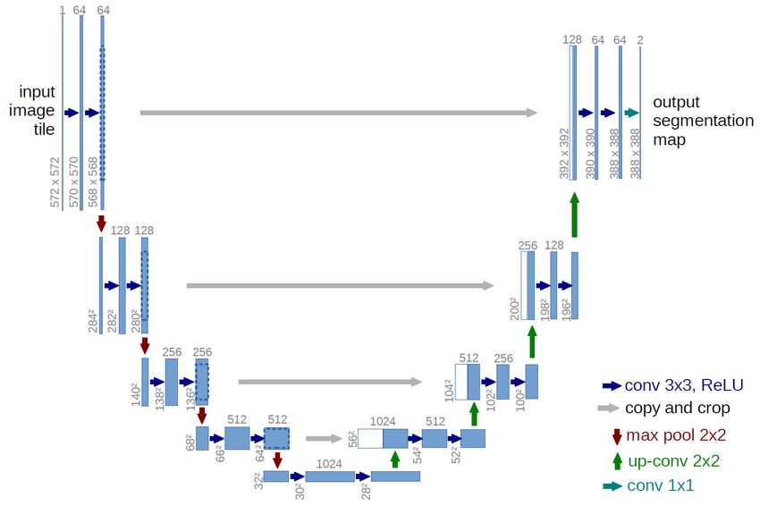
2                                                              Sanket Badhe et al.

1 Purpose

The purpose of this study is to develop an automated, validated algorithm
for vertebral body segmentation on lateral chest radiographs using deep
learning. Successful, automated segmentation of vertebral bodies could lead
to the detection of spinal fractures and the precise quantification of vertebral
body heights. Automated vertebral body height measurements appropriately
applied to a large, diverse dataset could stratify mean values and standard
deviations of vertebral heights by patient age, height, sex, and other clinical
parameters.

2 Introduction

In the United States, an estimated 750,000 new spinal compression fractures
occur every year. On routine lateral chest radiography, vertebral fractures
and compression deformities may be under-diagnosed and sometimes difficult
to appreciate. The progression of undetected fractures places patients at an
increased risk for complications associated with significant morbidity and
mortality [1] An automated, validated solution to the detection of vertebral
body compression fractures on lateral chest radiographs may facilitate early
diagnosis and allow for timely intervention to unburden patients of preventable
compression fracture sequela.
    Prior automated solutions for vertebral segmentation using traditional
computer aided detection (CAD) approaches, demonstrated modest results,
likely due to the sheer heterogeneity of patient anatomical variation. A study
performed by Mysling et al. outperformed previous vertebral segmentation
attempts but demonstrated a 52% error rate in the segmentation of fractured
vertebrae and a 10% overall vertebral segmentation error rate. The efficacy of
this approach was analyzed incompletely, but segmentation error was defined
in this study as >2 millimeters point-to-countor distance, a length metric
between the ground truth manual segmentation and the algorithm-based
segmentation. Wong et al. proposed a live fluoroscopic vertebral segmentation
strategy, but demonstrated no performance data to assess the technique’s real-
world performance. [2]
    Deep learning techniques have demonstrated success in pixel-level label-
ing tasks using semantic segmentation, which is the partition of an image
into unique parts or objects, such as identifying all cars or people within an
image. [3] In medical images, this has been applied to the segmentation of
pancreatic tumors on CT [4] , brain tumors on MRI [5], and stroke lesions
on MRI for example. [6] Some deep learning methods for semantic segmenta-
tion include fully convolutional neural networks (FCN) [7] convolutional au-
toencoders such as the U-Net [8] and DeepLab [9] . As opposed to semantic
segmentation, there also exist solutions using deep learning for instance seg-
mentation, where each object instance is identified within an image (e.g. car
1, car 2, car 3, etc...). For example, with regard to vertebrae, an instance seg-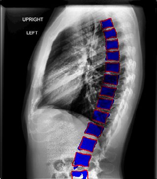
Title Suppressed Due to Excessive Length                                      3

mentation solution could identify each vertebral body such as T1, T2, T3 and
color-code them for example. Some popular instance segmentation solution
includes Mask-RCNN. [10]
    In this study, we choose to employ semantic segmentation using a standard
U-Net [8] , as it has shown to be relatively accurate with regard to segmen-
tation in medical imaging, including segmentation of brain tumors, kidneys
and pulmonary nodules. [11–13] The U-Net network architecture is structured
into an encoder and a decoder. The encoder follows the classic architecture
of the convolutional neural network, with convolutional blocks each followed
by a rectified linear unit (ReLU) and a max polling operation to encode im-
age features at different levels of the network. The decoder up-samples the
feature map with subsequent up-convolutions and concatenations with the
corresponding encoder blocks. This network style architecture helps to better
localize and extract image features and assembles a more precise output based
on encoder information.
    The model is first created using the training dataset by comparing the
training data with expected output to establish optimal weights with back-
propagation rules. Validation data is then used to establish the optimal number
of hidden units to verify a stopping point for the back-propagation algorithm
of the trained model essential for model selection. The test dataset is utilized
to establish the accuracy of the model from the fully trained final model
weights. The test and validation datasets are categorized independently to
ensure accuracy as the final model is biased toward the validation data used
to make final model selection.

Fig. 1 U-Net Architecture
4 Sanket Badhe et al.
In Figure 1, each blue box corresponds to a multi-channel feature map.
The number of channels is denoted on top of the box. The x-y-size is provided
at the lower left edge of the box. White boxes represent copied feature maps.
The arrows denote the different operations.
As vertebrae typically represent less than 20% of the total area in each lat-
eral radiograph, vertebral segmentation data are highly imbalanced. Previous
studies demonstrate that neural network performance deteriorates as class im-
balance increases. [14] The resulting imbalanced data model would be highly
trained for the features of the majority class but poorly trained for the features
of the minority class, causing instability in early training. Despite a probable
technically high accuracy from the generated model, the output results would
be poor segmentations.
A dice loss function is a type of loss function specifically designed to
mitigate dataset class imbalance and is frequently used for medical imaging
algorithms. [7] The dice score is measured as an overlap of the output mask
with ground truth to assess each segmentation task and is specifically designed
for use in volumetric segmentation in medical imaging. [7] The coefficient
measures the overlap between set X, the ground truth, and Y, the predicted
mask. For binary class segmentation, the dice score is expressed as the
following:
2 ∗ |X ∩ Y |
DiceScore =
|X| + |Y |
Intersection-over-union (IoU), also known as the Jaccard index, is a coefficient
that calculates overlap in segmentation tasks. [15] IoU also measures the
overlap between set X, the ground truth, and Y, the predicted mask. Some
studies indicate that IoU values typically exceed corresponding dice coefficients
except when coefficient values equal 0 or 1. [16] The IoU is calculated as
the intersection between two images divided by their union, expressed as the
following:
Intersection − over − union = |X∩Y |
|X∪Y |
If the ground truth and predicted mask are identical, then the dice
coefficient and IoU both equal 1. If the ground truth and predicted mask share
no elements, then both coefficients equal 0. If the ground truth and predicted
mask are neither identical nor absolutely incongruent, then each coefficient
value falls between 0 and 1.
3 Materials and Methods
An IRB-exempt study using 124 de-identified HIPAA-compliant lateral chest
radiographs on unique patients was performed. Images were pre-processed
using contrast-adaptive histogram equalization (CLAHE) to standardize the
appearance and improve image contrast. Images were subsequently down-
sampled from the original size (4238 × 3480 pixels) to 512 x 512 pixels using
bilinear interpolation. Down-sampling was performed to aid back-propagationTitle Suppressed Due to Excessive Length 5
and neural network learning within graphics processor unit (GPU) memory
constraints. Segmentations of visible vertebrae were manually performed using
ImageJ version 1.50i (National Institutes of Health, USA) by both a medical
student (JL) and a board-certified radiologist (PL). All segmentations of the
images were verified and adjusted as needed by a board-certified radiologist
(PL)
The resulting binary mask was additionally down-sampled to 512 x 512
pixels. Ground truth-labels were color-coded with black for vertebrae and
white for each image background, as seen in Figure 2. Class imbalance in
the vertebral segmentations was preempted in this study with the use of a
dice loss function.
Fig. 2 (a) Down-sampled lateral chest radiograph (b) Binary ground truth label with black
vertebrae and white background
The U-Net based convolutional neural network was employed to segment
vertebrae from lateral chest radiographs. 74 images (59.68%) were used for the
training dataset, 10 images (8.1%) were used for the validation dataset, and
40 images (32.25%) were used for the test dataset. The model was built using
Keras 2.06 (https://keras.io/) with TensorFlow 1.1 (Google LLC, Mountain
View, CA) and CUDA 8.1 (Nvidia Corporation, Santa Clara, CA).
Differential learning rates and optimizers were trialed to enhance update
weight and bias values to maximize model performance. Based on multiple
experiments, the best performance resulted from using the Adam optimizer
with a learning rate of 0.0001. The dice score and IoU were utilized on the
validation dataset to augment model selection. The model was trained until6 Sanket Badhe et al.
a plateau in validation loss, which occurred at 10 epochs on a CUDA-enabled
Nvidia 1080Ti 11GB graphics processing unit. (Nvidia Corporation, Santa
Clara, CA)
The dice score and IoU were used to assess model performance. As the
segmentation task was binary, loss function was applied as the summation of
dice score loss and binary cross entropy.
4 Results
In the holdout test dataset, an average dice coefficient value of 90.5 and an
average IoU of 81.75 were obtained. Ground truth masks, predicted masks,
and overlay segmentation masks were successfully generated. In Figure 3, the
ground truth mask represents the manual segmentation and the predicted
mask is the resulting output of the deep learning model.
Fig. 3 (a) Original image (b) Ground truth mask (c) Predicted mask
In the overlay-segmented mask seen in Figure 4, the original radiograph is
superimposed with the ground truth mask in red and the predicted mask in
blue.
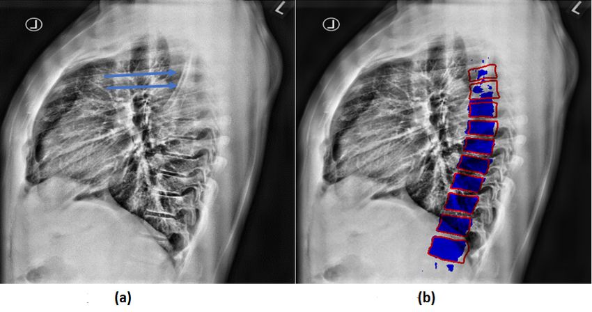
Figure 5 demonstrates an example of an inferior segmentation result. Two
adjacent thoracic vertebrae, T3 and T4, as indicated by the blue arrows, are
poorly visualized on the original radiograph and poorly segmented by the U-
Net model.
5 Discussion
The U-NET segmentation network resulted in a model with a dice score of
90.12 and an IoU of 82.10 for automated segmentation of vertebrae on lateral
chest radiography. These findings demonstrate that deep learning techniques
for pixel-wise segmentation may require a relatively small number of imagesTitle Suppressed Due to Excessive Length 7
Fig. 4 Overlay segmented mask
Fig. 5 (a) Original Image (b) Overlay segmented mask
to accurately segment vertebral bodies on radiography, compared to deep
learning for whole-image classification, which typically needs thousand or
more images per class. [17] This solution represents novel success with regard
to a vertebral segmentation algorithm on lateral chest radiographs though
there has been previous success in the classification and identification of spine
fractures on 3D CT. [18] Previous failures in automated radiographic vertebral
segmentation may be due to the comparatively low-resolution of radiographs
when compared with CT, especially in regard to patients with osteoporosis and
other conditions that limit vertebral visualization and background contrast.
While the U-NET was relatively successful in segmenting the thoracic
vertebre, there were some cases in which the segmentation failed or was less8 Sanket Badhe et al.
optimal. For example,Figure 5 demonstrates one lateral radiograph where
there was an improper segmentation of some of the upper thoracic vertebra.
This was most likely due to the poor contrast resolution and relatively
obscurity of the upper thoracic spine on that image related surrounding
structures . As there were few cases with complex low-level spinal features,
it is likely that a larger, more diverse dataset will improve the model’s ability
to train on more complex cases and apply that learning to complex test cases.
Because vertebrae typically represent a minority of the total pixels on
a lateral radiograph, there was a high risk for class imbalance between the
segmented vertebrae and background. To combat this risk of class imbalance,
the dice coefficient as a loss function was employed , and data augmentation
strategies and appropriate loss functions were utilized to mitigate model
overfitting. Though not explicitly assessed, it is likely that the use of the dice
coefficient and data augmentation strategies improved model performance.
One can consider other strategies to improve performance, including the
use of a U-Net variant with Inception-inspired architecture, other FCNs, or
RCNNs. [19] A larger, more clinically diverse dataset would also likely improve
performance.
In the future, we plan to use this model in conjunction with image
processing techniques that count the number of uniquely segmented vertebre
starting from the first readily visible vertebrae, likely from T3 or T4, and
ending at the most caudal vertebrae visible on the image. In the future, it
would be interesting to try an instance segmentation solution, such as a Mask
R-CNN where each vertebra is labeled and separately segmented (e.g. T3, T4,
T5, etc...). This has the advantage of a one-step deep learning solution that
can identify and segment each vertebra. [10]
As two-view chest radiography represents the most frequently utilized
imaging modality in the United States, an automated screening solution to
detect vertebral fractures would provide tremendous benefit to patients and
could mitigate systemic healthcare expenditure. [20]- [21] The retrospective
application of this algorithm to a large database of lateral chest radiographs
could also determine normative values for mean vertebral body height, strat-
ified by pertinent patient data, such as age, sex, height, weight and other
demographic and clinical parameters. This has implications to better individ-
ualize patient care and prevent future spinal fractures.
6 Conclusion
Deep learning using a U-NET demonstrates promise in the automated seg-
mentation of vertebrae on lateral chest radiographs.
References
1. Adams JE, Lenchik L, Roux C, Genant HK. Radiological as-
sessment of vertebral fracture. International osteoporosis founda-Title Suppressed Due to Excessive Length 9
tion vertebral fracture initiative resource document part II; 2010.
Available from: https://www.iofbonehealth.org/sites/default/files/PDFs/
Vertebral%20Fracture%20Initiative/IOF_VFI-Part_II-Manuscript.pdf.
2. Wong SF, Wong KYK, Wong WNK, Leong CYJ, undefined D K K Luk.
Tracking Lumbar Vertebrae in Digital Videofluoroscopic Video Automat-
ically . Medical Imaging and Augmented Reality. 2004;p. 154–162.
3. Long J, Shelhamer E, Darrell T. Fully convolutional networks for semantic
segmentation;.
4. Roth HR, Lu L, Farag A, Shin HC, Liu J, Turkbey EB, et al. DeepOrgan:
Multi-level Deep Convolutional Networks for Automated Pancreas Seg-
mentation. In: Medical Image Computing and Computer-Assisted Inter-
vention - MICCAI 2015. Cham: Springer International Publishing; 2015.
p. 556–564.
5. Pereira S, Pinto A, Alves V, Silva C. Brain Tumor Segmentation Using
Convolutional Neural Networks in MRI Images. IEEE Transactions on
Medical Imaging. 2016 03;35:1–1. Available from: 10.1109/TMI.2016.
2538465.
6. Yanran W, Aggelos KK, Xue W, Todd BP. A deep symmetry convnet for
stroke lesion segmentation. 2016 IEEE International Conference on Image
Processing (ICIP). 2016;p. 111–115.
7. Evan S, Jonathan L, Trevor D. Fully Convolutional Networks for Semantic
Segmentation. IEEE Trans Pattern Anal Mach Intell. 2017;39(4):640–651.
Available from: 10.1109/TPAMI.2016.2572683.
8. Ronneberger O, Fischer P, Brox T. U-Net: Convolutional Networks for
Biomedical Image Segmentation. CoRR. 2015;abs/1505.04597.
9. L C, G P, I K, K M, A LY. DeepLab: Semantic Image Segmentation
with Deep Convolutional Nets, Atrous Convolution, and Fully Connected
CRFs. IEEE Transactions on Pattern Analysis & Machine Intelligence.
2018 April;40(4):834–848. Available from: 10.1109/TPAMI.2017.2699184.
10. He K, Gkioxari G, Dollár P, Girshick R. Mask r-cnn; 2017. p. 2961–2969.
11. Çiçek Ö, Abdulkadir A, Lienkamp SS, Brox T, Ronneberger O. 3D U-Net:
Learning Dense Volumetric Segmentation from Sparse Annotation. In:
Medical Image Computing and Computer-Assisted Intervention - MICCAI
2016. Cham: Springer International Publishing; 2016. p. 424–432.
12. Dong H, Yang G, Liu F, Mo Y, Guo Y. Automatic Brain Tumor Detection
and Segmentation Using U-Net Based Fully Convolutional Networks; 2017.
p. 506–517. Available from: 10.1007/978-3-319-60964-5_44.
13. Tong G, Li Y, Chen H, Zhang Q, Jiang H. Improved U-NET network
for pulmonary nodules segmentation. Optik. 2018;174:460–469. Available
from: https://doi.org/10.1016/j.ijleo.2018.08.086.
14. Mazurowski MA, Habas PA, Zurada JM, Lo JY, Baker JA, Tourassi GD.
Training neural network classifiers for medical decision making: the effects
of imbalanced datasets on classification performance. Neural networks : the
official journal of the International Neural Network Society. 2007;21:427–
36.10 Sanket Badhe et al.
15. Jaccard P. The distribution of the flora in the alpine zone. New
Phytologist. 1912;2(37–50). Available from: https://doi.org/10.1111/j.
1469-8137.1912.tb05611.x.
16. Taha AA, Hanbury A, Hanbury A. Metrics for evaluating 3D medical
image segmentation: analysis, selection, and tool. BMC Med Imaging.
BMC medical imaging . 2015;15(29). Available from: 10.1186/s12880-015-
0068-x.
17. Deng J, Dong W, Socher R, Li LJ, Li K, Fei-Fei L. Imagenet: A large-scale
hierarchical image database. In: IEEE conference on computer vision and
pattern recognition. Ieee; 2009. p. 248–255.
18. Joseph EB, Jianhua Y, Ronald MS. Vertebral Body Compression Frac-
tures and Bone Density: Automated Detection and Classification on CT
Images. Radiology. 2017;284 3:788–797.
19. Fausto M, Nassir N, Seyed-Ahmad A. V-Net: Fully Convolutional Neural
Networks for Volumetric Medical Image Segmentation. 2016 Fourth
International Conference on 3D Vision (3DV). 2016;p. 565–571.
20. Dixon S. Diagnostic Imaging Dataset Statistical Release. NHS England .
2016;July:1–16.
21. Maehara C, Jacobson F, Andriole K, Khorasani R. Utilization Effect of
Integrating a Chest Radiography Room into a Thoracic Surgery Ward.
Journal of the American College of Radiology . 2012;9(6):421–425.You can also read