Mycobacterium tuberculosis Complex DNA from an Extinct Bison Dated 17,000 Years before the Present
←
→
Page content transcription
If your browser does not render page correctly, please read the page content below
MAJOR ARTICLE

Mycobacterium tuberculosis Complex DNA
from an Extinct Bison Dated 17,000 Years
before the Present
Bruce M. Rothschild,1,2,3,4 Larry D. Martin,4 Galit Lev,5 Helen Bercovier,5 Gila Kahila Bar-Gal,5 Charles Greenblatt,8
Helen Donoghue,5 Mark Spigelman,5 and David Brittain6
1
 Arthritis Center of Northeast Ohio, Youngstown, 2Department of Internal Medicine, Northeastern Ohio Universities College of Medicine,
Rootstown, Ohio; 3The Carnegie Museum, Pittsburgh; 4University of Kansas Museum of Natural History, Lawrence, Kansas; 5Department
of Bacteriology, Royal Free Hospital and University College London, London; 6Veterinary Sciences Division, Department of Agriculture
and Rural Development, Belfast; and 8Kuvin Center for the Study of Infectious and Tropical Diseases and Ancient DNA, Hadassah
Medical School, Hebrew University, Jerusalem

In order to assess the presence of tuberculosis in Pleistocene bison and the origin of tuberculosis in North
America, 2 separate DNA extractions were performed by 2 separate laboratories on samples from the metacarpal
of an extinct long-horned bison that was radiocarbon dated at 17,870  230 years before present and that
had pathological changes suggestive of tuberculosis. Polymerase chain reaction amplification isolated fragments
of tuberculosis DNA, which were sequenced, and on which spoligotyping was also performed to help determine
its relationship to the various members of the Mycobacterium tuberculosis complex. Extensive precautions
against contamination with modern M. tuberculosis complex DNA were employed, including analysis of pa-
leontologic and modern specimens in 2 geographically separate laboratories.

Recognition of tuberculosis-compatible pathology in                                   Clarification of the evolutionary history of infectious
bones of North American Pleistocene bovids has sug-                                diseases affords great opportunity for understanding
gested that tuberculosis was present from an early date                            the factors that have created current epidemiological
on that continent. This is now confirmed by the results                            patterns. Study of pathognomonic lesions in fossils has
of DNA sequencing for a bone sample from a bison                                   allowed progress in this endeavor [1–3], but for many
dated to 17,000 years before the present (BP). The pres-                           lesions a specific etiology cannot be determined. PCR
ence of similar pathology in fossil bighorn sheep and                              and application of DNA spoligotyping and sequencing
musk ox suggests that Mycobacterium tuberculosis com-                              techniques [4–8] allow that limitation to be tran-
plex organisms were widespread in bovids that immi-                                scended, but the very sensitivity of PCR raises the ad-
grated to North America over the Bering Strait con-                                ditional challenge of contamination [7, 8]. Contami-
nection and widespread during the late Pleistocene. It
                                                                                   nation of ancient by modern DNA is now sufficiently
suggests Holarctic distribution and that bovids were the
                                                                                   recognizable (and usually preventable) that confident
likely vector and reservoir for dispersion of what be-
                                                                                   diagnosis of ancient DNA is possible [7, 8].
came known as the white plague.
                                                                                      The resurgence of tuberculosis in recent years em-
                                                                                   phasizes the current limits to its control [9, 10]. Knowl-
                                                                                   edge of the evolutionary history and the antibiotic sus-
    Received 9 November 2000; electronically published 5 July 2001.
    Financial support: The Center for the Study of Emerging Diseases, Jerusalem.
                                                                                   ceptibilities of the M. tuberculosis complex may provide
  Reprints or correspondence: Dr. Bruce M. Rothschild, Arthritis Center of         insights that will lead to better control in the future.
Northeast Ohio, 5500 Market St., Youngstown, OH 44512 (bmr@neoucom.edu).           The issue is complex, however. The standard DNA
Clinical Infectious Diseases 2001; 33:305–11
                                                                                   probes for tuberculosis recognize what has been called
 2001 by the Infectious Diseases Society of America. All rights reserved.
1058-4838/2001/3303-0006$03.00                                                     the “M. tuberculosis complex” [11, 12], which consists

                                                                                      Origins of Tuberculosis in North America • CID 2001:33 (1 August) • 305
of M. tuberculosis, Mycobacterium bovis, Mycobacterium afri-           variably fatal and also keeps the temperature nearly constant
canum, and Mycobacterium microti [13]. The term “M. tuber-             at 4C–5C.
culosis complex” distinguishes this group of organisms from               During a survey of all of the approximately 40,000 bones
saprophytic mycobacteria (e.g., soil organisms) and atypical           recovered from Natural Trap Cave, a classic but relatively rare
mycobacteria such as Mycobacterium avium–intracellulare [14].          skeletal expression of granulomatous infection, involving un-
   Additional analysis is required to distinguish among the            dermining of subchondral surfaces, was noted in bovids (i.e.,
members of the M. tuberculosis complex. Spoligotyping clearly          bighorn sheep, extinct musk ox, and bison). These lesions were
distinguishes modern M. tuberculosis from modern M. bovis              absent in the very large horse and American pronghorn an-
and from non–M. tuberculosis complex mycobacteria [15].                telope samples from that site [31]. Radiological analysis revealed
   Documentation of M. tuberculosis complex in humans on               peri-erosive osteopenia (decrease in the bone density around
both sides of the Atlantic Ocean who lived 13 millennia ago            the involved area). The most likely etiologic agent of this pa-
[2, 16–18] has led to a great deal of speculation about its origins.   leopathology was M. tuberculosis.
Such work suggests that at least some of the previously reported          Samples and controls. Two separate samples were taken
presumptive diagnoses [16–30] were accurate. The presence of           from the undermined articular surface region of a metacarpal
M. tuberculosis complex on both sides of the Atlantic at such          of an extinct long-horned bison (Bison cf. antiquus) that was
early dates suggests that it either arrived with early settlers in     enclosed in sediments dated by radiocarbon analysis at
North America or was present in North America when they                17,870  230 years BP. Areas distinct from the pathological
arrived. The latter hypothesis is supported by suggestive evi-         lesion were also examined. Fossil controls consisted of lesion-
dence in the fossils of North American bovids [31].                    free bones from Canis species and Equus species.
   One of the classic findings in tuberculosis is the undermined          DNA extraction. Two separate DNA extractions were in-
subchondral articular surface [3, 32]. That finding, which is          dependently analyzed by 2 geographically separate laboratories.
relatively specific for the diagnosis of tuberculosis, is common       Cell lysis with guanidinium thiocyanate and DNA capture onto
in fossil bones from the Late Pleistocene in North America             silica was used on nondecalcified samples in the Jerusalem lab-
[31].                                                                  oratory and on decalcified samples in the London laboratory
   This study involved the use of molecular DNA techniques             [34–36]. Stringent precautions were taken at both laboratories
to identify the presence of M. tuberculosis complex in a skeletal      to avoid cross-contamination. DNA extraction and PCR were
specimen with lesions suggestive of tuberculosis from Natural          performed with standard precautions, including the use of
Trap Cave (Wyoming). That unique site was an unavoidable               ultraviolet-irradiated safety cabinets, ultrapure reagents, and
hazard on a game trail, providing an unbiased sampling of the          sterile disposables under full isolation procedures. Control sam-
many passing species. Identification of the DNA fragments was          ples that contained no bone material and bone material from
done by spoligotyping and direct DNA sequence determination.           nonlesional regions and from unaffected species were used to
Analysis was conducted at 2 separate laboratories (Department          assess contamination during the extraction and amplification
of Bacteriology, Royal Free Hospital and University College,           processes.
London, and Department of Parasitology, Hebrew University,                DNA amplification. Different PCR systems were used in
Jerusalem) to ensure the reproducibility of the findings and           the 2 laboratories. The Jerusalem laboratory used primers from
eliminate the possibility of contamination.                            the ribosomal protein S12 [5, 6] to amplify a 204-bp DNA
   Our results provide the first definitive diagnosis of M. tu-        fragment: forward 5-TCGTCGGGACAAGATCAGTAAG-3
berculosis complex in a fossil. They suggest that bovids were          (position 33–54) and reverse 5-ATCGAGTGCTCCTGCAGG-
the vectors that transported the primordial organism that              TTG-3 (position 216–236). In addition, a primer based on the
caused what we today call the white plague: tuberculosis.              insertion sequence IS6110 [37] was also used to amplify a region
                                                                       of 245 bp: forward 5-CGTGAGGGCATCGAGGTGGC-3 (po-
                                                                       sition 633–652) and reverse 5-GCGACGTAGGCGTCGGTGA-
METHODS                                                                CAAA-3 (primer position 880–858).
                                                                          The London laboratory used primers to amplify a smaller
Sample source. Bones were recovered in Wyoming from the                segment from within the IS6110. A 2-stage nested PCR was
Natural Trap Cave, a chamber nearly 30 m deep with a 4-m               used: the first reaction amplified a 123-bp region of IS6110
opening. The cave lies across an ancient game trail that leads         [14], and a second nested reaction amplified a 92-bp inner
to and from low-altitude pastures. For 1100,000 years it has           segment [38]. The latter segment was as follows: TTCGGACC-
trapped animals such as bears, wolves, foxes, wolverines, mar-         ACCAGCACCTAA (bases 777–796 of X17398, aka IS3) and
tens, cheetahs, lions, horses, mammoths, camels, sheep, musk           TCGGTGACAAAGGCCACGTA (bases 868–849 of X17398,
oxen, and bison [33]. The depth of the cave makes falls in-            aka IS4).

306 • CID 2001:33 (1 August) • Rothschild et al.
Amplification was performed in a solution of 20 mM of Tris- this technique to ancient specimens has been established by
HCL (pH, 7.5), 40 mM of NaCl, 2 mM of sodium phosphate, Taylor et al. [38].
0.1 mM of EDTA, 1 mM of dithiothreitol stabilizers (Sigma-
Aldrich), 50% (v/v) glycerol, 1.5 mM of MgCl2, 40 pM of each
primer, 0.2 mM each of nuclear triphosphates (MBI, Fermen- RESULTS
tas), and 1.25 U of Platinum Taq DNA polymerase (GibcoBRL
DNA recovery and sequencing. PCR amplification enabled
Life Technologies). Amplification consisted of initial denatur-
recovery of M. tuberculosis complex at both laboratories. Se-
ation at 94C for 2 min, followed by 39 cycles of 94C for 30
quencing revealed that the PCR product that was obtained
s, 61C for 45 s, 1 min at 72C, and, finally, 10 min at 72C.
belonged to a species in the M. tuberculosis complex. With use
DNA sequence analysis. The Jerusalem laboratory per-
of primers for the S12 ribosomal gene, the isolated gel bands
formed direct sequencing using the Termo-Sequenase kit
yielded a perfect match for a 163-bp fragment of the M. tu-
(Amersham), followed by agarose gel electrophoresis of the berculosis complex (of a possible 204 bp). IS6110 sequencing
PCR product. The band was cut from Nu-Sieve low-melting- revealed a base-pair substitution difference. The London lab-
point agar and used as such in the labeling mix. The London oratory 92-bp segment had a 12-bp section missing (822–832
laboratory removed the selected band from the gel and ex- of X17348) in the forward strand, but the flanking sequences
tracted it using a MERmaid Spin Kit (10–200 nucleotides; An- of 35 bp and 31 bp were identical to those in the database. A
achem). The purified DNA was then fluorescence-labeled with sequence for the reverse strand was obtained from bases
the ABI PRISM Dye Terminator Cycle sequencing kit with 782–853, including the missing region on the positive strand.
AmpliTaq DNA Polymerase, FS (PE Applied Biosystems). The The total length sequenced was 92 bp, which included the total
sequence was analyzed in an ABI PRISM 310 automated se- length between primers IS3 and IS4. Base discrepancy was spe-
quencer (PE Applied Biosystems). cifically noted in the Jerusalem laboratory at 798 (T for C).
Spoligotyping. To determine the species of Mycobacterium However, the sequence obtained in London agreed with that
within the M. tuberculosis complex, the spoligotyping system in the database.
(Isogen) was applied in the Jerusalem laboratory. “Spoligotyp- Extraction control samples and PCR control samples at both
ing” stands for “spacer-oligos.” The process is based on PCR laboratories yielded no tuberculosis DNA. For all the other
amplification of a series of 43 nonrepetitive short spacer se- animal bone samples and specimens from the affected bone at
quences (of between 36 and 41 bp), located between small locations away from the lesions, multiple extractions and PCR
repeats (DRs) in the DR locus of the Mycobacterium genome sequencing analyses found no evidence of tuberculosis DNA.
[39]. This system differentiates between modern M. bovis and Spoligotyping. Spoligotyping revealed that the Pleistocene
M. tuberculosis, as demonstrated by the array of the oligonu- bison lesions contained M. tuberculosis complex segments not
cleotide sequences attached to a membrane [15]. The spacers associated with modern M. bovis or M. microti [37]. The pres-
are arranged on the blot in the order in which they appear in ence of spacers 39, 40, 41, and 43 eliminates the possibility that
the H37Rv DR sequence (spacers 1–19, 22–32, and 37–43) and the DNA is from modern M. bovis [9, 10] or from the related
BCG [9, 10], and thus eliminates the possibility that the sample
the sequence of M. bovis BCG (spacers 20, 21, and 33–36).
was contaminated by DNA from a BCG-vaccinated individual.
Subsequent sequencing of the DR regions of other M. tuber-
On the basis of identification of the spoligotype pattern as a
culosis, M. bovis, M. microti, and Mycobacterium canetti isolates
43-digit binary number, the pattern can be assigned descriptive
has elucidated 65 novel spacer sequences (26 from M. canetti).
nomenclature: taking a negative as 0 and positive as 1 and
The order of spacers in all isolates is very well conserved, apart
dividing the pattern into 6 blocks of 7 or 8 digits results in 6
from a few duplications of spacers [5, 6, 40]. One primer is
blocks containing spacers 1–7, 8–14, 15–21, 22–28, 29–36, and
biotinylated, and the amplified products are hybridized under
37–43. The binary number in each block can then be converted
stringent conditions to the membrane furnished in the Isogen to the hexidecimal base. The result is a code of 6 ⫻ 2 digits
kit. The final readout is made exquisitely sensitive by the bind- [41]. The “hex code” for the bison pattern is thus 7F-6E-7E-
ing of streptavidin–horseradish peroxidase to the biotinylated 7F-F8-7D.
hybridized spacers, followed by use of a chemiluminescent de- The bison pattern was compared with a combined database
tection system (Amersham). of patterns collated by the National Institute of Public Health
Similar spoligotyping results for different DNA extractions and Environment (RIVM), Utrecht, The Netherlands, and the
can imply an accurate result based on the spacer regions of the Veterinary Sciences Division, Department of Agriculture and
genome or the spacer regions of a consistent pattern of frag- Rural Development, Belfast. The pattern did not exactly match
mentation, perhaps because the molecule’s stability varies at any pattern in the database. Computer analysis use of Bio-
different sites along the DNA strand [38]. The applicability of Numerics 1.5 software (Applied Maths) was carried out to cal-
Origins of Tuberculosis in North America • CID 2001:33 (1 August) • 307culate similarities by means of the Dice coefficient. The bison
pattern was found to be most similar to pattern ST203 (hex
code 7F-7F-7E-7F-F0-7F), with a similarity value of 93.2%. This
pattern has been obtained from 2 M. tuberculosis isolates.
The pattern from the bison sample was then compared with
a library of species-defined units. The highest average similarity
to the bison pattern was obtained from the M. africanum unit
(82.3%), followed by M. tuberculosis (76.6%) and M. bovis
(72.7%; table 1). Therefore, the bison pattern fits well within
the M. africanum and M. tuberculosis units. Further study of
the bison spoligotype pattern by discriminant analysis shows
that the bison pattern plots more closely to the M. tuberculosis
group than to the M. bovis and M. africanum groups (figure
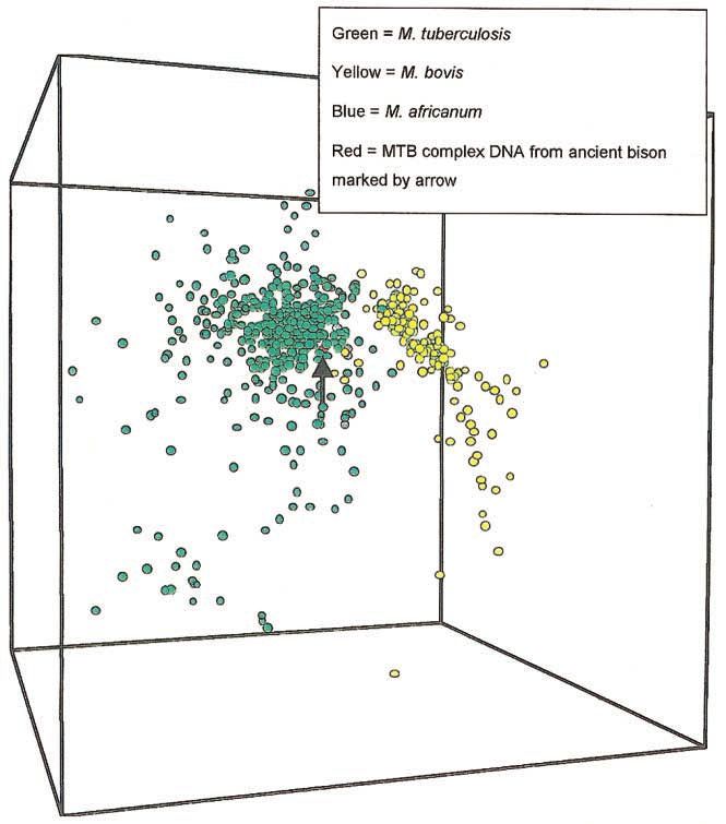
1). Principal-components analysis of the spoligotype patterns
in the database, excluding those spacers that were negative for
the bison pattern, shows that the bison pattern continues to
group with M. tuberculosis and M. africanum rather than with
M. bovis (figure 2). This demonstrates that grouping the bison
pattern with M. tuberculosis and M. africanum is not related to
any difficulties in obtaining a full spoligotype pattern.
Figure 1. Pseudo–3-dimensional representation of discriminant anal-
ysis of 634 Mycobacterium bovis, Mycobacterium tuberculosis, and My-
DISCUSSION cobacterium africanum spoligotype patterns from a combined database
of modern patterns collated by the National Institute of Public Health
Clarification of the evolutionary history of infectious diseases
and Environment (RIVM), Utrecht, The Netherlands, and the Veterinary
is advanced by comparison of DNA in ancient and modern Sciences Division, Belfast. MTB, M. tuberculosis complex.
specimens. Our understanding of the antiquity of tuberculosis
was previously based upon presumptive, macroscopic pathol-
[39]. The results of our study further specified the particular
ogy–based diagnosis of skeletal remains [16–30]. The work of
species of this 4-member complex that is responsible for disease
Allison et al. [19], Cockburn et al. [42], Garcia-Frias [43], and
in bison.
Zimmerman [20] allowed histologic confirmation of Myco-
M. bovis and relationships. It is clear from the presence
bacterium-related infection [18].
of spacer elements 39, 40, 41, and 43 that the bison DNA is
In the current study, DNA analysis of a 17,000-year-old skel-
not modern M. bovis DNA. However, M. bovis, M. tuberculosis,
etal specimen from Natural Trap Cave confirms the diagnosis
M. africanum, and M. microti probably evolved from an M.
of M. tuberculosis complex infection. IS6110 DNA fingerprint-
tuberculosis precursor ∼15,000–20,000 years BP [44], so the
ing is now the standard method for molecular analysis of tu-
bison sample may be representative of the precursor or of group
berculosis epidemiology, because this sequence is apparently
1 species shortly after speciation.
restricted to species that belong to the M. tuberculosis complex
Information from the M. tuberculosis genome sequence has
very recently been used to construct a DNA microarray, in
Table 1. Average similarity of bison spoligotype pat-
tern to a library of spoligotype patterns, divided into which almost every open-reading frame is displayed, allowing
units based on species. a global analysis of genetic differences between M. tuberculosis,
M. bovis, and BCG substrains. Multilocus sequencing [44] had
Average similarity implied that there was a high degree of genetic conservation
Pattern, by species to pattern Confidence between M. bovis and M. tuberculosis. The new findings suggest
a
of Mycobacterium from bison, % factor
that genetic diversity within the M. tuberculosis complex may
M. africanum 82.3 1.33
be much greater than previously supposed and that gene de-
M. tuberculosis 76.6 1.01
letion, rather than point mutation, may be a key source of
M. bovis 72.7 2.22
genetic variation [9, 10].
M. microti 17.4 20.31
M. microti. Van Soolingen et al. [37] demonstrated that
a
Calculated by comparing the average similarity between the M. microti could be recognized and distinguished on the basis
bison pattern and the library unit’s entries and the average sim-
ilarities of the library unit’s entries with each other, thus taking
of IS6110 restriction fragment–length polymorphism patterns
account of the heterogeneity of the library units. and by means of spoligotyping. Although the former technique
308 • CID 2001:33 (1 August) • Rothschild et al.form of the organisms today recognized as M. tuberculosis and
M. bovis.
M. tuberculosis complex. The Eisenach/Taylor primers are
specific for the M. tuberculosis complex. Larger IS6110 primers
and those for S12 are not. Hence, any product with the nested
PCR is specific, as further documented by sequencing studies.
It is intriguing that the spoligotyping pattern of the bison
isolate (as determined by comparison with the extensive col-
lection of spoligotyping patterns in data banks) is closer to the
pattern of modern M. africanum and M. tuberculosis than to
the patterns of the other members of the M. tuberculosis com-
plex. The patterns were compared to a database containing
several hundred different patterns obtained from M. tuberculosis
isolates, 1170 M. bovis patterns, 34 M. microti patterns, and 6
M. africanum patterns. The pattern obtained from the bison
material most closely matches with an M. tuberculosis pattern
(figure 2). However, the bison pattern fits most closely with
the mean of the M. africanum patterns as a group. The bison
isolate, however, cannot be assigned to a particular species of
the M. tuberculosis complex on the basis of spoligotyping data.
Assignment of this isolate to any one of the species in the M.
tuberculosis complex must be done with reservations, because
speciation of the M. tuberculosis complex may not yet have
Figure 2. Pseudo–3-dimensional representation of principle-compo-
occurred by 17,000 years BP. The M. tuberculosis complex is
nents analysis of 634 Mycobacterium bovis, Mycobacterium tuberculosis,
and Mycobacterium africanum spoligotype patterns from a combined da- very homogeneous at the DNA level and may have speciated
tabase of modern patterns collated by the National Institute of Public from a progenitor organism only very recently [44].
Health and Environment (RIVM), Utrecht, The Netherlands, and the Vet- Caveats. The slight base-pair difference found at one lab-
erinary Sciences Division, Belfast. Analysis excludes spacers absent from oratory between the ancient DNA from the bison specimen
spoligotyping of DNA from ancient bison (i.e., spacers 8, 10, 25–37, and
and modern sequences requires confirmation. It should be
43). MTB, M. tuberculosis complex.
noted that there have been no reports of base changes in the
92-bp sequence of IS6110, although other tested samples have
may be of limited value for application to ancient DNA because been considerably less ancient.
of taphonomic changes, the short direct-repeat regions of M. Base-pair differences do, however, provide an additional
microti identified by spoligotyping allow its recognition and proof that contamination is not a factor. Kolman and Tuross
elimination as a causative organism for tuberculosis in Pleis- [7] have criticized previous work because the investigators
found ancient DNA that had sequences identical to those of
tocene bison. Although M. microti is not known to affect bovids,
modern DNA. They state that “the finding of identical se-
it does affect mice. Isolation of M. tuberculosis complex DNA
quences in an archaeological specimen and the control DNA
from the lesion only and not from unaffected species and bones
sample precludes convincing proof that ancient DNA was ex-
suggests that contamination of the site by mice is unlikely.
tracted and analyzed” [7, p. 16]. The lack of a perfect match
Results of spoligotyping totally eliminate the possibility of such
between the DNA extracted from the ancient bison and the
contamination.
DNA from contemporary organisms not only documents ab-
M. africanum. M. africanum was originally described by
sence of contamination, but the character of that DNA suggests
Castets et al. [45]. It is responsible for 60% of cases of tuber-
that it may represent the primordial organisms that developed
culosis in Africa. Hass and des Prez [34] refer to M. africanum
into the organisms that are today used as positive controls.
as intermediate between M. tuberculosis and M. bovis. Spoli-
gotyping data support this. The possibly close relationship be-
tween the isolated organism and M. africanum is therefore per- CONCLUSION
haps not surprising. If the oldest identified organism (that
described in this study) is more closely related to M. africanum This is the first DNA-based documentation of a macroscopically
than to other members of the M. tuberculosis complex, it would recognized tuberculosis infection in the fossil record. The con-
seem reasonable to consider this organism to be the primordial ditions of Natural Trap Cave favored DNA preservation, be-
Origins of Tuberculosis in North America • CID 2001:33 (1 August) • 309cause the temperature has remained nearly constant and rel- 7. Kolman CJ, Tuross N. Ancient DNA analysis of human populations.
Am J Phys Anthropol 2000; 111:5–23.
atively low, and the climate is semiarid with little water 8. Kolman CJ, Centurion-Lara A, Lukehart SA, et al. Identification of
circulation. M. tuberculosis complex bacteria have hydrophobic Treponema pallidum subspecies pallidum in a 200-year-old skeletal
cell walls, rich in long-chain mycolic acids. Location within specimen. J Infect Dis 1999; 180:2060–3.
9. Behr MA, Wilson MA, Gill WP, et al. Comparative genomics of BCG
bones provides further protection of DNA from environmental
vaccines by whole-genome DNA microarray. Science 1999; 284:1520–3.
taphonomic loss [46]. 10. Dye C, Scheele S, Dolin P, et al. Global burden of tuberculosis: esti-
Because this documentation is of tuberculosis occurring in mated incidence, prevalence, and mortality by country. JAMA 1999;
the Pleistocene long before domestication of cattle, it suggests 282:677–86.
11. Musial CE, Tice LS, Stockman L, Roberts GD. Identification of my-
a different scenario for the spread of tuberculosis than is usually cobacteria from culture using the Gen-Probe rapid diagnostic system
conceived [4, 21]. The presence of characteristic lesions in sheep for Mycobacterium avium complex and Mycobacterium tuberculosis
(Ovis canadensis catclawensis) and extinct musk ox (Bootherium complex. J Clin Microbiol 1988; 26:2120–3.
12. van der Heijden IM, Wilbrink B, Schouls LM, van Embden JD, Breed-
bombifrons) [31] and in bison—now confirmed as tubercular veld FC, Tak PP. Detection of mycobacteria in joint samples from
in origin—indicates that the disease was widespread throughout patients with arthritis using a genus-specific polymerase chain reaction
the Pleistocene. It is suggested that tuberculosis in bovids had and sequence analysis. Rheumatology 1999; 38:547–53.
13. Frothingham R, Strickland PL, Bretzel G, Ramaswamy S, Musser JM,
a Holarctic pattern and that it reached North America at least
Williams DL. Phenotypic and genotypic characterization of Mycobac-
20,000 years BP. Absence of tubercular lesions in pronghorn terium africanum isolates from West Africa. J Clin Microbiol 1999; 37:
Antilocapra americanum and Equus species suggests that it was 1921–6.
absent in North America before the immigration of the ad- 14. Eisenach KD, Cave MD, Bates JH, Crawford JT. Polymerase chain
reaction amplification of a repetitive DNA sequence specific for My-
vanced northern bovids. cobacterium tuberculosis. J Infect Dis 1990; 161:977–81.
Analysis of additional specimens that have tuberculosis le- 15. Cousins DV, Williams SN, Dawson DJ. Tuberculosis due to Mycobac-
sions is now indicated to elucidate the evolution of tuberculosis. terium bovis in the Australian population: DNA typing of isolates,
1970–1994. Int J Tuberc Lung Dis 1999; 3:722–31.
It is likely that the bison isolate contains some “novel” spoli- 16. Verano JW, Ubelaker DH. Disease and demography in the Americas.
gotyping spacers—and perhaps spacers as yet not identi- Washington, DC: Smithsonian Institution Press, 1992.
fied—between the spacers that appeared on the blot for our 17. Widmer L, Perzigian AJ. The ecology and etiology of skeletal lesions
in late prehistoric populations from eastern North America. North-
specimen. The paleontologic record provides an excellent geo-
western Univ Archeol Prog Sci Papers 1981; 5:99–113.
graphic and chronological database for understanding disease 18. Salo WL, Aufderheide AC, Buikstra J, Holcomb TA. Identification of
origins and spread, especially for diseases with significant os- Mycobacterium tuberculosis DNA in a pre-Columbian Peruvian mum-
seous impact. my. Proc Natl Acad Sci USA 1994; 91:2091–4.
19. Allison MJ, Mendoza D, Pezzia A. Documentation of a case of tuber-
culosis in pre-Columbian America. Am Rev Respir Dis 1973; 107:
985–991.
Acknowledgments 20. Zimmerman MR. Pulmonary and osseous tuberculosis in an Egyptian
mummy. Bull N Y Acad Med 1979; 55:604–8.
The support of The Center for the Study of Emerging Dis- 21. Buikstra JE. The Caribou Eskimo: general and specific disease. Am J
Phys Anthropol 1976; 45:351–68.
eases, Jerusalem, is gratefully acknowledged. Dr. Dick van Sool-
22. Aceves-Avila FJ, Baez-Molgado S, Medina F, Fraga A. Paleopathology
ingen’s cogent input is acknowledged with appreciation. in osseous remains from the XVI century: a survey of rheumatic dis-
eases. J Rheumatol (in press).
23. Aufderheide AC, Rodriguez-Martin C. The Cambridge encyclopedia
of human paleopathology. Cambridge, UK: Cambridge University
References
Press, 1998.
1. Greenblatt CL, ed. Digging for pathogens: ancient emerging diseases: 24. Formicola V. X-linked hypophosphatemic rickets: a probable Upper
their evolutionary, anthropological and archeological context. Jerusa- Paleolithic case. Am J Phys Anthropol 1995; 98:403–9.
lem: Balaban Publishers, 1998. 25. Morse D. Tuberculosis. In: Brothwell D, Sandison AT, eds. Diseases in
2. Martin LD, Rothschild BM. Earth history and the evolution of sickness. antiquity. Springfield, IL: Charles C. Thomas, 1967:249–71.
In: Greenblatt CL, ed. Digging for pathogens: ancient emerging dis- 26. Palfi G. The osteo-archaeological evidence of vertebral tuberculosis in
eases: their evolutionary, anthropological and archeological context. the 8th century. Acta Biologica Szeged 1991; 37:101–5.
Jerusalem: Balaban Publishers, 1998:15–46. 27. Sager P, Schalimtzek M, Moller-Christensen V. A case of spondylitis
3. Rothschild BM, Martin LD. Paleopathology: disease in the fossil record. tuberculosa in the Danish Neolithic age. Danish Med Bull 1972; 19:
London: CRC Press, 1999. 176–80.
4. Arriaza BT, et al. Pre-Columbian tuberculosis in northern Chile: mo- 28. Strouhal E. Vertebral tuberculosis in ancient Egypt and Nubia. In:
lecular and skeletal evidence. Am J Phys Anthropol 1995; 98:37–45. Ortner DJ, Aufderheide AC, eds. Human paleopathology: current syn-
5. Cole ST, Bosch R, Parkhill J, et al. Deciphering the biology of Myco- theses and future options. Washington, DC: Smithsonian Institute
bacterium tuberculosis from the complete genome sequence. Nature Press, 1991:181–94.
1998; 393:537–44. 29. Wells C. Bones, bodies and diseases. London: Thames and Hudson,
6. Finken M, Kirschner P, Meier A, Wrede A, Bottger EC. Molecular basis 1964.
of streptomycin resistance in Mycobacterium tuberculosis: alterations of 30. Bartels P. Tuberkulose (Wirbelkaries) in der jungeren Steinzeit. Arch
the ribosomal protein S12 gene and point mutation within a functional Anthropol 1907; 6:243–55.
S16 ribosomal RNA pseudoknot. Mol Microbiol 1993; 9:1239–46. 31. Martin LD, Rothschild BM. Frequency of pathology in a large natural
310 • CID 2001:33 (1 August) • Rothschild et al.sample from Natural Trap Cave (late Pleistocene). J Vert Paleontol tuberculosis IS6110 restriction fragment length polymorphism patterns
l989;9:31A. and spoligotypes determined by analyzing serial isolated from patients
32. Resnick D, Niwayama G. Diagnosis of bone and joint disorders. 2d with drug-resistant tuberculosis. J Clin Microbiol 1999; 37:409–12.
ed. Philadelphia: Saunders, 1988. 40. van Embden JD, van Gorkom T, Kremer T, Jansen R, van der Zeijst
33. Wang X, Martin LD. Natural Trap Cave. Natl Geographic Soc Res Rep BA, Schoulds LM. Genetic variation and evolutionary origin of the
1993; 9:422. direct repeat locus of Mycobacterium tuberculosis complex bacteria. J
34. Hass DW, des Prez RM. Mycobacterium tuberculosis. In: Mandell GL, Bacteriol (2000); 182:2393–401.
Bennett JE, Dolin R, eds. Principles and practice of infectious diseases. 41. Dale JW, Brittain D, Cataldi AA, et al. Spacer oligonucleotide typing
4th ed. New York: Churchill Livingstone, 1995:2213–43. of bacteria of the Mycobacterium tuberculosis complex: recommenda-
35. Boom R, Sol A, Salimans M, Jansen C, Wertheim-van Dillen P, van tions for standardised nomenclature. Int J Tuberc Lung Dis 2001; 5:
der Noordaa J. Rapid and simple method for purification of nucleic 216–9.
acid. J Clin Microbiol 1990; 28:495–503. 42. Cockburn A, Cockburn E, Reyman TA. Mummies, disease and ancient
36. Donoghue HD, Spigelman M, Zias J, Gernaey-Child AM, Minnikin cultures. Cambridge, UK: Cambridge University Press, 1998.
DE. Demonstration of Mycobacterium tuberculosis complex DNA in 43. Garcia-Frias J. La tuberculosis en los antiguos Peruanos. Actualidad
calcified pleura from remains 1400 years old. Lett Appl Microbiol Medicina Peruana 1940; 5:274–91.
1998; 27:265–9. 44. Streevatsan S, Pan X, Stockbauer KE, et al. Restricted structural gene
37. van Soolingen D, van der Zanden AG, de Haas PE, et al. Diagnosis of polymorphism in the Mycobacterium tuberculosis complex indicates
Mycobacterium microti infections among humans by using novel ge- evolutionarily recent global dissemination. Proc Natl Acad Sci USA
netic markers. J Clin Microbiol 1998; 36:1840–5. 1997; 94:9869–74.
38. Taylor GM, Goyal M, Legge AJ, Shaw RJ, Young D. Genotyping analysis 45. Castets M, Boisvert H, Grumbach F, Brunel M, Rist N. Tuberculosis
of Mycobacterium tuberculosis from medieval human remains. Micro- bacilli of the African type. Rev Tuberc Pneumonol 1968; 32:179–84.
biology 1999; 145:899–904. 46. Eglinton G, Logan GA. Molecular preservation. Philos Trans R Soc
39. Niemann S, Richter E, Rusch-Gerdes S. Stability of Mycobacterium Lond B Biol Sci 1991; 333:315–28.
Origins of Tuberculosis in North America • CID 2001:33 (1 August) • 311You can also read