Physiological and motion signatures in static and time-varying functional connectivity and their subject identifiability
←
→
Page content transcription
If your browser does not render page correctly, please read the page content below
RESEARCH ARTICLE

                                     Physiological and motion signatures in
                                     static and time-varying functional
                                     connectivity and their subject
                                     identifiability
                                     Alba Xifra-Porxas1†*, Michalis Kassinopoulos1†, Georgios D Mitsis2*
                                     1
                                      Graduate Program in Biological and Biomedical Engineering, McGill University,
                                     Montréal, Canada; 2Bioengineering Department, McGill University, Montréal,
                                     Canada

                                     Abstract Human brain connectivity yields significant potential as a noninvasive biomarker.
                                     Several studies have used fMRI-based connectivity fingerprinting to characterize individual patterns
                                     of brain activity. However, it is not clear whether these patterns mainly reflect neural activity or the
                                     effect of physiological and motion processes. To answer this question, we capitalize on a large data
                                     sample from the Human Connectome Project and rigorously investigate the contribution of the
                                     aforementioned processes on functional connectivity (FC) and time-varying FC, as well as their
                                     contribution to subject identifiability. We find that head motion, as well as heart rate and breathing
                                     fluctuations, induce artifactual connectivity within distinct resting-state networks and that they
                                     correlate with recurrent patterns in time-varying FC. Even though the spatiotemporal signatures of
                                     these processes yield above-chance levels in subject identifiability, removing their effects at the
                                     preprocessing stage improves identifiability, suggesting a neural component underpinning the
*For correspondence:
                                     inter-individual differences in connectivity.
alba.xifraporxas@mail.mcgill.ca
(AX-P);
georgios.mitsis@mcgill.ca (GDM)
†
 These authors contributed
equally to this work                 Introduction
                                     Functional magnetic resonance imaging (fMRI) is based on the blood-oxygenation-level-dependent
Competing interests: The
                                     (BOLD) contrast mechanism (Ogawa et al., 1990), and is widely viewed as the gold standard for
authors declare that no
                                     studying brain function because of its high spatial resolution and non-invasive nature. The BOLD sig-
competing interests exist.
                                     nal exhibits low frequency (~0.01–0.15 Hz) fluctuations that are synchronized across different regions
Funding: See page 28                 of the brain, a phenomenon known as functional connectivity (FC). FC has been observed even in
Preprinted: 05 February 2020         the absence of any explicit stimulus or task, giving rise to the so-called resting-state networks (RSNs)
Received: 21 August 2020             (Biswal et al., 1995; Fox and Raichle, 2007; Smith et al., 2009). Initially, FC was viewed as a sta-
Accepted: 02 August 2021             tionary phenomenon (static FC) and was commonly measured as the correlation between brain
Published: 03 August 2021            regions over an entire scan. However, several researchers challenged this assumption (Chang and
Reviewing editor: Alex Fornito,
                                     Glover, 2010; Sakoğlu et al., 2010), and recent studies have been focusing on FC dynamics, quanti-
Monash University, Australia         fied over shorter time scales than the scan duration (time-varying FC) (Hutchison et al., 2013;
                                     Lurie et al., 2020).
   Copyright Xifra-Porxas et al.
                                        Although the neurophysiological basis of resting-state FC measured with fMRI is not yet fully
This article is distributed under
                                     understood, many studies have provided evidence to support its neuronal origin. For instance, in ani-
the terms of the Creative
Commons Attribution License,         mal models, a strong association between spontaneous BOLD fluctuations and neural activity, in par-
which permits unrestricted use       ticular band-limited local field potentials and firing rates, has been reported (Logothetis et al.,
and redistribution provided that     2001; Schölvinck et al., 2010; Shmuel and Leopold, 2008; Thompson et al., 2013b). Furthermore,
the original author and source are   a recent study suggested a close correspondence between windowed FC calculated from simulta-
credited.                            neously recorded hemodynamic signals and calcium transients (Matsui et al., 2019). In human

Xifra-Porxas, Kassinopoulos, et al. eLife 2021;10:e62324. DOI: https://doi.org/10.7554/eLife.62324                                    1 of 36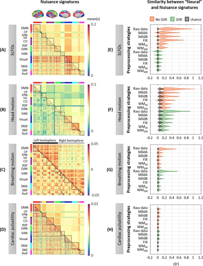
Research article                                                                                                       Neuroscience

                                    studies, direct measurements of macroscale neural activity have revealed a spatial correlation struc-
                                    ture similar to that of spontaneous BOLD fluctuations (Brookes et al., 2011; Hacker et al., 2017;
                                    He et al., 2008; Hipp et al., 2012; Kucyi et al., 2018), even during transient (50–200 ms) events
                                    (Baker et al., 2014a; Hunyadi et al., 2019; Vidaurre et al., 2018). Therefore, it is widely assumed
                                    that resting-state FC measured using BOLD fMRI reflects spontaneous co-fluctuations of the underly-
                                    ing neuronal networks.
                                        However, BOLD signals rely on changes in local cerebral blood flow (CBF) to infer underlying
                                    changes in neuronal activity, and according to a recent study, at least 50% of the spontaneous
                                    hemodynamic signal is unrelated to ongoing neural activity (Winder et al., 2017). For instance, sys-
                                    temic physiological functions can induce variations in global and local CBF, which in turn result in
                                    BOLD signal fluctuations. In particular, low frequency variations in breathing activity (Birn et al.,
                                    2008b; Birn et al., 2006; Power et al., 2017), arterial blood pressure (Whittaker et al., 2019), arte-
                                    rial CO2 concentration (Prokopiou et al., 2019; Wise et al., 2004), and heart rate (Chang et al.,
                                    2009; Shmueli et al., 2007) are known to account for a considerable fraction of variance of the
                                    BOLD signal, presumably through changes in CBF. In addition, the BOLD signal intensity is distorted
                                    by high-frequency physiological fluctuations, such as cardiovascular pulsation and breathing, through
                                    displacement of the brain tissues and perturbations of the B0 magnetic field (Dagli et al., 1999;
                                    Glover et al., 2000). Further, head motion is well-known to have a substantial impact on fMRI
                                    through partial volume, magnetic inhomogeneity and spin-history effects (Friston et al., 1996;
                                    Power et al., 2012). These non-neuronal factors may introduce common variance components in
                                    signals recorded from different brain regions and subsequently induce spurious correlations between
                                    these areas (Chen et al., 2020). Therefore, to account for motion-related and physiological con-
                                    founds, nuisance regressors are typically obtained using model-based and data-driven techniques,
                                    and regressed out from the fMRI data before further analysis (Caballero-Gaudes and Reynolds,
                                    2017).
                                        Static and time-varying resting-state FC have shown promise for providing concise descriptions of
                                    how the brain changes across the lifespan (Battaglia et al., 2020; Chan et al., 2014; Ferreira et al.,
                                    2016; Geerligs et al., 2015; Sala-Llonch et al., 2015; Xia et al., 2019), and to assay neural differen-
                                    ces that are associated with disease (Baker et al., 2014b; Chen et al., 2017; Damaraju et al., 2014;
                                    Demirtaş et al., 2016; Drysdale et al., 2017; Du et al., 2016; Gratton et al., 2019; Hahamy et al.,
                                    2015; Mash et al., 2019; Morgan et al., 2017; Xia et al., 2018). However, recent studies assessing
                                    the performance of a large range of preprocessing strategies found that there is always a trade-off
                                    between adequately removing confounds from fMRI data and preserving the signal of interest
                                    (Ciric et al., 2017; Kassinopoulos and Mitsis, 2021a; Parkes et al., 2018). Importantly, these stud-
                                    ies found that widely used techniques for the preprocessing of fMRI data may not efficiently remove
                                    physiological and motion artifacts. The latter raises a concern, as it is still not clear how nuisance fluc-
                                    tuations may impact the outcome of FC studies.
                                        Several studies have examined whether physiological fluctuations across the brain could give rise
                                    to structured spatial patterns that resemble common RSNs, based on the evidence that vascular
                                    responses following systemic changes are spatially heterogenous (Chang et al., 2009; Pinto et al.,
                                    2017), or account for the observed time-varying interactions between RSNs. For instance,
                                    Bright and Murphy, 2015 applied independent component analysis (ICA) to the fraction of the fMRI
                                    data explained by nuisance regressors related to head motion and physiological variability and
                                    revealed a characteristic network structure similar to previously reported RSNs. Similarly,
                                    Tong et al., 2013; Tong and Frederick, 2014 found significant contributions of systemic fluctuations
                                    on ICA time courses related to the visual, sensorimotor and auditory networks. Recently,
                                    Chen et al., 2020 generated BOLD data containing only slow respiratory-related dynamics and
                                    showed that respiratory variation can give rise to apparent neurally-related connectivity patterns.
                                    Further, recent investigations have shown that physiological confounds can modulate time-varying
                                    FC measures (Chang et al., 2013; Nalci et al., 2019; Nikolaou et al., 2016). These results suggest
                                    that changes in brain physiology, breathing patterns, heart rhythms and head motion across ses-
                                    sions, within-subject or across populations, may introduce artifactual inter-individual and group-
                                    related differences in FC independent of any underlying differences in neural activity. For instance,
                                    cardiac autonomic dysregulation has been associated to a variety of psychiatric disorders
                                    (Alvares et al., 2016; Benjamin et al., 2020), which could in principle lead to group differences in
                                    connectivity patterns between patients and controls if the effects of heart rate are not accounted

Xifra-Porxas, Kassinopoulos, et al. eLife 2021;10:e62324. DOI: https://doi.org/10.7554/eLife.62324                                       2 of 36
Research article                                                                                                     Neuroscience

                                    for. Therefore, the disentanglement of the neural and physiological correlates of resting-state FC is
                                    crucial for maximizing its clinical impact.
                                        While the previous findings provide evidence for the dual-nature of RSNs in both static and time-
                                    varying scenarios, only specific physiological processes and/or particular brain networks were evalu-
                                    ated in each of the aforementioned studies. A more holistic assessment of the impact of these non-
                                    neural processes on FC measures is needed to better understand whether and how systemic fluctua-
                                    tions, as well as head or breathing motion affect inter-individual and group differences. Importantly,
                                    the wide range of possible preprocessing strategies needs to be reassessed taking into consider-
                                    ation the effects of several non-neural processes on FC measures rather than accounting only for the
                                    effects of a specific process (e.g. head motion).
                                        The varying efficiency of different preprocessing pipelines with respect to removing the effect of
                                    physiological fluctuations and motion has also implications for studies investigating properties of FC
                                    at the individual level. Recent studies have shown that connectivity profiles vary substantially
                                    between individuals, acting as an identifying fingerprint (Finn et al., 2015; Miranda-
                                    Dominguez et al., 2014) that is stable over long periods of time (Horien et al., 2019). However, the
                                    high subject discriminability of connectivity profiles may arise partly as a result of physiological pro-
                                    cesses (Batchvarov et al., 2002; Golestani et al., 2015; Malik et al., 2008; Pinna et al., 2007;
                                    Pitzalis et al., 1996; Power et al., 2020; Reland et al., 2005) and head motion (Van Dijk et al.,
                                    2012; Zeng et al., 2014) being highly subject-specific. Evidence supporting this hypothesis comes
                                    from studies showing that the mean of intraclass correlation values of functional connections, associ-
                                    ated to test-retest reliability across sessions, is reduced when a relatively aggressive pipeline is used
                                    (Birn et al., 2014; Kassinopoulos and Mitsis, 2021a; Parkes et al., 2018), suggesting that artifacts
                                    exhibit high subject specificity. However, the relation between subject discrimination and inter-indi-
                                    vidual differences in physiological processes and head motion has yet to be addressed.
                                        In the present work, we capitalize on 3T resting-state fMRI data from the Human Connectome
                                    Project (HCP) to uncover the whole-brain connectome profiles of systemic low-frequency oscillations
                                    (SLFOs) associated with heart rate and breathing patterns, cardiac pulsatility, breathing motion and
                                    head motion on estimates of static and time-varying FC. To quantify the contributions of physiologi-
                                    cal processes and head motion on FC, we employ model-based techniques with externally recorded
                                    physiological measurements, and subsequently generate nuisance datasets that only contain non-
                                    neural fluctuations. Using these datasets, we provide a comprehensive examination of the regional
                                    variability of the impact of the considered nuisance processes on the BOLD signal, as well as an
                                    investigation of the group consistency and inter-individual differences of their characteristic signa-
                                    tures on FC. We further evaluate several fMRI preprocessing strategies to assess the extent to which
                                    different techniques remove the physiological and motion FC signatures from the fMRI data. Finally,
                                    we investigate the potential effect of physiological processes and head motion on individual discrim-
                                    inability in the context of connectome fingerprinting.
                                        Using the proposed approach, we show that SLFOs and head motion have a larger impact on FC
                                    measures compared to breathing motion and cardiac pulsatility, and we highlight the functional con-
                                    nections that are more prone to exhibiting biases. Furthermore, our findings suggest that the recur-
                                    rent whole-brain connectivity patterns observed in time-varying FC can be partly attributed to
                                    SLFOs and head motion. Finally, we show that connectome fingerprinting accuracies are higher
                                    when non-neural confounds are reduced, suggesting a neural component underpinning the individ-
                                    ual nature of FC patterns.
                                        The codes that were employed to carry out the analyses described in the present study are pub-
                                    licly available and can be found on github.com/axifra/Nuisance_signatures_FC. (copy archived at
                                    swh:1:rev:52781e743d4b4eb491b9330210dac52dcd46fd10), Xifra-Porxas, 2021a.

                                    Results
                                    Contributions of nuisance processes to the BOLD signal
                                    We examined regional differences in the influence of physiological processes and head motion to
                                    the BOLD signal. The physiological processes evaluated here were breathing motion, cardiac pulsa-
                                    tility, and SLFOs associated with changes in heart rate and breathing patterns (see Materials and
                                    methods – Nuisance processes evaluated). Scans with LR and RL phase encoding were examined

Xifra-Porxas, Kassinopoulos, et al. eLife 2021;10:e62324. DOI: https://doi.org/10.7554/eLife.62324                                    3 of 36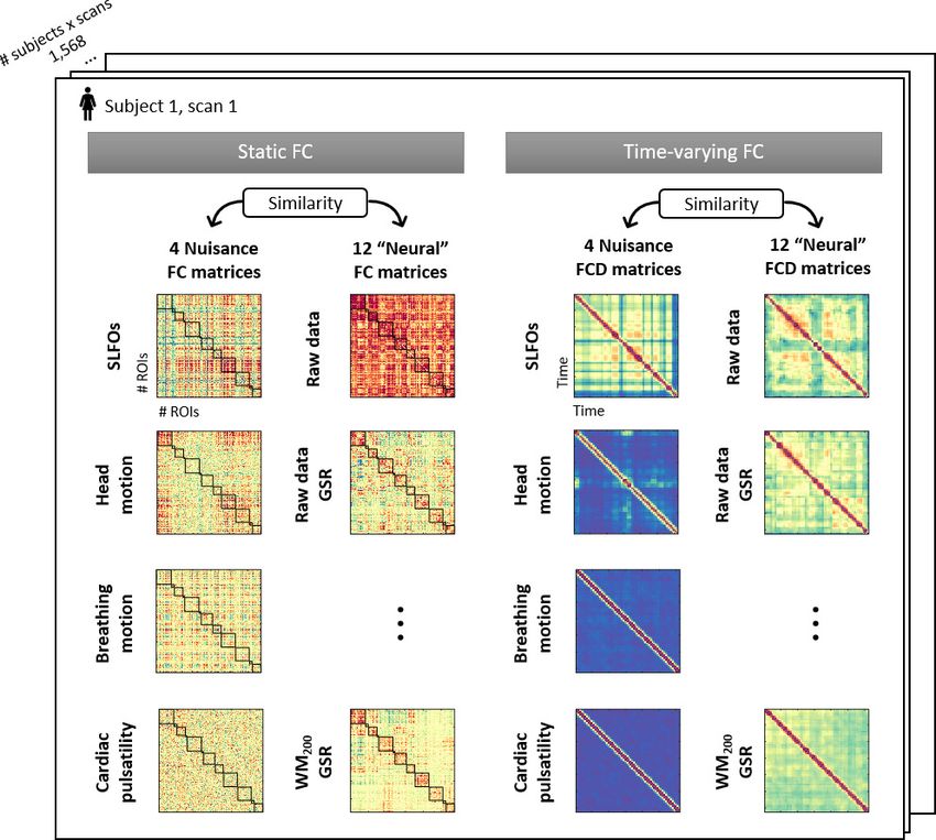
Research article                                                                                                      Neuroscience

                                    separately as it has been suggested that breathing motion artifacts vary across scans with different
                                    phase encoding directions (Raj et al., 2001), and thus we aimed to examine whether other pro-
                                    cesses such as head motion demonstrate a similar dependence. The contributions of each nuisance
                                    process on BOLD signal fluctuations were quantified as the correlation between the nuisance fluctua-
                                    tions of the process in question, modeled using externally recorded physiological measurements,
                                    and the BOLD fluctuations ’cleaned’ of all other nuisance fluctuations (denoted as rnuis in the Materi-
                                    als and methods section – Isolation of nuisance fluctuations from fMRI data, see also Figure 7). We
                                    computed these contributions for each scan and then tested for the presence of consistent patterns
                                    across scans with the same phase encoding direction (significance testing using inter-subject surro-
                                    gates, two-sample t-test, p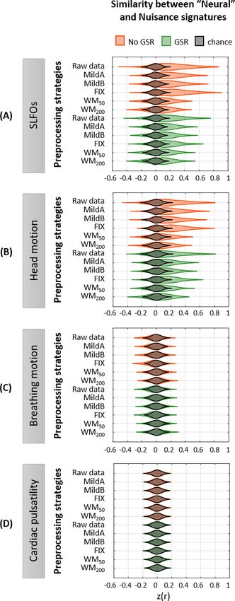
Research article                                                                                                                Neuroscience

                                    Figure 1. Contributions of nuisance processes to the resting-state BOLD signal. T-score maps of the correlation
                                    between each nuisance process and BOLD fMRI fluctuations (raw data) for (A) SLFOs, (B) head motion, (C)
                                    breathing motion, and (D) cardiac pulsatility, computed within each parcel of the Gordon atlas (nonparametric
                                    permutation test, p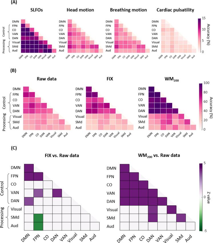
Research article                                                                                                               Neuroscience

Figure 2. Whole-brain connectome patterns induced by nuisance processes and effect of preprocessing strategies. (A–D) Group averaged nuisance FC
matrices across all 1568 scans for (A) SLFOs, (B) head motion, (C) breathing motion, and (D) cardiac pulsatility. The lower triangular matrices show the
FC value for each network pair. The upper triangular matrices show FC values from the lower triangular averaged within network pairs (A, B, D panels),
or the FC value for each network pair reordered according to left/right hemisphere (C panel). These results demonstrate that nuisance fluctuations
Figure 2 continued on next page

Xifra-Porxas, Kassinopoulos, et al. eLife 2021;10:e62324. DOI: https://doi.org/10.7554/eLife.62324                                               6 of 36

Research article                                                                                                                 Neuroscience

Figure 2 continued
induce heterogeneous whole-brain connectivity profiles which, if unaccounted for, can result in biased estimates functional connectivity. (E–H)
Distribution of Pearson correlation coefficients across all 1568 scans between the ‘neural’ FC matrix for different preprocessing strategies and nuisance
FC matrices associated to (E) SLFOs, (F) head motion, (G) breathing motion, and (H) cardiac pulsatility. Correlation values were Fisher z transformed.
SLFOs, head motion and breathing motion were found to confound the FC matrices more severely (E–G). GSR effectively removed the effects of
SLFOs, while more aggressive preprocessing pipelines mitigated the effects of head motion, breathing motion and cardiac pulsatility.
The online version of this article includes the following figure supplement(s) for figure 2:
Figure supplement 1. Whole-brain connectome patterns induced by nuisance processes, separately for scans with LR and RL phase encoding.
Figure supplement 2. Effectiveness of preprocessing strategies in reducing the whole-brain connectivity profiles effects of SLFOs for two different
global signal calculation methods.
Figure supplement 3. Effectiveness of preprocessing strategies in reducing the whole-brain connectivity effect of each nuisance process, with and
without including model-based regressors.
Figure supplement 4. Investigating the negative correlations between the ‘neural’ and SLFOs signatures after GSR.
Figure supplement 5. Whole-brain connectome patterns induced by nuisance processes and effect of preprocessing strategies, using the Seitzman
atlas.

                                     physiological and head motion artifacts. A distribution of the similarity values across scans, in this
                                     case Pearson’s correlation coefficients, is shown in Figure 2E–H for each preprocessing strategy and
                                     nuisance process. We found that SLFOs, head motion and breathing motion had the strongest influ-
                                     ence on static FC, based on the similarity of their connectome profiles with the ‘neural’ connectome
                                     profiles from the raw data (Figure 2E–H).
                                         The signature induced by SLFOs remained after the MildA, MildB, and FIX pipelines were
                                     applied, but was greatly reduced by the WM50 and WM200 strategies (Figure 2E). The observation
                                     that FIX, which is a rather aggressive preprocessing strategy, was unable to remove most of the
                                     SLFOs is consistent with recent studies showing that global artifactual fluctuations are still prominent
                                     after FIX denoising (Burgess et al., 2016; Glasser et al., 2018; Kassinopoulos and Mitsis, 2019;
                                     Power et al., 2018; Power et al., 2017). Notably, GSR seemed to be an effective technique for
                                     removing the physiological signature from SLFOs on static FC, albeit for some scans it appeared to
                                     introduce a negative correlation between the SLFOs and ‘neural’ FC matrices (Figure 2E). This effect
                                     was greater when the global signal was computed across the whole brain in volumetric space, com-
                                     pared to across vertices in surface space (Figure 2—figure supplement 2A). The effect of the signa-
                                     ture related to head motion was reduced with more aggressive preprocessing strategies, but none
                                     of the examined approaches completely eradicated the head motion effects in static FC (Figure 2F).
                                     GSR slightly reduced the similarity between the head motion and ‘neural’ connectome profiles. The
                                     signature induced by breathing motion was greatly reduced by all preprocessing strategies, and par-
                                     ticularly by FIX denoising, which yielded almost chance level (Figure 2G). Still, none of the prepro-
                                     cessing strategies entirely eliminated the breathing motion signature in static FC. The confounds
                                     introduced by cardiac pulsatility were overall small and effectively removed by the FIX, WM50, and
                                     WM200 strategies (Figure 2H). GSR did not have any effect on the removal of breathing motion and
                                     cardiac pulsatility connectome profiles.
                                         Finally, we evaluated the addition of model-based nuisance regressors to the preprocessing strat-
                                     egies. Specifically, we added the physiological regressors used to model SLFOs (Kassinopoulos and
                                     Mitsis, 2019), cardiac pulsatility and breathing motion (Glover et al., 2000). We found that includ-
                                     ing the regressor that models SLFOs reduces their effect on static FC for all preprocessing strategies
                                     apart from WM50 and WM200, but, in contrast to GSR, the similarity remains well above chance levels
                                     (Figure 2—figure supplement 3A). Including the RETROICOR regressors related to breathing
                                     motion considerably reduced the breathing motion signature in the raw data and when only using
                                     GSR as a preprocessing method; however, none of the preprocessing strategies benefited from
                                     including these regressors (Figure 2—figure supplement 3C). On the contrary, including the RET-
                                     ROICOR regressors related to cardiac pulsatility completely removed the effect of the latter for the
                                     raw data and the MildA and MildB strategies (Figure 2—figure supplement 3D), suggesting that
                                     conservative preprocessing strategies greatly benefit by adding the model-based regressors for car-
                                     diac pulsatility.

Xifra-Porxas, Kassinopoulos, et al. eLife 2021;10:e62324. DOI: https://doi.org/10.7554/eLife.62324                                                 7 of 36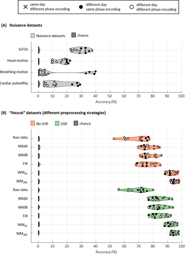
Research article                                                                                                       Neuroscience

                                    Connectome-based identification of individuals
                                    We next investigated the extent to which FC matrices associated to physiological processes and
                                    head motion can identify an individual subject, and whether the accuracy of connectome-based fin-
                                    gerprinting is inflated by the examined nuisance processes (see Materials and methods – Connec-
                                    tome-based identification of individual subjects).
                                        We initially considered all the edges of the FC matrices for subject identification (Gordon atlas:
                                    40,755 edges). Accuracy was above chance for all database-target combinations for the nuisance
                                    processes, with rates up to 40% (Figure 3A). Breathing motion exhibited an intriguing bimodal dis-
                                    tribution: database-target pairs that had the same phase encoding yielded much higher identifica-
                                    tion rates than database-target pairs with different phase encodings, even if the latter were acquired
                                    on the same day. This effect was also observed, although to a lesser extent, for cardiac pulsatility
                                    and head motion.
                                        Identification accuracy was much higher for the ‘neural’ datasets compared to the nuisance data-
                                    sets, with rates ranging from 52% to 99% (Figure 3B). The MildA, MildB, and FIX techniques consid-
                                    erably improved the accuracy compared to the raw data, and the WM50 and WM200 techniques
                                    significantly outperformed all other preprocessing strategies. GSR considerably improved identifica-
                                    tion accuracy for the MildA, MildB and FIX strategies. Furthermore, we observed that for the raw
                                    data, database-target pairs from different days with the same phase encoding showed identification
                                    rates as high as the ones from the same day but different phase encoding. In contrast, for all other
                                    preprocessing strategies the database-target pairs from the same day were always higher.
                                        We subsequently tested identification accuracy on the basis of within and between edges of spe-
                                    cific functional networks to examine whether certain functional connections had a more pronounced
                                    contribution to individual subject discriminability. Results for the nuisance datasets are shown in
                                    Figure 4A, where it can be seen that nuisance processes yielded a markedly lower identification
                                    accuracy when using specific edges compared to using all edges (Figure 3A). Furthermore, func-
                                    tional connections between networks seemed to contribute more to the subject discriminability of
                                    SLFOs compared to connections within brain networks (p
Research article Neuroscience Figure 3. Connectome fingerprinting results. (A) Fingerprinting accuracy obtained using the static FC matrices from the generated nuisance datasets whereby non-neural fluctuations were isolated from the BOLD data. Above-chance level accuracy values were obtained for all nuisance processes, suggesting some degree of subject specificity in whole-brain connectivity profiles arises from nuisance fluctuations. (B) Fingerprinting accuracy obtained using the static FC matrices generated from each of the preprocessing strategies evaluated. The pairs of resting-state scans are indicated with different symbols, depending on whether they belong to the same or different day session, as well as whether they have the same phase encoding. Higher fingerprinting accuracy values were observed for white matter denoising approaches (WM50, WM200) compared to milder pipelines and FIX denoising. Both mild and more aggressive pipelines yielded higher subject discriminability for pairs of scans acquired on the same day. GSR increased the fingerprinting accuracy of milder strategies and FIX denoising. The online version of this article includes the following figure supplement(s) for figure 3: Figure 3 continued on next page Xifra-Porxas, Kassinopoulos, et al. eLife 2021;10:e62324. DOI: https://doi.org/10.7554/eLife.62324 9 of 36
Research article                                                                                                            Neuroscience

Figure 3 continued
Figure supplement 1. Schematic of the methodology for connectome fingerprinting.
Figure supplement 2. Connectome fingerprinting results for the Seitzman atlas.

                                    zero, indicating the absence of systematic effects on ‘neural’ FCD matrices (Figure 5). Including
                                    model-based regressors in the preprocessing pipelines led to a small decrease in the similarity
                                    between neural and SLFOs FCD matrices, particularly for mild pipelines and FIX without GSR, even

Figure 4. Connectome fingerprinting results using edges within and between networks. (A) Fingerprinting accuracy for SLFOs, head motion, breathing
motion and cardiac pulsatility, averaged across all database-target pairs. (B) Fingerprinting accuracy for the raw data, FIX and WM200 pipelines,
averaged across all database-target pairs. (C) Significant differences in fingerprinting accuracy obtained when using the FIX and WM200 pipelines as
compared to the raw data (p
Research article Neuroscience
Figure 5. Effectiveness of preprocessing strategies in reducing functional connectivity dynamics (FCD) profiles
induced by physiological and motion processes. Distribution of Pearson correlation coefficients across all 1568
scans between the ‘neural’ functional connectivity dynamics (FCD) matrix after each preprocessing pipeline and
nuisance FCD matrices associated to (A) SLFOs, (B) head motion, (C) breathing motion, and (D) cardiac pulsatility.
Figure 5 continued on next page
Xifra-Porxas, Kassinopoulos, et al. eLife 2021;10:e62324. DOI: https://doi.org/10.7554/eLife.62324 11 of 36Research article Neuroscience
Figure 5 continued
Correlation values were Fisher z transformed. Results shown in the top row of each subpanel (raw data) suggest
that SLFOs and head motion most severely confound the FC matrices, whereas breathing motion and cardiac
pulsatility do not induce artifactual dynamics. None of the examined strategies completely eliminated these
effects.
The online version of this article includes the following figure supplement(s) for figure 5:
Figure supplement 1. Effectiveness of preprocessing strategies in reducing functional connectivity dynamics
(FCD) profiles induced by physiological and motion processes, with and without including model-based
regressors.
Figure supplement 2. Effectiveness of preprocessing strategies in reducing functional connectivity dynamics
(FCD) profiles induced by physiological and motion processes, using the Seitzman atlas.
though these decreased similarity values were still above chance levels (Figure 5—figure supple-
ment 1A).
None of the preprocessing pipelines was able to vanish the effects of SLFOs and head motion.
However, these effects were considerably reduced by the WM50 and WM200 strategies (Figure 5A–
B, Figure 6). FIX denoising was the least successful strategy in terms of reducing the SLFOs’ signa-
ture (Figure 5A, Figure 6), similarly to static FC (Figure 2E), and only achieved the same levels of
performance as other strategies after GSR. However, even after GSR none of the strategies reached
chance levels (Figure 5A), in contrast with the static FC results (Figure 2E). GSR led also to a slight
reduction in the similarity between the head motion and ‘neural’ FCD matrices (Figure 5B), as in the
case of static FC (Figure 2F).
Discussion
In this work, we characterized the effects of physiological processes and head motion on static and
time-varying estimates of functional connectivity measured with BOLD fMRI. While the BOLD signal
is considered a proxy of neural activity via changes in local blood oxygenation, physiological pro-
cesses and motion artifacts can also induce variations in the BOLD signal, which can in turn lead to
confounds in estimates of functional connectivity. Here, we developed an innovative framework to
characterize the spatial signature of head motion and physiological processes (cardiac and breathing
activity) on estimates of functional connectivity. Our results demonstrated that functional connectiv-
ity measures can be influenced by non-neural processes. Specifically, we identified stereotyped
whole-brain functional connectivity profiles for SLFOs, head motion and breathing motion
(Figure 2A–C), suggesting that these processes introduce a systematic bias in estimates of functional
connectivity if they are not properly accounted for. Furthermore, we provided evidence that recur-
ring patterns in time-varying FC can be attributed, to some extent, to SLFOs and head motion (Fig-
ure 5, Figure 6). We also assessed the performance of several state-of-the-art preprocessing
strategies in mitigating the effects of nuisance processes, and showed that more aggressive prepro-
cessing strategies such as FIX (Salimi-Khorshidi et al., 2014) and WM denoising
(Kassinopoulos and Mitsis, 2021a) combined with GSR were the most effective with regard to
removing the effects of non-neural processes for both static and time-varying FC analyses
(Figure 2E–H, Figure 5, Figure 6). Finally, we evaluated the potential subject specificity of the con-
nectivity profiles associated with physiological and motion confounds, along with their role as hypo-
thetical contributors to connectome fingerprinting accuracy. Interestingly, we found that these non-
neural functional connectivity patterns are to some extent subject specific (Figure 3A); however,
fMRI data corrected for these confounds increased identification accuracy in connectome fingerprint-
ing (Figure 3B), suggesting that the inter-individual differences in FC that facilitate subject identifica-
tion are strongly neural and do not largely stem from physiological processes or head motion.
Spatially heterogeneous contributions of nuisance processes to the
BOLD signal
It is well established that head and breathing motion affect areas at the edges of the brain (Jo et al.,
2010; Patriat et al., 2015; Satterthwaite et al., 2013), whereas cardiac pulsatility affects areas near
the large cerebral arteries just above the neck (Glover et al., 2000; Kassinopoulos and Mitsis,
Xifra-Porxas, Kassinopoulos, et al. eLife 2021;10:e62324. DOI: https://doi.org/10.7554/eLife.62324 12 of 36Research article Neuroscience
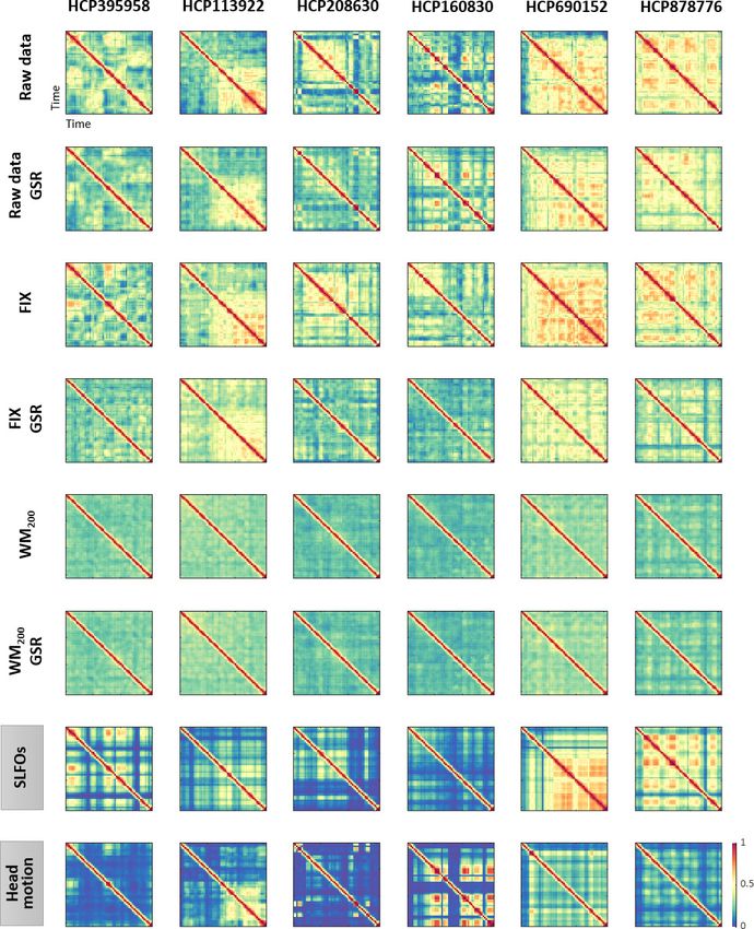
Figure 6. Functional connectivity dynamics (FCD) profiles associated with SLFOs and head motion resemble patterns commonly attributed to neural
processes. Illustrative examples of FCD matrices from specific HCP subjects as obtained from the fMRI data for several pre-processing pipelines (rows
1–6), as well as from SLFOs and head motion (rows 7 and 8, respectively). All the examples are from the HCP scan Rest1_LR. These examples show a
clear resemblance between FCD matrices computed from the ‘neural’ datasets and the nuisance processes (SLFOs and head motion). Note that the
size of FCD matrices is WW, where W is the number of sliding windows within a scan.
The online version of this article includes the following figure supplement(s) for figure 6:
Figure supplement 1. Examples of functional connectivity dynamics (FCD) profiles.
2021b). These observations are based on studies that typically examine the brain regions affected
by the aforementioned sources of noise on a voxel-wise basis. However, at the voxel level we cannot
easily assess whether the average fMRI signal from atlas-based ROIs includes significant contribu-
tions from these nuisance processes. In principle, it could be the case that the dynamics of artifacts
associated with a specific nuisance process demonstrate significant variability across voxels, and as a
Xifra-Porxas, Kassinopoulos, et al. eLife 2021;10:e62324. DOI: https://doi.org/10.7554/eLife.62324 13 of 36Research article Neuroscience
result their effects cancel out when averaging voxels within an ROI. In the present study, we assessed
the impact and regional variation of these nuisance processes in the Gordon parcellation
(Gordon et al., 2016), a widely used atlas in the literature.
SLFOs related to changes in heart rate and breathing patterns were found to affect mostly sen-
sory regions including the visual and somatosensory cortices (particularly of the face) (Figure 1A),
which correspond to regions with a high density of veins (Bernier et al., 2018; Huck et al., 2019).
The spatial pattern of SLFOs is very similar to statistical maps reported in prior works, which have
highlighted brain regions highly correlated with the global signal (Billings and Keilholz, 2018;
Glasser et al., 2016; Li et al., 2019a; Power et al., 2017; Tong et al., 2013; Zhang et al., 2020).
This is not surprising, since the global signal is strongly driven by fluctuations in heart rate and
breathing patterns (Birn et al., 2006; Chang and Glover, 2009a; Falahpour et al., 2013;
Kassinopoulos and Mitsis, 2019; Shmueli et al., 2007). Moreover, we show evidence that SLFOs
and cardiac pulsatility do not affect the same brain regions, consistent with (Chen et al., 2019;
Kassinopoulos and Mitsis, 2019; Tong and Frederick, 2014). Specifically, cardiac pulsatility was
more dominant in regions such as the insular and auditory cortices, which align with cortical
branches of the middle cerebral artery (Figure 1D) and are the regions with highest arterial density
(Bernier et al., 2018).
Regarding head motion, previous studies found that its effect was more pronounced in prefron-
tal, sensorimotor, and visual brain regions (Satterthwaite et al., 2013; Yan et al., 2013). However,
these studies did not remove breathing artifacts from the realignment parameters, which are present
even in single-band datasets (Gratton et al., 2020), and thus were unable to disentangle whether a
specific type of motion affected particular brain regions. In the present work, we regressed out
breathing motion from the realignment parameters, and observed that sensorimotor and visual areas
were strongly affected by head motion (Figure 1B), whereas breathing motion artifacts were more
pronounced in the prefrontal cortex and brain regions in the parietal and temporal cortices
(Figure 1C). Furthermore, Yan et al. showed that framewise displacement was positively correlated
with sensory regions and negatively correlated with prefrontal regions. Collectively, these findings
suggest that most regions exhibit an increase in the BOLD signal due to head and breathing motion,
whereas the prefrontal cortex may exhibit a decrease in the BOLD signal likely due to breathing-
related chest movements.
Physiological and head motion signatures in static FC
Head motion is considered the biggest source of confound for FC fMRI studies and there is a signifi-
cant effort from the neuroimaging community toward developing and evaluating preprocessing
strategies that mitigate its effects (Ciric et al., 2017; Parkes et al., 2018; Power et al., 2015). On
the other hand, while it has been shown that SLFOs affect the default-mode network (Birn et al.,
2014; Birn et al., 2008a; Chang and Glover, 2009a), and high frequency cardiac and breathing arti-
facts influence the BOLD signal (Glover et al., 2000; Power, 2019), a systematic investigation of the
effects of physiological processes in the context of whole-brain FC is lacking in the literature. In the
present study, we evaluated collectively the impact of the aforementioned sources of noise on
whole-brain fMRI resting-state FC.
Our results revealed that all four nuisance datasets exhibited mainly positive correlations between
ROIs (Figure 2A–D), suggesting that the presence of nuisance fluctuations in a conventional fMRI
dataset typically leads to a shift of correlation values toward more positive numbers. In other words,
in the case of an fMRI dataset that has not been corrected for nuisance fluctuations, two ROIs for
which neural-related fluctuations are negatively correlated could be found to be positively correlated
due to the presence of similar nuisance fluctuations in the ROIs. Furthermore, we observed that
SLFOs and head motion confounded FC to a larger degree compared to breathing motion and car-
diac pulsatility (Figure 2E–H).
Our results suggest that SLFOs due to spontaneous changes in heart rate or breathing patterns
inflate connectivity (toward more positive values) across the whole brain but particularly for edges
within the visual network, as well as edges between the visual and the rest of the networks
(Figure 2A). It is well known that the visual cortex is characterized by the highest venous density
(Bernier et al., 2018), possibly due to its functional importance (Collins et al., 2010). In addition, it
has been shown that brain regions with higher vascular density exhibit larger amplitude of spontane-
ous BOLD fluctuations (Vigneau-Roy et al., 2014). Therefore, it is likely that the structure of the
Xifra-Porxas, Kassinopoulos, et al. eLife 2021;10:e62324. DOI: https://doi.org/10.7554/eLife.62324 14 of 36Research article Neuroscience
SLFOs’ connectome profile may largely reflect the underlying vascular architecture. The effect of
SLFOs on static FC was considerably reduced after WM denoising, while additionally performing
GSR almost removed this effect (Figure 4E). Notably, FIX denoising without GSR was unable to
remove the confounds introduced by SLFOs, which is consistent with recent studies showing that
global artifactual fluctuations are still prominent after FIX denoising (Burgess et al., 2016;
Glasser et al., 2018; Kassinopoulos and Mitsis, 2019; Power et al., 2018; Power et al., 2017).
Head motion was found to influence the connectivity within the visual and sensorimotor networks
(Figure 2B), in line with previous studies (Power et al., 2012; Satterthwaite et al., 2012; Van Dijk
et al., 2012). Our results showed that only regressing out the realignment parameters and average
WM/CSF signals (with or without expansion terms) is not sufficient to remove the effects of head
motion (Figure 2F, MildA and MildB pipelines), which is consistent with findings in Parkes et al.,
2018. Among all preprocessing strategies, WM denoising yielded the largest reduction of motion
effects (Figure 2F). The two pipelines WM50 and WM200 refer to the removal of 50 and 200 white
matter regressors from the data (i.e. principal components obtained from the white matter compart-
ment). In our previous study, we showed that while both pipelines yielded high large-scale network
identifiability compared to other pipelines, the more aggressive WM200 resulted in a larger reduction
of motion artifacts compared to WM50 (Kassinopoulos and Mitsis, 2021a). The results of the current
study also show a stronger reduction of head motion effects for the former compared to the latter
(Figure 2F), which may explain the higher accuracy in connectome fingerprinting observed for the
pipeline WM200 compared to WM50 (Figure 3B).
A natural concern regarding the head motion connectome profile is that it may reflect motor-
related activity (Yan et al., 2013). Even though motor-related neural activity would be expected to
lag the instantaneous motion traces due to the sluggishness of the hemodynamic response, we can-
not exclude the scenario that the head motion connectome profile reflects the neural correlates of
the executed movements and eye adjustments to fixate on the cross. Nonetheless, even if prepro-
cessing strategies remove neural activity associated with spontaneous head movements, this source
of neural activity is typically of no interest in resting-state fMRI studies.
Furthermore, we provide evidence that head and breathing motion do not affect functional con-
nectivity in the same manner. Specifically, breathing motion was found to inflate within-hemisphere
connectivity (Figure 2C). This bias seems to arise as a result of factitious motion rather than real
motion of the head, since it is related to the LR/RL phase encoding direction (see section 4.4 for
more details). All preprocessing strategies yielded a substantial reduction of artifacts related to
breathing motion, with FIX denoising being the most effective (Figure 2G).
In our dataset, cardiac pulsatility did not seem to have a large effect on FC, neither in cortical nor
subcortical regions (Figure 2D, Figure 2—figure supplement 5D), and its effect was entirely
removed with more aggressive pipelines such as FIX and WM denoising (Figure 2H), as well as with
model-based techniques (Figure 2—figure supplement 3D). However, it has been recently reported
that the 3T HCP dataset has poor temporal signal-to-noise ratio in the subcortex (Ji et al., 2019;
Seitzman et al., 2020). Therefore, it is possible that we may have underestimated the effect of car-
diac pulsatility in functional connections involving subcortical regions.
It is important to note that our proposed methodology assumes that the stereotyped nuisance
connectome profiles do not resemble the true neural connectome profiles. However, in principle,
nuisance fluctuations could give rise to similar spatial patterns as neurally-driven fluctuations. A
recent study by Bright et al., 2020 provided evidence that physiological fluctuations (end-tidal CO2)
give rise to networks that spatially resemble neurally driven networks linked to working memory and
visual stimuli. The authors suggested that this phenomenon may be due to the vasculature adapting
to the neural network architecture, as vascular and neuronal growth processes evolve concurrently
during development (Quaegebeur et al., 2011). These findings suggest a possible caveat of our
methodology when assessing pre-processing strategies, as pipelines that yield the lowest similarity
between nuisance and ‘neural’ FC matrices might also remove some signal of interest. Nonetheless,
the pre-processing strategies that were found in this study to reduce the nuisance effects the most
(i.e. FIX and WM denoising combined with GSR) have been shown to demonstrate the highest
improvement in large-scale network identifiability in an earlier study (Kassinopoulos and Mitsis,
2021a). In addition, these pipelines were found to exhibit the highest accuracy in connectome fin-
gerprinting (Figure 3B). These results suggest that they are able to adequately remove the effects
of nuisance processes while also preserving the signal of interest.
Xifra-Porxas, Kassinopoulos, et al. eLife 2021;10:e62324. DOI: https://doi.org/10.7554/eLife.62324 15 of 36Research article Neuroscience
Physiological and head motion signatures in time-varying FC
The investigation of neural dynamics using resting-state fMRI is a promising avenue of research that
has gained increasing attention lately (Hutchison et al., 2013; Lurie et al., 2020). Yet, there is skep-
ticism regarding its validity and underlying origins. For instance, variations in FC over shorter time-
scales (i.e. minutes) could largely be explained by sampling error, acquisition artifacts and subject
arousal (Hindriks et al., 2016; Laumann et al., 2017; Savva et al., 2020), as well as head motion
and physiological processes (Nalci et al., 2019; Nikolaou et al., 2016).
In the present study, we sought to evaluate whether non-neural fluctuations could partly explain
the recurrent connectivity patterns observed in fMRI studies. To this end, we computed time-
resolved FC dynamics (Hansen et al., 2015) for all four nuisance datasets, and assessed their similar-
ity with time-resolved FC dynamics obtained from fMRI data preprocessed employing widely used
denoising strategies (‘neural’ datasets). The FC dynamics of head motion and SLFOs datasets were
markedly similar to the FC dynamics observed in preprocessed fMRI data (Figure 5A–B), albeit the
similarity was smaller compared to static FC (Figure 2E–F). When observing the time-resolved FC
matrices (Figure 6), it becomes apparent that a large component of variability in FC patterns is due
to non-neural processes, and that these patterns remain after implementing popular preprocessing
pipelines such as MildA, MildB and FIX. These results are aligned with the observation that even
after regressing out nuisance processes from the BOLD signal, correlations between time-varying FC
measures and nuisance fluctuations remain (Nalci et al., 2019). WM denoising was found to be the
most efficient strategy in terms of mitigating the influence of nuisance processes on time-varying FC
(Figure 5A–B).
After WM denoising, variability in FC patterns was greatly diminished (Figure 6), even when those
patterns could not be directly associated with any nuisance process (Figure 6—figure supplement
1). These results can be interpreted in two ways that are not mutually exclusive: (1) A significant frac-
tion of the variability in FC patterns is a result of non-neural confounds and WM denoising is able to
remove most of these confounds. This is supported by the fact that many other nuisance processes,
which we did not examine here (e.g. arterial blood pressure, CO2 concentration, scanner instabil-
ities), can influence the BOLD signal and time-varying FC patterns (Nikolaou et al., 2016;
Whittaker et al., 2019; Wise et al., 2004). (2) WM denoising removes a considerable fraction of var-
iance of neural origin. Future work with concurrent direct measurements of neuronal activity (e.g.
electroencephalography, calcium imaging) and additional physiological recordings would be instru-
mental for resolving to which extent time-varying FC is the result of underlying neural dynamics.
While head motion and SLFOs were found to be strongly associated to recurrent connectivity pat-
terns, breathing motion and cardiac pulsatility do not seem to be a main concern for time-varying
FC studies (Figure 5C–D). Likely, the effects of breathing motion and cardiac pulsatility do not influ-
ence time-varying FC, because their effect on the BOLD signal does not change from window to win-
dow, possibly due to their quasi-periodic nature. In contrast, the levels of head motion vary across
time windows, which can modulate time-varying FC patterns. Heart rate and breathing patterns can
be relatively constant during some time periods, whereby SLFOs are not expected to influence the
BOLD signal and, in turn, the FC measures across time windows. On the other hand, in other instan-
ces heart rate and breathing patterns may change considerably over time, whereby SLFOs are
expected to influence the BOLD signal and thus modulate the FC measures across time windows. In
other words, ROIs sensitive to head motion and SLFOs are likely to exhibit a time-varying signal-to-
noise ratio depending on the presence of these sources of noise, which eventually leads to con-
founds in time-varying FC measures.
Importantly, neither data-driven nor model-based preprocessing strategies were able to
completely remove these confounds (Figure 5—figure supplement 1). It was only recently that
researchers have started to examine the performance of pre-processing pipelines in the context of
time-varying FC (Lydon-Staley et al., 2019), albeit with a focus on motion effects, thus more work is
needed to identify effective data cleaning strategies for resting-state time-varying FC studies.
Global signal regression
The practice of removing the GS from fMRI data (i.e. GSR) has been adopted by many fMRI investi-
gators as it has been linked to head motion artifacts and fluctuations in heart rate and breathing pat-
terns (Birn et al., 2006; Byrge and Kennedy, 2018; Chang and Glover, 2009a; Falahpour et al.,
Xifra-Porxas, Kassinopoulos, et al. eLife 2021;10:e62324. DOI: https://doi.org/10.7554/eLife.62324 16 of 36Research article Neuroscience
2013; Kassinopoulos and Mitsis, 2019; Power et al., 2018; Power et al., 2014; Shmueli et al.,
2007). Further, GSR has been shown to increase the neuronal-hemodynamic correspondence of FC
measures extracted from BOLD signals and electrophysiological high gamma recordings
(Keller et al., 2013), as well as strengthen the association between FC and behavior (Li et al.,
2019b). On the other hand, studies capitalizing on EEG-fMRI data have reported an association
between the GS amplitude and vigilance measures (Wong et al., 2016; Wong et al., 2013) and indi-
vidual differences in the global signal topography have been related to behavior and cognition
(Li et al., 2019a). Thus, as there is evidence that GSR may remove neuronal-related activity in addi-
tion to nuisance-related fluctuations, GSR still remains a controversial pre-processing step (Liu et al.,
2017; Murphy et al., 2009; Murphy and Fox, 2017).
Our results provide evidence that, in the context of static FC, GSR removes physiological fluctua-
tions related to SLFOs and to a lesser extent head motion artifacts (Figure 2E–F). Note that GSR
does not account for breathing motion artifacts (Figure 2G) but rather changes in breathing patterns
and deep breaths, which are related to SLFOs and possibly the head motion component at ~0.12 Hz
(Power et al., 2019). Furthermore, GSR improved connectome fingerprinting accuracy (Figure 3B),
which suggests that by removing nuisance fluctuations due to SLFOs and head motion, GSR enhan-
ces the individual specificity of connectivity profiles. Overall, our results suggest that the strong
reduction in the effects of SLFOs and head motion achieved by GSR outweighs the possible loss of
neuronal-driven fluctuations when examining FC patterns. GSR is particularly important when using
ICA-based noise correction techniques such as FIX and AROMA (Pruim et al., 2015; Salimi-
Khorshidi et al., 2014), since ICA components related to SLFOs frequently exhibit similar spatial
patterns and frequency profile to neural components and thus are classified as non-artifactual and
remain in the data after denoising.
For some scans, GSR led to a negative similarity between the SLFOs and neural signatures
(Figure 2E). Further investigation revealed that scans that exhibited a negative SLFOs-neural similar-
ity after GSR were scans where a large fraction of the GS variance was attributed to SLFOs (Fig-
ure 2—figure supplement 4A). In contrast, scans with low correlation between GS and SLFOs,
which are likely scans with fairly constant cardiac and breathing rhythms, were not affected by GSR.
In addition, we observed that in the case of scans with negative SLFOs-neural similarity after GSR,
the estimated SLFOs signature yielded relatively low contributions to the somatosensory and audi-
tory network compared to other networks (Figure 2—figure supplement 4B, left), while these two
networks exhibited high correlation values for the neural signature after GSR (Figure 2—figure sup-
plement 4B, right). In contrast, scans whose SLFOs-neural similarity was close to zero after GSR (Fig-
ure 2—figure supplement 4C) did not exhibit increased FC in the somatosensory and auditory
networks (Figure 2—figure supplement 4D). Thus, the different levels of correlation in these two
networks, as compared to other networks, were likely responsible for the negative GS-SLFOs similar-
ity after GSR. A possible explanation for the low contribution of SLFOs in the somatosensory and
auditory networks is the use of a single global regressor for the estimation of the SLFOs signature,
which does not account for variability in the dynamics of SLFOs across brain regions. Interestingly,
the primary sensory areas that belong to the two aforementioned networks were also reported in
the work by Chen et al., 2020 as exhibiting a different respiration response function (RRF) compared
to other areas. These findings suggest that modeling SLFOs at the individual level could potentially
be improved if spatial variability in RRF is also accounted for. Moreover, these results indicate that
the effects of SLFOs cannot be corrected equally well in all areas solely by regressing out the GS. It
should be noted, though, that the RRF spatial variability reported by Chen et al., 2020 was
observed at the group level using a relatively flexible model with 10 free parameters. Future studies
are still needed to address how the SLFOs can be modeled at the individual level in a manner that
takes into consideration both subject and spatial specificity, while avoiding overfitting. Related to
this, we also observed that the negative SLFOs-neural similarity was substantially decreased when
GSR is combined with white matter denoising, which may suggest that the nuisance regressors
obtained from white matter can reduce fluctuations induced by SLFOs in all areas, including primary
sensory areas, better than GSR alone.
Regarding time-varying FC, GSR did not reduce the effect of nuisance processes equally well
compared to static FC (Figure 2E–F vs. Figure 5A–B). Nonetheless, a recent study evaluating pre-
processing strategies in the context of time-varying FC showed that incorporating GSR in the pre-
processing improved the identification of modularity in functional networks (Lydon-Staley et al.,
Xifra-Porxas, Kassinopoulos, et al. eLife 2021;10:e62324. DOI: https://doi.org/10.7554/eLife.62324 17 of 36Research article Neuroscience
2019). This may indicate that GSR was able to remove nuisance processes that we did not evaluate
in the current study. These processes may be related to scanner instabilities, CO2 concentration
(Power et al., 2017; Wise et al., 2004) and finger skin vascular tone (Kassinopoulos and Mitsis,
2021b; Özbay et al., 2019), which are known to be reflected on the GS.
Despite the effectiveness of GSR in reducing nuisance confounds from the data, we cannot
exclude the possibility of removing some neuronal-related fluctuations. Alternatives to GSR that
have been proposed to remove global artifacts include time delay analysis using ‘rapidtide’
(Tong et al., 2019), removal of the first principal component from the fMRI data (Carbonell et al.,
2011), removal of fluctuations associated to large clusters of coherent voxels (Aquino et al., 2020),
and the use of temporal ICA (Glasser et al., 2018), albeit the latter is only applicable to datasets
with a large number of subjects such as the HCP.
The effect of phase encoding direction in connectivity
Earlier studies have demonstrated that chest wall movements due to breathing perturb the B0 field
(Raj et al., 2001; Raj et al., 2000; Van de Moortele et al., 2002), which has consequences on EPI
fMRI data. While this phenomenon is not fully understood, it seems to have two main effects that
are observable along the phase encoding direction: (1) Breathing causes factitious motion of the
fMRI volumes in the phase encoding direction (Raj et al., 2001; Raj et al., 2000). This effect has
sparkled attention recently, since it has been recognised that it may have critical implications for
motion correction when performing censoring (i.e. removal of motion-contaminated fMRI volumes)
in multi-band (Fair et al., 2020; Power et al., 2019) and single-band (Gratton et al., 2020) data. (2)
Breathing induces artifacts on voxel timeseries that depend on the location of those voxels along
the phase encoding direction (Raj et al., 2001; Raj et al., 2000). Our results provide further evi-
dence in support of the latter effect. Specifically, we found that depending on the phase encoding
direction (LR or RL), breathing motion artifacts were more pronounced in the left or right hemisphere
respectively (Figure 1C). Moreover, we observed that breathing motion increased within-hemisphere
connectivity for both phase encoding scan types (Figure 2C, Figure 2—figure supplement 1C),
which implies that breathing induces artifactual fluctuations that are to a certain extent different
between hemispheres. However, note that the connectome profile of breathing motion exhibited
some differences between the two phase encoding directions (Figure 2—figure supplement 1C),
which explains the higher connectome fingerprinting accuracy in the breathing motion dataset when
examining pairs of scans with the same phase encoding direction, compared to scans with different
phase encoding direction (Figure 3A).
Our results point to a systematic effect of breathing on static FC through variations in the B0 mag-
netic field. Importantly, this systematic bias is contingent on the phase encoding direction, which
seems to indicate that factitious rather than real motion is the predominant source of respiration-
related motion artifacts in fMRI, as has been previously suggested (Brosch et al., 2002; Raj et al.,
2001). Even though common preprocessing pipelines greatly reduce these effects, they do not elim-
inate them (Figure 2G). Thus, studies that consider datasets with different phase encodings, should
be aware of the effect of phase encoding on FC, especially if data from different groups have been
acquired with different phase encodings.
Individual discriminability
Test-retest reliability is important for establishing the stability of inter-individual variation in fMRI FC
across time. However, apart from neural processes, nuisance processes can also have an impact on
test-retest reliability, given the subject-specific nature of physiological processes (Batchvarov et al.,
2002; Golestani et al., 2015; Malik et al., 2008; Pinna et al., 2007; Pitzalis et al., 1996;
Power et al., 2020; Reland et al., 2005) and head motion (Van Dijk et al., 2012; Zeng et al.,
2014). This leads to the concerning notion that nuisance processes may be artifactually driving the
reports of high reliability in FC measures. For instance, it has been reported that the median of intra-
class correlation values across functional connections, which is a metric of test-retest reliability, is
reduced when a relatively aggressive pipeline is used (Birn et al., 2014; Parkes et al., 2018). Fur-
thermore, motion can classify subjects at above-chance levels (Horien et al., 2019), and breathing
motion is more prominent in older individuals and those with a higher body mass index
Xifra-Porxas, Kassinopoulos, et al. eLife 2021;10:e62324. DOI: https://doi.org/10.7554/eLife.62324 18 of 36You can also read