Predicting pathogenicity for novel hearing loss mutations based on genetic and protein structure approaches
←
→
Page content transcription
If your browser does not render page correctly, please read the page content below
www.nature.com/scientificreports
OPEN Predicting pathogenicity for novel
hearing loss mutations based
on genetic and protein structure
approaches
Paula I. Buonfiglio 1,9, Carlos D. Bruque 2,9, Vanesa Lotersztein3, Leonela Luce 4
,
Florencia Giliberto 4, Sebastián Menazzi 5, Liliana Francipane5, Bibiana Paoli6,
Ernesto Goldschmidt7, Ana Belén Elgoyhen 1,8 & Viviana Dalamón 1*
Hearing loss is a heterogeneous disorder. Identification of causative mutations is demanding due to
genetic heterogeneity. In this study, we investigated the genetic cause of sensorineural hearing loss in
patients with severe/profound deafness. After the exclusion of GJB2-GJB6 mutations, we performed
whole exome sequencing in 32 unrelated Argentinean families. Mutations were detected in 16 known
deafness genes in 20 patients: ACTG1, ADGRV1 (GPR98), CDH23, COL4A3, COL4A5, DFNA5 (GSDDE),
EYA4, LARS2, LOXHD1, MITF, MYO6, MYO7A, TECTA, TMPRSS3, USH2A and WSF1. Notably, 11
variants affecting 9 different non-GJB2 genes resulted novel: c.12829C > T, p.(Arg4277*) in ADGRV1;
c.337del, p.(Asp109*) and c.3352del, p.(Gly1118Alafs*7) in CDH23; c.3500G > A, p.(Gly1167Glu) in
COL4A3; c.1183C > T, p.(Pro395Ser) and c.1759C > T, p.(Pro587Ser) in COL4A5; c.580 + 2 T > C in EYA4;
c.1481dup, p.(Leu495Profs*31) in LARS2; c.1939 T > C, p.(Phe647Leu), in MYO6; c.733C > T, p.(Gln245*)
in MYO7A and c.242C > G, p.(Ser81*) in TMPRSS3 genes. To predict the effect of these variants, novel
protein modeling and protein stability analysis were employed. These results highlight the value of
whole exome sequencing to identify candidate variants, as well as bioinformatic strategies to infer
their pathogenicity.
Deafness affects approximately 1 out of 500–1000 newborns and is mainly of genetic origin. The genetic causes
that lead to hearing loss can be categorized into syndromic and nonsyndromic conditions, which constitute 30%
and 70% of the genetic forms, respectively1. Over 400 distinct syndromes that include hearing impairment are
listed in Online Mendelian Inheritance in Man (OMIM), the most common including Alport, Pendred, Usher
and Waardenburg1–3. Despite the wide genetic heterogeneity of Nonsyndromic autosomal recessive hearing loss,
a few mutations in the GJB2 and GJB6 genes (encoding connexin-26 and 30, respectively) account for nearly 50%
of the cases in the Mediterranean p opulation4–8. To date, approximately 100 genes and more than 1000 mutations
causing nonsyndromic deafness have been reported: more than 72 genes associated with autosomal recessive non-
syndromic deafness (named locus DFNB1 to 108) and nearly 44 with autosomal dominant forms, named locus
DFNA1 to 73 (http://hereditaryhearingloss.org/). Molecular diagnosis of hearing loss (HL) remains a challenge
due to the high number of genes involved. Genetic screening consists of analyzing many and sometimes long
1
Laboratory of Physiology and Genetics of Hearing. Instituto de Investigaciones en Ingeniería Genética y Biología
Molecular “Dr. Héctor Torres”, Consejo Nacional de Investigaciones Científicas y Técnicas, INGEBI/CONICET,
Vuelta de Obligado 2490‑ (C1428ADN), Ciudad Autónoma de Buenos Aires, Argentina. 2Unidad de Conocimiento
Traslacional Hospitalaria Patagónica, Hospital de Alta Complejidad SAMIC - El Calafate, Provincia de Santa Cruz,
Argentina. 3Servicio de Genética, Hospital Militar Central “Dr. Cosme Argerich”, C1426 Ciudad Autónoma de Buenos
Aires, Argentina. 4Laboratorio de Distrofinopatías. Cátedra de Genética, Facultad de Farmacia y Bioquímica,
Universidad de Buenos Aires, C1113AAD Ciudad Autónoma de Buenos Aires, Argentina. 5División Genética,
Hospital de Clínicas “José de San Martín”, C1120AAR Ciudad Autónoma de Buenos Aires, Argentina. 6Sector
de Otorrinolaringología Infantil, Hospital de Clínicas “José de San Martín”, C1120AAR Ciudad Autónoma de
Buenos Aires, Argentina. 7Laboratorio Diagnogen, Ciudad Autónoma de Buenos Aires, Argentina. 8Instituto de
Farmacología, Facultad de Medicina, Universidad de Buenos Aires, C1121ABG Ciudad Autónoma de Buenos
Aires, Argentina. 9These authors contributed equally: Paula I. Buonfiglio and Carlos D. Bruque. *email: dalamon@
dna.uba.ar
Scientific Reports | (2022) 12:301 | https://doi.org/10.1038/s41598-021-04081-2 1
Vol.:(0123456789)
www.nature.com/scientificreports/
genes for mutations, making conventional methods (e.g., Sanger sequencing) expensive and time-consuming9.
Except for mutations in GJB2, most deafness mutations are seen in only a single or few f amilies10. Moreover, in a
limited number of cases, the characteristics of hearing impairment (audiogram shape, hearing impairment onset
and progression or family history) pinpoint the gene to test. Therefore, an approach that assays for most or all
of the known genes at the same time would ease and accelerate diagnosis. Whole-exome sequencing (WES) has
thus become an efficient and cost-effective alternative approach for molecular diagnosis of hearing impairment.
After the exclusion of GJB2 and GJB6 in patients, new genes and new genetic variants in reported genes have
been described using WES in the research and clinical molecular diagnosis of syndromic and nonsyndromic
forms of hearing impairment. Moreover, WES is gradually being integrated into the routine genetic diagnostics of
Mendelian diseases, including hereditary HL11–14. However, the follow-up of novel variants, in particular missense
changes, which can lead to a spectrum of phenotypes and unequivocal genotype-to-phenotype correlations, is
not always straightforward. This study presents a custom-designed multistep methodology to evaluate the impact
of genetic variants found in patients with HL on protein function. This highlights the importance of developing
combined molecular protein structure studies together with database analysis to evaluate and characterize the
impact of reported and novel gene variants.
Results
Validation of variants and genetic diagnosis. Thirty-two patients with different forms of hearing loss
were studied by WES, followed by data filtering based on 183 genes reported for this pathology. Approximately
90,000 variants were identified for each patient studied. Filtering reduced the list of candidate variants to 1–10
for each patient. After analysis following the American College of Medical Genetics and Genomics (ACMG)
and Hearing Loss Variant Curation Expert Panel (HL-EP) parameters, variants were classified from benign to
pathogenic in each patient and confirmed by Sanger sequencing and family segregation when available (full data
are detailed in Supplementary Figure S1).
Overall, 27 different mutations were identified in the following 16 genes: ACTG1, ADGRV1 (GPR98), CDH23,
COL4A3, COL4A5, DFNA5 (GSDDE), EYA4, LARS2, LOXHD1, MITF, MYO6, MYO7A, TECTA, TMPRSS3,
USH2A and WSF1 in 20 of the 32 studied patients (62.5%). The other 12 cases that remained undiagnosed had
recessive variants in the heterozygous state or variants that failed to segregate with the pathology in the family.
Therefore, since they did not fulfill the ACMG criteria, they were not reported as positive. Since neither large
insertions/deletions nor repetitions or deep intronic variants were studied, exome data remain available for
further analysis.
In positive cases, segregation within the family was performed in 16 out of the 20 cases, and variant inherit-
ance was established. All variants are listed in Table 2, together with the patients´ auditory phenotype (pedigrees
are detailed in Fig. 1 and Supplementary Figure S1).
Among the 27 different mutations identified in 9 genes, 11 were novel, since they were not reported in deaf-
ness and Leiden Open Variation Database (LOVD) databases or in PubMed as related to pathology: c.12829C > T,
p.(Arg4277*) in ADGRV1; c.337del, p.(Asp109*) and c.3352del, p.(Gly1118Alafs*7) in CDH23; c.3500G > A,
p.(Gly1167Glu) in COL4A3; c.1183C > T, p.(Pro395Ser) and c.1759C > T, p.(Pro587Ser) in COL4A5; c.580+2T > C
in EYA4; c.1481dup, p.(Leu495Profs*31) in LARS2; c.1939T > C, p.(Phe647Leu), in MYO6; c.733C > T, p.(Gln245*)
in MYO7A and c.242C > G, p.(Ser81*) in TMPRSS3 genes. Values for each bioinformatic predictor, as well as
its final interpretation using the Varsome platform manually adjusted, are detailed in Supplementary Table S4.
Five of the thirty-two patients under study presented features compatible with syndromic forms of hearing
loss: 2 Alport, 2 Usher and 1 Waardenburg type 2 (WS2) (Table 1). Both Alport and Usher cases resulted in
causative mutations in genes related to a syndromic phenotype, and the Waardenburg patient remained undi-
agnosed (nevertheless large deletions and/or insertions cannot be ruled out by this approach). Three additional
cases presenting nonsyndromic HL at consultation resulted in pathogenic variants in genes associated both
with syndromic and nonsyndromic forms: 2 Usher syndrome and 1 Perrault syndrome. These molecular genetic
results might modify the future clinical outcome, genetic counseling and clinical follow-up to the patients and
families. Of the 20 patients with identified variants, 13 required no further studies since they had nonsyndromic
hearing loss.
Clinical characteristics of relevant cases. Case #1. Two novel variants in CDH23 (NM_022124.5) were identi-
fied, c.337del, p.(Val113*) and c.3353del, p.(Gly1118Alafs*7), in a 23-year-old patient with cochlear implant
in childhood (Fig. 1A). Both variants were associated with Usher Type 1D and resulted in truncated proteins,
probably with no residual function. According to HL-EP recommendations, they were both interpreted as path-
ogenic. Both parents were healthy carriers by Sanger sequencing. The patient already showed some initial retin-
opathy compatible with Usher signs, further indicating that the mutations were causative of the phenotype and
that the signs will progress.
Case #2. The patient had postlingual bilateral moderate hearing loss with a U-shaped audiogram, which was
caused by a novel heterozygous variant in the MYO6 gene (NM_004999.4): c.1939T > C in exon 18 p.(Phe647Leu).
The patient had four affected siblings (not available for molecular diagnosis). Thus, the variant was linked to a
dominant form of inheritance (DFNA22). To further study the effect of the variant in the MYO6 structure, a
model of the affected motor domain bearing the p.(Phe647Leu) mutation was generated (Fig. 3).
Case #3. The patient had postlingual bilateral moderate hearing loss that was caused by a novel heterozygous
variant in EYA4: c.580+2T > C (splicing site). The variant cosegregated with the affected mother in the family,
consistent with a dominant form of inheritance (Fig. 1C).
Scientific Reports | (2022) 12:301 | https://doi.org/10.1038/s41598-021-04081-2 2
Vol:.(1234567890)www.nature.com/scientificreports/
Figure 1. Pedigrees and audiograms of some of the families. All variants were identified by whole-exome
sequencing and confirmed by Sanger sequencing. (A) Two novel variants in the CDH23 gene were identified,
c.337del, p.(Val113*) and c.3353del, p.(Gly1118Alafs*7), in a patient with Usher signs. (B) Two previously
reported variants were identified in CDH23: c.1515-12G > A, reclassified as likely pathogenic after manual
curation, and c.1096 G > A, p.(Ala366Thr) classified as benign based on its high population frequency. (C)
Postlingual bilateral moderate hearing loss caused by a novel heterozygous variant in EYA4: c.580+2T > C
(splicing). (D) One-year-old boy with nonsyndromic isolated prelingual hearing loss and no retinal or
vestibular pathologies at the time of study. Novel variants c.733C > T, p.(Gln245*) and c.1344-2A > G (splicing
site mutation previously reported in ClinVar) in MYO7A were detected. (E) Two congenital bilateral profound
cochlear implanted sisters with variants in LARS2: novel c.1481dup, p.(Leu495Thrfs*31*) and previously
reported c.1886C > T, p.(Thr629Met). (F) Previously reported nonsense mutation c.877C > T, p.(Arg293*) in
MITF cosegregated with pathology in four affected members of the family with nonsyndromic hearing loss.
Characteristic Number
Sex
Male 14
Female 18
No family history 16
Family History 16
Autosomal recessive 3
Autosomal dominant 13
Onset
Congenital 16
Postlingual 16
Physical exam
No other signs 27
Syndromic 5
Alport 2
Usher 2
Waardenburg 1
Table.1. Reported phenotype characteristics of the 32 patients evaluated in this study.
Case #4. Two variants in MYO7A (NM_000260.3) were identified: c.1344-2A > G (splicing site muta-
tion reported in ClinVar for Usher Syndrome type I, either in homozygous or compound heterozygous state;
rs111033415), in trans with the novel nonsense variant c.733C > T, p.(Gln245*). As this nonsense variant pro-
duces a stop codon, it will most likely result in a truncated protein. Thus, both of the variants meet the criteria to
be classified as pathogenic for Usher syndrome type 1B or DFNB2 in an autosomal recessive manner. Segrega-
Scientific Reports | (2022) 12:301 | https://doi.org/10.1038/s41598-021-04081-2 3
Vol.:(0123456789)www.nature.com/scientificreports/
Figure 2. Domain architecture, mapping of variants and structural analysis of the LeuRS protein (LARS2
gene). (A) Linear representation of the LeuRS protein with its domains and motifs: HIGH motif (pink), catalytic
domain (light green), editing domain (cyan), LS domain (orange), KMSKS motif (purple), anticodon domain
(red), and C-terminal domain (yellow). Red lines depict the location of the p.Thr629Met variant of case # 5
patient and 19 other pathogenic/likely pathogenic variants found in databases. (B) Human LeuRS molecular
homology model, with the representation of domains and motifs. The zoom in shows variant analysis performed
in three domains: light green for the catalytic (C,E), cyan for the editing (D) and orange for the leucine-specific
(LS) (F) regions. For each domain mutations affect the electrostatic surface of the protein as well as the distance
between neighboring residues. The p.Thr629Met variant lies in the LS domain between the hairpin of beta
strand I–I, altering the folding of this loop and compromising the stability of the region (Zoom F). Detailed
information regarding the genetic variants analyzed can be found in Table 3.
tion in the family was confirmed by Sanger sequencing. The affected proband, a 1-year-old boy, presented iso-
lated prelingual hearing loss at the time of genetic diagnosis, with no retinal or vestibular pathologies (Fig. 1D).
Since syndromic and nonsyndromic forms have been reported due to mutations in the MYO7A gene, patient
clinical follow-up was recommended.
Case #5. A familial case with two affected cochlear implanted siblings (8- and 12-year-old boys). Two vari-
ants were identified in LARS2 (NM_015340.3): c.1886C > T, p.Thr629Met was previously reported only once in
ClinVar related to Perrault syndrome, and the novel c.1481dup, p.(Leu495Thrfs*31) (Fig. 1E). This latter novel
frameshift mutation is predicted to be pathogenic based on its truncating effect on LeuRS, leading to the loss of
its catalytic, leucine-specific and anticodon-binding domains. Pathogenicity was further confirmed by the del-
eterious effect of the mutation on the LeuRS structure through molecular modeling analysis (Fig. 2). Segregation
analysis indicated that the parents were carriers for the mutations.
Case #7. A family case with a proband diagnosed with prelingual bilateral profound sensorineural hearing
loss and three other affected members with a similar phenotype (son, sister and mother), who were cochlear
implanted. The reported nonsense mutation c.877C > T (NM_000248.3) in MITF was found and predicted to be
pathogenic, leading to an early truncated and nonfunctional protein p.(Arg293*). The variant cosegregated with
the pathology in all of the affected members of the family. The same mutation has been reported in a family with
Scientific Reports | (2022) 12:301 | https://doi.org/10.1038/s41598-021-04081-2 4
Vol:.(1234567890)www.nature.com/scientificreports/
Waardenburg syndrome type 2 (WS2)15. It is striking that no Waardenburg signs were observed in any of the
members of the family (Fig. 1F).
Case #8:. An affected girl (15 years old) with isolated postlingual hearing loss and a sloping audiogram was
diagnosed at 11 years old. Heterozygous variants were detected in TMPRSS3 (NM_024022.2): c.1276 G > A,
p.(Ala426Thr), reported several times as likely pathogenic in ClinVar supporting a deleterious effect (rs56264519)
and the novel c.733C > T, p.(Ser81*) mutation. Parents were found to be carriers of these mutations, consistent
with recessive inheritance.
Case #11. A 27-year-old patient with high frequency hearing loss. Two variants were identified in CDH23:
c.1515-12G > A, previously reported as variant of uncertain significance (VUS) and now reclassified as likely
pathogenic, and c.1096G > A, p.(Ala366Thr) classified as benign based on its high population frequency (BA1
applied) (Fig. 1B). It remains unclear whether these genetic findings are related to the pathology in this family,
since the proband had no retinopathies or vestibular abnormalities, as seen in Usher Type D syndromes associ-
ated with mutations in CDH23 (OMIM #601067).
Variant curation. After WES analysis, 28 different variants were found in 20 patients. Sixteen variants
were already reported in the ClinVar database: 5 pathogenic, 6 pathogenic/likely pathogenic, 1 VUS, 3 con-
flicting interpretations and 1 benign (Table 2). We reanalyzed the 16 reported variants according to the Expert
Panel specified recommendations for ACMG rules and/or manually adjusted them with evidence of segregation
within the family and data from the literature. Eleven of these 16 variants changed their previous category (69%)
(Table 2).
For instance, one variant changed from pathogenic to likely pathogenic, [p.(Thr629Met) in LARS2], two from
likely pathogenic to pathogenic [p.(Arg1890Cys) in TECTA] and [p.(Lys118Met) in ACTG1], three from likely
pathogenic/pathogenic to pathogenic [p.Ala426Thr in TMPRSS3, p.(Thr3571Met) in USH2A, p.(Arg1494*) in
LOXHD1] and one from likely pathogenic/pathogenic to likely pathogenic [p.(Glu864Lys in WFS1]. Interestingly,
two variants changed from conflicting interpretation to benign after reanalysis [p.(Lys41Glufs*113) in DFNA5
and p.(Glu776Val) in WFS1], and another changed to VUS [p.(Val1550Gly) in COL4A3]. Variant c.1515-12G > A
in CDH23 reported as VUS in the ClinVar database was reclassified as likely pathogenic, reinforcing its causal
relationship with the pathology.
In the case of novel variants, classification was established following the standard protocol as per ACMG/
AMP for variant interpretation for genetic hearing loss and the updated recommendations of the ClinGen Hear-
ing Loss Expert Panel, and in some cases (LARS2 and MYO6), their final classification was further established
through molecular modeling and in silico strategies.
Combined in silico analysis. Functional assays are essential for the interpretation of missense variants
associated with pathology. However, experiments for functional validation are time consuming and not always
feasible in the clinical context. Therefore, bioinformatic tools that predict protein malfunction appear to be
valid predictable tools of pathogenicity. We implemented full modeling and domain modeling as bioinformatic
approaches to determine the in silico implications of missense variants.
Full modeling of LeuRS (LARS2 gene). The LARS2 gene encodes a mitochondrial leucyl-tRNA synthetase
(LeuRS) that catalyzes the aminoacylation of a specific tRNA. The protein architecture of LeuRS includes motifs
that are catalytically important (HIGH and KMSKS) and different domains: catalytic, editing, leucine-specific
(LS), anticodon-recognition, and the C-terminal domains (C-ter) (Fig. 2). Sequence variants in LARS2 have
been previously associated with Perrault syndrome, characterized by premature ovarian failure, hearing loss and
other severe multisystem metabolic disorders (OMIM #604544).
In Case #9, we identified two variants: c.1481dup, p.(Leu495Thrfs*31) and c.1886C > T (p.Thr629Met). The
former is novel and predicted to yield a truncated nonfunctional protein. Although the latter variant has already
been reported, we performed a deeper follow-up analysis to understand the impact of the mutation on the
translated protein. Thus, we conducted molecular modeling of the entire human LeuRS and analyzed its stabil-
ity, electrostatic surface and tRNA interaction. In addition to the p.Thr629Met variant found in the proband of
Case #5, we included in our analysis 17 additional missense variants reported as likely pathogenic or pathogenic
for the LARS2 gene in LOVD, Deafness Variation Database and ClinVar (2 frameshift and 1 nonsense reported
variant were not included). These variants are identified above the primary structure of LeuRS (Fig. 2A). Most
of the variants were located in the catalytic, LS and editing domains, and none were located in the C-terminal
or anticodon domain.
The model shows that protein stability was altered in 7 of the variants analyzed (41%), the electrostatic charge
in 3 (18%) and the tRNA-protein interaction in 3 (18%) of the previously reported mutations. The analysis was
nonconclusive for 4 variants (24%) (Table 3).
The p.Thr629Met variant detected in the patient of Case #5 lies in the LS domain between the hairpin of beta
strand I–I altering the folding of this loop, compromising the stability of the region (Fig. 2F and Table 3). An
additional previously reported variant, p.(Glu638Lys), also lies in the LS domain and alters the interaction with
de t-RNA through the change of a negative to a positive side chain of the residue (Fig. 2F). Ten genetic vari-
ants are located in the catalytic domain of the protein. According to the model, p.Met117 (light green helix in
Fig. 2C) interacts with residues p.Trp800, p.Val820, p.Trp825 and p.Val786, of the anticodon domain (red helix
in Fig. 2C). The methionine to isoleucine change in p.(Met117Ile) results in altered protein stability (Table 3),
Scientific Reports | (2022) 12:301 | https://doi.org/10.1038/s41598-021-04081-2 5
Vol.:(0123456789)www.nature.com/scientificreports/
Gene Phenotype of Segregation Criteria
ID (Transcript ID) Genotype Change dbSNP patient (inheritance) Reference ClinVar report After curation applied
Usher syn- PM2, PVS1,
c.337del p.(Val113*) – Maternal This work – Pathogenic
CDH23 drome PM3, PP4
1
(NM_022124.5) PL, PR, B, PM2, PVS1,
c.3353del p.(Gly1118Alafs*7) – PF, CI Paternal This work – Pathogenic
PM3 and PP4
MYO6
2 c.1939T > C p.(Phe647Leu) rs752585373 PL, B, M Non available This work – VUS PM2, PP3
(NM_004999.4)
EYA4
PM2, PVS1,
3 NG_011596.2 c.580+2T > C splicing – PL, B, M Maternal This work – Pathogenic
PP1_Sup
(NM_004100.5)
PVS1, PM2,
MYO7A c.733C > T p.(Gln245*) – Maternal This work – Pathogenic
PM3
4 NG_009086.2 C, PL, B, PF, CI
(NM_000260.4) c.1344-2A > G PM2, PVS1,
Splicing rs111033415 Paternal 65
Pathogenic Pathogenic
PM3_S, PP4
PVS1, PM2,
c.1481dup p.(Leu495Thrfs*31) rs762797278 Paternal This work – Pathogenic
LARS2 PM3, PP1_Sup
5 C, B, PF, CI
(NM_015340.3) 16 Likely Patho- PM2, PM3_S,
c.1886C > T p.Thr629Met rs398123036 Maternal Pathogenic
genic PP1_Sup, PP4
PVS1, PM2,
Non available
c.12829C > T p.(Arg4277*) – This work – Pathogenic PM3, PP1_Sup,
ADGRV1 Usher syn- father
PP4
6 /GPR98 drome
(NM_032119.3) B, P, PR PVS1, PM2,
c.956dup p.(Asn319Lysfs*6) rs752179149 Maternal 66
Pathogenic Pathogenic PM3, PP1_Sup,
PP4
MITF Segregation 15 PM2, PVS1_S,
7 c.877C > T p.(Arg293*) – C, B, PF, CI – Pathogenic
(NM_000248.3) confirmed PP1_S
BS1_Sup, PM3_
Maternal inher- 67 Pathogenic/
c.1276G > A p.Ala426Thr rs56264519 Pathogenic VS, PP1_S,
TMPRSS3 PL, B. sloping itance Likely Path
8 PS3_Sup
(NM_024022.3) audiometry
Paternal inher- PVS1, PM2,
c.242C > G p.(Ser81*) rs757110501 This work – Pathogenic
itance PM3, PP1_Sup
WFS1 Pathogenic/ Likely Patho- PM2, PS4_M,
9 c.2590G > A p.(Glu864Lys) rs74315205 B, CI 68
(NM_006005.3) Likely Path genic PP1_Mod, PP3
PM2, PM3_
Maternal inher- 69
USH2A c.1841-2A > G Splicing rs397518003 Pathogenic Pathogenic VS,PP4 PP1_M,
PR, B, M. No itance
10 NG_009497.2 PS3_S
retinopathies
(NM_206933.4) Paternal inher- 70 Pathogenic/ PM2, PM3_VS,
c.10712C > T p.(Thr3571Met) rs202175091 Pathogenic
itance Likely Path PP4, PP1_M
c.1096G > A p.(Ala366Thr) rs143282422 B, High-fre- Maternal 71
Benign Benign BA1
CDH23
quency affected PM2_Sup, PM3,
11 NG_008835.1 VUS (validated Likely Patho-
No retinopa-
(NM_022124.5) c.1515-12G > A splicing rs369396703 Paternal –
by HL–EP) genic
PP1_Sup, PP3,
thies PP4
De novo PM2, PS2, PM1,
c.3500G > A p.(Gly1167Glu) – This work – Pathogenic
Alport (maternal) PM5, PP3
COL4A3
12 Syndrome. Conflicting
(NM_000091.5)
c.4649T > G p.(Val1550Gly) rs200655479 Hematuria 72
Interpretation VUS PM2_Sup, PP3
(VUS/LP)
Conflicting
DFNA5 No familial
13 c.119dup p.(Lys41Glufs*113) rs758488919 De novo – Interpretation Benign BA1, PS2
(NM_004403.2) history
(VUS/LB)
COL4A5
14 c.1183C > T p.(Pro395Ser) – C, B, S This work – VUS PM2
(NM_000495.3)
COL4A5
15 c.1759C > T p.(Pro587Ser) – PL, PR, B, M This work – VUS PM2, PP3
(NM_000495.3)
Alport
COL4A5 PM2, PM1, PP3,
16 c.3659G > A p.(Gly1220Asp) rs104886251 Syndrome. 73
Pathogenic Pathogenic
(NM_000495.3) PP4, PS4_Sup
Hematuria
Conflicting
WFS1 B, M, PR. High Maternal inher- 74 BA1, PP3,
17 c.2327A > T p.(Glu776Val) rs56002719 Interpretation Benign
(NM_006005.3) frequencies itance PP1_Sup, BS4
(VUS/B/LB)
Segregation
TECTA confirmed. 75 Likely patho- PM2, PP1_VS,
18 c.5668C > T p.(Arg1890Cys) rs121909063 PL, M-S Pathogenic
(NM_005422.4) Paternal Inher- genic PS4_Sup
itance
LOXHD1 c.4480C > T Segregation 76 Pathogenic/ PVS1, BS1_Sup,
19 p.(Arg1494*) rs201587138 C, B, PF Pathogenic
(NM_144612.6) (homozygous) confirmed Likely Path PM3_S, PP1_M
Segregation
ACTG1 confirmed. 77 Likely patho- PS4_Sup, PM2,
20 c.353A > T p.(Lys118Met) rs104894544 PL, B, M-S Pathogenic
(NM_001614.5) Paternal Inher- genic PP5, PP1_S,PP3
itance
Table.2. Relevant Variants identified by WES. All variants were curated following the Hearing Loss Expert Panel
recommendations. The phenotype of the patients is indicated as follows: C congenital, PL postlingual, PR progressive,
B bilateral, M moderate, PF profound, S severe, CI cochlear implanted.
Scientific Reports | (2022) 12:301 | https://doi.org/10.1038/s41598-021-04081-2 6
Vol:.(1234567890)www.nature.com/scientificreports/
Variant and amino acid Deafness Variation Hearing loss expert panel
change (NM_015340.3) Effect † Stability + ClinVar Database/LOVD classification + Modeling Reference
c.351G > C; Likely Pathogenic
Stability 4.11 ± 0.63 – P/LP PMID: 26,970,254
p.(Met117Ile) (PM2, PM3, PP1, PP4,PP3)
c.371A > T; Likely Pathogenic
Non conclusive 0.41 ± 0.66 P P/– PMID: 28,708,303
p.(Asn124Ile) (PM2, PM3_Strong, PP4)
Likely Pathogenic
c.440A > C; p.(Gln147Pro) Stability 1.49 ± 0.12 LP LP/– (PM2, PM3, PP4, BP4_Sup- SCV000994657.1
porting, PP3)
Likely Pathogenic
c.457A > C; p.(Asn153His) Stability 3.36 ± 0.82 LP LP/– PMID: 32,423,379
(PM2, PM3 PP3, PP4)
Likely Pathogenic
c.683G > A p.(Arg228His) Electrostatic Surface 0.40 ± 0.02 LP LP/LP (PM2, PM3_Supporting, PMID: 28,000,701
PP3, PP4)
Likely Pathogenic
PMID: 28,000,701; 3,276,773;
c.880G > A; p.(Glu294Lys) Electrostatic surface 0.67 ± 0.14 – P/LP (PM2, PM3_Strong, PP4,
29,205,794
PP3)
Likely Pathogenic
c.899C > T; p.(Thr300Met) tRNA interaction 0.12 ± 0.84 P P/P PMID: 26,657,938
(PM2, PM3, PP1, PP3, PP4)
Pathogenic
c.1077del; p.Ile360fs LoF – P P/P (PVS1, PS3_Supporting, PMID: 23,541,342
PM2, PM3, PP4)
Pathogenic
c.1115C > G; p.(Ser372*) LoF – LP LP/– SCV000891207.1
(PVS1, PM2, PP4)
VUS
c.1237G > A; p.(Glu413Lys) Electrostatic surface 0.14 ± 0.02 LP LP/– SCV001244305.1
(PM2, BP4, PP4)
Likely Pathogenic
Electrostatic surface/Stabil-
c.1358G > A; p.(Arg453Gln) 1.42 ± 0.3 – P/P (PM2_Supporting, PM3, PMID: 27,650,058
ity?
PP3, PP4)
Pathogenic
c.1481dup; p.(Leu495fs) LoF – – P/– (PVS1, PM2, PM3, PP1_Sup- This study paper
porting)
Likely Pathogenic
c.1520C > G; p.(Pro507Arg) Stability 2.20 ± 0.07 LP LP/– (PM2, PM3, PP1_Support- SCV000731430.1
ing, PP3)
Likely Pathogenic
c.1556C > T; p.(Thr519Met) & − 0.44 ± 0.25 – P/– (PM2, PM3, PP1_Support- PMID: 29,205,794
ing, PP3, PP4)
Likely Pathogenic
(PM2_Supporting, PM3_
c.1565C > A; p.Thr522Asn & − 0.59 ± 0.08 LP P/LP PMID: 23,541,342
Strong, PS3_Supporting,
PP3, PP4)
Likely Pathogenic
c.1607C > T; p.(Pro536Leu) Stability 6.82 ± 1.87 LP LP/– (PM2, PS3_Supporting, Accession: SCV000994658.1
PM3, PP3, PP4)
Likely Pathogenic
c.1886C > T; p.Thr629Met Stability 2.56 ± 0.19 P P/P (PM2, PM3_Strong, PP1_ PMID: 23,541,342
Sup, PP4)
Likely Pathogenic
c.1912G > A; p.(Glu638Lys) tRNA interaction − 0.06 ± 0.50 P P/P (PM2, PM3, PP1_Support- PMID: 26,657,938
ing, PP3, PP4)
Likely Pathogenic
c.1987C > T; p.(Arg663Trp) Stability 2.94 ± 0.32 P P/– (PM2, PS3_Supporting, PMID: 28,708,303
PM3_Strong, PP3, PP4)
Likely Pathogenic
c.2108T > C; p.(Ile703Thr) tRNA interaction 0.67 ± 0.11 – LP/– PMID: 32,767,731
(PM2, PM3, PP3, PP4)
Table.3. Evaluation of genetic variants in LeuRS. † Classification of variants according to structural
criteria. + ΔΔG Energy evaluation for pathogenic and likely pathogenic genetic variants, FOLDX: |X|± SD
(n = 5). LoF loss of function, P pathogenic, LP: likely pathogenic. This information was compiled from the
LOVD3, ClinVar and deafness variation databases until 26 January 2021. &: both residues are oriented facing
the core, causing a probable steric effect. The new model of LeuRS was considered a new parameter (PP3 score
applied) to classify the variants reported in databases according to the Hearing Loss Expert Panel classification.
as do p.Gln147Pro and p.Asn153His variants found in the H4 α-Helix. In addition, the p.(Asn153His) change
affects protein stability, particularly the interaction between p.Pro93, p.Ser94 and p.Gly95 of the HIGH motif.
The variant p.(Arg228His) abolishes a negative charge, altering the protein electrostatic surface (bottom Fig. 2C).
Under the magnifying glass of the present model, variant p.Arg453Gln lies within the catalytic domain of the
protein, in contrast with the previously reported model that positioned it in the t-RNA binding domain 16. Thus,
the pathogenicity of p.(Arg453Gln) is related to a combination of a change in surface electrostatic charge and a
steric effect of this residue in the region (Table 3 and Fig. 2E). Mutations p.(Thr519Met) and p.Thr522Asn affect
Scientific Reports | (2022) 12:301 | https://doi.org/10.1038/s41598-021-04081-2 7
Vol.:(0123456789)www.nature.com/scientificreports/
the structure of the catalytic domain through a steric effect. In addition, p.(Pro507Arg) affects the structure of the
loop between the G-G β-strand, and p.(Pro536Leu) affects the structure of the H19-H20 α-helix (Fig. 2E, green).
Three missense variants were found in the editing domain. Variants p.(Glu294Lys) and p.(Glu413Lys) gener-
ate a change from glutamic acid to arginine, decreasing the electrostatic surface charge. The p.Thr300 residue,
mutated to M in a previously reported p atient17, is crucial for leucine-tRNA edition in the β-strand of this
domain, as observed in the center of Fig. 2D.
The new model of LeuRS presented in the present work can be considered a new parameter (PP3 score
applied) to classify variants reported in databases according to the Hearing Loss Expert Panel classification. The
final classification of reported LARS2 variants is detailed in Table 3.
Modeling of the N‑terminal motor (head) domain of the MYO6 protein. Myosins are actin-based motor mol-
ecules with ATPase activity. Myosin VI is a reverse-direction motor protein that moves toward the minus-end
of actin filaments and plays a crucial role in the organization of the stereociliary bundle and the maintenance of
the cuticular plate anchoring of stereocilia rootlets in hair cells18. Mutation p.(Phe647Leu) in MYO6 detected in
Case #6 was not previously reported in ClinVar and was not found in the GnomAd (Genome Aggregation Data-
base) or 1000 Genomes databases. To determine the potential effect of the missense variant on MYO6 function,
we further performed in silico studies. The bioinformatic analysis predicted that c.1939T > C, (p.Phe647Leu) is
a disease causing variant by Mutation Taster (0,81), damaging by PolyPhen-2 HumDiv (score 0.986), damaging
by SIFT (0), deleterious by CADD, damaging by PROVEAN (-5,7) and pathogenic by the UMD-Predictor (score
75), REVEL (0,9).
With the aim of further analyzing the impact of the mutation on the protein, specific protein domain mod-
eling was performed in two conformations. The root mean-square deviation (RMSD) values of the protein back-
bone distances between the two MYO6 motor domain conformations (pre-powerstroke and PI release) were first
calculated to determine whether the residue is located in a motil region. In particular, two high RMSD values
were obtained for amino acid positions 395 to 405 and 621 to 649 in the two conformations. Therefore, amino
acid changes in these regions could affect the interaction with nearby residues, affecting the proper function of
the protein (Fig. 3B). Our analysis indicates that residue Phe647 is indeed located in a motile region between
both conformations, resulting in a significant influence on the structure of the protein. This change leads to a
steric effect in the amino acid side chain, altering interactions with nearby nonpolar residues. In particular, when
Phe647 is mutated to Leu, the distance between the side chains of the three nearest residues (less than 5 Å in the
wild type), Ile457, Cys476 and Leu651, is significantly increased. For the pre-powerstroke state conformation,
the average distance between residue 647 and these amino acids is 4.98 Å (Phe) and 8.42 Å (Leu). For the Pi
release state conformation, the average is 3.95 Å (Phe) and 7.64 Å (Leu). Thus, in both the pre-powerstroke and
Pi release conformations, the average distance was almost twofold increased in the Leu647 variant (distances
are detailed in Fig. 3D). In addition, model analysis shows that the alpha helix containing the Phe647 residue is
adjacent to the actin binding region (Fig. 3C, in green β-strandand). Hence, it is possible to infer that alterations
in the surrounding areas may affect the structure and/or function of the actin binding site.
Discussion
Genotype–phenotype characterization in HL patients is not a straightforward endeavor. Nonequivocal geno-
typic information is crucial for the clinical care and genetic counseling of HI patients. Gene variants leading to
frameshift and nonsense mutations or affecting canonical splice sites most likely lead to a null translation of the
mutated allele. However, predicting the effect of DNA substitutions leading to missense mutations is far more
complicated. These can lead to a myriad of effects that end in protein malfunction, including altered stability,
nonfunctional protein domains and lack of catalytic function. Compiled by field experts gathered in the Hear-
ing Loss Expert Panel, genotype validation is accelerating. In the present work, we report new gene variants in
a cohort of Argentinian HI patients. These were curated following the ACMG and Hearing Loss Expert Panel
guidelines. Moreover, the pathogenicity of some of the variants was validated through in silico protein analysis.
In addition, the pathogenicity of previously reported variants in the ClinVar database was reclassified. The present
work adds to the standardization of HI variant interpretation as a crucial step to provide consistent and accu-
rate diagnoses for families and professionals involved, as well as for a better understanding of the mechanisms
underlying disease pathogenesis.
Overall, 27 different mutations were identified in 16 hearing loss genes in 20 out of the 32 patients studied.
The rate of genetic diagnosis was 63%, significantly higher than the 36% standard of care (GJB2/6 sequencing
only), previously reported in our l aboratory8,19,20. There was a significant diversity in the overall diagnostic rate.
In patients with a family history, the diagnostic rate was 50% (8/16), while in isolated cases, 69% were diag-
nosed (11/16). This bias can be attributed to the fact that the isolated patients who were diagnosed had mostly
syndromic forms, where the genetic target to be studied is more enclosed and has a higher diagnostic success
rate. The curation procedure was effective since eleven of the sixteen (69%) reported variants changed or refined
their category previously reported in ClinVar. In this regard, rigorous variant manual curation demonstrates its
importance in accurate variant interpretation and hence precise genetic counseling to patients. These outcomes
are in accordance with our previous report19.
The two sisters of Case #11 with nonsyndromic hearing loss exhibited two variants in CDH23, a gene that
encodes a putative cell adhesion protein with multiple cadherin-like domains. CDH23 is responsible for both
Usher syndrome and DFNB12 nonsyndromic deafness1. It has been established that nonsense, splice-site, or
frameshift mutations in CHD23 are related to Usher 1D, whereas missense mutations are related to a milder
phenotype (nonsyndromic HL, DFNB12). Moreover, concerning the type of hearing loss, previous genotype–phe-
notype reports showed that the majority of patients have some residual hearing at lower frequencies and a
Scientific Reports | (2022) 12:301 | https://doi.org/10.1038/s41598-021-04081-2 8
Vol:.(1234567890)www.nature.com/scientificreports/
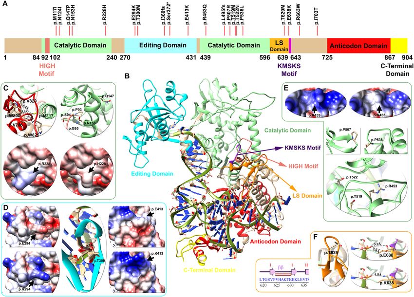
Figure 3. Motor head domain analysis of MYO6 protein. (A) Ideogram of MYO6. (B) RSMD analysis of the
motor head domain shows peaks with high RMSD values, revealing that two areas (one including the p.(F647L))
are involved in regions with great motility between pre-powerstroke and Pi release conformations. (C) Motor
head domain modeling. Pre-powerstroke conformation in light blue and Pi release in brown. The p.(Phe647Leu)
is shown with a black arrow. (D) Zoom in of the alpha-helix at the 647 region. The interaction of the wild-
type Phe647 residue or the mutated version Leu647 with the three nearest residues is shown for each protein
conformation (Pi release in brown and pre-powerstroke in light blue). The β-strand in green represents the actin
binding region. Distances in Å are detailed under each bubble, showing the increase in the distances for the
mutated Leu647 residue in both conformations.
characteristic high frequency hearing loss sloping pattern. This is in accordance with the audiogram of our
studied siblings21,22. After the curation process, variant c.1515-12G > A in CDH23, applying criteria PM2_Sup-
porting, PP3, PM3, PP4 and PP1_Supporting, changed its previous classification in ClinVar from VUS to Likely
Pathogenic. Further functional analysis, as well as new reports in Usher patients would provide confident evi-
dence concerning its pathogenicity. Variant c.1096G > A; p.(Ala366Thr) was classified as benign due to its high
population frequency in the Ashkenazi population (BA1 criteria applied). Hidden variants in gene regions that we
did not explore, such as deep intronic mutations that can disrupt transcription regulatory motifs and/or noncod-
ing RNA regions, might underlie the observed p henotype22,23. Taking into account all the evidence presented we
cannot unequivocally conclude that these two variants are the cause of the hearing loss in this family. Neverthe-
less, due to the reclassification of c.1515-12G > A to likely pathogenic, the case is worth mentioning and discussed.
In Case #10 a nonsense variant was detected in MITF. Most mutations in MITF have been mostly associated
with Waardenburg syndrome type 2, a dominant syndromic form of hearing loss. It is associated with hypo-
pigmentation of the skin, hair and eyes, since MITF has a regulatory effect on TYRtranscription, a key enzyme
involved in melanin s ynthesis24. The variant identified in our patients had already been reported in a Waarden-
burg type 2 family c ase15. Notably, none of the four affected members of this family presented any other signs in
addition to profound HL. This finding is in accordance with some reports in which variants in the MITF gene
cause only hearing loss25,26. The variable phenotype expression could be explained by the presence of modifier
genes, as well as interactions with environmental f actors27,28.
Several bioinformatic algorithms have been developed to predict the functional consequences of single
nucleotide variants in protein coding regions. These in silico approaches are an alternative to tedious and time-
consuming experimental approaches to infer pathogenicity. Thus, the new model presented in the present work
for the LeuRS human protein aided in defining the plausible pathogenicity of the identified p.Thr629Met vari-
ant in Case #9. Moreover, it can be further used to predict the pathogenicity of other reported variants. The
p.Thr629Met mutation in the patient lies in the beta hairpin of LeuRS between strands I-I of the LS domains
and leads to the disruption of the motif and protein stability. This is in accordance with the observation that
beta hairpin motifs are implicated in protein s tability29,30. Moreover, the substitution of p.Ala508 in Escherichia
coli LeuRS (analogous to human LeuRS p.Thr629) with a nonpolar methionine disrupts the structure and/or
Scientific Reports | (2022) 12:301 | https://doi.org/10.1038/s41598-021-04081-2 9
Vol.:(0123456789)www.nature.com/scientificreports/
position of the leucine-specific domain and thus shifts the location of the KMSKS loop and reduces the catalytic
efficiency16,31. It should be noted that the present model of human LeuRS is an improved version of those previ-
ously reported16,32 since we used a variety of selected crystals. In this regard, the present model shows that most
of the variants in the catalytic domain produce a severe effect on protein stability, affecting the proper function of
the protein. Our new model shows that variants reported as pathogenic in databases effectively induce significant
structural changes of the protein that would cause potential functional changes, playing an important role in
genotype/phenotype prediction. Thus, we propose that this model should be used for future in silico predictions
of variant pathogenicity in patients with Perrault syndrome, becoming a bridge between genomics and structural
data to guide the interpretation of human genetic variants.
In Case #6, we identified a novel MYO6 variant c.1939T > C, p.(Phe647Leu) in a family with late-onset auto-
somal dominant nonsyndromic hearing loss. Most MYO6-known genetic variants present progressive hearing
loss33–35 and myosin VI is required for structural integrity and proper functioning of inner ear hair c ells18,36.
More than half of the known pathogenic mutations are located in the motor head domain, indicating that it
plays an important role in the function of M YO634. The motor-head domain modeling approach applied in the
present work reveals that mutation p.(Phe647Leu) alters the proper function, most likely through the increased
distance of this amino acid from the three nearby nonpolar residues. Moreover, the structural change caused by
the mutation in the alpha-helix of this highly motile region could potentially affect the contiguous actin binding
region. In this regard, mutations in the MYO6 motor domain alter anchoring of the membrane of stereocilia to
actin filaments, leading to disruption of hair bundle o rganization36,37.
In conclusion, the present work highlights the importance of the curation of genetic variants leading to HL
following recommendations of experts for the correct phenotype-genotype correlation. Moreover, we show the
importance of the incorporation of integrated workflows for predicting the biomedical impact of the variations
identified by exome analysis. Most importantly, we propose a multitarget approach including genomics, protein
structure and data analysis to guide the interpretation and standardization of human genetic variants leading
to hearing loss.
Materials and methods
Subjects and selection criteria. Thirty-two unrelated Argentinean families were included in this study
(Fig. 1 and Supplementary Figure S1). Hearing loss was bilateral and moderate to severe (45–95 dB) or pro-
found (> 95 dB), and onset was either congenital or postlingual progressive (average 20–40 years). Audiological
evaluation included pure-tone audiometry at four frequencies (0.5, 1, 2, and 4 kHz). The pure-tone average
(PTA) was calculated from the audiometric thresholds. The HL patients were divided into three groups based
on severity: moderate (41–70 dB HL), severe (71–95 dB HL), and profound (> 95 dB HL). The audiometric con-
figurations were classified into low-frequency, middle-frequency (U-shaped), high-frequency and flat types38.
Patients underwent auditory brainstem response (ABR), tympanometry, fundus examination, and cardiac and
renal ultrasonography to detect undiagnosed syndromic forms. Clinical examinations revealed symptoms sug-
gesting a syndromic form of deafness in five patients: two with visual defects, two others with hematuria, and
one with hair pigment abnormalities. All data were reviewed by a clinical geneticist. The study was conducted
in accordance with the Declaration of Helsinki, and the protocol was approved by the Ethics Committee of
“Administración Nacional de Laboratorios e Institutos de Salud” (ANLIS-19122018) and “Fundación para la
Lucha contra las Enfermedades Neurológicas de la Infancia” (FLENI -04,092,020). Written informed consent for
testing and publication was obtained from patients or parents in the case of minors.
Genomic DNA was extracted from peripheral blood samples using the standard method. Quality and con-
centration were measured by agarose gel electrophoresis and absorbance-based nucleic acid quantification
(Thermo Scientific–NanoDrop™). A total of 695 patients with different forms of deafness were recruited for
GJB2/GJB6 mutation screening, identifying biallelic pathogenic mutations in 103 of them (15%). Thirty-two
patients undiagnosed for GJB2/GJB6 mutations were selected for WES screening. The age of the patients varied
between 6 months and 50 years. Seventeen out of the 32 patients (53%) were females, and fifteen (47%) were
males. Among the 32 probands, 50% (16/32) were sporadic and 50% (16/32) had at least two affected relatives
with HL (familial cases).
Whole exome sequencing and bioinformatics analysis. Massive parallel sequencing was carried
out on an Illumina NovaSeq6000. Base calling, read mapping and annotation of variants were performed by
Macrogen Genome Sequencing Services (Macrogen, Korea) and data were processed according to the Genome-
Analysis-Toolkit (GATK) best practices workflow. Variants for 183 deafness-related genes were filtered from
the WES on samples from affected patients and relatives when available (analyzed genes are listed in Supple-
mentary Table S1). The average read length covered 148 bp, with an average exon depth coverage in analyzed
genes of 100X; 97% of targeted reads had > tenfold coverage. Aligned reads were compared to the human refer-
ence genome (GRCh37/hg19). After variant annotation, mutations that arose from known deafness genes were
selected by filtering with an in silico panel using a homemade Python script pipeline. The missense, nonsense,
insertion/deletion and splicing variants were selected from the detected variants. The minor allele frequency
threshold (MAF) considered was ≤ 0.01 and 0.005 for recessive and dominant alleles, respectively, when com-
pared with those reported in gnomAD—http://gnomad.broadinstitute.org/) and 1000 genomes (https://www.
internationalgenome.org/1000-genomes-browsers/). The pathogenic potential of selected variants was analyzed
using bioinformatic programs and databases: Mutation Taster—http://www.mutationtasetr.org39, PolyPhen-
2—http://genetics.bwh.harvard.edu/pph/40, CADD (Combined Annotation Dependent Depletion)—http://
cadd.gs.washington.edu/, UMD-Predictor—http://umd-predictor.eu.41, ClinVar—https://www.ncbi.nlm.nih.
gov/clinvar/, LOVD—https://www.lovd.nl/, The Human Gene Mutation Database—http://www.hgmd.cf.ac.uk/,
Scientific Reports | (2022) 12:301 | https://doi.org/10.1038/s41598-021-04081-2 10
Vol:.(1234567890)www.nature.com/scientificreports/
dbNSFP—https://sites.google.com/site/jpopgen/dbNSFP. All information was compiled and criteria rules were
combined to reach a variant classification according to data retrieved from InterVar -http://wintervar.wglab.org/,
Varsome—https://varsome.com/ and REVEL—https://sites.google.com/site/revelgenomics42. Once annotation
of variants was performed, the number of significant variations was reduced from more than eighty thousand
to a small, manageable number (1 to 10) of putative candidate disease-causative mutations for further valida-
tion. This process included various parameters, such as mode of inheritance, mutation localization, mutation
type (nonsynonymous variants, splice acceptor or donor site mutations, and coding noninframe In/Dels), fre-
quency, pathogenicity of variants, published reports, and so forth. Pathogenicity prediction and variant clas-
sification were assessed taking into account criteria defined by the ACMG and further modifications, as well as
recommendations of the HL-EP and data from deafness variation databases (http://deafnessvariationdatabase.
org/)43–45. Variants were classified as pathogenic, likely pathogenic, VUS, likely benign or benign. Patients were
informed of the results, and clinical follow-up was recommended when necessary, since some likely patho-
genic variants were identified in syndromic genes, before the onset of additional clinical manifestations. Positive
results were always confirmed by Sanger sequencing prior to reporting, and segregation through family was
performed when possible.
Variant curation. To further analyze and validate the identified variants in patients, a combined in silico
analysis was performed. In the first curation step, the prediction of pathogenicity of the variants previously
reported in ClinVar was reanalyzed following the guidelines mentioned before43, collecting new data regarding
segregation, new PubMed reports or publications and new updated data of frequency and phenotype/genotype
arsome46.
correlations. The putative pathogenic effect of novel variants was manually scored using V
Bioinformatic analysis. To predict the potential consequences of some of the missense variants on protein
function, different bioinformatic analyses were performed. These included a molecular homology modeling
(MHM) approach and structural analysis of the mutated proteins. The line protein graphs in figures were gener-
ated with the PyGame library in the Python 2.7 programming language (http://www.python.org)47.
Molecular modeling and structural analysis. MHM was performed for mitochondrial (LeuRS) (UniProt_ID
Q15031-1) and the MYO6 motor-head domain (UniProt_ID Q9UM54-3, between amino acid residues 4 and
771). The evaluation criteria to select the template for the MHM were as follows: (1) protein sequence identity
as template, (2) X-ray crystal resolution less than 2.4 Å, (3) crystallography conditions, (4) chain length and
amount of residues of each template with respect to sequence identity and gaps, (5) structural conformation of
each protein template, and (6) species of the template. The alignment between template and target sequences was
performed with structural alignments (Expresso extension) in the T-Coffee web server48,49, MODELLER align-
ment scripts and Kluskal-Wallis in MEGA5 software, taking into account the secondary structures and topology
of the r egions50. Homology modeling was generated using the MODELLER p rogram51. The models were first
optimized with the variable target function method with conjugate gradients and then refined using molecular
dynamics with simulated annealing52. Model quality evaluation was performed using DOPE53, QMEAN/QME-
ANDisCo54,55 and P roSA56 to predict local pairwise residue-residue distances to the assessed model. The UCSF
57
Chimera program and the backbone-dependent rotamer library were used for structural interpretation and
visualization58.
For the LeuRS protein model, template structures were selected from the RCSB Protein Data Base59 and Uni-
Prot (https://www.uniprot.org/). Fifteen potential templates were considered using a cutoff in sequence identity
larger than 36% and a resolution less than 2.5 Å. For the alignment between templates and the LeuRS sequence,
the N-terminal mitochondrial targeting signals (presequences) were deleted using MitoFates60. Structural regions
that did not have reference templates were not considered for the structural analysis and are not shown in the
visualization of the structure. After evaluation, six structures in the editing conformation were selected (Sup-
plementary Table S2). Thirty molecular homology models with the selected templates were generated, and the
final structure of the tRNA was determined with the 4AS1 template.
The MYO6 protein structure is divided into three regions: an N-terminal motor-head-domain (which com-
prises the N-terminal SH3-like, myosin motor, actin binding site and ATP catalytic region), followed by a neck
domain (with a calmodulin-binding linker domain and a single IQ motif), and a C-terminal tail region (with a
three-helix bundle region, an SAH domain and a unique globular domain required for interaction with other
proteins such as cargo-binding)61 (Fig. 3A). The modeled N-terminal motor (head) contains the N-terminal
SH3-like (aa 4–53) and myosin motor (aa 57–771) domains with 98.15% identity between the target sequence
and templates and a resolution of less than 2.5 Å. Modeling was performed in two MYO6 conformations accord-
ing to ATPase cycle states: the pre-powerstroke, which is the key force-generating step (PDBID: 2V26, 4DBR,
4E7Z), and the next state with the release of phosphate, Pi release (PDBID: 4PFP, 4PFO, 4PJN)62 (Supplementary
Table S3).
Stability analysis. To evaluate the effect of mutations on the stability/folding of LeuRS, we first analyzed the
structural and energetic details of the interactions for each mutated residue using FoldX (http://foldxsuite.crg.
eu/).63. Structures were optimized to the FoldX force field command from the molecular homology models. The
∆∆G values were estimated as the difference between the energy of the wild-type protein and the average of five
replicas for each point mutation64. Values above 1,6 kcal/mol (twice the standard deviation) were considered to
significantly destabilize the protein. To favor the wild-type conformation, all residues involved in tRNA interac-
tions (5 Å distance) were fixed when optimizing the structures to the FoldX force field.
Scientific Reports | (2022) 12:301 | https://doi.org/10.1038/s41598-021-04081-2 11
Vol.:(0123456789)www.nature.com/scientificreports/
Data availability
The genetic variant data that support the findings of this study are openly available in ClinVar at [http://www.
clinvar.com/], reference number SUB10104745. New LeuRS and MYO6 protein models are openly available in
https://www.modelarchive.org/, https://doi.org/10.5452/ma-rc0b9 (LeuRS), https://doi.org/10.5452/ma-d28xp
(MYO6 pre-powerstroke conformation) and https://d oi.o
rg/1 0.5 452/m
a-w
e6d7 (MYO6 Pi release conformation).
Received: 23 August 2021; Accepted: 14 December 2021
References
1. Shearer, A. E., Hildebrand, M. S. & Smith, R. J. H. Hereditary hearing loss and deafness overview. In GeneReviews (eds Adam, M.
P. et al.) (University of Washington, 1999).
2. Morton, C. C. & Nance, W. E. Newborn hearing screening—a silent revolution. N. Engl. J. Med. 354, 2151–2164 (2006).
3. Wémeau, J.-L. & Kopp, P. Pendred syndrome. Best Pract. Res. Clin. Endocrinol. Metab. 31, 213–224 (2017).
4. Hilgert, N., Smith, R. J. H. & Van Camp, G. Forty-six genes causing nonsyndromic hearing impairment: Which ones should be
analyzed in DNA diagnostics?. Mutat. Res. 681, 189–196 (2009).
5. Feldmann, D. et al. Large deletion of the GJB6 gene in deaf patients heterozygous for the GJB2 gene mutation: Genotypic and
phenotypic analysis. Am. J. Med. Genet. A 127A, 263–267 (2004).
6. Snoeckx, R. L. et al. GJB2 mutations and degree of hearing loss: A multicenter study. Am. J. Hum. Genet. 77, 945–957 (2005).
7. Del Castillo, I. et al. Prevalence and evolutionary origins of the del(GJB6-D13S1830) mutation in the DFNB1 locus in hearing-
impaired subjects: A multicenter study. Am. J. Hum. Genet. 73, 1452–1458 (2003).
8. Dalamón, V. et al. GJB2 and GJB6 genes: Molecular study and identification of novel GJB2 mutations in the hearing-impaired
Argentinean population. Audiol. Neurootol. 15, 194–202 (2010).
9. Diaz-Horta, O. et al. Whole-exome sequencing efficiently detects rare mutations in autosomal recessive nonsyndromic hearing
loss. PLoS ONE 7, e50628 (2012).
10. Duman, D. & Tekin, M. Autosomal recessive nonsyndromic deafness genes: A review. Front. Biosci. 17, 2213–2236 (2012).
11. Atik, T. et al. Comprehensive analysis of deafness genes in families with autosomal recessive nonsyndromic hearing loss. PLoS
ONE 10, e0142154 (2015).
12. Choi, H. J. et al. Whole-exome sequencing identified a missense mutation in WFS1 causing low-frequency hearing loss: A case
report. BMC Med. Genet. 18, 151 (2017).
13. Shearer, A. E., Eliot Shearer, A., Hildebrand, M. S., Sloan, C. M. & Smith, R. J. H. Deafness in the genomics era. Hear. Res. 282,
1–9 (2011).
14. Parzefall, T. et al. Whole-exome sequencing to identify the cause of congenital sensorineural hearing loss in carriers of a heterozy-
gous GJB2 mutation. Eur. Arch. Otorhinolaryngol. 274, 3619–3625 (2017).
15. Jalilian, N. et al. A novel pathogenic variant in the MITF gene segregating with a unique spectrum of ocular findings in an extended
Iranian Waardenburg syndrome kindred. Mol. Syndromol. 8, 195–200 (2017).
16. Pierce, S. B. et al. Mutations in LARS2, encoding mitochondrial leucyl-tRNA synthetase, lead to premature ovarian failure and
hearing loss in Perrault syndrome. Am. J. Hum. Genet. 92, 614–620 (2013).
17. Soldà, G. et al. First independent replication of the involvement of LARS2 in Perrault syndrome by whole-exome sequencing of
an Italian family. J. Hum. Genet. 61, 295–300 (2016).
18. Avraham, K. B. et al. Characterization of unconventional MYO6, the human homologue of the gene responsible for deafness in
Snell’s waltzer mice. Hum. Mol. Genet. 6, 1225–1231 (1997).
19. Buonfiglio, P. et al. GJB2 and GJB6 genetic variant curation in an argentinean non-syndromic hearing-impaired cohort. Genes 11,
2 (2020).
20. Dalamón, V. et al. Identification of four novel connexin 26 mutations in non-syndromic deaf patients: Genotype-phenotype analysis
in moderate cases. Mol. Biol. Rep. 40, 6945–6955 (2013).
21. Miyagawa, M., Nishio, S.-Y. & Usami, S.-I. Prevalence and clinical features of hearing loss patients with CDH23 mutations: A large
cohort study. PLoS ONE 7, e40366 (2012).
22. Astuto, L. M. et al. CDH23 mutation and phenotype heterogeneity: A profile of 107 diverse families with Usher syndrome and
nonsyndromic deafness. Am. J. Hum. Genet. 71, 262–275 (2002).
23. Meng, X., Liu, X., Li, Y., Guo, T. & Yang, L. Correlation between genotype and phenotype in 69 Chinese patients with USH2A
mutations: A comparative study of the patients with Usher Syndrome and Nonsyndromic Retinitis Pigmentosa. Acta Ophthalmol.
https://doi.org/10.1111/aos.14626 (2020).
24. Yasumoto, K., Yokoyama, K., Shibata, K., Tomita, Y. & Shibahara, S. Microphthalmia-associated transcription factor as a regulator
for melanocyte-specific transcription of the human tyrosinase gene. Mol. Cell. Biol. 15, 1833 (1995).
25. Zhang, Z. et al. A novel variant in MITF in a child from Yunnan-Guizhou Plateau with autosomal dominant inheritance of non-
syndromic hearing loss: A case report. Mol. Med. Rep. 17, 6054–6058 (2018).
26. Thongpradit, S. et al. MITF variants cause nonsyndromic sensorineural hearing loss with autosomal recessive inheritance. Sci.
Rep. 10, 12712 (2020).
27. Sun, L. et al. Molecular etiology and genotype-phenotype correlation of Chinese Han deaf patients with type I and type II Waarden-
burg Syndrome. Sci. Rep. 6, 35498 (2016).
28. Pandya, A. Phenotypic variation in Waardenburg syndrome: mutational heterogeneity, modifier genes or polygenic background?.
Hum. Mol. Genet. 5, 497–502 (1996).
29. DuPai, C. D., Davies, B. W. & Wilke, C. O. A systematic analysis of the beta hairpin motif in the protein data bank. bioRxiv https://
doi.org/10.1101/2020.10.28.359612 (2020).
30. DuPai, C. D., Cunningham, A. L., Conrado, A. R., Wilke, C. O. & Davies, B. W. TsrA regulates virulence and intestinal colonization
in. mSphere 5, 2 (2020).
31. Zafar, S. et al. Novel mutations in CLPP, LARS2, CDH23, and COL4A5 identified in familial cases of prelingual hearing loss. Genes
11, 978 (2020).
32. Yan, W. et al. Modulation of aminoacylation and editing properties of leucyl-tRNA synthetase by a conserved structural module.
J. Biol. Chem. 290, 12256–12267 (2015).
33. Miyagawa, M., Nishio, S.-Y., Kumakawa, K. & Usami, S.-I. Massively parallel DNA sequencing successfully identified seven families
with deafness-associated MYO6 mutations: The mutational spectrum and clinical characteristics. Ann. Otol. Rhinol. Laryngol.
124(Suppl 1), 148S-S157 (2015).
34. Tian, T. et al. Identification of a novel MYO6 mutation associated with autosomal dominant non-syndromic hearing loss in a
Chinese family by whole-exome sequencing. Genes Genet. Syst. 93, 171–179 (2018).
35. Cheng, J. et al. Exome sequencing identifies a novel frameshift mutation ofMYO6as the cause of autosomal dominant nonsyndromic
hearing loss in a Chinese family. Ann. Hum. Genet. 78, 410–423 (2014).
Scientific Reports | (2022) 12:301 | https://doi.org/10.1038/s41598-021-04081-2 12
Vol:.(1234567890)You can also read