Switch-like control of helicase processivity by single-stranded DNA binding protein
←
→
Page content transcription
If your browser does not render page correctly, please read the page content below

RESEARCH ARTICLE

                                    Switch-like control of helicase processivity
                                    by single-stranded DNA binding protein
                                    Barbara Stekas1, Steve Yeo2, Alice Troitskaia2, Masayoshi Honda3, Sei Sho3,
                                    Maria Spies3, Yann R Chemla1,2,4*
                                    1
                                     Department of Physics, University of Illinois, Urbana-Champaign, Urbana, United
                                    States; 2Center for Biophysics and Quantitative Biology, University of Illinois,
                                    Urbana-Champaign, Urbana, United States; 3Department of Biochemistry, Carver
                                    College of Medicine, University of Iowa, Iowa City, United States; 4Center for the
                                    Physics of Living Cells, University of Illinois, Urbana-Champaign, Urbana, United
                                    States

                                    Abstract Helicases utilize nucleotide triphosphate (NTP) hydrolysis to translocate along single-
                                    stranded nucleic acids (NA) and unwind the duplex. In the cell, helicases function in the context of
                                    other NA-associated proteins such as single-stranded DNA binding proteins. Such encounters
                                    regulate helicase function, although the underlying mechanisms remain largely unknown.
                                    Ferroplasma acidarmanus xeroderma pigmentosum group D (XPD) helicase serves as a model for
                                    understanding the molecular mechanisms of superfamily 2B helicases, and its activity is enhanced
                                    by the cognate single-stranded DNA binding protein replication protein A 2 (RPA2). Here, optical
                                    trap measurements of the unwinding activity of a single XPD helicase in the presence of RPA2
                                    reveal a mechanism in which XPD interconverts between two states with different processivities
                                    and transient RPA2 interactions stabilize the more processive state, activating a latent ‘processivity
                                    switch’ in XPD. A point mutation at a regulatory DNA binding site on XPD similarly activates this
                                    switch. These findings provide new insights on mechanisms of helicase regulation by accessory
                                    proteins.

*For correspondence:
ychemla@illinois.edu
                                    Introduction
Competing interest: See
                                    Helicases are molecular machines that use the energy of NTP hydrolysis to separate the strands of
page 20
                                    nucleic acid (NA) duplexes. A large class of helicases unwind by translocating directionally along a
Funding: See page 20                single NA strand and can utilize this translocation activity to displace NA-bound proteins
Received: 29 June 2020              (Delagoutte and von Hippel, 2002; Delagoutte and von Hippel, 2003; Singleton et al., 2007;
Accepted: 18 March 2021             Lohman et al., 2008). In the cell, helicases are involved in many essential genome maintenance pro-
Published: 19 March 2021            cesses, including replication, recombination, and repair (McGlynn, 2013; Daley et al., 2013;
                                    Kuper and Kisker, 2013). In a number of instances, the same helicase is known to carry out several
Reviewing editor: James M
                                    of these disparate functions (Lohman et al., 2008; Wu and Spies, 2013; Beyer et al., 2013). Thus,
Berger, Johns Hopkins University
School of Medicine, United
                                    helicase activity must be tightly regulated not only to prevent indiscriminate unwinding of duplex
States                              DNA in the cell, which could lead to genome instability, but also to define the context-dependent
                                    role of the helicase. How this regulation occurs is often unclear, but growing evidence points to
   Copyright Stekas et al. This
                                    interactions with protein partners as one regulatory mechanism (Lohman et al., 2008; Wu and
article is distributed under the
                                    Spies, 2013; Beyer et al., 2013).
terms of the Creative Commons
Attribution License, which             Xeroderma pigmentosum group D (XPD) protein is a 50 to 30 DNA helicase that serves as a model
permits unrestricted use and        for understanding members of the structural superfamily 2B of helicases, a group that includes yeast
redistribution provided that the    Rad3 and human FANCJ, RTEL, and CHLR1 (Fairman-Williams et al., 2010; Byrd, 2012; White and
original author and source are      Dillingham, 2012; Beyer et al., 2013). Human XPD is part of transcription factor IIH and plays a vital
credited.                           role in nucleotide excision repair (Egly and Coin, 2011; Fuss and Tainer, 2011; Kuper et al., 2014;

Stekas et al. eLife 2021;10:e60515. DOI: https://doi.org/10.7554/eLife.60515                                                        1 of 23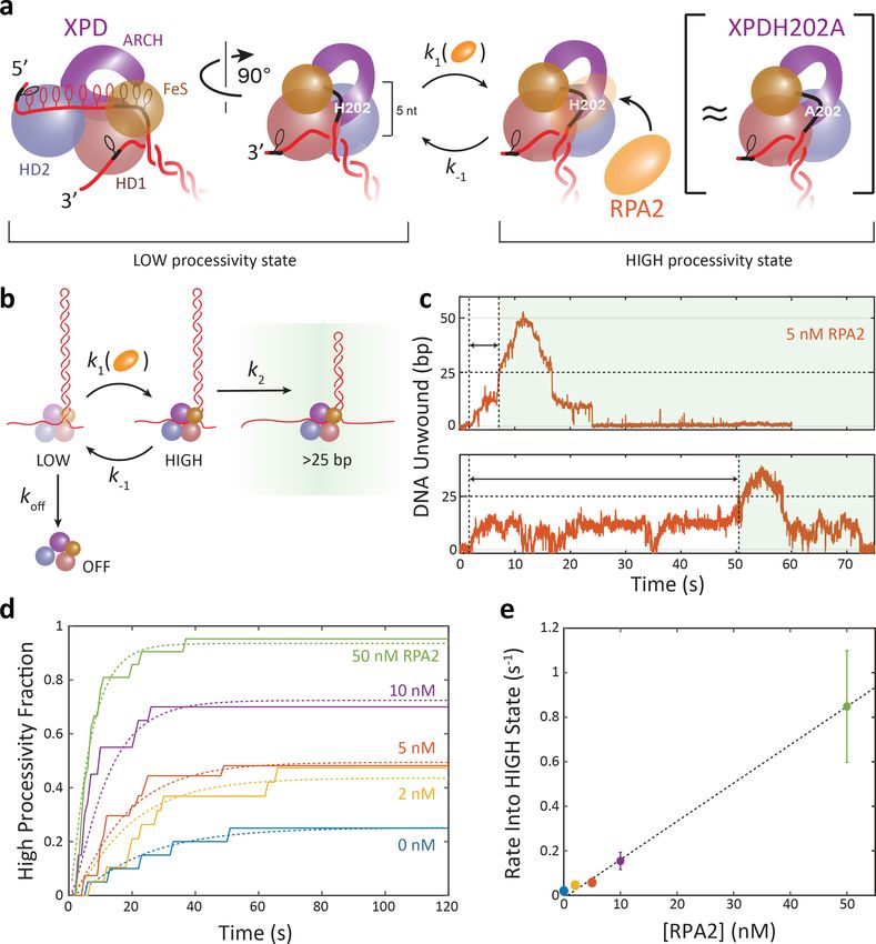
Research article                                                                                Structural Biology and Molecular Biophysics

                                     Houten et al., 2016). It has also been shown to participate in chromosome segregation (Ito et al.,
                                     2010) and in the cell’s defense against retroviral infection (Yoder et al., 2006). Structurally, XPD
                                     consists of a conserved motor core (Figure 1a)—helicase domains 1 and 2 (HD1 and HD2)—that
                                     couples ATP binding and hydrolysis to translocation on ssDNA, an FeS cluster-containing domain,
                                     and a unique ARCH domain that encircles the translocating strand (Fan et al., 2008; Liu et al.,
                                     2008; Wolski et al., 2008; Kuper et al., 2012; Spies, 2014).
                                        As it functions on ssDNA, XPD is expected to encounter other proteins such as replication protein
                                     A (RPA), which binds single-stranded DNA non-sequence specifically. Single-stranded DNA binding
                                     proteins function as protectors of ssDNA against nucleolytic damage and play important regulatory
                                     roles (Shereda et al., 2008; Caldwell and Spies, 2020). Due to their ubiquity, frequent interactions
                                     with other DNA-bound proteins are likely (Spies and Ha, 2010). Single-stranded binding proteins
                                     are known to stimulate the DNA unwinding activity of SF2 helicases, although the mechanisms of
                                     enhancement remain elusive (Harmon and Kowalczykowski, 2001; Rajagopal and Patel, 2008;
                                     Cadman and McGlynn, 2004; Cui et al., 2004; Gupta et al., 2007; Pugh et al., 2008;

Figure 1. RPA2 increases xeroderma pigmentosum group D (XPD) helicase processivity. (a) Schematic of FacXPD based on Sulfolobus acidocaldarius
XPD structure (PDB 3CRV). XPD is composed of four domains: helicase domains 1 and 2 (HD1, pink; HD2, blue), which form the motor core, and the
ARCH domain (purple) and FeS cluster (brown). A model of the DNA fork (red) shows ~10 nt bound to the motor core and secondary contacts (black) to
each strand of the fork. (b) Schematic of FacRPA2 based on partial crystal structure of Methanococcus maripaludis replication protein A (RPA) (PDB
2K5V). RPA2 consists of a single OB-fold that binds to ssDNA in the C-shaped cavity. (c) Single-molecule hairpin unwinding assay. A DNA hairpin
consisting of an 89 bp stem and (dT)4 loop is tethered between trapped beads by biotin–streptavidin (yellow cross) and digoxigenin-antibody linkages
(pentagon) and held at a constant force. A 10 nt poly-dT site at the 50 end of the hairpin allows one XPD molecule to load, and an abasic site at the 30
end prevents XPD unwinding past the hairpin. Unwinding of the hairpin by XPD increases the end-to-end extension of the construct by an amount, Dx,
proportional to the number of base pairs unwound. Arrows indicate the 50 to 30 direction of XPD translocation along ssDNA. (d) Representative traces
of a single molecule of XPD unwinding in the presence of varying concentrations of RPA2 (0–50 nM) at constant force (F = 12 pN). ATP and RPA2 are
added at t = 0 s. XPD processivity increases with RPA2 concentration.
The online version of this article includes the following figure supplement(s) for figure 1:
Figure supplement 1. DNA hairpin construct.
Figure supplement 2. Laminar flow chamber.

Stekas et al. eLife 2021;10:e60515. DOI: https://doi.org/10.7554/eLife.60515                                                                     2 of 23
Research article                                                                         Structural Biology and Molecular Biophysics

                                    Bétous et al., 2013). In the present work, we investigate the effect of single-stranded DNA binding
                                    proteins on helicase unwinding activity using XPD from Ferroplasma acidarmanus as a model system
                                    due to the availability of biochemical (Pugh et al., 2008) and single-molecule kinetic data
                                    (Honda et al., 2009; Qi et al., 2013; Ghoneim and Spies, 2014), as well as structural information
                                    from homologs (Liu et al., 2008; Wolski et al., 2008; Kuper et al., 2012; Wolski et al., 2010). Prior
                                    work (Pugh et al., 2008) has shown that FacXPD unwinding is enhanced greatly by one type of cog-
                                    nate ssDNA binding protein in F. acidarmanus, RPA2, more so than a second cognate protein,
                                    RPA1, or by heterologous proteins. FacRPA2 has a simple, single-domain architecture with one OB-
                                    fold that occludes ~4 nt of ssDNA (Figure 1b; Pugh et al., 2008). Single-molecule experiments dem-
                                    onstrated that XPD can occupy the same DNA strand as RPA2 and bypass RPA2-bound ssDNA
                                    (Honda et al., 2009). Crucially, no direct, specific interactions between RPA2 and XPD in solution
                                    have been observed (Pugh et al., 2008).
                                        How an ssDNA binding protein with no known protein–protein contacts to the helicase stimulates
                                    helicase-mediated DNA unwinding activity remains an open question and makes FacXPD and
                                    FacRPA2 an intriguing system for studying the stimulatory effects of ssDNA binding proteins on heli-
                                    cases. Mechanisms for enhancement of unwinding activity can loosely be placed in three categories
                                    (Pugh et al., 2008). First, an ssDNA binding protein could destabilize the DNA duplex at the
                                    ssDNA–dsDNA junction ahead of the helicase, facilitating its motion forward. Second, it could
                                    enhance unwinding by sequestering ssDNA and providing a physical barrier to helicase backsliding,
                                    that is, rectifying helicase forward motion. Lastly, it could activate the helicase for processive unwind-
                                    ing through direct interactions with the helicase-DNA complex.
                                        Here, we use a single-molecule optical trap assay to observe individual molecules of XPD unwind-
                                    ing DNA and to analyze the effects of RPA2 on their unwinding activity. While previous reports have
                                    shown that RPA2 is able to enhance XPD activity (Pugh et al., 2008; Honda et al., 2009), they have
                                    not defined what aspects of activity are enhanced or provided a mechanism. Our results show that
                                    RPA2 primarily increases XPD processivity or the maximum number of base pairs unwound. XPD
                                    exhibits repeated attempts to unwind duplex DNA, and RPA2 increases the frequency of attempts
                                    that have high processivity. While RPA2 can transiently destabilize duplex DNA, this does not pro-
                                    mote XPD unwinding. Instead, data point to a mechanism in which XPD possesses a latent processiv-
                                    ity ‘switch’, which is activated specifically by RPA2. We propose that an integral component of this
                                    processivity switch is the regulatory interaction between DNA and a secondary binding site on XPD,
                                    as supported by a point mutation at this site that enhances XPD processivity similarly to RPA2. Our
                                    measurements shed new light on the mechanisms by which accessory proteins such as single-
                                    stranded DNA binding proteins can enhance helicase activity.

                                    Results
                                    XPD processivity increases in the presence of RPA2
                                    As shown in Figure 1c, we monitored the activity of a single XPD in the absence and presence of
                                    RPA2 in solution from the unwinding of an 89 bp DNA hairpin (Figure 1—figure supplement 1)
                                    tethered between two optically trapped beads and stretched under constant force, as described
                                    previously (Qi et al., 2013). Measurements were carried out over a range of forces (7–12 pN) at
                                    which single-molecule XPD activity was previously reported (Qi et al., 2013) and below that required
                                    to mechanically unfold the hairpin (15 pN; Figure 1—figure supplement 1). Data were collected at
                                    a rate of 89 Hz. Unwinding of the hairpin was detected from the increase in the end-to-end exten-
                                    sion of the DNA tether as each broken base pair released two nucleotides (see
                                    Materials and methods). A 10 dT ssDNA site—approximately equal to the footprint of XPD
                                    (Qi et al., 2013; Kokic et al., 2019)—at the 50 end of the hairpin served as a binding site for a single
                                    XPD molecule, and an abasic site positioned at the 30 end of the hairpin prevented XPD from
                                    unwinding the long (1.5 kb) dsDNA handle used to separate the hairpin from the trapped beads
                                    (Qi et al., 2013; Figure 1—figure supplement 1). To control the loading of XPD and RPA2 onto the
                                    hairpin, we used a custom flow chamber consisting of parallel laminar-flow streams containing differ-
                                    ent buffers, as described previously (Whitley et al., 2017) (see Materials and methods; Figure 1—
                                    figure supplement 2a). The hairpin was placed in a protein ‘loading’ stream containing XPD but no
                                    ATP for ~1 min., which allowed a single XPD to bind to the 10 dT loading site without unwinding the

Stekas et al. eLife 2021;10:e60515. DOI: https://doi.org/10.7554/eLife.60515                                                            3 of 23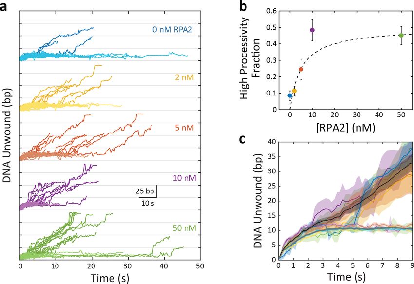
Research article                                                                          Structural Biology and Molecular Biophysics

                                    DNA. Unwinding was initiated by moving the hairpin and bound XPD into the ‘unwinding’ stream
                                    containing saturating ATP (500 mM), varying concentrations of RPA2 (0–50 nM), and no additional
                                    XPD in solution (Figure 1—figure supplement 2b, c). This procedure ensured that only a single XPD
                                    was loaded at one time, and that the unwinding observed resulted from that of a single XPD helicase
                                    in the absence or presence of many RPA2 molecules in solution (see Materials and methods).
                                        Figure 1d shows representative traces of a single XPD protein unwinding in increasing [RPA2] at
                                    a force of 12 pN. As previously reported, XPD unwound in repeated ‘bursts’, comprising cycles of
                                    forward unwinding motion followed by backward rezipping (Qi et al., 2013), repeating a number of
                                    times (5 ± 1) until the protein dissociated (e.g., t = 95 s for 0 nM RPA2 or t = 160 s for 2 nM RPA2 in
                                    Figure 1d). Unwinding never resumed once a dissociation event occurred, indicating that the single
                                    molecule of XPD was flushed away in the flow chamber. In the absence of RPA2, we observe that
                                    the processivity—the maximum hairpin position reached by XPD during any one burst—was low, on
                                    average 15–20 out of 89 bp, consistent with our prior work (Qi et al., 2013). With increasing [RPA2],
                                    XPD unwound farther into the hairpin, occasionally unwinding the entire 89 bp hairpin stem (e.g.,
                                    t = 20 s for 5 nM RPA2 or t = 30 s, 70 s for 50 nM RPA2 in Figure 1d).
                                        Each burst represents an attempt by one XPD molecule to unwind the hairpin, and we found that
                                    processivity varied greatly from burst to burst. Each burst consisted of varying extents of duplex
                                    unwinding followed by rezipping, often to the base of the hairpin. As shown in the representative
                                    trace in Figure 2a, several molecular events comprised backward motion: XPD backstepping or
                                    backsliding via temporary disengagement from the unwinding DNA strand (Figure 2b, diagrams 1a
                                    and 1b), or translocation on the other hairpin strand away from the fork junction (2 and 3), all leading
                                    to DNA rezipping under its regression force. An example of the latter occurred when XPD
                                    completely unwound the hairpin stem and translocated past the dT tetraloop cap onto the opposing
                                    strand, allowing the stem to rezip gradually in the protein’s wake (Figure 2b, diagram 2). In a con-
                                    trasting example, we observed gradual rezipping of the duplex mid-hairpin, which we attributed to
                                    XPD disengaging from its DNA strand to switch to the opposing strand (Figure 2b, diagram 3), a
                                    behavior reported for other helicases (Dessinges et al., 2004; Comstock et al., 2015). This interpre-
                                    tation is corroborated by the observation that gradual rezipping was typically followed by a stall
                                    at ~10 bp (Figure 2a, t = 58 s) the size of XPD’s footprint, consistent with the protein stalling at the
                                    abasic site on the 30 strand at the base of the hairpin. Subsequent unwinding bursts were presum-
                                    ably due to XPD strand-switching back to the original strand. In contrast, rapid rezipping events due
                                    to backsliding usually occurred to the base of the hairpin (Figure 2a, t = 44 s), consistent with the
                                    protein remaining on the hairpin 50 strand.
                                        We analyzed the processivity of each burst at 12 pN as a function of [RPA2], as shown in the scat-
                                    ter plot in Figure 2c. In the absence of RPA2, nearly all the individual burst processivities cluster
                                    below 25 bp, with a small fraction (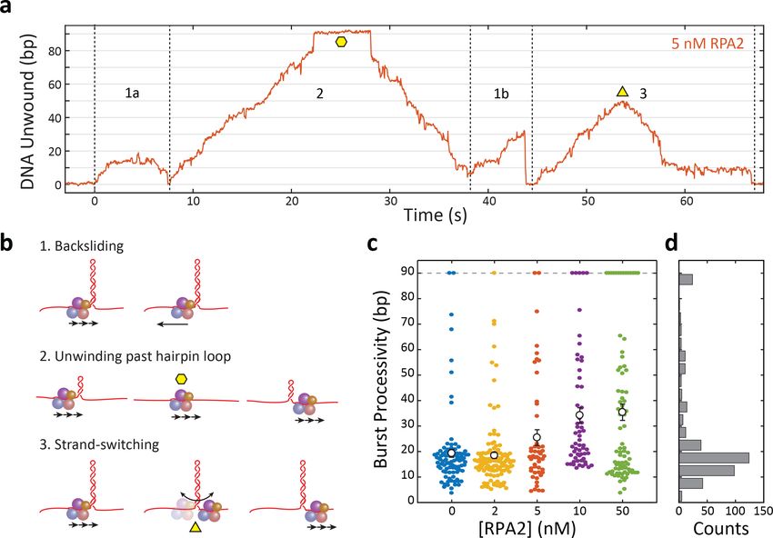
Research article                                                                                  Structural Biology and Molecular Biophysics

Figure 2. Xeroderma pigmentosum group D (XPD) unwinds in bursts of varying processivity whose average increases with replication protein
A 2 (RPA2) concentration. (a) Representative trace of a single molecule of XPD unwinding in the presence of RPA2 (5 nM) at constant force (F = 12 pN).
One XPD exhibits repetitive bursts of activity, making multiple attempts to unwind hairpin DNA. Processivity can vary widely from burst to burst.
Example time trace with (1a) 20 bp- and (1b) 30 bp-processivity bursts composed of forward unwinding followed by backsliding to the hairpin base;
(2) a high-processivity burst during which XPD completely unwinds the 90 bp hairpin past the hairpin loop (time point indicated by yellow hexagon) and
translocates on the opposing strand, allowing the hairpin to rezip; and (3) a 50 bp-processivity burst during which XPD unwinds and switches strand
mid-hairpin (indicated by yellow triangle), allowing the hairpin to rezip. (b) Schematics representing the behaviors in (a). (c) Processivity of each burst
(colored circles) vs. RPA2 concentration. The mean processivity (open circles) increases with RPA2 concentration. Error bars represent s.e.m. (d)
Histogram of all burst processivities at all RPA2 concentrations.
The online version of this article includes the following source data and figure supplement(s) for figure 2:
Source data 1. XPD burst processivity vs. [RPA2].
Figure supplement 1. Xeroderma pigmentosum group D (XPD) burst processivity increases with replication protein A 2 (RPA2) concentration at a force
of 9 pN.

                                      (Pugh et al., 2008), RPA2 would presumably bind at the ssDNA–dsDNA junction and melt several
                                      base pairs ahead of XPD. Because XPD relies heavily on thermal fluctuations to help it break the
                                      base pairing bonds ahead of it (Qi et al., 2013), RPA2 lowering the energy barrier to unzipping the
                                      duplex could plausibly enhance XPD’s unwinding activity.
                                         As shown in Figure 3a, RPA2 is capable of transiently destabilizing hairpin DNA under force of 12
                                      pN. In experiments with RPA2 but without XPD, short melting events are observed, corresponding
                                      to the hairpin opening by ~5–6 bp, comparable to the footprint of RPA2 (Pugh et al., 2008), and
                                      reannealing shortly thereafter (~20 ms). (Such transient melting is also observed in Figure 1d after
                                      XPD dissociation [e.g., at t > 110 s for 50 nM RPA2].) These melting events become more frequent

Stekas et al. eLife 2021;10:e60515. DOI: https://doi.org/10.7554/eLife.60515                                                                        5 of 23
Research article                                                                      Structural Biology and Molecular Biophysics

Table 1. Data statistics.
Force (pN)                         7–8                 9                              12
XPD                                wt                  wt               H202A         wt                                          H202A
[RPA2] (nM)                                            0       10       0       10    0       2         5       10       50       0         10
[gp32] (nM)                        0         250
No. of XPD molecules               10        8         6       25       11      11    20      19        27      20       21       29        16
No. of bursts                      141       23        54      74       38      67    94      123       79      60       82       123       79
No. of bursts/XPD                  14.1      2.9       9       3        3.5     6.1   4.7     6.5       2.9     3.0      3.9      4.2       4.9
No. of low-processivity bursts     141       23        54      60       36      63    86      109       61      31       45       46        36
No. of high-processivity bursts    0         0         0       14       2       4     8       14        18      29       37       77        43
XPD: xeroderma pigmentosum group D; RPA2: replication protein A 2; gp32: gene protein 32.

                                    as the concentration of RPA2 is increased (Figure 3—figure supplement 1), in support of melting
                                    being RPA2-mediated. Analyzing the times between melting events, we estimate an effective sec-
                                    ond-order rate constant of (1.2 ± 0.3)108 M 1 s 1 for binding.
                                        RPA2-mediated melting events are also observable as XPD unwinds in the presence of RPA2
                                    (Figure 3b). Such events are identifiable over XPD unwinding dynamics due to their characteristic
                                    size (~5–6 bp) and short lifetime (~20 ms) (Figure 3b, inset; see Materials and methods and Fig-
                                    ure 3—figure supplement 2). While some false positives are detected in the absence of RPA2, they
                                    are uncommon and the frequency of detected melting events increases with [RPA2] (Figure 3c), con-
                                    firming their connection to RPA2 activity. This finding indicates that RPA2 is capable of binding at
                                    the same fork junction as XPD and melting DNA ahead of the helicase. However, the question
                                    remains whether RPA2 transient melting aids XPD in unwinding.
                                        To answer this question, we determined how RPA2-mediated melting events impact the forward
                                    progress of the helicase. If duplex destabilization aids in unwinding, we suspected XPD may be
                                    more likely to advance along the DNA during melting events. For example, RPA2 melting could
                                    maintain the DNA fork open by ~5 bp, allowing XPD to translocate rapidly to catch up to the fork
                                    position and then to take the lead unwinding at the fork junction. However, analyzing the occur-
                                    rences of ~5 bp melting events followed by XPD unwinding by its step size of 1 bp (Qi et al.,
                                    2013; Figure 3—figure supplement 2; light blue box), we found that such pairs of events become
                                    less frequent as RPA2 concentration increases (8.0 ± 0.5% in the absence of RPA2 vs. 4.0 ± 0.2% at
                                    50 nM RPA2), inconsistent with this mechanism.
                                        To determine if XPD could advance during RPA2-mediated melting, we examined the position of
                                    the helicase on the hairpin prior to each RPA2 melting event and after the duplex reannealed. We
                                    identified many (N » 400) individual melting events during XPD unwinding and aligned them along
                                    the position axis such that each event began at 0 bp before melting occurred (Figure 3d, sche-
                                    matic). We then independently aligned the melting and reannealing transition of each event in time
                                    at t = tmelt and t = tanneal, respectively (Figure 3d, ‘before’ and ‘after’ schematic). As shown in
                                    Figure 3e, the resulting probability density of these aligned traces reveals the average position of
                                    XPD prior to and following an RPA2 melting event (see Materials and methods). After a melting
                                    event, we find that the hairpin is most likely to reanneal all the way back to the same position as
                                    before the event, indicating that XPD has not moved from its initial position. Importantly, this result
                                    shows that there is no significant forward movement of XPD as a result of RPA2 duplex melting, and
                                    that XPD does not exploit RPA2’s melting activity to enhance its own unwinding. Figure 3f shows a
                                    control in which the alignment process above was repeated at random points during unwinding,
                                    each point paired with one t = 0.06 s after, corresponding to the time window over which we
                                    searched and identified RPA2 melting events (see Materials and methods). Again, XPD on average
                                    shows no net progress in this amount of time. Comparing Figure 3e and f shows that the RPA2-
                                    mediated melting events have little to no impact on XPD movement.
                                        Our data collected at lower forces further disfavor a melting mechanism. As force is decreased,
                                    RPA2-mediated melting occurs less frequently, with almost no melting detected at 9 pN and below
                                    (Figure 3—figure supplement 3). If RPA2 melting were necessary to increase the unwinding

Stekas et al. eLife 2021;10:e60515. DOI: https://doi.org/10.7554/eLife.60515                                                            6 of 23
Research article Structural Biology and Molecular Biophysics Figure 3. Replication protein A 2 (RPA2) transiently melts hairpin duplex but does not assist xeroderma pigmentosum group D (XPD) unwinding. (a) Representative time trace of RPA2 transiently destabilizing hairpin dsDNA at a constant force (F = 12 pN). Inset: RPA2 melts ~8 bp, which then rapidly reanneals (see Figure 3—figure supplement 1). (b) RPA2 is able to melt dsDNA at a fork occupied with XPD. Inset: transient RPA2-like melting events during XPD unwinding (see Figure 3—figure supplement 2). (c) The frequency of RPA2 melting events increases with RPA2 concentration. Error bars Figure 3 continued on next page Stekas et al. eLife 2021;10:e60515. DOI: https://doi.org/10.7554/eLife.60515 7 of 23
Research article                                                                                   Structural Biology and Molecular Biophysics

Figure 3 continued
represent s.e.m. (d) Analysis of RPA2 melting events and their effect on XPD unwinding. The schematic shows all RPA2 melting events identified and
divided into ‘melting’ and ‘reannealing’ transitions. Each transition is aligned temporally such that all melting transitions begin at the same time tmelt
and corresponding reannealing transitions end at the same time tanneal. Both types of transitions are aligned spatially relative to the starting extension
before melting (x = 0). (e) Probability distribution of all aligned RPA2 melting events. Although RPA2 melts an average of 5 bp of hairpin DNA (left), it
reanneals by the same amount (right), and there is no net progress of XPD due to melting. (f) Probability distribution at a random time point (left) and
0.06 s later (right). Net progress of XPD after an RPA2 melting event is indistinguishable from net progress at a random time point. Probability
distributions were obtained using kernel density estimation (see Materials and methods).
The online version of this article includes the following source data and figure supplement(s) for figure 3:
Source data 1. RPA2 melting statistics and XPD burst processivity vs. [gp32].
Figure supplement 1. Replication protein A 2 (RPA2) transiently melts DNA under force.
Figure supplement 2. Detecting replication protein A 2 (RPA2) melting events during xeroderma pigmentosum group D (XPD) unwinding.
Figure supplement 3. Replication protein A 2 (RPA2) transient melting of DNA depends on force.
Figure supplement 4. T4 gene protein 32 (gp32) transiently melts hairpin duplex but does not assist xeroderma pigmentosum group D (XPD)
unwinding.

                                      processivity of XPD, we would expect little to no change in activity at these forces. However, XPD
                                      activity was enhanced by RPA2 at 9 pN (Figure 2—figure supplement 1), with a significantly higher
                                      fraction of bursts with processivities in the long tail extending from 25 to 89 bp.
                                         Lastly, we measured XPD unwinding in the presence of bacteriophage T4 gene protein 32 (gp32),
                                      a non-cognate single-stranded DNA binding protein capable of destabilizing the DNA duplex
                                      (Pugh et al., 2008; Pant et al., 2003). Like RPA2, gp32 is a monomeric protein containing a single
                                      OB-fold and thus possesses a similar footprint as RPA2 (Robbins et al., 2005; Shamoo et al., 1995;
                                      Theobald et al., 2003). Prior ensemble studies showed that gp32 can melt short forked duplexes,
                                      but enhances XPD unwinding to a much lower extent than RPA2 under the same conditions
                                      (Pugh et al., 2008). If transient duplex melting were sufficient to enhance XPD processivity, we
                                      would expect gp32 to yield similar enhancements under conditions where it melts the hairpin to a
                                      similar extent as RPA2. We determined that at forces of 7–8 pN and a concentration of 250 nM
                                      gp32 melts the hairpin transiently by ~5–10 bp, comparable to RPA2 at 12 pN (Figure 3—figure
                                      supplement 4a). We estimate that the extent of melting by gp32 under these conditions is equiva-
                                      lent to that of 10–35 nM RPA2 at 12 pN. (Hairpin DNA is transiently melted 2.5% and 7.0% of the
                                      time at 10 and 35 nM RPA2, respectively, compared to 4.1% of the time for gp32.) However, gp32
                                      has a minimal impact on XPD processivity under these conditions. We observed no bursts in the >25
                                      bp, high-processivity tail of the distribution (Figure 3—figure supplement 4b), in marked contrast
                                      to the corresponding conditions with RPA2 (Figure 2d). Taken together, these data strongly indicate
                                      that enhancement of XPD processivity by RPA2 is mediated by a mechanism other than duplex
                                      melting.

                                      XPD exhibits two distinct types of activity, the fraction of which
                                      depends on RPA2
                                      The burst processivities at different [RPA2] (Figure 2c) show that a persistent and significant fraction
                                      of bursts exhibit relatively low processivity, <
                                                                                     ~ 25 bp. Bursts with higher processivity extending from
                                      25 to 89 bp also exist at all [RPA2], with their number apparently increasing as [RPA2] increases. An
                                      important question is whether there exist any characteristic differences between bursts during which
                                      more or less DNA is unwound.
                                          To answer this question, we grouped bursts into two categories: ‘high processivity’ or
                                      ‘low processivity’ depending on whether or not a threshold of 25 bp was exceeded, this value cho-
                                      sen based on where the tail in the distribution of processivities occurred (Figure 2c). Figure 4a
                                      shows the unwinding portions of many bursts at a force of 12 pN aligned to begin at the same time
                                      (t = 0), color coded by [RPA2] and processivity category (light color for low processivity; dark color
                                      for high processivity; see Table 1 for the number of data traces). When grouped in this manner,
                                      some notable features emerge. The fraction of bursts categorized as high processivity increases
                                      clearly with RPA2 concentration (Figure 4a) saturating to ~50% at RPA2 concentrations >10 nM
                                      (Figure 4b). Furthermore, long stalls (sometimes exceeding tens of seconds in duration) around 10–
                                      15 bp are evident at all concentrations and in both high- and low-processivity traces. This stalling

Stekas et al. eLife 2021;10:e60515. DOI: https://doi.org/10.7554/eLife.60515                                                                        8 of 23
Research article Structural Biology and Molecular Biophysics
Figure 4. Xeroderma pigmentosum group D (XPD) exhibits two burst types, the fraction of which is replication protein A 2 (RPA2) dependent. (a) Plot of
XPD unwinding bursts, aligned to start at t = 0 and grouped by RPA2 concentration (colored traces). XPD unwinding bursts come in two types: low
processivity, never unwinding more than 25 bp (light colors); and high processivity, unwinding more than 25 bp (dark colors). (b) The fraction of high-
processivity bursts (>25 bp) increases with RPA2 concentration. Fit to model described in the text (dashed line; see Materials and methods). (c)
Averages of all low-processivity bursts (light colored lines) and all high-processivity bursts (dark colored lines) at each RPA2 concentration. Comparison
to averages of low- and high-processivity burst types over all RPA2 concentrations (dark gray and black lines, respectively). Shaded regions represent s.
e.m. throughout. Unwinding behavior within each burst category remains the same over all RPA2 concentrations.
The online version of this article includes the following source data and figure supplement(s) for figure 4:
Source data 1. High-processivity fraction and burst duration vs. [RPA2], and XPD speed vs. position.
Figure supplement 1. Unwinding velocity varies with processivity type but not replication protein A 2 (RPA2) concentration.
Figure supplement 2. Xeroderma pigmentosum group D (XPD) backward motion varies with processivity type but not replication protein A 2 (RPA2)
concentration.
behavior has been reported previously (Qi et al., 2013) and attributed to XPD’s encounter with GC-
rich regions—which are more difficult to unwind due to higher base-pairing energy—at this position
in the hairpin. All low-processivity bursts exhibit these stalls, after which XPD either backslides to the
base of the hairpin or dissociates, ending the burst. However, we have observed XPD to continue
unwinding past 25 bp after a long stall and be categorized as ‘high processivity’.
Importantly, we do not observe significant differences between bursts of the same processivity
category obtained at different RPA2 concentrations. Figure 4c shows all unwinding bursts at 12 pN
for high- and low-processivity types averaged together at each RPA2 concentration (color coded the
same as in Figure 4a) and across all RPA2 concentrations (black and gray lines, respectively). The
averaged unwinding traces exhibit the same behavior independent of RPA2 concentration. Low-
Stekas et al. eLife 2021;10:e60515. DOI: https://doi.org/10.7554/eLife.60515 9 of 23Research article Structural Biology and Molecular Biophysics
processivity bursts all display stalls at ~10 bp, whereas high-processivity bursts unwind past this
region. Burst-to-burst differences are reflected by the shaded areas and are consistently smaller than
the differences between the two processivity categories. This finding is corroborated when analyzing
the velocity of XPD. Figure 4—figure supplement 1 shows the average unwinding velocity deter-
mined separately for low- and high-processivity bursts at each RPA2 concentration as a function of
XPD’s position on the hairpin. While XPD on average slows down near 10 bp due to the high GC
content of this section of the hairpin, the velocity of high-processivity bursts is consistently higher
than for low-processivity bursts at this position. On the other hand, differences in velocity are insig-
nificant across varying [RPA2] within the same processivity category.
We also find that both low- and high-processivity burst durations remain constant with RPA2 con-
centration (Figure 4—figure supplement 2, light-colored and open data points, respectively). The
more processive bursts tend to have longer durations since XPD travels a longer distance on DNA,
and processivity increases with [RPA2]. The increase in burst duration observed when combining
both types of bursts is explained simply from the increase in the fraction of high-processivity bursts
with increasing [RPA2].
Our results show that high-processivity unwinding in the absence of RPA2 is indistinguishable
from that in the presence of RPA2 and likewise for low-processivity unwinding. All parameters we
quantified by burst in the same processivity category are independent of [RPA2]. Instead, [RPA2]
increases the probability of high-processivity bursts. These findings suggest that high- and low-proc-
essivity unwinding correspond to intrinsic states of XPD, with RPA2 increasing the likelihood of XPD
being in a state competent for high-processivity unwinding.
A mutant of XPD recapitulates the behavior of wild-type XPD in the
presence or RPA2
In a previous study, point mutations in the homologous Thermoplasma acidophilum XPD were iden-
tified that yielded increased unwinding activity and force generation (Pugh et al., 2012). We
enquired if mutants of FacXPD could act similarly to the high-processivity state proposed above in
overcoming energetic barriers in the DNA sequence (such as the high-GC section of the hairpin near
10 bp) and what the effect of RPA2 on their activity would be. We thus synthesized the mutant
FacXPDH202A, which targeted the site analogous to H198 in TacXPD that had been shown to elicit
the largest positive effect on unwinding activity (Pugh et al., 2012). This residue is located on
domain HD1, within a secondary binding site thought to interact with the translocating strand of
DNA in the groove between HD1 and the FeS domains (see Figure 1a, black DNA contacts behind
the FeS domain). It is distinct from the residues shown to be responsible for XPD damage verification
(Mathieu et al., 2013) (see Discussion).
Figure 5a shows representative time traces of XPDH202A unwinding alone (cyan) and in the pres-
ence of 10 nM RPA2 (magenta) at 12 pN of force. Like wild-type (wt) XPD, XPDH202A also unwinds
DNA in repeated bursts. However, on its own, XPDH202A exhibits higher processivity and can regu-
larly unwind the entire hairpin during its multiple bursts on the DNA hairpin. As shown in the scatter
plot in Figure 5b, the average burst processivity of XPDH202A greatly exceeds that of wt XPD and
is also slightly higher than that of wt XPD in the presence of 10 nM RPA2. Similarly, the fraction of
high-processivity bursts for XPDH202A is much higher than for wt XPD and comparable to that for
wt XPD with RPA2 (Figure 5c). Interestingly and unlike wt XPD, the unwinding processivity of
XPDH202A does not increase appreciably in the presence of 10 nM RPA2 (Figure 5b, c). We
observed similar trends at a lower force of 9 pN (Figure 5—figure supplement 1). Overall, the proc-
essivity of XPDH202A (alone or in the presence of RPA2) is similar to that of wt XPD with RPA2, sug-
gesting that RPA2 elicits the same effect on XPD as the H202A mutation.
Discussion
Our single-molecule measurements of XPD with RPA2 provide important clues to decipher the
underlying mechanism of processivity enhancement. We have already explored some mechanisms in
which RPA2 binding to DNA could enhance XPD activity. Our results in Figure 3 (and also Figure 2—
figure supplement 1 and Figure 3—figure supplement 4) rule out RPA2 melting of DNA as a plau-
sible mechanism. The data also disfavor a sequestration mechanism, in which RPA2 binding to
ssDNA behind XPD inhibits retrograde motion. By acting as a physical barrier against XPD
Stekas et al. eLife 2021;10:e60515. DOI: https://doi.org/10.7554/eLife.60515 10 of 23Research article Structural Biology and Molecular Biophysics Figure 5. A point mutation in xeroderma pigmentosum group D (XPD) enhances its processivity similarly to replication protein A 2 (RPA2). (a) Representative traces of a single molecule of the mutant XPDH202A unwinding at constant force (F = 12 pN) alone (cyan) and in the presence of RPA2 (10 nM; magenta). (b) Processivity of individual bursts (colored circles) at a constant force of 12 pN for wild-type XPD alone (blue) and with 10 nM RPA2 (purple), and for XPDH202A alone (cyan) and with 10 nM RPA2 (magenta). The H202A mutation increases XPD’s mean processivity (open circles); Figure 5 continued on next page Stekas et al. eLife 2021;10:e60515. DOI: https://doi.org/10.7554/eLife.60515 11 of 23
Research article                                                                                Structural Biology and Molecular Biophysics

Figure 5 continued
addition of RPA2 does not enhance processivity further. (c) The fraction of high-processivity bursts (>25 bp) corresponding to (b). The H202A mutation
increases the fraction of high-processivity bursts to a level similar to that of XPD with RPA2. Error bars represent s.e.m. throughout.
The online version of this article includes the following source data and figure supplement(s) for figure 5:
Source data 1. XPD and mutant burst processivity vs. [RPA2], and complex formation statistics .
Figure supplement 1. A point mutation in xeroderma pigmentosum group D (XPD) enhances its processivity similarly to replication protein A 2 (RPA2)
at a force of 9 pN.
Figure supplement 2. Test of stable complex formation.

                                     backstepping or backsliding (Figure 4—figure supplement 2a), two behaviors often exhibited by
                                     XPD during unwinding (Qi et al., 2013), ssDNA sequestration could in principle enhance processiv-
                                     ity. However, identifying XPD backsteps and backslides in unwinding traces (e.g., Figure 3—figure
                                     supplement 2) shows that RPA2 did not reduce their frequency. There is a marginal increase in the
                                     fraction of backsteps (which we defined as steps between 2 and 0 bp) from 19 ± 1% to 25 ± 1%
                                     and almost no change in the fraction of backslides (defined as steps smaller than 2 bp) from
                                     18 ± 1% to 19 ± 1% as RPA2 increased from 0 to 50 nM. Furthermore, since each burst involves
                                     XPD-mediated DNA unwinding followed by rezipping to the base of the hairpin, the duration of a
                                     burst provides information on the amount of retrograde motion. We expect bursts with less frequent
                                     backsteps and backslides to be longer in duration since XPD would unwind further into the hairpin
                                     and thus take longer to return to the hairpin base. However, as shown in Figure 4—figure supple-
                                     ment 2b, we do not observe the mean duration of either the low- or high-processivity bursts to
                                     increase with RPA2, as predicted by a sequestration mechanism.
                                         Alternately, RPA2 may enhance unwinding through direct interactions with XPD (Figure 5—figure
                                     supplement 2). Direct RPA–helicase interactions have been detected for the superfamily 2 human
                                     helicases WRN, BLM, RECQ, FANCJ, and SMARCAL1 and shown to increase helicase activity
                                     (Cui et al., 2004; Gupta et al., 2007; Bétous et al., 2013; Brosh et al., 1999; Brosh et al., 2000;
                                     Doherty et al., 2005). The inability of the non-cognate T4 gp32 to enhance XPD unwinding
                                     (Pugh et al., 2008 and Figure 3—figure supplement 4) strongly suggests that specific interactions
                                     between XPD and RPA2 are essential. While no strong interactions between FacXPD and FacRPA2
                                     have been reported in solution (Pugh et al., 2008), formation of an XPD–RPA2 complex may require
                                     pre-assembly on DNA.
                                         Our measurements of the mutant XPDH202A point to the potential nature of specific XPD–RPA2–
                                     DNA interactions. As shown in Figure 6a, XPD has several secondary DNA binding sites, and the
                                     residue H202 lies within one site on the HD1 domain, in the groove between the HD1 and FeS
                                     domains (Kuper et al., 2012; Pugh et al., 2012). XPD–ssDNA contacts at this site are believed to
                                     be important for DNA fork positioning and to play an inhibitory role, acting as a throttle to unwind-
                                     ing (Pugh et al., 2012). Disruption of contact points by mutagenesis in both TacXPD (Pugh et al.,
                                     2012) and FacXPD (Figure 5) enhances unwinding. Structural analysis of XPD and homologs
                                     maps ~5 nt of the translocating DNA strand to this secondary site between the DNA fork at the FeS
                                     domain and the entry pore into the motor core (Qi et al., 2013; Kokic et al., 2019; Pugh et al.,
                                     2012; Figure 6a). We propose that RPA2 binding of these 5 nt of ssDNA could interfere with the
                                     inhibitory XPD–ssDNA contacts at this secondary site and activate XPD unwinding to a similar extent
                                     as mutation. As we have shown, RPA2 can bind at a DNA junction occupied with XPD (Figure 3),
                                     and the 5 nt matches the footprint of RPA2 (Pugh et al., 2008). Such a mechanism is consistent with
                                     our observation that XPDH202A processivity is comparable to that of wt XPD in the presence of
                                     RPA2, and that RPA2 does not enhance XPDH202A processivity further (Figure 5, Figure 5—figure
                                     supplement 1). In further support of this model, prior single-molecule measurements (Qi et al.,
                                     2013) showed that individual XPD exhibits 5 bp backward/forward step pairs (see also Figure 3—
                                     figure supplement 2; dark blue box), attributed to the transient release/recapture of 5 nt of ssDNA
                                     from the secondary site. In the presence of RPA2, we observe the frequency of such events to
                                     decrease (Figure 3—figure supplement 2; compare dark blue box for 0 nM to 50 nm RPA2), consis-
                                     tent with the idea that RPA2 may affect DNA contacts to this regulatory site. Lastly, the fact that
                                     gp32 has no effect on XPD despite having a similar footprint as RPA2 indicates that binding of

Stekas et al. eLife 2021;10:e60515. DOI: https://doi.org/10.7554/eLife.60515                                                                   12 of 23
Research article Structural Biology and Molecular Biophysics Figure 6. Replication protein A 2 (RPA2) activates a high-processivity state of xeroderma pigmentosum group D (XPD). (a) Model of XPD enhancement by RPA2. Schematic of XPD–DNA complex (left; side and front views), with 5 nt of ssDNA (black) bound at a regulatory secondary binding site on HD1 that contains H202. XPD can adopt one of two intrinsic states—a low (left) and high (right) processivity state—that correspond to different DNA fork conformations. Interconversion between both states occurs either spontaneously or through RPA2 binding of the 5 nt at the regulatory site. XPDH202A adopts a similar conformation (right). (b) Kinetic model of XPD processivity and the effect of RPA2. XPD interconverts between low and high states with rates k1 and k-1, and RPA2 shifts the equilibrium toward the high state. XPD can dissociate from DNA only from the low state with rate koff. Only in the Figure 6 continued on next page Stekas et al. eLife 2021;10:e60515. DOI: https://doi.org/10.7554/eLife.60515 13 of 23
Research article                                                                                  Structural Biology and Molecular Biophysics

Figure 6 continued
high state is XPD able to unwind in excess of 25 bp, which occurs at a rate k2. Once an XPD molecule unwinds >25 bp, it is scored as being in the high-
processivity state. (c) Representative traces of XPD exhibiting high-processivity unwinding. The time t>25 denotes the first time at which XPD crosses the
25 bp threshold. (d) Fraction of all XPD molecules that have reached high processivity (>25 bp) after time t for each RPA2 concentration. The curves are
globally fit to the model in (a) using as parameters the rate constants k1, k-1, k2, and koff. (e) The rate of entry into the high-processivity state, k1,
depends linearly on RPA2 concentration. For more details on the kinetic model, see Materials and methods.
The online version of this article includes the following source data for figure 6:
Source data 1. High-processivity fraction vs. time and high-processivity state entry rate vs. [RPA2].

                                      ssDNA at the secondary site is insufficient by itself; specific protein–protein contacts between RPA2
                                      and XPD must also be required.
                                           As the above arguments point to direct interactions between XPD and RPA2 mediated by DNA,
                                      we tested whether we could pre-form a stable XPD–DNA complex on DNA. We carried out an
                                      experiment in which we allowed XPD and RPA2 to bind to DNA in the absence of ATP, then pro-
                                      vided ATP for unwinding. Here, we utilized a different flow chamber configuration in which one
                                      stream contained XPD and RPA2 (Figure 5—figure supplement 2b; ‘loading’ stream) but no ATP,
                                      and another stream contained ATP but no protein (‘unwinding’ stream). We first placed the DNA
                                      hairpin in the protein ‘loading’ stream for 40–60 s, then moved it into the ATP-containing ‘unwind-
                                      ing’ stream to observe unwinding. If XPD and RPA2 were to form a complex with higher processivity
                                      than XPD alone, we would expect more processive activity at higher RPA2 concentrations as the
                                      probability of a complex forming should increase. However, we observed no effect on processivity
                                      across RPA2 concentration (Figure 5—figure supplement 2c); the fraction of bursts that exhibited
                                      high processivity (>25 bp) remained constant. This result indicates that any complex formed would
                                      be short-lived and would not survive the move between the protein and ATP streams, which takes
                                      up to ~10 s.
                                           Integrating all our results, Figure 6a shows our proposed mechanism. XPD can adopt two intrin-
                                      sic states, one competent for high-processivity unwinding and the other not. We propose that these
                                      two states correspond to different conformations of the DNA fork in its interactions with the second-
                                      ary binding site on XPD. Both states are populated independently of RPA2; highly processive
                                      unwinding activity can occur in the absence of RPA2 and is indistinguishable from that detected in
                                      the presence of RPA2. RPA2 does not assist XPD unwinding by directly destabilizing the duplex, but
                                      rather by transiently shifting the equilibrium toward the state competent for high processivity. RPA2
                                      binding to ssDNA at the fork interferes with its interactions with the secondary site in a similar way
                                      as the H202A mutation, activating high-processivity unwinding. In the data we have collected, the
                                      two states are effectively hidden, and we can infer their presence only from the processivity exhib-
                                      ited by XPD, that is, whether XPD unwinds beyond a threshold we have selected as 25 bp, past the
                                      first high-GC energetic barrier in the hairpin sequence that XPD must overcome. As a result, the cor-
                                      respondence between conformational state and processivity need not be exact.
                                           Figure 6b shows a simple kinetic scheme that describes this model quantitatively. XPD can switch
                                      between ‘low’- and ‘high’-processivity states with rates k1 and k-1. Here, we assume k1 to be depen-
                                      dent on RPA2 concentration, whereas all other rate constants are independent of RPA2. In the high-
                                      processivity state, XPD reaches the 25 bp threshold—and is scored as high processivity—at a rate
                                      k2, corresponding to the time XPD takes to unwind 25 bp, which encompasses all forward and back-
                                      ward steps to this position. Finally, we assume that XPD can dissociate from the DNA in the low-
                                      processivity state with rate constant koff. Given this kinetic scheme, we derive an expression for the
                                      probability, P>25(t), that a given XPD molecule unwinds >25 bp by time t (see
                                      Materials and methods), which can be compared directly to experimental data (Figure 6d), obtained
                                      from the fraction of XPD molecules that have crossed the 25 bp threshold at each time point
                                      (Figure 6c).
                                           Figure 6d shows a global fit to our data using different values of k1 for each RPA2 concentration
                                      but a common set of values of k-1, k2, and koff. (We considered an alternative model in which XPD
                                      can dissociate in both its low- and high-processivity states by introducing an additional rate constant
                                        0                                                                                        0
                                      koff ; see Materials and methods. The best fit to the data yielded negligible values for koff , indicating
                                      that XPD dissociation occurs in its low-processivity state but is unlikely from its high-processivity
                                      state.) The parameter values can be found in Table 2 and are consistent with independent

Stekas et al. eLife 2021;10:e60515. DOI: https://doi.org/10.7554/eLife.60515                                                                      14 of 23
Research article Structural Biology and Molecular Biophysics
Table 2. Model fit parameters.
1
Rate constant Fit value (s )
k-1 0.18 ± 0.13
k2 0.170 ± 0.017
koff 0.037 ± 0.019
0
koff 0
k1, [RPA2]=0 nM 0.02 ± 0.006
k1, 2 nM 0.059 ± 0.017
k1, 5 nM 0.065 ± 0.019
k1, 10 nM 0.200 ± 0.059
k1, 50 nM 1.15 ± 0.39
Error bars represent 95% confidence intervals.
RPA2: replication protein A 2.
measurements. The value obtained for koff predicts that XPD remains bound to DNA for an average
lifetime >25 s, consistent with observation. A value of k2 = 0.17 ± 0.02 s 1 matches that expected
for XPD to unwind 25 bp at an average speed of 4–5 bp/s (Figure 4—figure supplement 1a), a
speed consistent with previous measurements (Qi et al., 2013). The rate constant k-1 in our kinetic
model determines the lifetime of the high-processivity state, and its value gives a lifetime of ~8 s,
which agrees with interpretations of a short-lived complex lastingResearch article Structural Biology and Molecular Biophysics
Nguyen et al., 2017). Identifying the molecular details of the conformational states that regulate
helicase activity is a rich subject ripe for future investigation.
Materials and methods
Protein synthesis and purification
FacXPD and FacRPA2 were purified as described previously by Pugh et al., 2008. The H202A
mutant was constructed by site-directed mutagenesis. A QuikChange Lightning site-directed muta-
genesis kit (Agilent) was used to introduce an H202A substitution into the pET28a-FacXPD plasmid
using CGGTTCAATGTTTTTGCTGGCACCGAAGGGG and CCCCTTCGGTGCCAGCAAAAACA
TTGAACCG custom synthesized primers (Integrated DNA Technologies). Successful construction of
the pET28a-FacXPD H202A plasmid was confirmed by sequencing at the DNA Core Sequencing
Facility (University of Iowa IIHG Genomics Division). Mutant protein was expressed and purified iden-
tically to the wt XPD. T4 gp32 was obtained from a commercial source (New England Biolabs
#M0300S).
DNA construct synthesis
The hairpin construct was synthesized as described in Qi et al., 2013. The construct consists of three
separate dsDNA fragments ligated together after synthesis and purification (Figure 1—figure sup-
plement 1): a 1.5 kb ‘right handle’, an 89 bp ‘hairpin’ stem capped by a (dT)4 loop, and a 1.5 kb ‘left
handle’. ‘Right handle’ and ‘left handle’ are functionalized with a 50 digoxigenin and biotin, respec-
tively, for binding to anti-digoxigenin and streptavidin-coated beads. The hairpin stem sequence
used for all experiments was identical to ‘sequence 1’ used in Qi et al., 2013, which contains a ran-
dom 49% GC sequence: 50 -GGC TGA TAG CTG AGC GGT CGG TAT TTC AAA AGT CAA CGT ACT
GAT CAC GCT GGA TCC TAG AGT CAA CGT ACT GAT CAC GCT GGA TCC TA-30 . The hairpin
stem is flanked by a 50 10 dT binding site for loading XPD and a 30 abasic site to prevent XD unwind-
ing into the handle. All oligonucleotides were purchased from Integrated DNA Technologies (Coral-
ville, IA).
Flow chamber for optical tweezers measurements
All measurements were carried out in laminar flow chambers described in Whitley et al., 2017.
Chambers were made from NescoFilm (Karlan, Phoenix, AZ) melted between two glass coverslips
and laser-engraved with channels for buffers. Chambers for all measurements had three channels
(Figure 1—figure supplement 2). Top and bottom channels contained anti-digoxigenin and DNA-
coated streptavidin beads, respectively. Small (100 mm OD) glass capillaries connected the top and
bottom channels to the central channel for trapping and allow a controlled flow of beads. Three sep-
arate streams converged into the central trapping channel. Because the flow in each stream is lami-
nar, mixing between different buffer streams is minimal and a reasonably sharp boundary between
buffer conditions was maintained (Figure 1—figure supplement 2a, inset). This chamber design
allowed moving freely between different streams via motorized sample stage and changing buffer
conditions during an experiment, as described below.
Optical trap measurements
Two high-resolution dual-trap optical tweezers instruments based on previously reported designs
(Whitley et al., 2017; Comstock et al., 2011; Bustamante et al., 2008) were used to study XPD
helicase unwinding in the presence of RPA2. The optical traps were calibrated by standard proce-
dures (Whitley et al., 2017; Berg-Sørensen and Flyvbjerg, 2004) and both traps had a typical stiff-
ness of k = 0.3 pN/nm in all experiments. All data were acquired using custom LabVIEW software
(National Instruments, Austin, TX) available in an external repository (Chemla, 2020a;
Chemla, 2020b). Most data were collected on one instrument at a 267 Hz sampling rate and boxcar
filtered to a lower frequency, usually 89 Hz or as indicated in the text. Measurements with
XPDH202A and gp32 were made on a second instrument at a 100 Hz sampling rate. All measure-
ments were carried out at a constant force (ranging between 7 and 12 pN, as indicated in the text)
using a feedback loop to maintain constant force.
Stekas et al. eLife 2021;10:e60515. DOI: https://doi.org/10.7554/eLife.60515 16 of 23Research article Structural Biology and Molecular Biophysics
Single XPD unwinding experiments were performed in a similar manner to that described in
Qi et al., 2013, with some amendments. The trapping buffer consisted of 100 mM Tris-HCl (pH 7.6),
20 mM NaCl, 20 mM DTT, 3 mM MgCl2, 0.1 mg/mL BSA, and oxygen scavenging system
(Swoboda et al., 2012) (0.5 mg/mL pyranose oxidase [Sigma-Aldrich, St. Louis, MO], 0.1 mg/mL cat-
alase [Sigma-Aldrich, St. Louis, MO], and 0.4% glucose) to increase the lifetime of the DNA tethers
(Landry et al., 2009). To this buffer, varying concentrations of XPD, ATP, ATP-gS, and RPA2 or T4
gp32 were added.
During a typical experiment, the XPD–DNA complex was assembled by moving the traps
between the three streams of the central trapping channel of the sample flow cell (Figure 1—figure
supplement 2a). First, a single DNA hairpin was tethered between an optically trapped streptavidin-
coated bead and an anti-digoxigenin-coated bead in the ‘blank’ stream, containing 500 mM ATP-gS
but no protein or ATP (step 1 in Figure 1—figure supplement 2b, c). A low force (~2 pN) was
applied to the tether, and the traps and tethered DNA were moved by motorized sample stage into
the ‘loading’ stream containing 60 nM XPD + 500 mM ATP-gS, where they incubated for ~40–60 s to
allow a single XPD to bind to the 10 dT ssDNA loading site but not unwind the hairpin (step 2). ATP-
gS was used to increase the binding efficiency of XPD to the DNA loading site (Honda et al., 2009).
Following incubation, the force on the tether was increased to a constant force (7–12 pN, as indi-
cated) and the tether was moved into the ‘unwinding’ stream containing 500 mM ATP + 0–50 nM
RPA2 or 250 nM T4 gp32 (step 3). (An exception to this protocol was used for testing XPD–RPA2
complex formation [Figure 5—figure supplement 2], in which case the loading stream contained 0–
200 nM RPA2 in addition to XPD + ATP-gS, and the unwinding stream had no RPA2.) Upon exposure
to ATP, a single XPD molecule bound at the loading site unwound the hairpin ahead of it, which, at
constant force, resulted in an increase in the end-to-end extension of the DNA tether (Figure 1—fig-
ure supplement 2c). Unwinding data were typically collected until the tether broke or XPD
dissociated.
For most measurements, the blank stream contained 10 nM of the dye molecule Cy3 in addition
to 500 mM ATP-gS. Cy3 was added to this stream to detect, via fluorescence imaging, the precise
locations of the stream boundaries (Figure 1—figure supplement 2c). Fluorescence detection of
the Cy3 stream was achieved using a confocal microscope incorporated into the optical trap instru-
ment, as described in Whitley et al., 2017 and Comstock et al., 2011. Measurements with
XPDH202A and gp32 were made on an instrument that did not have fluorescence capabilities, and
thus Cy3 was not included.
Analysis of DNA hairpins and determining base pairs unwound by XPD
A force–extension curve of each tether was usually taken in the middle stream containing no protein
to verify proper synthesis. A properly synthesized hairpin unzips mechanically at an applied force
of ~15 pN (Figure 1—figure supplement 1b), and its force–extension curve is well fit to the follow-
ing model:
xðF Þ ¼ Nds ds ðF Þ þ Nss ss ðF Þ; (1)
where ds ðF Þ and ss ðF Þ are the extension of 1 bp of dsDNA and 1 nt of ssDNA at a given force, F,
respectively, and Nds and Nss are the number of dsDNA base pairs and ssDNA nucleotides, respec-
tively, in the construct. ds ðF Þ and ss ðF Þ are given by the extensible worm-like chain model of elastic-
ity using the following parameters: persistence length Pds = 50 nm and Pss = 1.0 nm, contour length
per base pair/nucleotide hds = 0.34 nm bp 1 and hss = 0.59 nm nt 1, and stretch modulus
Sds = 1,000 pN and Sss = 1,000 pN (Qi et al., 2013; Camunas-Soler et al., 2016). For the closed
hairpin at forces < 15 pN, the model Equation (1) was used with Nds = 3050 bp, corresponding to
the sum of the handle lengths, and Nss = 10 nt, corresponding to the helicase loading site length
(Figure 1—figure supplement 1b, black dashed line). For the open hairpin at forces > 15 pN, values
Nds = 3050 bp and Nss = 192 nt were used (Figure 1—figure supplement 1b, gray dashed line).
As a helicase unwinds the tethered DNA hairpin at a constant force, the tether extension
increases due to the release of 2 nt of ssDNA for each base pair of the hairpin dsDNA unwound. The
number of base pairs unwound at each time point was obtained from the relation
Stekas et al. eLife 2021;10:e60515. DOI: https://doi.org/10.7554/eLife.60515 17 of 23You can also read