Primary chaetotaxy and morphometry of the head capsule and head appendages of first instar larvae of Chaetarthria bruchi Coleoptera: ...
←
→
Page content transcription
If your browser does not render page correctly, please read the page content below
Revista de la Sociedad Entomológica Argentina
                                                              ISSN: 0373-5680
                                                              ISSN: 1851-7471
                                                              santiago@cepave.edu.ar
                                                              Sociedad Entomológica Argentina
                                                              Argentina

Primary chaetotaxy and morphometry of
the head capsule and head appendages
of first instar larvae of Chaetarthria
bruchi (Coleoptera: Hydrophilidae:
Chaetarthriinae: Chaetarthriini)
ARCHANGELSKY, Miguel
Primary chaetotaxy and morphometry of the head capsule and head appendages of first instar larvae of
Chaetarthria bruchi (Coleoptera: Hydrophilidae: Chaetarthriinae: Chaetarthriini)
Revista de la Sociedad Entomológica Argentina, vol. 80, núm. 2, 2021
Sociedad Entomológica Argentina, Argentina
Disponible en: https://www.redalyc.org/articulo.oa?id=322066981004

                                   PDF generado a partir de XML-JATS4R por Redalyc
                    Proyecto académico sin fines de lucro, desarrollado bajo la iniciativa de acceso abierto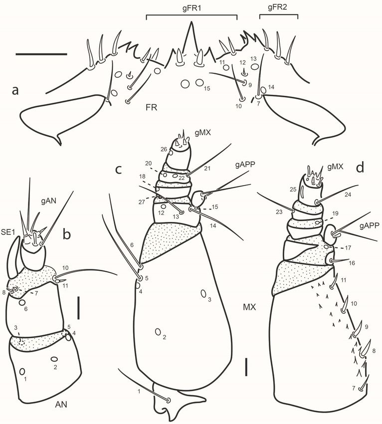
Artículos
Primary chaetotaxy and morphometry
of the head capsule and head appendages
of first instar larvae of Chaetarthria
bruchi (Coleoptera: Hydrophilidae:
Chaetarthriinae: Chaetarthriini)
Quetotaxia primaria y morfometría de la cápsula cefálica y
apéndices cefálicos del primer estadio larval de Chaetarthria
bruchi (Coleoptera: Hydrophilidae: Chaetarthriinae:
Chaetarthriini)
Miguel ARCHANGELSKY hydrophilinae@gmail.com
Laboratorio de Investigaciones en Ecología y Sistemática Animal
(LIESA). CIEMEP (CONICET-Universidad Nacional de La Patagonia,
San Juan Bosco)., Argentina
Revista de la Sociedad Entomológica
Argentina, vol. 80, núm. 2, 2021
Sociedad Entomológica Argentina,
Argentina Abstract: e primary chaetotaxy of the head capsule and head appendages of the larva
Recepción: 10 Diciembre 2020 of Chaetarthria bruchi Balfour-Browne is described for the first time. Morphometric
Aprobación: 15 Abril 2021 characters derived from the head capsule and mouthparts are included, together with
Publicación: 30 Junio 2021 detailed illustrations of all characters. Chaetotaxy of C. bruchi is compared to that of C.
seminulum (Herbst), the only other Chaetarthriini species for which the chaetotaxy has
Redalyc: https://www.redalyc.org/ been described.
articulo.oa?id=322066981004
Keywords: Aquatic beetles, Argentina, Morphology, Taxonomy.
Resumen: La quetotaxia primaria de la cápsula cefálica y de los apéndices cefálicos
de la larva de Chaetarthria bruchi Balfour-Browne se describen por primera vez.
Caracteres morfométricos derivados de la cabeza y sus apéndices son incluidos junto con
ilustraciones detalladas de todos los caracteres. La quetotaxia de C. bruchi se compara
con la de C. seminulum (Herbst), la única otra especie de Chaetarthriini para la cual se
ha descripto la quetotaxia.
Palabras clave: Argentina, Escarabajos acuáticos, Morfología, Taxonomía.
INTRODUCTION
Chaetarthria Stephens is a water scavenger beetle genus included in
the subfamily Chaetarthriinae, tribe Chaetarthriini (Short & Fikáček,
2013). Formally the tribe includes four genera in the Neotropical
region: Apurebium García, Chaetarthria, Guyanobius Spangler and
Venezuelobium García (Spangler, 1986; García, 2002; Gustafson & Short,
2010). Nonetheless, it has been suggested by Short (2009) that the genera
Apurebium and Venezuelobium include species that seem to be variants of
Chaetarthria, in which case the tribe would have only two genera in the
Neotropical region, Guyanobius and Chaetarthria (Clarkson et al., 2018).
e other two genera belonging to this tribe, Hemisphaera Pandellé and
ysanarthria Orchymont, are restricted to the Ethiopian, Palearctic and
PDF generado a partir de XML-JATS4R por Redalyc
Proyecto académico sin fines de lucro, desarrollado bajo la iniciativa de acceso abierto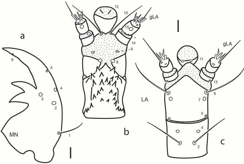
Revista de la Sociedad Entomológica Argentina, 2021, vol. 80, núm. 2, Abril-Junio, ISSN: 0373-5680 / 1851-7471
Oriental regions. is tribe is easily recognized by a unique characteristic
of the adults that consists of a fringe of setae arising at the anterior margin
of the first abdominal ventrite that covers a large depression usually filled
with a hyaline substance of unknown function.
Chaetarthria has a worldwide distribution, comprising 49 species (52
species if Apurebium and Venezuelobium are considered synonyms of
Chaetarthria), most of which are restricted to the New World. Larval
knowledge of the New World Chaetarthriini is better than that of
other regions since larvae of both Chaetarthria and Guyanobius have
been described (Spangler, 1986; Archangelsky, 1997, 2002). Nonetheless,
the original descriptions only deal with the general morphology of
these larvae, and no attempts were made to include chaetotaxic and
morphometric characters. In this contribution, the primary chaetotaxy of
the first instar larva of Chaetarthria bruchi Balfour-Browne is described
in detail and morphometric characters are also included. e chaetotaxy
of C. bruchi is compared to that of the European species C. seminulum
(Herbst) presented by Fikáček (2006). Larvae of Hemisphaera and
ysanarthria remain unknown.
MATERIAL AND METHODS
Source of material. Two first instar larvae of C. bruchi were studied for
the descriptions. P. N. Talampaya, arroyo Shimpa, 29° 44’ 43” S, 67° 44’
52” W, 9-XI-1999. e material studied is kept in the larval collection of
the author and will be deposited in the larval collection of the Laboratory
of Entomology, Buenos Aires University, Argentina.
Methods. Larval specimens were cleared in warm lactic acid, dissected,
and mounted on glass slides with Hoyer’s medium. Observations (up
to 1000×), photographs and drawings were done with a Leica S6D
dissecting microscope and Leica DMLB compound microscope both
with camera lucida and a photographic camera attached.
Morphometry. Different measurements of the head capsule and head
appendages were taken with a micrometer. Measurements were used
to calculate ratios, which are practical to characterize shapes. Measured
structures were adjusted as parallel as possible to the plane of the
objective. e following measurements were taken: TL: total body
length; MW: maximum body width, measured at level of prothorax;
HL: head length, measured medially along epicranial stem from anterior
margin of frontoclypeus to occipital foramen; HW: maximum head
width; AL: length of antenna, derived by adding the lengths of the first
(A1L), second (A2L) and third (A3L) antennomeres; SeL: length of
antennal sensorium; SL: length of stipes; MPL: length of maxillary palpus,
obtained by adding the lengths of the first (MP1L), second (MP2L), third
(MP3L) and fourth (MP4L) palpomeres; ML: length of maxilla, derived
by adding SL and MPL; cardo omitted; LPL: length of labial palpus,
obtained by adding the lengths of the first (LP1L) and second (LP2L)
palpomeres; LigL: length of ligula; MtW: maximum width of mentum;
PDF generado a partir de XML-JATS4R por Redalyc
Proyecto académico sin fines de lucro, desarrollado bajo la iniciativa de acceso abierto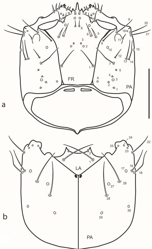
Miguel ARCHANGELSKY. Primary chaetotaxy and morphometry of the head capsule and head appendages of first instar larvae of Chaetarthria
bruch...
PrmtL: length of prementum, measured from its base to the base of LP1;
PrmtW: maximum width of prementum.
Chaetotaxy. Primary (present in first-instar larva) setae and pores
were identified in the cephalic capsule and head appendages following
system developed by Fikáček et al. (2008) and Byttebier & Torres (2009).
Homologies were established using the criterion of similarity of position
(Wiley, 1981). Sensilla were coded with a number and two capital letters,
usually corresponding to the first two letters of the name of the structure
on which they are located. e following abbreviations were used: AN:
antenna; FR: frontale; LA: labium; MN: mandible; MX: maxilla; PA:
parietale; gAN: group of antennal sensilla; gAPP: group of sensilla on the
inner appendage of the maxilla; gFR1, gFR2: groups of sensilla on the
frontale; gLA: group of sensilla on the labial palp; gMX: group of sensilla
on the maxillary palp.
Table I. Measurements (in mm) and ratios for different
structures of first instar larvae of Chaetarthria bruchi.
Abbreviations: see Material and Methods section.
PDF generado a partir de XML-JATS4R por Redalyc
Proyecto académico sin fines de lucro, desarrollado bajo la iniciativa de acceso abiertoRevista de la Sociedad Entomológica Argentina, 2021, vol. 80, núm. 2, Abril-Junio, ISSN: 0373-5680 / 1851-7471
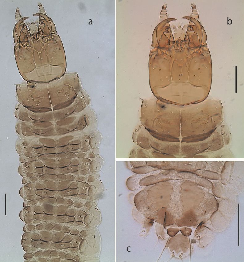
Fig. 1. Chaetarthria bruchi, first instar larva. a. habitus, dorsal view. b. detail of head
capsule and prothorax, dorsal view. c. detail of abdominal segment VIII, dorsal view.
Scale bars = 0.1 mm
RESULTS
Description of the first instar larva of C. bruchi
Diagnosis of Chaetarthria larvae (first instar). e following
combination of characters distinguish larvae of Chaetarthria from other
known Hydrophilidae larvae. Larval morphology (Figs. 1-4). Head
capsule subquadrate; frontal lines subparallel, reaching occipital foramen
widely apart, coronal line absent; clypeolabrum symmetrical, nasale
bearing three sharp teeth, middle one larger than lateral ones; lateral
lobes of epistome symmetrical, not projected farther than nasale, bearing
a few sharp spines projecting mesally; posterior tentorial grooves close
to midline, subapical. Stemmata closely aggregated, difficult to count.
Cervical sclerites present, narrow. Antenna short, first and second
antennomeres subequal in length, basal one slightly wider. Mandibles
symmetrical, with two inner teeth similar in size. Maxilla with stout
stipes, longer than palpus, with sharp cuticular spines along inner
face dorsally; first palpomere wider than long, incompletely sclerotized
dorsally, smooth, second and third palpomeres short, fourth palpomere
slightly longer than first. Labium stout, submentum wide, subpentagonal;
mentum and prementum closely united, subequal in width; mentum
narrow, subquadrate, with strong cuticular projections on dorsal face;
prementum incompletely sclerotized dorsally; palpi smooth, basal
palpomere the shortest; ligula as long as palpi, spatulate. Prothoracic plate
large, covering most of pronotum, with sagittal line, prosternal sclerite
poorly sclerotized, mostly membranous; meso- and metathorax with
pleural areas slightly lobed, bearing one pair of narrow subrectangular
PDF generado a partir de XML-JATS4R por Redalyc
Proyecto académico sin fines de lucro, desarrollado bajo la iniciativa de acceso abiertoMiguel ARCHANGELSKY. Primary chaetotaxy and morphometry of the head capsule and head appendages of first instar larvae of Chaetarthria
bruch...
tergites on anterior margin, those of mesothorax larger. Legs short,
reduced, three-segmented. Abdominal segments poorly sclerotized, with
one pair of small oval sclerites dorsally and pleural areas slightly lobed;
segment eight bearing two longitudinal plates; meso-, metathorax and
abdominal segments I-VII bearing small membranous oval tubercles
covered by microtrichia. Chaetotaxy (Figs. 2-4). Frons with gFR1 bearing
six setae; gFR2 with three or four setae; pores FR15 closely aggregated;
setae FR5 and FR6 stout; seta FR3 minute; pore FR2 and seta FR3 closely
aggregated; seta FR1 long. Parietale with PA6 sub-basal, not touching
frontal lines; PA1-PA5 arranged in a zigzag; setae PA13 and PA14 closely
aggregated; setae PA16 and PA18 long, closely aggregated; pore PA29
posterior to pore PA30. Antenna with AN9 absent; SE1 as long as or
slightly longer than A3. Mandible with seta MN1 long, sub-basal; seta
MN5 closer to pore MN4 than to apex. Maxilla with seta MX1 very
long; setae MX8-11 simple distally. Labium with LA10 close to base of
ligula; LA11 subapical or sub-basal. Morphometric measures are detailed
in Table I.
Fig. 2. Chaetarthria bruchi, first instar larva, head capsule. a. dorsal view. b. ventral view.
Scale bar = 0.1 mm.
Chaetotaxy (Figs. 2-4). Head capsule (Figs. 2, 3a). Frontale with 42
sensilla: two long setae at basal fourth close to frontal lines (FR1); two
pores (FR2) and two minute setae (FR3) closer to midline at midlength;
two pairs of stout setae (FR5 and FR6) and one pore (FR4) behind inner
margin of antennal socket; one rather long seta (FR7) on inner margin of
antennal socket; distal area of frontale with three setae (FR9 and FR10
PDF generado a partir de XML-JATS4R por Redalyc
Proyecto académico sin fines de lucro, desarrollado bajo la iniciativa de acceso abiertoRevista de la Sociedad Entomológica Argentina, 2021, vol. 80, núm. 2, Abril-Junio, ISSN: 0373-5680 / 1851-7471
rather long, FR12 minute) and three pores (FR11, FR13 and FR14);
central area behind nasale with one pair of pores (FR15) and one pair of
short setae (FR8); nasale dorsally with six short and stout setae (gFR1);
each epistomal lobe with three rather stout setae (gFR2). Each parietale
with 30 sensilla. Dorsal surface with a basal zigzag row of four minute
setae (PA1, PA2, PA4, PA5) and one pore (PA3); one sub-basal pore
(PA6) close to but not touching frontal line; four setae arranged in a
transverse row behind stemmata (PA7 rather long and slender, closer
to frontal line, PA12 short, PA13 and PA14 long and slender on outer
face); one long seta (PA8) close to frontal line on distal third of parietale;
one pore (PA10) between setae PA8 and PA12; three long setae (PA9,
PA20, PA21) and one pore (PA19) distal to stemmata; one short seta
(PA11) arising among stemmata. Ventral surface with three pores (PA23,
PA24, PA25) and one long seta (PA22) on anterolateral corner, behind
mandibular acetabulum; two long setae (PA16, PA18) and two pores
(PA17, PA30) along outer margin; central area of parietale with two long
setae (PA26 and PA28) and two pores, PA27 (between setae PA26 and
PA28) and PA29 (behind seta PA28). Antenna (Fig. 3b). A1 with five
pores, three dorsal (AN1 at midlength closer to outer margin, AN2 at
center, AN4 distally on inner margin) and two ventral on apical margin
(AN3 on outer corner, AN5 on inner corner). A2 with one dorsal pore
(AN6) and four setae on membrane connecting with A3, two minute
apical setae on outer margin (AN7, AN8) close to base of SE1, and two
apical setae on inner margin (AN10 long, AN11 very short); AN9 absent.
A3 bearing a group of several setae of different lengths (gAN); SE1 as
long as or slightly longer than A3. Mandible (Fig. 4a). Bearing six sensilla;
three dorsal pores at level on retinacula arranged in a triangle (MN2 and
MN4 on outer margin, MN3 at base of distal retinaculum); one rather
long seta (MN1) on outer face at basal fourth; one minute seta (MN5) on
outer margin at distal third; one subapical pore (MN6) on inner margin.
Maxilla (Fig. 3c-d). Cardo with one long seta (MX1) ventrally; stipes with
an inner row of five short setae (MX7-11), MX7 slender, remaining ones
stout and simple apically, ventrally with three pores (MX2 at basal third,
MX3 at midlength close to inner margin, MX4 at distal third on outer
margin) and two long setae on outer margin (MX6 apical, MX5 subapical,
close to MX4). MP1 dorsally with one basal setiform seta (MX16) and
one pore at base of appendage (MX17), ventrally with two long subapical
setae (MX13, MX14) and two pores (MX12 close to outer margin and
MX15 at base of appendage); inner appendage with two long setae and
two short sensoria (gAPP). MP2 with two pores, one ventral on outer face
(MX18) and one dorsal on membrane connecting with MP3 (MX19) on
inner face; minute seta MX27 basal on outer margin. MP3 with two long
setae (MX21 on inner margin, MX23 on outer margin) and two ventral
pores (MX20, MX22). MP4 with one basal long seta (MX24) on inner
margin and two subapical pores on outer face (MX25 digitiform and
dorsal, MX26 ventral); a group of at least seven short sensoria constitute
gMX. Labium (Figs. 2b, 4b-c). Submentum with two pairs of setae, one
long (LA1), the other very short, on anterior margin (LA2). Mentum
PDF generado a partir de XML-JATS4R por Redalyc
Proyecto académico sin fines de lucro, desarrollado bajo la iniciativa de acceso abiertoMiguel ARCHANGELSKY. Primary chaetotaxy and morphometry of the head capsule and head appendages of first instar larvae of Chaetarthria
bruch...
ventrally with two rather long sub-basal setae (LA3) and two pores (LA4)
close to anterolateral angle. Prementum ventrally with two pairs of setae
(LA6 long on anterolateral corner, LA5 short on basal corner) and one
pair of pores (LA7) behind setae LA6; dorsally with one pair of pores on
disc (LA8) and one pair of minute seta-like sensilla (LA9) on membrane
connecting with labial palpi. Ligula with three pairs of sensilla, one pair of
very long basal setae (LA10) and two pairs of pores (LA11 sub-basal and
ventral, LA12 subapical and dorsal). LP1 with one minute seta (LA13,
ventral) and one distal pore (LA14, dorsal) on membrane connecting
with LP2; LP2 dorsally with one subapical pore on outer face (LA15);
distally with a group of at least six or seven sensoria (gLA).
Fig. 3. Chaetarthria bruchi, first instar larva. a. detail of clipeolabrum, dorsal view.
b. le antenna, dorsal view. c. le maxilla, ventral view. d. le maxilla, dorsal view.
Scale bars: a = 0.025 mm, b-d = 0.01 mm.
PDF generado a partir de XML-JATS4R por Redalyc
Proyecto académico sin fines de lucro, desarrollado bajo la iniciativa de acceso abiertoRevista de la Sociedad Entomológica Argentina, 2021, vol. 80, núm. 2, Abril-Junio, ISSN: 0373-5680 / 1851-7471
Fig. 4. Chaetarthria bruchi, first instar larva. a. right mandible,
dorsal view. b. labium, dorsal view. c. labium, ventral view.
Scale bars = 0.01 mm.
DISCUSSION
On a worldwide basis larval knowledge of Chaetarthriini is rather limited
(Short & Fikáček, 2013; Clarkson et al., 2018) since only larvae of
Chaetarthria and Guyanobius are known. In the New World larvae of
Guyanobius adocetus Spangler were described by Spangler (1986) and
later by Archangelsky (1997); an unidentified north American larva of
Chaetarthria and larvae of C. bruchi were described by Archangelsky
(1997, 2002). With regards to the chaetotaxy of the tribe, the only species
for which the chaetotaxy is known in detail is C. seminulum (Fikáček,
2006). erefore, the chaetotaxy of C. bruchi can only be compared to
that of C. seminulum.
Both larvae are very similar morphologically and also in their
chaetotaxy, nonetheless several chaetotaxic differences can be mentioned.
In the frontale, gFR2 has three setae in C. bruchi (four in C. seminulum);
the distance between both pores FR2 is greater than that between both
setae FR3 in C. bruchi (distance between both pores FR2 smaller than
that between both setae FR3 in C. seminulum); FR5 and FR6 are short
and very stout in C. bruchi (very long and rather stout in C. seminulum).
e parietale also shows some differences between these two species, in
C. bruchi seta FR12 is short (long in C. seminulum); pore PA17 is in line
with setae PA16 and PA18 in C. bruchi (pore PA17 distal to setae PA16
and PA18 in C. seminulum); seta PA26 is placed behind pore PA17 in
C. bruchi (distal to pore PA17 in C. seminulum); pore PA30 is posterior
to seta PA28 in C. bruchi (pore PA30 is in line with seta PA28 in C.
seminulum). e chaetotaxy of the antennae is very similar and no evident
differences could be found. e mandible in C. bruchi has pore MN6, in
C. seminulum this pore was not illustrated by Fikáček (2006) but later it
was identified and included in a paper describing the chaetotaxic ground
PDF generado a partir de XML-JATS4R por Redalyc
Proyecto académico sin fines de lucro, desarrollado bajo la iniciativa de acceso abiertoMiguel ARCHANGELSKY. Primary chaetotaxy and morphometry of the head capsule and head appendages of first instar larvae of Chaetarthria
bruch...
plan of Hydrophilidae (Fikáček et al., 2008); this pore is very small and
sometimes difficult to detect. In maxillary palpomere one of C bruchi seta
MX16 is short (long in C. seminulum); in palpomere two seta MX27 in
C. bruchi is present, (absent in C. seminulum, probably overlooked). In
the labium two evident differences could be identified, seta LA9 is minute
in C. bruchi (long in C. seminulum); pore LA11 is placed at midlength of
ligula in C. bruchi (subapical in C. seminulum); it can also be mentioned
that seta LA5 is present in C. bruchi (absent in C. seminulum, but it
could have been overlooked since these setae are very small and sometimes
difficult to see).
From what has been mentioned above it seems clear that even though
morphologically Chaetarthria larvae are very similar, detailed chaetotaxic
studies are useful to differentiate between larvae of different species. Of
all these differences the most obvious is the number of setae in gFR2;
in C. bruchi, and also in an unidentified third instar larva described by
Archangelsky (1997), gFR2 has 3 setae, while the European C. seminulum
has four setae in gFR2 (Fikáček, 2006). It would be interesting to see if this
character could help to tell apart Chaetarthria larvae of the New World
from those of the Old World; that will require further larval studies
on new Chaetarthria species from different regions. e information
herein presented will be useful for future chaetotaxic studies of the tribe
Chaetarthriini and also for phylogenetic studies using larval characters.
ACKNOWLEDGMENTS
CONICET (Consejo Nacional de Investigaciones Científicas y
Técnicas) is acknowledged for supporting systematic research.
APN (Administración de Parques Nacionales) and CONICET are
acknowledged for allowing fieldwork funded by the Project PIP 0568/98
(Diversidad, conservación y manejo de la fauna del Parque Talampaya, La
Rioja). e anonymous reviewers are also acknowledged for their critical
review.
REFERENCES
Archangelsky, M. (1997) Studies on the biology, ecology, and systematics
of the immature stages of New World Hydrophiloidea (Coleoptera:
Staphyliniformia). Ohio Biological Survey Bulletin New Series, 12, 1-207.
Archangelsky, M. (2002) Nuevas larvas de Hydrophilidae (Coleoptera:
Hydrophiloidea): Hemiosus multimaculatus y Chaetarthria bruchi.
Revista de la Sociedad Entomológica Argentina, 61, 89-97.
Byttebier, B., & Torres, P.L.M. (2009) Description of the preimaginal stages of
Enochrus (Hugoscottia) variegatus (Steinheil, 1869) and E. (Methydrus)
vulgaris (Steinheil, 1869) (Coleoptera: Hydrophilidae), with emphasis on
larval morphometry and chaetotaxy. Zootaxa, 2139, 1-22.
Clarkson, B., Archangelsky, M., Torres, P.L.M., & Short, A.E.Z. (2018)
Family Hydrophilidae. Keys to Neotropical Hexapoda. orp and Covich’s
PDF generado a partir de XML-JATS4R por Redalyc
Proyecto académico sin fines de lucro, desarrollado bajo la iniciativa de acceso abiertoRevista de la Sociedad Entomológica Argentina, 2021, vol. 80, núm. 2, Abril-Junio, ISSN: 0373-5680 / 1851-7471
Freshwater Invertebrates (Fourth Edition), Volume III. (ed. Hamada, N.,
orp, J.H. & Rogers, D. C.), pp. 561-576. Academic Press, London.
Fikáček, M. (2006) Primary chaetotaxy of the larval head of the
hydrophiloid beetles (Coleoptera: Hydrophiloidea). Unpublished M.Sc.
thesis, Department of Zoology, Faculty of Science, Charles University in
Prague, Praha.
Fikáček, M., Archangelsky, M., & Torres, P.L.M. (2008) Primary chaetotaxy
of the larval head capsule and head appendages of the Hydrophilidae
(Coleoptera) based on larva of Hydrobius fuscipes (Linnaeus, 1758).
Zootaxa, 1874, 16-34.
García, M. (2002) Nuevos escarabajos Chaetarthriini (Coleoptera;
Hydrophilidae; Hydrophilinae) de Apure, extremo suroccidental de
Venezuela. Boletín del Centro de Investigaciones Biológicas, 36, 185-204.
Gustafson, G.T., & Short, A.E.Z. (2010) Revision of the Neotropical
water scavenger beetle genus Guyanobius Spangler, 1986 (Coleoptera:
Hydrophilidae: Chaetarthriini). Aquatic Insects, 32, 245-258.
Short, A.E.Z. (2009) Description of Micramphiops gen. n. from Madagascar
(Coleoptera: Hydrophilidae). Koleopterologische Rundschau, 79, 189-195.
Short, A.E.Z., & Fikáček, M. (2013) Molecular phylogeny, evolution and
classification of the Hydrophilidae (Coleoptera). Systematic Entomology,
38, 723-752.
Spangler, P.J. (1986) A new genus and species of water scavenger
beetle, Guyanobius adocetus, from Guyana and its larva (Coleoptera:
Hydrophilidae: Hydrobiinae). Proceedings of the Entomological Society of
Washington, 88, 585-594.
Wiley, E.O. (1981) Phylogenetics. e theory and practice of phylogenetic
systematics. John Wiley & Sons, New York.
Notas de autor
hydrophilinae@gmail.com
PDF generado a partir de XML-JATS4R por Redalyc
Proyecto académico sin fines de lucro, desarrollado bajo la iniciativa de acceso abiertoYou can also read