Primary Pericardial Angiosarcoma: A Case Report and Literature Review
←
→
Page content transcription
If your browser does not render page correctly, please read the page content below
Primary Pericardial Angiosarcoma: A Case Report
and Literature Review
hong sun
Hangzhou First People's Hospital
min zhao ( sh17816024455@163.com )
Hangzhou First People's Hospital https://orcid.org/0000-0001-6568-8174
Case report
Keywords: cardiac mass, angiosarcoma, Radiotherapy
Posted Date: June 8th, 2021
DOI: https://doi.org/10.21203/rs.3.rs-569939/v1
License: This work is licensed under a Creative Commons Attribution 4.0 International License.
Read Full License
Page 1/8Abstract
Primary angiosarcoma is extremely rare malignant tumor that has no typical symptoms and progress
rapidly with poor prognosis. It is mesenchymal in origin and observed most frequently in the right atrium,
cases in the pericardium is much more rare. Only few can detected in the early-stage allowing complete
radical resection with a mean survival of 3 months to 1 year. There is few pericardial angiosarcoma
reported among these years. The present study reports a case of a 44-year-old woman with primary
pericardial angiosarcoma, who underwent a wide range of imaging methods, including transthoracic
echocardiography, contrast-enhanced computed tomography (CT) and positron emission tomography-
magnetic resonance imaging (PET-MRI). The patient recovered well after operation in two years and died
due to the recrudescence and pulmonary metastases in April, 2020. We report the case for its rarity and
revealing the early detection of primary pericardial angiosarcoma with imaging examinations is critical
for prognosis. Finally a literature review is done.
Introduction
Primary cardiac tumors are extremely rare with an incidence from 0.001%-0.003%[1]. Angiosarcomas are
the most common malignant cardiac sarcoma, with the characteristics of high degree of malignancy and
poor prognosis. They are observed most frequently in adult with a mean age of 41 years at presentation.
Chemotherapy and radiotherapy have well-established postoperative roles whereas its high metastatic
potential. However, there is no specific clinical symptoms in the early-stage, the prognosis is poor with a
mean survival of 3 months to 1 year due to the diagnostic delay[2]. Timely and accurate diagnosis is vital
to the patients. The report presents one case with clinical, imaging, treatment process and pathological
data of primary pericardial angiosarcoma confirmed by surgery. In addition, relevant literatures review is
provided.
Clinical Information
Clinical data
A 44-year-old female was admitted to the hospital with complaint of chest pain since last year. The
patient’s medical history was inconclusive. Upon physical examination, the area of chest pain was mainly
located in the latent sternal and relief after rest, no cyanosis, no jugular venous distention founded. There
was no positive result of laboratory. In order to the further diagnosis, an electrocardiogram and
transthoracic echocardiography (TTE) were performed. The electrocardiogram showed a heart rate of 76
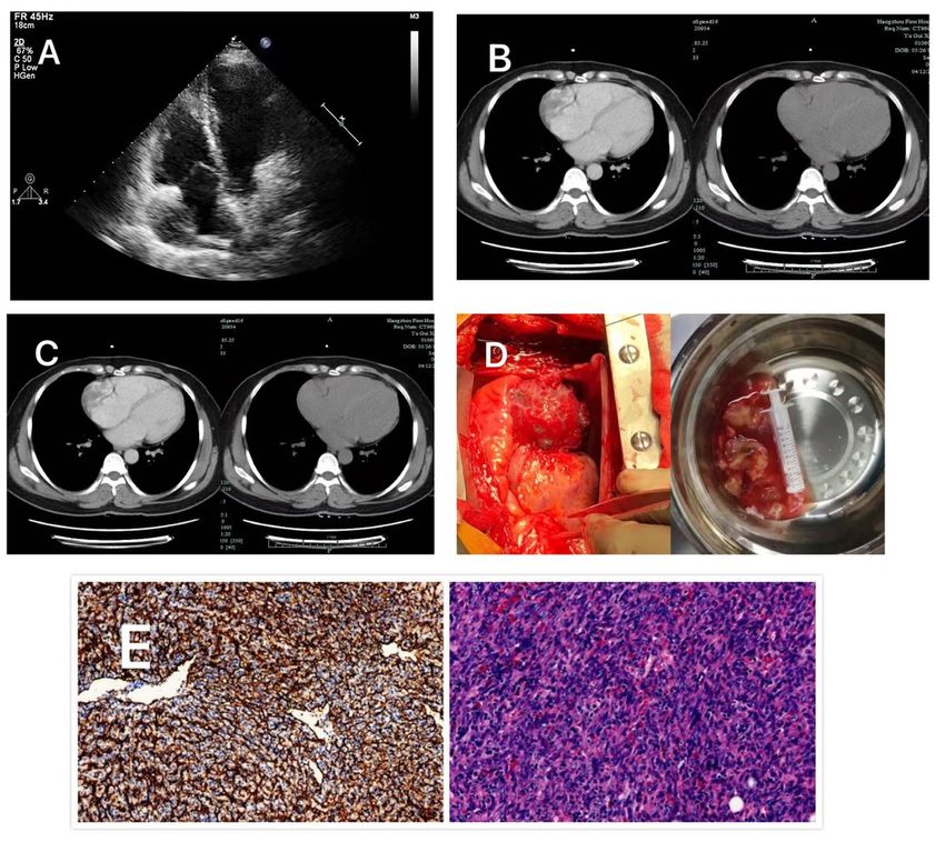
beats per min with early cardiac repolarization. TTE revealed there was a cauliflower-like mass with the
diameter of 4.5×3.2×4.0 cm attached to the right atrium with pericardial effusion. The mass was
considered as a space-occupying lesion of the heart which might be malignant with the presents of TTE
images (fig. 1A).
Page 2/8CT was performed to better define the location and extension of the mass. The plain scan showed a 5.2
×2.9 × 4.0 cm lesion with the density of tumor parenchyma in the pericardial with no clearly demarcation
between right atrium and pericardial. Enhanced scan showed uneven enhancement of it accompanied by
the pericardial and inferior vena cava invaded(fig. 1B). No abnormal enlarged lymph nodes were found in
hilum and mediastinum. In addition, the patient was examined by MRI. MRI indicated a heterogeneous
neoplasm with abnormal signal of iso-T1 and iso-T2 in the right anterior pericardium. The boundary
between the mass and the right atrium was unclear. Small nodules with iso-T1 and iso-T2 signals were
also founded in the right atrium (fig. 1C).
Besides, the FDG-MRI demonstrated an elevated FDG uptake mass on the pericardium and protruded into
the right atrium, which was about 3.8*3.9cm in size. The lump showed slightly high DWI signal and
unclear boundary. Combined with the image information above, a possible low-grade malignant tumor
was diagnosed.
Surgical data
In consideration of the results of the imaging examinations pointed to the malignant lesions, on May,
2018, surgical resection was performed under general anesthesia. During the operation, a tumor was
found in the pericardial accompany by 500 ml blood fluid in the pericardial cavity. The inferior vena cava
and right atrium were all invaded(fig.1D). The tumors was completely removed, appeared to have a rough
surface, soft texture.
Pathological Findings
Histological features
The tumors was completely removed and the final diagnosis of primary pericardial angiosarcoma was
confirmed by histopathology(fig.1E). The tumor tissue was arranged in solid structure. The tumor cells in
solid structure area were spindle shaped, and the cells were heteromorphic. Part tumor tissues were
arranged in anastomotic lacunar.
Immunohistochemistry
Immunohistochemical results showed: CK[-]、Vim[+]、CD31[+] CD34[+]、FVⅢ[+]、ERG[+]、D2-40[-]、SMA[-]、
Desmin[-]、S-100[-]、CK5/6[-]、Calretinin[-]、P53[+]、Ki-67[+]10-15%.
Follow up
Chemotherapy and radiotherapy were added after operation. Her CT and TTE were reexamined on June,
2018 and March, 2019 respectively with no lumps founded, and the patient was asymptomatic after
therapies. Unfortunately, recrudescence of the sarcoma and multiple pulmonary metastases were
founded and caused the death of the patient in 2020.
Page 3/8Discussion
The incidence of primary cardiac tumor is reporter as 0.0017%-0.033% and sarcomas make up for the
majority of them. However, the recognition of a primary cardiac sarcoma can be hard and progress
rapidly in a short time. The prognosis for patients with surgery only is poor with a mean survival of 3
months to 1 year. Although most angiosarcomas are resistant to both chemotherapy the radiation, it’s
meaningful to adopt them due to the high possibilities of the further metastases. It should be treated as
early as possible if suspected in imaging examinations and symptoms, however there’s no specific
clinical symptoms to detect it on time, dyspnea is the most common feature followed by chest pain as we
mentioned in the case above. Cardiac angiosarcoma can grow in any part of heart, usually originates
from the right atrium, rarely occurs in pericardium, and the most common sites of metastasis are the
lungs and bones[3].
Non-invasive imaging examinations such as echocardiography, CT and MRI, has contributed to the early
diagnosis or detection of the tumors. The common manifestation of pericardial angiosarcoma in echo
including the hemorrhagic pericardial effusion, abnormal mass attaching to the pericardium which
usually thickened. The echo could not only provide the specific location, morphology, internal echo and
attachment site of the tumor, but also observe the movement of the mass in the cavity and the degree of
obstruction dynamically, the relationship between the neoplasm and other cavity structures, evaluate the
secondary hemodynamic changes to supply the reliable first-hand information for clinical diagnosis and
treatment. The identify of benign and malignant tumors was preliminary based on the image
characteristics, such as inhomogeneous masses with poor border definition, invasion into extracardiac
structures, or the presence of a pericardial effusion, but it’s still difficult to differentiate benign and
malignant tumors by ultrasound only, and the large amount of pericardial effusion may lead to missed
diagnosis of the lesion occasional.
CT and enhanced CT are the most practical and valuable methods to show the size and location of the
tumor. Primary cardiac angiosarcoma often presents as homogeneous or inhomogeneous density on
unenhanced CT scans and heterogeneous centripetal enhancement on enhanced images[4]. Delayed
imaging could offer more information about the visualization of the mass given late contrast
enhancement. CT imaging can also provide more detailed information such as pericardial thickening and
effusion, the scope of tumor invasion, and the relationship between the lesion and surrounding structures
to guide the clinical operation. With percardial involvement, there is ussally “sheet-like” thickening due to
the distribution of mass cells. In cases with percardial diffusion, linear contrast material enhancement
along vascular lakes have described as a “sunray” appearance[5]. But it is not reliable for prediction of the
degree of malignancy of mass comparing with MRI due to the poorer histological resolution.
MRI examination has better tissue specificity and soft tissue contrast rate, more accuracy in the size,
location and shape of cardiac tumor, which has great significance in judging the degree of benign and
malignant invasion of neoplasm. The lesion often shows as heterogeneous intensity, with hyperintense
hemorrhage foci on both T1-and T2-weighted images compared with the myocardium. It is recommended
Page 4/8as the evaluation of heart function, surgical assessment, and postoperative follow-up. PET/CT reveals
abnormal FDG activity in part of the pericardium. It may help in the diagnosis in differentiating the benign
from a malignant lesion with a 100% sensitivity and about 86% specificity, using a cutoff of SUVmax of
3.5[6]. So it can be used for early detection of tumor and determining whether metastasis occurs.
In principle, cardiac tumor should be operated within a time limit once diagnosed to avoid arterial
embolism and (or) sudden death, and sometimes emergency operation is needed. Surgical indications
should be strictly grasped for patients with poor preoperative cardiac function and distant metastasis.
Generally, the tumor can be resected under cardiopulmonary bypass, the mass should be completely
resected as well the endocardium and myocardial tissue at the root and attachment of the tumor pedicle.
For the defects caused by surgery, the autologous pericardium or artificial patch can be used for repair,
and valve replacement when necessary[7]. At present, the treatment of cardiac malignant tumors tends to
the multidisciplinary comprehensive treatment of surgery with radiotherapy and chemotherapy followed
by.
Conclusion
Primary pericardial angiosarcoma is extremely rare with poor prognosis. Despite advances in imaging
techniques and increasing clinical availability, atypical clinical symptoms are the main reason for delayed
diagnosis and treatment. Transthoracic echocardiography is the first-line modality used to identify
cardiac mass. CT and MRI could provide more information about the invasion and metastasis.
Multimodality imaging methods should be combined for clinicians. Surgery is still the main treatment, it
can distinguish the nature of the tumor, relieve compression and prolong life, which is convenient for
further radiotherapy and chemotherapy. However, the effect of radiotherapy and chemotherapy is not
optimistic, and the quality of life of patients is still significantly reduced.
Declarations
Availability of data and materials
Not applicable.
Acknowledgements
Not appplicable.
Funding
Not applicable.
Author information
Affliations
Page 5/8Department of ultrasound, Hangzhou first people's Hospital Affiliated to Medical College of Zhejiang
University
Hong Sun, Min Zhao
Contributions
Hong Sun wrote the first draft of the case. Min Zhao revised and edited successive drafts of the
manuscript. All authors read and approved the final manuscript.
Corresponding author
Correspondence to Min Zhao.
Ethics declarations
Written informed consent was obtained from the patient for publication of this case report and
accompanying images.
Consent for publication
Written informed consent for publication was obtained.
Competing interests
The authors declare that they have no competing interests.
References
1. Lam KY, Dickens P, Chan AC. Tumors of the heart. A 20-year experience with a review of 12,485
consecutive autopsies. Arch Pathol Lab Med 1993;117:1027-31.
2. Butany J, Nair V, Naseemuddin A, Nair GM, Catton C, Yau T. Cardiac tumours: diagnosis and
management. Lancet Oncol. 2005;219-28.
3. Fatima J, Duncan AA, Maleszewski JJ, Kalra M, Oderich GS, Gloviczki P, et al. Primary angiosarcoma
of the aorta, great vessels, and the heart. J Vasc Surg. 2013;57:756–64.
4. Yu, J.F.; Cui, H.; Ji, G.M.; Li, S.Q.; Huang, Y.; Wang, R.N.; Xiaop, W.F. Clinical and imaging
manifestations of primary cardiac angiosarcoma. BMC Med. Imaging 2019, 19, 16.
5. Rahbar K, Seifarth H, Schäfers M, Stegger L, Hoffmeier A, Spieker T, et al. Differentiation of malignant
and benign cardiac tumors using 18F-FDG PET/CT. J Nucl Med 2012;53:856–63.
6. Yahata S, Endo T, Honma H, et al. Sunray apperance on enhanced magnetic resonance image of
cardiac angiosarcoma with pericardial obliteration. Am Heart J 1994; 127:468-471.
7. Chaves, V.M.; Pereira, C.; Andrade, M.; von Hafe, P.; Almeida, J.S. Cardiac Angiosarcoma From Cardiac
Tamponade to Ischaemic Stroke—A Diagnostic Challenge. Eur. J. Case. Rep. Intern. Med. 2019, 6,
Page 6/8001079.
Figures
Figure 1
A TTE revealed there was a cauliflower-like mass with the diameter of 4.5×3.2×4.0 cm attached to the
right atrium with pericardial effusion. B CT plain scan showed a 5.2 ×2.9 × 4.0 cm lesion with the density
of tumor parenchyma in the pericardial with no clearly demarcation between right atrium and pericardial.
C MRI indicated a heterogeneous mass with abnormal signal of iso-T1 and iso-T2 in the right anterior
pericardium. D 4 A tumor was found in the pericardial with about 500ml blood fluid in the pericardial
Page 7/8cavity during the operation and surgical specimens. E The tumor cells were heteromorphic, and the
vascular cavity was lined with heteromorphic endothelial cells
Supplementary Files
This is a list of supplementary files associated with this preprint. Click to download.
disclosureofpotentialconflictsofinterest.pdf
Page 8/8You can also read